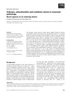
Báo cáo khoa học: Intrinsic disorder and coiled-coil formation in prostate apoptosis response factor 4 pptx
Bạn đang xem bản rút gọn của tài liệu. Xem và tải ngay bản đầy đủ của tài liệu tại đây (2.02 MB, 19 trang )
Intrinsic disorder and coiled-coil formation in prostate
apoptosis response factor 4
David S. Libich
1,
*, Martin Schwalbe
1,
*, Sachin Kate
1
, Hariprasad Venugopal
1
, Jolyon K. Claridge
1
,
Patrick J. B. Edwards
1
, Kaushik Dutta
2
and Steven M. Pascal
1
1 Centre for Structural Biology, Institute of Fundamental Sciences and Department of Physics, Massey University, Palmerston North,
New Zealand
2 New York Structural Biology Centre, NY, USA
Introduction
Prostate apoptosis response factor-4 (Par-4) is an ubi-
quitously expressed and evolutionary conserved protein
that was initially identified as a pro-apoptotic factor in
rat AT-3 androgen-independent prostate cancer cells
exposed to ionomycin [1,2]. The identified pro-apopto-
tic and tumour-suppressive roles of Par-4 are consid-
ered to be its most important cellular functions and,
accordingly, Par-4 is downregulated in various cancers
[3]. The anti-cancer strategy employed by Par-4 is
achieved by direct activation of the cell-death machinery
(e.g. Fas ⁄ FasL) [4] and inhibition of pro-survival fac-
tors (e.g. nuclear factor-kappa B) [5]. Furthermore,
ectopic over-expression of Par-4 can either directly
induce apoptosis or sensitize cancer cells to apoptotic
stimuli, dependent on cell type [6]. Primarily a cyto-
plasmic protein, translocation of Par-4 to the nucleus
is linked with the direct induction of apoptosis in
cancer cells [3,7,8]. Initially characterized in prostate
cancer, Par-4 has also been demonstrated to function
in renal cell carcinomas [9], leukaemia [10] and
Keywords
circular dichroism; coiled-coil; intrinsically
disordered protein; prostate apoptosis
response factor 4; solution NMR
spectroscopy
Correspondence
D. S. Libich or S. M. Pascal, Institute of
Fundamental Sciences, Massey University,
Turitea Site, Private Bag 11222, Palmerston
North 4442, New Zealand
Fax: +64 6 350 5682
Tel: +64 6 356 9099
E-mails: ;
*These authors contributed equally to this
work
(Received 24 March 2009, accepted 6 May
2009)
doi:10.1111/j.1742-4658.2009.07087.x
Prostate apoptosis response factor-4 (Par-4) is an ubiquitously expressed
pro-apoptotic and tumour suppressive protein that can both activate cell-
death mechanisms and inhibit pro-survival factors. Par-4 contains a highly
conserved coiled-coil region that serves as the primary recognition domain
for a large number of binding partners. Par-4 is also tightly regulated by
the aforementioned binding partners and by post-translational modifica-
tions. Biophysical data obtained in the present study indicate that Par-4
primarily comprises an intrinsically disordered protein. Bioinformatic
analysis of the highly conserved Par-4 reveals low sequence complexity and
enrichment in polar and charged amino acids. The high proteolytic suscep-
tibility and an increased hydrodynamic radius are consistent with a largely
extended structure in solution. Spectroscopic measurements using CD and
NMR also reveal characteristic features of intrinsic disorder. Under physio-
logical conditions, the data obtained show that Par-4 self-associates via the
C-terminal domain, forming a coiled-coil. Interruption of self-association
by urea also resulted in loss of secondary structure. These results are
consistent with the stabilization of the coiled-coil motif through an intra-
molecular association.
Abbreviations
CREB, cAMP-responsive element-binding protein; DLS, dynamic light scattering; GST, glutathione S-transferase; HSQC, heteronuclear single
quantum coherence; IDP, intrinsically disordered protein; IPTG, isopropyl thio-b-
D-galactoside; LZ, leucine zipper; NLS, nuclear localization
sequence; Par-4, prostate apoptosis response factor 4; PK, protein kinase; SAC, selective apoptosis of cancer cells.
3710 FEBS Journal 276 (2009) 3710–3728 ª 2009 The Authors Journal compilation ª 2009 FEBS
neuroblastomas [11], as well as endometrial [12],
pancreatic [13] and gastric [8] cancers.
In addition to its role in cancer, Par-4 is thought to
assist in normal neuronal development by preventing
the hyper-proliferation of nerve tissues, in turn con-
trolling the number of neurones and glial cells in both
the peripheral and central nervous systems [14,15].
Par-4 is upregulated in several neurodegenerative dis-
eases, such as Alzheimer’s disease [16,17], Parkinson’s
disease [18], Huntington’s disease [19] and amyotrophic
lateral sclerosis [20]. Par-4 is also reportedly involved
in immune response modulation [21], synaptic function
modulation [22] and apoptosis of neurones that have
received a traumatic insult [23].
The C-terminal quarter of Par-4 (Fig. 1) is highly
conserved and shares some homology with the death
domains of other apoptotic proteins, such as Fas,
receptor-interacting protein, Fas-associated death
domain protein and tumour necrosis factor receptor-
associated death domain protein [24,25]. This region
functions as the primary recognition and binding site
for various partners of Par-4, including Wilms’ tumour
1 [7], Akt1 ⁄ protein kinase (PK) B [26], atypical PKCs
(PKCs f and k ⁄ i) [24], p62 [27], death-associated pro-
tein-like ⁄ zipper interacting kinase [28], THAP [29],
Amida [30], E2F1 [31], D
2
dopamine receptor [32],
b-site amyloid precursor protein cleaving enzyme 1
[17], apoptosis-antagonizing transcription factor [33]
and topoisomerase 1 [34]. In addition, several binding
partners have been shown to interact at various sites
N-terminal to the aforementioned C-terminal segment,
including the androgen receptor [35], F-actin [36],
14-3-3 [26] and the SPRY domain-containing suppressor
of cytokine signalling box proteins 1, 2 and 4 [37].
Par-4 contains several conserved phosphorylation
sites that are modified by kinases, such as PKA, PKC,
casein kinase II and Akt1, adding a further level of
regulation of the function of Par-4 [38]. Phosphoryla-
tion of an absolutely conserved threonine (rat T155,
human T163 or mouse T156; Fig. 1) by PKA is
required for nuclear translocation [8]. Phosphorylation
of a C-terminal serine residue (rat S249, human or
mouse S231; Fig. 1) by Akt1 effectively inactivates
Par-4 by allowing the chaperone protein 14-3-3 to bind
and sequester it in the cytoplasm, even if it is
phosphorylated on T155 [26].
These multiple interactions coupled with a high
degree of sequence conservation and post-translational
modification suggest that the in vivo role(s) of Par-4
are highly temporally and spatially regulated. Simi-
larly, the ubiquitous expression, post-translational
modifications and a plethora of binding partners are
characteristics common to many intrinsically disor-
dered proteins (IDPs) [39]. In the present study, we
demonstrate that residual structure exists in Par-4
because the measured hydrodynamic radius increased
under denaturing conditions, suggesting that the
ensemble becomes less compact. CD and NMR indi-
cate that Par-4 is primarily intrinsically disordered
under physiological conditions and exists as an ensem-
ble of fast-averaging (on the NMR time-scale) struc-
tures. Furthermore, Par-4 forms a stable coiled-coil
through a self-association event mediated by the C-ter-
minus. The coiled-coil was probed using increasing
concentrations of chaotropic agents and was found to
be very stable. Using NMR, the segment of Par-4 not
involved in the coiled-coil was shown to have spectral
features that were similar to those of a C-terminal
deletion mutant. This is important because it suggests
that Par-4 is able to bind more than one partner at a
time and thus could function as a hub linking the
functions of several proteins. The coiled-coil region of
Par-4 represents an important functional domain that
is an example of a gain of structure upon binding tran-
sition, which is another important feature of IDPs [40].
Results
All sequence numbering is made with reference to rat
Par-4, to reflect the recombinant rat (rrPar-4) constructs
used in these studies. Three constructs were created;
rrPar-4FL (Par-4 full-length, residues 1–332), rrPar-
4DLZ (deleted leucine zipper, residues 1–290) and rrPar-
4SAC [selective apoptosis of cancer cells (SAC) domain
construct, residues 137–195] (Fig. 2A). The sequence
identity expressed relative to rat Par-4 of mouse and
human is 92% and 76%, respectively, whereas African
clawed frog and zebra fish share 52% and 47%
sequence identity with rat, respectively (Fig. 1).
The nuclear localization sequences (NLS) 1 (residues
20–25) and 2 (residues 137–153) are strictly conserved
in all known Par-4 sequences (Fig. 1). The SAC
domain, which includes NLS2, is the minimum frag-
ment of Par-4 that is absolutely required for apoptosis
[6] and is completely conserved amongst mammals
(Fig. 1). Furthermore, there is a high degree of
sequence conservation in the C-terminal quarter of Par-
4, which contains primarily a coiled-coil-like sequence
(residues 254–332; Figs 1 and 2A). In particular, a leu-
cine zipper (residues 292–330), which is a subset of the
coiled-coil domain, is almost conserved in all known
Par-4 sequences, suggesting a common functionality
(Figs 1 and 2A). Relatively few Par-4 genes have been
sequenced. It has been suggested that the general pat-
tern of sequence conservation shown in Fig. 1 is likely
to be conserved across other mammalian sequences [1].
D. S. Libich et al. Intrinsic disorder in Par-4
FEBS Journal 276 (2009) 3710–3728 ª 2009 The Authors Journal compilation ª 2009 FEBS 3711
Based on disembl analysis [41], the majority (> 70%)
of Par-4 is predicted to be disordered. The putative
regions of order in Par-4, as indicated by grey bars in a
disembl plot (Fig. 2B), align with or occur within func-
tionally important regions of Par-4 (Fig. 2A), namely
NLS1, NLS2, SAC and the coiled-coil. Secondary
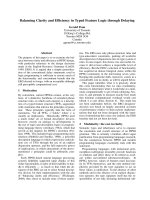
Fig. 1. Sequence alignment of the prostate apoptosis response factor 4 (Par-4). A BLASTP ⁄ CLUSTALW [102,103] alignment of sequences of
Par-4 from various species: rat (Rattus norvegicus), mouse (Mus musculus), human (Homo sapiens), African clawed frog (Xenopus laevis)
and zebra fish (Danio rero). The amino acids are coloured: red (nonpolar side chains: G, A, V, L, I, M, P, F and W), blue (polar side chains: S,
T, N, Q, Y and C) and green (polar, charged side chains: K, R, H, D and E). Symbols: residues in that column are identical in all sequences
(*); substitutions are conservative (:); and substitutions are semi-conservative (.). The high degree of sequence conservation of Par-4
suggests functional significance and thus resistance to evolutionary pressure. With reference to the numbering of rat Par-4, several seg-
ments are of notable interest: two nuclear localization sequences [NLS1 (20–25) and 2 (137–153)], which are completely conserved among
all known Par-4s, and the SAC domain (137–195), which is defined by being the absolute minimum fragment required for apoptosis and
includes NLS2 [6]. The C-terminal domain (254–332) is a coiled-coil (CC) motif that encompasses a LZ (292–330) as a subset. Two important
phosphorylation sites, T155 and S249, are denoted by red arrows.
Intrinsic disorder in Par-4 D. S. Libich et al.
3712 FEBS Journal 276 (2009) 3710–3728 ª 2009 The Authors Journal compilation ª 2009 FEBS
structure prediction using gor4 [42] shows that the
regions with the highest helical propensity also occur in
the aforementioned regions and align with the disembl
predicted ordered regions (Fig. 2C). The hydrophobic
cluster analysis [43] of Fig. 2D indicates that the most
hydrophobic regions align with the putative ordered and
predicted helical regions.
A plot of mean net charge against mean hydropho-
bicity determined from a protein’s primary structure
may be used to classify it as folded or intrinsically dis-
ordered. Plot space is divided by an empirically deter-
mined line (
R ¼ 2:785
H À 1:151) based on an analysis
by Uversky et al. [44]. The three constructs used in this
study are plotted in Fig. 3A along with several ‘classi-
cally folded’ proteins. Here, rrPar-4FL, rrPar-4DLZ
and rrPar-4SAC clearly fall into disordered space gen-
erally characterized by low mean hydrophobicity and
high net charge. The construct representing the SAC
domain (rrPar-4SAC), with 14 positively charged and
13 negatively charged residues but few hydrophobic
residues, lies further in the disordered region.
Figure 3B describes the sequence complexity of
rrPar-4FL by comparison with the percent difference
between the amino acid usage of a set of known IDPs
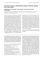
Fig. 2. (A) A block diagram of the three constructs of rrPar-4 used in the present study. Marked on each construct are the primary regions
of functional importance, including the nuclear localization sequences [NLS1 (20–25) and 2 (137–153), coloured green], the region necessary
for SAC (137–195), the coiled-coil C-terminal domain (CC, 254-332, coloured red) and the LZ (292–330, shown with hatching). The rrPar-
4DLZ construct lacks residues 291–332, which is approximately one-half of the coiled-coil and the entire leucine zipper. The rrPar-4SAC con-
struct represents residues 137–195 of Par-4, including NLS2. All three constructs used in the present study have an N-terminal GGS tag, a
remnant from the cleavage of the purification tag, which is omitted here for simplicity. (B)
DISEMBL predicts regions of order ⁄ disorder in pro-
teins using neural networks trained on multiple definitions of disorder [41]. The dashed line in (B) represents a threshold value separating
order and disorder. (C) Secondary structure (a-helix only shown) prediction using
GOR4 [42] and (D) hydrophobic cluster analysis (HCA) [43], a
visually enhanced representation of the primary sequence that highlights clustering of hydrophobic residues using symbols (
,T; ,S;¤,G;
w, P) and colours (red: P and acidic residues D, E, N, Q; blue: basic residues, H, K, R; green: hydrophobic residues, V, L, I, F, W, M, Y;
black: all other residues, G, S, T, C, A). The grey bars indicate the predicted regions of order in (B) and, for comparison, are extended over
(C) and (D).
D. S. Libich et al. Intrinsic disorder in Par-4
FEBS Journal 276 (2009) 3710–3728 ª 2009 The Authors Journal compilation ª 2009 FEBS 3713
versus a set of folded proteins (black bars). Positive
values indicate a depletion, whereas negative bars indi-
cate an enrichment relative to folded proteins. The pat-
tern of amino acid usage for rrPar-4FL (grey bars) is
in accordance with that generally observed for IDPs
[45,46], namely a depletion of order-promoting amino
acids (L, N, F, Y, I, W, C) and enrichment of dis-
order-promoting residues (S, Q, K, P, E). The amino
acid usage for rrPar-4DLZ and rrPar-4SAC follows a
similar pattern (not shown).
As calculated (i.e. from sequence) and experimen-
tally determined [i.e. from MS, Tricine-PAGE and
dynamic light scattering (DLS)], the molecular weights
for rrPar-4FL, rrPar-4DLZ and rrPar-4SAC are given
in Table 1. Because DLS measures the Stokes radius
(R
S
) of a particle, the equation log(R
S
) = 0.357 ·
log(MW) ) 0.204 was used to convert R
S
to MW for
comparative purposes [47,48]. Although this approxi-
mate calculation does not take into account the shape
of the particle (i.e. it assumes a sphere), the result is
useful for illustrating the degree of extended structure
in the protein.
The primary structure predicts MWs of 36.1, 31.1 and
7.0 kDa for rrPar-4FL, rrPar-4DLZ and rrPar-4SAC,
respectively. MALDI-TOF mass spectroscopy was used
to assess the purity and determine the sizes of the con-
structs produced. The sizes determined for rrPar-4DLZ
(44.5 Da difference between expected and observed) and
rrPar-4SAC (6.6 Da difference between expected and
observed after accounting for
15
N labelling of the sample
used for MS analysis) agree within error (approximately
0.1%) with the sizes predicted from sequence analysis
(Table 1). MS revealed that the rrPar-4FL construct is
approximately 0.2 kDa larger than expected.
Relative mobility analysis of the electrophoretic pro-
files of rrPar-4FL, rrPar-4DLZ and rrPar-4SAC using
a denaturing Tricine-PAGE system (see Experimental
procedures) determined apparent molecular weights of
49.1, 41.5 and 12.4 kDa, respectively. These sizes are
significantly larger (36%, 33% and 77% larger for
rrPar-4FL, rrPar-4DLZ and rrPar-4SAC, respectively)
than the expected MWs determined from the primary
structure or MS (Table 1).
The results of DLS experiments are shown in Table 2
and summarized in Table 1. The measured R
S
for
rrPar-4FL was 189 A
˚
, which is much larger than
expected for a monomeric random coil, suggesting a
polymeric state for rrPar-4FL under these conditions.
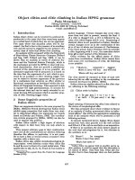
Fig. 3. (A) Charge ⁄ hydrophobicity plot of rrPar-4FL (335 residues),
rrPar-4DLZ (293 residues), and rrPar-4SAC (61 residues). The divid-
ing line
R ¼ 2:785
H À 1:151 represents an empirically determined
divisor between intrinsically disordered (high charge, low hydropho-
bicity) and structured (low charge, high hydrophobicity) space.
Proteins such as aprotinin [104], actin [105], ubiquitin [106] and 3C
protease [107] are plotted as examples of classically folded
proteins. (B) Sequence complexity of rrPar-4FL (grey bars) com-
pared with the average amino acid distribution of IDPs (black bars)
relative to the average amino acid distribution of globular proteins.
The relative distributions were sampled from proteins (both IDPs
and folded) deposited in the Protein Data Bank. Positive and nega-
tive values indicate an enrichment or depletion, respectively, of a
particular residue relative to globular proteins. Residues marked
with an asterisk occur two-fold more or less frequently, on average,
in IDPs than in globular proteins [46].
Table 1. Hydrodynamic properties of rrPar-4 constructs using vari-
ous biophysical techniques. MW (kDa) and hydrodynamic radius (A
˚
)
are shown in the format MW (R
S
) for three constructs using four
techniques. R
S
and MW were calculated from the primary structure
in reference to a folded conformation using log(R
S
) = 0.357 ·
log(MW) ) 0.204.
Construct Sequence
Method of analysis
MS PAGE DLS
rrPar-4FL 36.1 (26.5) 36.2 (26.5) 49.5 (29.6) 8899 (189)
rrPar-4-DLZ 31.1 (25.1) 31.2 (25.1) 41.5 (27.8) 64.1 (32.5)
rrPar-4 SAC 7.0 (14.8) 7.1 (14.8) 12.5 (18.1) 18.7 (20.9)
Intrinsic disorder in Par-4 D. S. Libich et al.
3714 FEBS Journal 276 (2009) 3710–3728 ª 2009 The Authors Journal compilation ª 2009 FEBS
For comparison, the R
S
(calculated) for rrPar-4FL as
either monomeric globular (i.e. folded), molten globule,
pre-molten globule, extended chain or urea-denatured
states are given in Table 2. The experimentally deter-
mined R
S
for rrPar-4DLZ (32.5 A
˚
) and rrPar-4SAC
(20.9 A
˚
) are larger than the expected folded R
S
(25.1
and 14.8 A
˚
, respectively) but still smaller than the cal-
culated random coil R
S
for either protein (Table 2).
This suggests that these constructs exist in an unfolded
yet monomeric form under these conditions. The
volume weighted distributions for rrPar-4FL, rrPar-
4DLZ and rrPar-4SAC are shown in the Supporting
information (Fig. S1A). The relatively broad distribu-
tion of sizes recorded for all three proteins is consistent
with an ensemble of interconverting conformations
rather than one single conformation.
Upon addition of 1 m urea, the measured R
S
for
both rrPar-4DLZ and rrPar-4SAC slightly increases
(Table 2; see also Fig. S1B,C). Conversely, the intro-
duction of 1 m urea to rrPar-4FL decreases the mea-
sured R
S
from 189 to 78.4 A
˚
, yet it remains larger
than the calculated R
S
of a random coil protein
(Table 2; see also Fig. S1A).
A classically folded protein is less susceptible to
proteolysis than an IDP upon equilateral exposure to
a protease such as trypsin because most of its cleavage
sites are protected by tertiary folding [49,50]. The
results of a limited trypsin digest of rrPar-4FL, rrPar-
4DLZ, rrPar-4SAC and BSA are shown in Fig. 4.
After 15 min of exposure to trypsin rrPar-4DLZ was
more than 95% digested, rrPar-4FL and rrPar-4SAC
were over 80% digested, whereas BSA was only 10%
digested. BSA was chosen for comparison because it
has a similar percentage of predicted cut sites to that
of the Par-4 constructs.
Figure 5 shows the full range (5 °C steps from
5–75 °C) and a sub-set of four spectra (5, 25, 45 and
65 °C) of a temperature series recorded by CD spec-
troscopy for rrPar-4FL (Fig. 5A,B), rrPar-4DLZ
(Fig. 5C,D) and rrPar-4SAC (Fig. 5E,F). Significant
a-helical character in rrPar-4FL is immediately evident
and remains stable up to 65 °C (Fig. 5A,B). By con-
trast, the CD spectra for rrPar-4DLZ (Fig. 5C,D) and
rrPar-4SAC (Fig. 5E,F) show a typical profile of IDPs
with a deep transition at 200 nm [51].
Pairwise overlays of
1
H-
15
N heteronuclear single
quantum coherence (HSQC) spectra for rrPar-4FL,
rrPar-4DLZ and rrPar-4SAC are shown in Fig. 6. The
spectra of all three proteins display the features that
characterize disorder in proteins, namely sharp peaks
and narrow
1
H chemical shift dispersion [51,52].
Chemical shift similarities indicate some structural
similarity between rrPar-4FL and rrPar-4DLZ
(Fig. 6A). Fewer peaks share similar chemical shifts
when comparing rrPar-4FL or rrPar-4DLZ with rrPar-
4SAC (Fig. 6B,C). Thus, the majority of residues in
rrPar-4SAC experience a different local environment
and possibly a different conformation than the SAC
domain in the context of either the rrPar-4FL or
rrPar-4DLZ constructs. Only 160 of the 308 peaks
expected (335 – N-terminal residue – 26 prolyl resi-
dues) for rrPar-4FL and 152 of the 266 expected peaks
(293 – N-terminal residue – 26 prolyl residues) for
rrPar-4DLZ are readily picked. Conversely, 58 peaks
Table 2. Comparison of experimental and theoretical values of Stoke’s radii (R
S
). Measured R
S
was recorded in 10 mM Tris (pH 7.0), 20 mM
NaCl in the presence or absence of urea. Calculated R
S
was obtained using the mean values from equations given in Uversky [47] for globu-
lar (folded) (G), molten globule (MG), pre-molten globule (PMG), random coil (RC) and urea-denatured (U) states.
Construct MW (kDa)
Measured R
S
(A
˚
) Calculated R
S
(A
˚
)
0
M urea 1 M urea G MG PMG RC U
rrPar-4FL 36.1 189 78.4 26.5 29.4 37.7 49.6 53.1
rrPar-4DLZ 31.1 32.5 43.6 25.1 28.0 35.6 46.1 49.2
rrPar-4SAC 7.0 20.9 28.1 14.8 17.1 19.9 22.2 22.7
Fig. 4. Limited proteolysis of rrPar-4FL (filled circles), rrPar-4DLZ
(open circles), rrPar-4SAC (filled triangles) and BSA (open triangles).
The proteins were dissolved in 20 m
M NaPO
4
(pH 7.5), 50 mM NaCl
and exposed to trypsin in a 280 : 1 (w ⁄ w) ratio.
D. S. Libich et al. Intrinsic disorder in Par-4
FEBS Journal 276 (2009) 3710–3728 ª 2009 The Authors Journal compilation ª 2009 FEBS 3715
of the expected 60 (62 – N-terminal residue – one pro-
lyl residue) were readily identifiable for rrPar-4SAC
with only two glycyl residues being unobservable.
To assess the degree of a-helicity in the rrPar-4FL
C-terminus, a CD difference spectrum between rrPar-
4DLZ and rrPar-4FL (25 °C) is shown in Fig. 7A. This
spectrum indicates a well-defined coiled-coil type struc-
ture (Fig. 7A). The two constructs differ in the dele-
tion of the leucine zipper (Fig. 2A); thus, rrPar-4FL
forms a stable coiled-coil under these conditions and
the majority of the a-helical character observed in
rrPar-4FL (Fig. 5A,B) may be attributed to this struc-
ture. The melting temperatures (based on the reduction
of the 222 nm transition in CD spectra) for rrPar-4FL
are 75, 55 and 25 °C when dissolved in native buffer,
native buffer + 1 m urea or native buffer + 6 m urea,
respectively (Fig. 7B). The results of the DLS experi-
ments on rrPar-4FL under the same conditions are
shown in Fig. 7C. As the concentration of urea is
increased from 1 to 6 m, the effective R
S
for rrPar-4FL
is reduced from 189 to 58.5 A
˚
. The latter value is very
close to the predicted R
S
of a random coil of the same
molecular weight (for comparison, see Table 2).
Discussion
The structure-defines-function paradigm of molecular
biology is currently under scrutiny because many
Fig. 5. Temperature dependence of the CD spectrum of (A, B) rrPar-4FL, (C, D) rrPar-4DLZ and (E, F) rrPar-4SAC. Data for all constructs
were recorded in 10 m
M Tris (pH 7.0), 20 mM NaCl over a temperature range of 5–75 °C. Traces for each temperature recorded in the exper-
iment are shown in (A, C, E). For clarity, four equally spaced temperatures from the sampled range are shown in (B, D, F).
Intrinsic disorder in Par-4 D. S. Libich et al.
3716 FEBS Journal 276 (2009) 3710–3728 ª 2009 The Authors Journal compilation ª 2009 FEBS
proteins have been identified that are functional with-
out the need for well-defined secondary or tertiary
structure [46,53,54]. The occurrence of intrinsic disor-
der in proteomes is correlated to the complexity of the
cell; thus, eukaryotic proteins have a higher proportion
of disorder (35–51% of proteins with disordered
regions of 40 residues or longer) than proteins from
prokaryotes and archaea (6–33%) [55].
The prevalence of intrinsic disorder is higher in
proteins that are involved in cell signalling, cytoskeletal
organization and ribosomal or cancer-related processes
[56]. Disorder in proteins that control these processes
appears to be of functional importance because these
events are often tightly controlled and highly dynamic
and often become deregulated in cancerous cells [57].
Many signalling proteins function in pathways associ-
ated with cancer. For example, the well-characterized
IDP p53 functions as a transcription regulator during
the G1 cell cycle phase. Critical mutations of p53 lead
to loss of its transcriptional control and thus lead to
inappropriate survival of damaged or mutated cells [58].
Sequence analysis of Par-4
Bioinformatic analysis of rrPar-4FL reveal characteris-
tic features of IDPs, including high net charge, low
mean hydrophobicity and low sequence complexity
[45,59]. Relative to the amino acid usage observed in
folded proteins, rrPar-4FL, rrPar-4DLZ and rrPar-
4SAC are depleted in order-promoting amino acids
and enriched in disorder promoting residues (Fig. 3B).
The lack of hydrophobic residues inhibits the forma-
tion of a hydrophobic core and thus the formation of
stable tertiary structure (Fig. 2D) [46].
More than 70% of rrPar-4FL is predicted to be dis-
ordered by disembl, providing a strong argument
against the formation of stable global tertiary structure
(Fig. 2B). Hydrophobic cluster analysis is a method of
displaying the primary structure such that the cluster-
ing of hydrophobic residues and thus regions of possi-
ble order become evident [43]. The regions of greatest
hydrophobic clustering in rrPar-4FL correlate well
with the predicted regions of order (Fig. 2B) and with
the secondary structure predictions (Fig. 2C).
Although the majority of rrPar-4FL is predicted to be
disordered, this does not preclude the formation of
short regions of structure or larger but transient sec-
ondary structure elements. Indeed, gor4 predictions of
a-helical structure (Fig. 2C) coincide with the more
ordered regions of rrPar-4FL and fall within the highly
conserved segments of the protein (Fig. 1). Thus,
regions of rrPar-4FL may be capable of forming
a-helices either independently or upon association with
binding partners. Furthermore, the predicted regions
of order in rrPar-4LZ occur within the functionally
relevant regions, namely NLS1 and 2, SAC and the
Fig. 6. Pairwise overlays of
1
H-
15
N HSQC spectra of (A) rrPar-4FL (black contours) and rrPar-4DLZ (blue contours), (B) rrPar-4DLZ (blue
contours) and rrPar-4SAC (red contours) and (C) rrPar-4FL (black contours) and rrPar-4SAC (red contours). The compositions of the samples
were: rrPar-4FL, 0.48 m
M in 10 mM Tris (pH 7.0), 20 mM NaCl, 5% D
2
O,
15
N-rrPar-4DLZ, 0.09 mM in 20 mM NaPO
4
(pH 7.5), 100 mM NaCl,
1m
M dithiothreitol, 5% D
2
O and
15
N-rrPar-4SAC, 0.34 mM in 10 mM Tris (pH 7.0), 20 mM NaCl, 5% D
2
O. All spectra were recorded at 5 °C
and the processing parameters (see Experimental procedures) were identical for qualitative comparison.
D. S. Libich et al. Intrinsic disorder in Par-4
FEBS Journal 276 (2009) 3710–3728 ª 2009 The Authors Journal compilation ª 2009 FEBS 3717
coiled-coil (Fig. 2) raising the possibility that Par-4
function may be associated with structure stabilization
in these regions.
Intrinsic disorder in proteins is often erroneously
considered to be a featureless random coil, although
proteins do not achieve a completely random confor-
mation even in strongly denaturing conditions [60]. A
more accurate depiction is that IDPs exist as ensembles
of rapidly interchanging conformers that sample vary-
ing regions of secondary structure space [46]. IDPs can
be broadly categorized into three non-exclusive groups
(i.e. a single IDP may fall into more than one
category): random coil, pre-molten globule or molten
globule [61].
Because of the high percentage of rrPar-4FL that is
predicted as disordered, a random coil-like classifica-
tion of the ensemble would appear to be the most
appropriate. Similar to the structural ensemble
described for activator for thyroid hormone and reti-
noid receptors [62], in the absence of interacting part-
ners, rrPar-4FL exists predominantly unfolded in
solution. The kinase-inducible transcriptional-activa-
tion domain of cAMP-responsive element-binding pro-
tein (CREB) has been shown to be an IDP that folds
into an orthogonal a-helix structure upon association
with CREB binding protein [63,64]. The intrinsically
disordered nature along with the CREB binding
protein-induced helical regions could be accurately pre-
dicted from its primary structure [53]. Similarly, the
primarily intrinsically disordered nature and potential
helical regions of Par-4 are predicted here (Figs 2
and 3).
Par-4 displays aberrant electrophoretic mobility
and is susceptible to proteolysis
Aberrant electrophoretic mobility in a denaturing
PAGE system is a hallmark of IDPs because their
unique amino acid composition reduces the amount
of sodium dodecyl sulphate that is able to bind
[46,51,65]. Aberrant mobility on PAGE gels of Par-4
and Par-4 constructs (i.e. deletion mutants) has been
demonstrated, although it is not known whether
the effects of IDP amino acid composition were con-
sidered [34,36]. In the present study, slower than
expected migration of the Par-4 constructs resulted
in apparent MWs that were 1.3- (rrPar-4FL and
rrPar-4DLZ) to 1.8- (rrPar-4SAC) fold larger than
that predicted from sequence or measured using MS
(Table 1).
Limited proteolysis can be used to distinguish
ordered and disordered proteins based on their relative
sensitivity to cleavage by proteases such as trypsin
Fig. 7. (A) Difference between the 25 °C traces of rrPar-4FL and
rrPar-4DLZ (Fig. 5). The difference spectrum is characteristic of a
well-defined coiled-coil displaying a h
222
⁄ h
208
ratio > 1. (B) Temper-
ature dependence of the molar elipticity measured at 222 nm for
rrPar-4FL in buffer (10 m
M Tris, pH 7.0, 20 mM NaCl) only (filled
circles), buffer + 1
M urea (open diamonds) and buffer + 6 M urea
(open triangles). (C) Volume distribution of DLS measurements of
rrPar-4FL showing the apparent hydrodynamic radius of the parti-
cles: buffer (10 m
M Tris, pH 7.0, 20 mM NaCl) only (white bars),
buffer + 1
M urea (grey bars) and buffer + 6 M urea (hatched bars).
The reduction of the apparent R
S
upon increasing urea concentra-
tion suggests the disruption of a polymeric complex.
Intrinsic disorder in Par-4 D. S. Libich et al.
3718 FEBS Journal 276 (2009) 3710–3728 ª 2009 The Authors Journal compilation ª 2009 FEBS
[45,50,66]. Although rrPar-4FL, rrPar-4DLZ and BSA
contain an approximately equal percentage of trypsin
cut sites, BSA is digested at a much slower rate
(Fig. 4). This implies that a significant portion of the
conformational ensemble of the rrPar-4 proteins are
more exposed to the solvent than BSA and largely lack
protection by folded and stable tertiary structure.
The hydrodynamic radius of Par-4 is larger than
that predicted by sequence analysis
The observable Stokes radius of a protein increases in
proportion to its degree of ‘unfoldedness’; thus, an
IDP will have an observable R
S
larger than a folded
globular protein of the same MW [48,67]. Some exam-
ples of IDPs with large R
S
values relative to MW have
been summarized previously [68]. In the present study,
the R
S
measured for rrPar-4FL, rrPar-4DLZ and
rrPar-4SAC correspond to MWs of 8.9 · 10
3
, 64.1 and
18.7 kDa, respectively, and are much larger (713, 129
and 141%) than what would be expected for a folded
globular protein of similar MW. The MW estimations
shown in Table 1 are used as a point of reference to
illustrate that the degree of ‘unfoldedness’ of Par-4 is
very high, which is similar to that expected for a coil-
like as opposed to a pre-molten globule ensemble [48].
The extremely large R
S
observed for rrPar-4FL relative
to the other two constructs clearly indicates a poly-
meric state, as discussed further below.
IDPs such as CREB and p27
Kip1
(i.e. cyclin-depen-
dent kinase inhibitor) exist as structurally intercon-
verting populations that have been demonstrated to
retain a nascent secondary structure to varying
degrees under physiological conditions [69]. The width
of the volume-weighted distributions for rrPar-4FL,
rrPar4DLZ and rrPar-4SAC (see Fig. S1) is consistent
with this type of conformational exchange. Interest-
ingly, although the addition of 1 m urea causes a sub-
tle but significant increase in R
S
for rrPar4DLZ and
rrPar-4SAC (Table 2), the width of the distributions
are largely unaltered. Together, these observations
suggest that 1 m urea can disrupt some folding
elements and bring the conformation of the ensemble
closer to random coil, but conformational exchange
continues.
Secondary structure of Par-4 assessed by CD and
NMR
The CD spectra for rrPar-4DLZ and rrPar-4SAC are
exemplary of IDPs with a deep transition at 200 nm
and a minor transition at 222 nm (Fig. 5) [51]. The
CD spectra of IDPs are often complicated by minor
contributions from secondary structure elements, such
as alpha or poly-proline type II helices [70]. Decon-
volution of the 25 °C spectra estimates 32%, 17% and
18% of combined regular and distorted a-helix for
rrPar-4FL, rrPar-4DLZ and rrPar-4SAC, respectively
[71]. All three constructs remain relatively stable
throughout the heating cycle because the 5–65 °C
traces exhibit similar features. Thus, in addition to the
coiled-coil region of rrPar-4FL, other regions of these
proteins may transiently populate a-helical or other
secondary structures. Interestingly, although the over-
all temperature-induced changes are minor, an isodich-
roic point at 210 nm is observed for rrPar-4SAC,
which may be interpreted as a two-state confor-
mational change (Fig. 5C). This could be the result of
secondary and ⁄ or tertiary structure that is thermally
disrupted.
The atomic resolution of NMR makes it uniquely
suited to assess the ‘orderedness’ of an IDP [51].
Because most residues of an IDP are solvent exposed
and inherently flexible, they share a similar chemical
environment and, consequently, share similar NMR
frequencies, resulting in significant overlap of reso-
nances (particularly for
1
H resonances) and popula-
tion-weighted average chemical shifts [72]. Mobility
also results in sharp peaks as a result of increased T
2
values [73]. The spectra of rrPar-4FL, rrPar-4DLZ and
rrPar-4SAC shown in Fig. 6 are characteristic of IDPs,
with ensemble averages, narrow peaks and poor chemi-
cal shift dispersion.
A total of 52% of residues for both rrPar-4FL and
rrPar-4DLZ are not readily observable in an
1
H-
15
N
HSQC. From the current data, it is impossible to
determine whether the same residues (or residues from
the same regions) are unobservable in these two con-
structs. Possible reasons for this feature include poor
chemical shift dispersion and intermediate exchange
[74]. A detailed examination of the dynamics of the
visible regions of the proteins (dependent on assign-
ments) may help to elucidate the time scales of motion
involved and thus more definitive statements could
then be made about particular residues or regions of
rrPar-4LZ and rrPar-4DLZ [75].
The spectrum of rrPar-4SAC (Fig. 6) is much more
complete than those recorded for rrPar-4FL and
rrPar-4DLZ. Nonetheless, a similar degree of disorder
is suggested by the peak shape and chemical shift dis-
persion. The size of the rrPar-4SAC (7 kDa) relative
to that of the other constructs (> 30 kDa) is likely to
be a contributing factor in the observance of these res-
onances because fewer residues equates to less chance
of spectral overlap and a lower likelihood of slow to
intermediate exchange.
D. S. Libich et al. Intrinsic disorder in Par-4
FEBS Journal 276 (2009) 3710–3728 ª 2009 The Authors Journal compilation ª 2009 FEBS 3719
Evidence for self-association of Par-4 mediated
by a coiled-coil
The putative coiled-coil region of Par-4 (residues 254–
332; Fig. 1) is the site of recognition and association
for the majority of known binding partners [76]: the
deletion of the leucine zipper renders Par-4 incapable
of binding to proteins such as Wilms’ tumour 1,
aPKCf, p62, death-associated protein-like ⁄ zipper
interacting kinase, Akt1, E2F1 and b-site amyloid
precursor protein cleaving enzyme 1 [17,31,76,77].
Coiled-coils are characterized by the heptad repeat
abcdefg with hydrophobic residues at the a and d
positions [78]. The leucine zipper is a subset type of
coiled-coil typified by the occurrence of a leucine at
every seventh position (d) [79,80]. The CD difference
spectra between rrPar-4FL and rrPar-4DLZ is charac-
teristic of a coiled-coil (h
222
⁄ h
208
> 1; Fig. 7A),
suggesting that at least two rrPar-4FL monomers self-
associate forming a coiled-coil. Lacking the residues
that comprise the leucine zipper region of the coiled-
coil (Fig. 2A), rrPar-4DLZ was not observed to have
strong a-helical character (Fig. 5C) or to self-associate
(see Fig. S2).
DLS measured an unusually large R
S
for rrPar-4FL
(189 A
˚
) dissolved in native buffer (Fig. 7C, Table 1).
Large Stokes radii have previously been measured in
rod-shaped proteins, including the winter flounder
anti-freeze protein [81], hydrophobin SC3 [82] and var-
ious coiled-coils, such as chromogranin A [83]. How-
ever, the apparent R
S
of rrPar-4FL was much larger
than expected if the ensemble consisted of coil-like
monomers. The constructs lacking the coiled-coil
region (rrPar-4DLZ and rrPar-4SAC) did not have
appreciably large R
S
relative to the estimated random
coil values (Table 2).
The reduction of the R
S
from 189 A
˚
to 78 A
˚
for
rrPar-4LZ in 1 m urea is likely to be a result of the
disruption of a noncovalent interaction. Furthermore,
the observed R
S
in 6 m urea (58 A
˚
) is close to the cal-
culated random coil value for rrPar-4FL (Fig. 7C).
Thus, as the concentration of urea is decreased, a poly-
meric state of rrPar-4FL forms in association with
increased stabilization of the coiled-coil. Melt curves
constructed from the reduction in intensity of the
222 nm CD transition demonstrate that the rrPar-4FL
complex is much less stable in increasing concentra-
tions of urea, and show that the melting temperature
for the rrPar-4FL complex increases from 25 °Cin6m
urea to 75 °C in native buffer (Fig. 7B). Taken
together with the the results of the CD (Fig. 7A) and
DLS measurements (Fig. 7C), these data clearly indi-
cate that rrPar-4FL self-associates via the putative
coiled-coil region, with the leucine zipper being of criti-
cal importance for self-association.
Recently, a 33 kDa isoform of Par-4 that lacks exon
3 (notably NLS2), but retains the coiled-coil region,
was reported by Wang et al. [84]. This isomer cannot
translocate to the nucleus and has been proposed to be
a negative regulator of Par-4 apoptotic activity by
binding to and sequestering Par-4 in the cytoplasm
[84]. Therefore, self-association of Par-4 may be physi-
ologically relevant as an additional regulator of its
pro-apoptotic activity.
Previous studies have demonstrated that a 47 residue
construct derived from the C-terminus (residues 286–
332) of Par-4, which includes all of the leucine zipper
but not all of the coiled-coil region, self-associates into
a coiled-coil structure under specific pH and tempera-
ture conditions [85]. Under native conditions (neutral
pH and moderate temperature), the construct was
predominantly disordered, as judged by CD spectra. It
was proposed that nonfavourable charge–charge inter-
actions at neutral pH prevented coiled-coil formation
and that mitigation of these nonfavourable contacts
through changes of pH, temperature, salt concentra-
tion or site-directed mutagenesis altered the ensemble
equilibrium in favour of a coiled-coil [86].
The results obtained in the present study show that,
in the context of rrPar-4FL, the C-terminus forms a
coiled-coil at neutral pH. This suggests that a region
N-terminal to the leucine zipper domain provides an
electrostatic surface to counter the negative charges in
the leucine zipper domain or otherwise helps to stabi-
lize the coiled-coil. Using a series of deletion mutants,
Gao et al. [35] demonstrated that an N-terminal region
of Par-4 is able to interact with the coiled-coil region.
This may be a required intramolecular interaction for
stabilization of the coiled-coil during self-association
or with other binding partners at neutral pH. Alterna-
tively, interactions with other parts of the protein,
including the N-terminal region of the coiled-coil (i.e.
N-terminal to the leucine zipper), may comprise a req-
uisite trigger sequence for coiled-coil formation [87].
Although the CD spectrum of rrPar-4FL showed
a-helical character (Fig. 5A), there is no obvious sign
of helix formation in the corresponding HSQC spec-
trum and, as noted, the total number of peaks
observed is only approximately 150. Recently, Liew
et al. [88] demonstrated that the signals observed in a
1
H-
15
N HSQC of a glutathione S-transferase (GST)
fusion peptide arose almost exclusively from the target
protein and not GST. They argued that because GST
forms a 52 kDa dimer, the signals arising from GST
would be broadened beyond observable limits and,
thus, the remaining resonances are from the flexible
Intrinsic disorder in Par-4 D. S. Libich et al.
3720 FEBS Journal 276 (2009) 3710–3728 ª 2009 The Authors Journal compilation ª 2009 FEBS
linker and target protein [88]. A similar phenomenon
may be occurring in the present study, where the elon-
gated and highly self-associated coiled-coil forms a
large core that behaves as a much larger protein. Thus,
its HSQC signals are unobservable as a result of line
broadening, whereas the attached intrinsically disor-
dered regions of the protein are observable because of
their relative flexibility. Intermediate conformational
exchange may also contribute to the broadening of the
coiled-coil peaks. All of the data presented in the pres-
ent study are consistent with this hypothesis. Also con-
sistent with this view, the overlay of rrPar-4FL and
rrPar-4DLZ shows that approximately 50 peaks have
identical or reasonably close chemical shifts, suggesting
some similarity in the local environment of these pro-
teins (Fig. 6A). Non-overlap of remaining peaks indi-
cates that a significant portion of the protein is
affected by coiled-coil formation, through direct or
relayed interactions.
One physiological implication of these observations
is that Par-4 may be well suited to bind one partner
via the coiled-coil, whereas much of the remainder of
the intrinsically disordered regions are available for
simultaneous interactions with another partner or part-
ners: Par-4 may function as a highly efficient linker
protein. The majority of the Par-4 binding partners
interact with the C-terminus, although a few, such as
the SPRY domain and F-actin, have been shown to
bind N-terminally [36,37]. Indeed, Par-4 has been dem-
onstrated to mediate the ternary complex between
aPKCf and p62 [27].
The advantage of disorder in dynamic processes
Intrinsic disorder may impart several advantages to
Par-4 in its role as a pro-apoptotic factor. Because dis-
ordered regions are solvent exposed, they are easily
accessible for post-translational modifications, such as
phosphorylation, ubiquitination or Ubl-conjugation,
etc., which enables precise control of function, localiza-
tion and turnover rate [39,89]. Notably for Par-4, the
phosphorylation of T155 (Fig. 1) is required for
nuclear translocation and subsequent initiation of
apoptosis [8]. The extreme proteolytic sensitivity of
IDPs offers an additional layer of cellular control via
rapid, controlled turnover [90]. Disordered regions also
confer an increased structural plasticity and, conse-
quently, IDPs are able to bind multiple targets with
high specificity yet in a readily reversible manor. The
‘fly-casting’ mechanism has been proposed to describe
how disordered segments bind their targets with
low affinity and fast association ⁄ dissociation rates
[45,51,69,91]. Two extensively studied proteins, p53
and high mobility group protein A, have been shown
to interact with multiple binding partners primarily
through disorder containing regions [56]. Similarly,
because of intrinsic disorder, Par-4 could contain mul-
tiple specific binding sites such that it binds to different
partners simultaneously, as discussed in the case of
aPKCf ⁄ p62. An extended conformation also has the
advantage of a large amount of accessible surface area
being available for intermolecular interactions, relative
to a globular protein of the same number of amino
acids [92].
It is noteworthy that all 26 prolines present in Par-4
occur in the first 255 residues (Fig. 1). Prolyl residues
favour open conformations and extended structures
such as polyproline-type II helices, which easily con-
vert to other conformational states [93]. Additionally,
proline is considered to promote inter-molecular recog-
nition as a result of the absence of intra-residue hydro-
gen bonds [94].
Because of the precise regulation through post-trans-
lational modifications and their promiscuous binding,
IDPs often form hubs or nodes that serve to link the
functions of several proteins and ⁄ or cofactors together
[95]. The high mobility group protein A is a chromo-
some and chromatin modulator that functions as a
hub in cancer and other related pathological processes
[96,97]. We raise the possibility of a similar role for
Par-4. The ubiquitous expression, tight temporal and
spatial regulation, rapid turnover, multiple binding
partners and inherent flexibility uniquely situate Par-4
to function as a control factor hub for apoptosis.
Conclusions
The data obtained in the present study indicate that
Par-4 can be classified as a predominantly intrinsically
disordered protein. Bioinformatic analysis shows that
highly conserved Par-4 has low sequence complexity, is
enriched in polar and charged amino acids and is
classified as disordered when plotted in charge-hydro-
phobicity space. disembl predicts that the majority of
Par-4 (> 70%) is disordered, yet ordered segments
align well with predicted secondary structure elements
(a-helix) and regions of hydrophobic clusters. Limited
proteolysis and DLS experiments demonstrate that
rrPar-4FL is primarily extended in solution, exhibiting
high susceptibility to trypsin and a large hydrodynamic
radius. Furthermore, CD and NMR experiments
revealed characteristic spectral features of intrinsic dis-
order. Taken together, these data demonstrate that
rrPar-4FL does not maintain a stabilized global
tertiary structure, but does not preclude the possible
formation of transient and ⁄ or local structure.
D. S. Libich et al. Intrinsic disorder in Par-4
FEBS Journal 276 (2009) 3710–3728 ª 2009 The Authors Journal compilation ª 2009 FEBS 3721
Although primarily disordered, rrPar-4FL is able to
self-associate via the C-terminus forming a stable
coiled-coil in this region. Self-association behaviour
was not observed for any of the other constructs used
in the present study, each of which lacked the C-termi-
nus. Although previous experiments with the leucine
zipper domain of Par-4 showed that self-association
required acidic pH, the same requirement was not
observed in the present study, possibly because of the
influence of other regions of Par-4 providing charge-
mediated or other forms of stabilization. Intrinsic dis-
order imparts many advantages to a multifunctional
protein such as Par-4. Protein–protein and protein–
ligand interactions can be highly specific yet readily
reversible, whereas post-translational modifications
allow for very tight control of the functions of Par-4.
The results obtained in the present study demonstrate
that the combined intrinsically disordered and coiled-
coil nature of Par-4 provides uniq ue structural properties
through which Par-4 can perform a multifunctional
role in various tissues and cellular processes.
Experimental procedures
The PCR primers used were purchased from Sigma Geno-
sys (Sigma-Aldrich PTY Ltd, Castle Hill, Australia). D
2
O
and
15
NH
4
Cl were obtained from Cambridge Isotope Labo-
ratories (Andover, MA, USA). Restriction enzymes were
purchased from Roche Diagnostic GmbH (Penzberg,
Germany). All other chemicals were of reagent grade or
higher and were acquired from either Sigma or Invitrogen
(Carlsbad, CA, USA).
Expression vector construction
A versatile expression vector was designed to be used to
either express Par-4 constructs alone or in conjunction with
putative binding partners in Escherichia coli cells. The
pET23a vector from Novagen (Merck Biosciences, Darms-
tadt, Germany) was used as a template to create pCFE-
TrxH-TEV, which enables the expression of targets as
fusion proteins with thioredoxin. A hexa-histidine tag is
included between thioredoxin and the N-terminus of the
target. An rTEV-protease cleavage site was included for
removal of the thioredoxin and hexa-histidine tags. The
fusion tag was PCR amplified using pET32a (Merck Bio-
sciences) as a template and subsequently inserted into the
NdeI ⁄ BamHI restriction sites of pET23a. The primers were:
forward 5¢-CTGGCATATGAGCGATAAAATTATTCAC-
3¢ and reverse 5¢-CCGGGGATCCCTGAAAATACAGG
TTTTCGGTCGTTGGGATATCGTAATCGTGATGGTG
ATGGTGATGCATATG-3¢.
The rrPar-4FL construct (residues 1–332, rat sequence
numbering) was prepared by PCR amplification using the
four primers 5¢-CAGGGATCCATGGCGACCGGCG
GCTATCGGAG-3¢,5¢-CTTGGCGGCTGGATCTCCGCC
GCTCGAAC-3¢,5¢-GTTCGAGCGGCGGAGATCCAGCC
GCCAAG-3¢ and 5¢-CAGGTCGACTTACCTTGTCAGC
TGCCCAACAAC-3¢ to remove an internal BamHI site on
the racine Par-4 cDNA. The PCR product was then cloned
into the BamHI ⁄ SalI sites of pCFE-TrxH-TEV. The rrPar-
4DLZ (residues 1–290) construct lacking the leucine
zipper was PCR amplified with the primers 5¢-CAG
GGATCCATGGCGACCGGCGGCTATCGGAG-3¢ and
5¢-CCGGAAGCTTTTATTCTTCTTTATCTTGCATCAG-
3¢ using the full-length construct as a template. The PCR
product was then cloned into the BamHI ⁄ HindIII sites of
pCFE-TrxH-TEV. The rrPar-4SAC (residues 137–195) con-
struct representing the SAC domain was cloned in the same
manner as rrPar-4DLZ using the primers 5¢-GAGGAT
CCAGGAAAGGCAAAGGGCAGATCG-3¢ and 5¢-GCA
AGCTTTTATGCTTCATTCTGGATGGTG-3¢.
Expression of Par-4
The three rrPar-4 expression vectors were used to transform
E. coli Rosetta(DE3) cells (Novagen). The cells were grown
in LB medium at 37 °C until D
600
of 0.6 was reached,
induced by the addition of 0.4 mm isopropyl thio-b-
d-galactoside (IPTG) and grown for a further 6 h at 25 °C.
Isotopic labels were introduced by growing cells in LB
medium at 37 °C until D
600
of 0.6 was reached. Cells were
pelleted by centrifugation and resuspended in one-half the
original volume of M9 minimal media using
15
NH
4
Cl as
the sole nitrogen source. After growth for 1 h at 30 °C,
expression was induced by the addition of 0.4 mm IPTG
and cells were grown for a further 6 h at 25 °C.
Cells were harvested by centrifugation, resuspended in
lysis buffer (50 mm Tris, pH 8.0, 100 mm NaCl, 25 mm
imidazole) or lysis buffer containing 8 m urea and lysed by
three passes through a French press (AMINCO, Silver
Spring, MD, USA). The resulting lysate was cleared by fil-
tration through a 0.8 lm syringe filter. The cleared lysate
was passed through a Ni-nitrilotriacetic acid (GE Health-
care, Uppsala, Sweden) column and eluted with 250 mm
imidazole in lysis buffer. To remove excess imidazole,
pooled fractions containing the rrPar-4 fusion proteins were
dialysed against lysis buffer. The purification tags (thiore-
doxin and hexa-histidine) were cleaved from the rrPar-4
proteins with rTEV at room temperature and passed again
over the Ni-nitrilotriacetic acid column. The cleavage leaves
a three residue (Gly-Gly-Ser) remnant at the N-terminus of
all the rrPar-4 constructs. The eluted fractions were subse-
quently dialysed against 10 mm Tris (pH 7.4) and 20 mm
NaCl.
Ion-exchange chromatography was used as a final purifi-
cation step for rrPar-4DLZ and rrPar-4SAC. The constructs
were purified on SP-sepharose column (GE Healthcare)
using a linear gradient of 0–100% high salt buffer over
Intrinsic disorder in Par-4 D. S. Libich et al.
3722 FEBS Journal 276 (2009) 3710–3728 ª 2009 The Authors Journal compilation ª 2009 FEBS
20 min. The low salt buffer contained 20 mm NaPO
4
(pH
6) and 50 mm NaCl; the high salt buffer contained 20 mm
Tris (pH 7.5) and 1 m NaCl. Fractions containing the
protein of interest were pooled and dialysed against 10 mm
Tris (pH 7.0), 20 mm NaCl and were concentrated by
centrifugation using a Vivaspin 20 device (Vivascience AG,
Hannover, Germany). The rrPar-4FL construct was further
purified by RP-HPLC using a Delta Pak C18-300 A
˚
,
300 · 3.9 mm column, (Waters Corporation, Milford, MA,
USA) with a linear gradient of 20–45% acetonitrile
containing 0.08% trifluoroacetic acid. The rrPar-4FL frac-
tions were lyopholized and resolubilized in 10 mm Tris (pH
7.0), 20 mm NaCl.
Protein concentration was determined by A
280
and A
205
measurements using the extinction coefficients (13 075
m
)1
Æcm
)1
(rrPar-4FL and rrPar-4DLZ) or 1490 m
)1
Æcm
)1
(rrPar-4SAC) and the relationship described by Scopes [98].
The purified samples were assessed by MALDI-TOF mass
spectroscopy (Centre for Protein Research, University of
Otago, Dunedin, New Zealand).
Limited proteolysis
The rrPar-4 constructs or BSA (Sigma-Aldrich, St Louis,
MO, USA) were incubated with trypsin at a protein to pro-
tease ratio of 280 : 1 (w ⁄ w) in 20 mm NaPO
4
(pH 7.5),
50 mm NaCl for 15 min at 37 °C. Aliquots were taken after
1, 2, 5, 10 and 15 min and the reaction was quenched by
the addition of Laemmli sample buffer and boiling for
5 min. Proteins were loaded on a 10% Tricine-PAGE gel
[99]. The extent of digestion was measured from the relative
intensities of the Tricine-PAGE gel band representing the
undigested band by densitometry using the Gel Doc Imager
and Quantity One software package (Bio-Rad, Hercules,
CA, USA).
DLS
The apparent Stokes radii of the rrPar-4 constructs were
analysed using a Zetasizer Nano ZS (Malvern Instruments,
Malvern, UK). Sample concentrations were 0.3 mgÆmL
)1
in
native buffer (10 mm Tris, pH 7.0, 20 mm NaCl) or native
buffer plus 1 or 6 m urea. DLS data were obtained at
25 °C using a low-volume disposable 1 cm pathlength plas-
tic cuvette (Sarstedt, Nu
¨
rnbrecht, Germany) and five
successive scans were collected and averaged for each pro-
tein sample. Samples were prepared 1 day in advance and
maintained overnight at 4 °C to allow any bubbles to dissi-
pate and were then allowed to equilibrate to 25 °C before
measurements were made. The diffusion coefficients were
extracted from the correlation curve and the hydrodynamic
radius was calculated using the Stokes–Einstein equation.
The highest peak of the resulting histogram recorded for
each sample was taken as the mean diameter for that
particular sample.
CD
Spectra were recorded on a Chirascan CD spectropolarime-
ter (Applied Photophysics, Leatherhead, UK) equipped with
a recirculating water bath. Samples were at a concentration
of 0.3 mgÆmL
–1
in native buffer (10 mm Tris, pH 7.0, 20 mm
NaCl) or native buffer plus 1 or 6 m urea. Spectra were
recorded in 0.5 nm steps from 260–190 nm with an integra-
tion time of 1 s at each wavelength. Three successive scans
were recorded, the sample blank was subtracted and the
scans were averaged and smoothed using a sliding window
function. Thermal stability was determined by acquiring CD
spectra as a function of temperature at 5 °C intervals from
5–75 °C with 2 min of equilibration time at each temperature
point. Deconvolution was performed using the continll
algorithm [100] through the dichroweb server interface [71].
NMR spectroscopy
NMR experiments were performed on a Bruker Avance
700 MHz spectrometer (Bruker BioSpin GmbH, Rheinstet-
ten, Germany) equipped with a cryoprobe, four rf-channels
and gradient pulse capabilities. All spectra were acquired at
5 °C on 300 lL samples containing 5% D
2
O in Shigemi
NMR tubes. The rrPar-4FL sample concentration was
0.48 mm in 10 mm Tris (pH 7.0), 20 mm NaCl. The rrPar-
4DLZ construct was uniformly
15
N labelled with a protein
concentration of 0.09 mm in 20 mm NaPO
4
(pH 7.5),
100 mm NaCl, 1 mm dithiothreitol. Similarly rrPar-4SAC
was uniformly
15
N labelled at a concentration of 0.34 mm
in 10 mm Tris (pH 7.0), 20 mM NaCl.
1
H-
15
N HSQC spectra were recorded with the settings:
rrPar-4FL: 200 transients, 2048 · 128 points (F
2
· F
1
) and
spectral widths of 8389.2 and 2128.9 Hz for F
2
and F
1
,
respectively; rrPar-4DLZ: 20 transients, 2048 · 128 points
(F
2
· F
1
) and spectral widths of 8389.2 and 2128.9 Hz for
F
2
and F
1
, respectively; rrPar-4SAC: 24 transients,
2048 · 256 points (F
2
· F
1
) and spectral widths of 8389.2
and 2128.9 Hz for F
2
and F
1
, respectively. All data sets
were linear predicted and zero-filled once in the indirect
dimension before Fourier transformation and final process-
ing. Spectra were apodised using a shifted (p ⁄ 6) squared
sinusoidal bell function using TopSpin 2.1 (Bruker BioSpin
GmbH, Rheinstetten, Germany). The
1
H and
15
N chemical
shifts were referenced to the water signal [101].
Acknowledgements
The authors wish to thank Professor Rangnekar. We
also acknowledge Mr Trevor Loo and Mrs Michelle
Tamehana for providing excellent technical assistance
and advice. Funding for this project was provided in
part by grants from the Royal Society of New Zealand
(Marsden Fund Award MAU0507) to S.M.P.
D. S. Libich et al. Intrinsic disorder in Par-4
FEBS Journal 276 (2009) 3710–3728 ª 2009 The Authors Journal compilation ª 2009 FEBS 3723
References
1 Boghaert ER, Sells SF, Walid AJ, Malone P, Williams
NM, Weinstein MH, Strange R & Rangnekar VM
(1997) Immunohistochemical analysis of the proapop-
totic protein Par-4 in normal rat tissues. Cell Growth
Differ 8, 881–890.
2 Sells SF, Wood DP Jr, Joshi-Barve SS, Muthukumar
S, Jacob RJ, Crist SA, Humphreys S & Rangnekar
VM (1994) Commonality of the gene programs
induced by effectors of apoptosis in androgen-depen-
dent and -independent prostate cells. Cell Growth
Differ 5, 457–466.
3 Ranganathan P & Rangnekar VM (2005) Regulation
of cancer cell survival by Par-4. Ann N Y Acad Sci
1059, 76–85.
4 Chakraborty M, Qiu SG, Vasudevan KM & Rangne-
kar VM (2001) Par-4 drives trafficking and activation
of Fas and Fasl to induce prostate cancer cell apopto-
sis and tumor regression. Cancer Res 61, 7255–7263.
5 Camandola S & Mattson MP (2000) Pro-apoptotic
action of PAR-4 involves inhibition of NF-kappaB
activity and suppression of BCL-2 expression. J Neuro-
sci Res 61, 134–139.
6 El-Guendy N, Zhao Y, Gurumurthy S, Burikhanov R
& Rangnekar VM (2003) Identification of a unique
core domain of par-4 sufficient for selective apoptosis
induction in cancer cells. Mol Cell Biol 23, 5516–5525.
7 Johnstone RW, See RH, Sells SF, Wang J, Muthuk-
kumar S, Englert C, Haber DA, Licht JD, Sugrue SP,
Roberts T et al. (1996) A novel repressor, par-4, mod-
ulates transcription and growth suppression functions
of the Wilms’ tumor suppressor WT1. Mol Cell Biol
16, 6945–6956.
8 Gurumurthy S, Goswami A, Vasudevan KM &
Rangnekar VM (2005) Phosphorylation of Par-4 by
protein kinase A is critical for apoptosis. Mol Cell Biol
25, 1146–1161.
9 Cook J, Krishnan S, Ananth S, Sells SF, Shi Y,
Walther MM, Linehan WM, Sukhatme VP, Weinstein
MH & Rangnekar VM (1999) Decreased expression of
the pro-apoptotic protein Par-4 in renal cell carcinoma.
Oncogene 18, 1205–1208.
10 Boehrer S, Chow KU, Puccetti E, Ruthardt M,
Godzisard S, Krapohl A, Schneider B, Hoelzer D,
Mitrou PS, Rangnekar VM et al. (2001) Deregulated
expression of prostate apoptosis response gene-4 in less
differentiated lymphocytes and inverse expressional
patterns of par-4 and bcl-2 in acute lymphocytic
leukemia. Hematol J 2, 103–107.
11 Kogel D, Reimertz C, Mech P, Poppe M, Fruhwald
MC, Engemann H, Scheidtmann KH & Prehn JH
(2001) Dlk ⁄ ZIP kinase-induced apoptosis in human
medulloblastoma cells: requirement of the mitochon-
drial apoptosis pathway. Br J Cancer 85, 1801–1808.
12 Moreno-Bueno G, Fernandez-Marcos PJ, Collado M,
Tendero MJ, Rodriguez-Pinilla SM, Garcia-Cao I,
Hardisson D, Diaz-Meco MT, Moscat J, Serrano M
et al. (2007) Inactivation of the candidate tumor sup-
pressor par-4 in endometrial cancer. Cancer Res 67,
1927–1934.
13 Azmi AS, Wang Z, Burikhanov R, Rangnekar VM,
Wang G, Chen J, Wang S, Sarkar FH & Mohammad
RM (2008) Critical role of prostate apoptosis response-
4 in determining the sensitivity of pancreatic cancer
cells to small-molecule inhibitor-induced apoptosis.
Mol Cancer Ther 7, 2884–2893.
14 Bieberich E, MacKinnon S, Silva J, Noggle S &
Condie BG (2003) Regulation of cell death in mitotic
neural progenitor cells by asymmetric distribution of
prostate apoptosis response 4 (PAR-4) and
simultaneous elevation of endogenous ceramide. J Cell
Biol 162, 469–479.
15 Blaschke AJ, Staley K & Chun J (1996) Widespread
programmed cell death in proliferative and postmitotic
regions of the fetal cerebral cortex. Development 122,
1165–1174.
16 Loo DT, Copani A, Pike CJ, Whittemore ER,
Walencewicz AJ & Cotman CW (1993) Apoptosis is
induced by beta-amyloid in cultured central
nervous system neurons. Proc Natl Acad Sci USA 90,
7951–7955.
17 Xie J & Guo Q (2005) PAR-4 is involved in regulation
of beta-secretase cleavage of the Alzheimer amyloid
precursor protein. J Biol Chem 280, 13824–13832.
18 Duan W, Rangnekar VM & Mattson MP (1999)
Prostate apoptosis response-4 production in synaptic
compartments following apoptotic and excitotoxic
insults: evidence for a pivotal role in mitochondrial
dysfunction and neuronal degeneration. J Neurochem
72, 2312–2322.
19 Zeitlin S, Liu JP, Chapman DL, Papaioannou VE &
Efstratiadis A (1995) Increased apoptosis and early
embryonic lethality in mice nullizygous for the
Huntington’s disease gene homologue. Nat Genet 11,
155–163.
20 Pedersen WA, Luo H, Kruman I, Kasarskis E &
Mattson MP (2000) The prostate apoptosis
response-4 protein participates in motor neuron
degeneration in amyotrophic lateral sclerosis. FASEB J
14, 913–924.
21 Lafuente MJ, Martin P, Garcia-Cao I, Diaz-Meco
MT, Serrano M & Moscat J (2003) Regulation of
mature T lymphocyte proliferation and differentiation
by Par-4. EMBO J 22, 4689–4698.
22 Guo Q, Xie J, Chang X, Zhang X & Du H (2001)
Par-4 is a synaptic protein that regulates neurite
outgrowth by altering calcium homeostasis and
transcription factor AP-1 activation. Brain Res 903,
13–25.
Intrinsic disorder in Par-4 D. S. Libich et al.
3724 FEBS Journal 276 (2009) 3710–3728 ª 2009 The Authors Journal compilation ª 2009 FEBS
23 Payette DJ, Xie J, Shirwany N & Guo Q (2008) Exac-
erbation of apoptosis of cortical neurons following
traumatic brain injury in par-4 transgenic mice. Int J
Clin Exp Pathol 1, 44–56.
24 Diaz-Meco MT, Municio MM, Frutos S, Sanchez P,
Lozano J, Sanz L & Moscat J (1996) The product of
par-4, a gene induced during apoptosis, interacts selec-
tively with the atypical isoforms of protein kinase C.
Cell 86, 777–786.
25 Valmiki MG & Ramos JW (2009) Death effector
domain-containing proteins. Cell Mol Life Sci 66,
814–830.
26 Goswami A, Burikhanov R, de Thonel A, Fujita N,
Goswami M, Zhao Y, Eriksson JE, Tsuruo T &
Rangnekar VM (2005) Binding and phosphorylation of
par-4 by akt is essential for cancer cell survival. Mol
Cell 20, 33–44.
27 Chang S, Kim JH & Shin J (2002) p62 forms a ternary
complex with PKCzeta and PAR-4 and antagonizes
PAR-4-induced PKCzeta inhibition. FEBS Lett 510,
57–61.
28 Page G, Kogel D, Rangnekar V & Scheidtmann KH
(1999) Interaction partners of Dlk ⁄ ZIP kinase:
co-expression of Dlk ⁄ ZIP kinase and Par-4 results in
cytoplasmic retention and apoptosis. Oncogene 18,
7265–7273.
29 Roussigne M, Cayrol C, Clouaire T, Amalric F &
Girard JP (2003) THAP1 is a nuclear proapoptotic
factor that links prostate-apoptosis-response-4 (Par-4)
to PML nuclear bodies. Oncogene 22, 2432–2442.
30 Boosen M, Vetterkind S, Koplin A, Illenberger S &
Preuss U (2005) Par-4-mediated recruitment of Amida
to the actin cytoskeleton leads to the induction of
apoptosis. Exp Cell Res 311, 177–191.
31 Lu C, Chen JQ, Zhou GP, Wu SH, Guan YF & Yuan
CS (2008) Multimolecular complex of Par-4 and E2F1
binding to Smac promoter contributes to glutamate-
induced apoptosis in human- bone mesenchymal stem
cells. Nucleic Acids Res 36, 5021–5032.
32 Park SK, Nguyen MD, Fischer A, Luke MP, Affarel
B, Dieffenbach PB, Tseng HC, Shi Y & Tsai LH
(2005) Par-4 links dopamine signaling and depression.
Cell 122, 275–287.
33 Guo Q & Xie J (2004) AATF inhibits aberrant produc-
tion of amyloid beta peptide 1–42 by interacting
directly with Par-4. J Biol Chem 279, 4596–4603.
34 Goswami A, Qiu S, Dexheimer TS, Ranganathan P,
Burikhanov R, Pommier Y & Rangnekar VM (2008)
Par-4 binds to topoisomerase 1 and attenuates
its DNA relaxation activity. Cancer Res 68, 6190–
6198.
35 Gao S, Wang H, Lee P, Melamed J, Li CX, Zhang F,
Wu H, Zhou L & Wang Z (2006) Androgen receptor
and prostate apoptosis response factor-4 target the
c-FLIP gene to determine survival and apoptosis in the
prostate gland. J Mol Endocrinol 36, 463–483.
36 Vetterkind S, Illenberger S, Kubicek J, Boosen M,
Appel S, Naim HY, Scheidtmann KH & Preuss U
(2005) Binding of Par-4 to the actin cytoskeleton is
essential for Par-4 ⁄ Dlk-mediated apoptosis. Exp Cell
Res 305, 392–408.
37 Masters SL, Yao S, Willson TA, Zhang JG, Palmer
KR, Smith BJ, Babon JJ, Nicola NA, Norton RS &
Nicholson SE (2006) The SPRY domain of SSB-2
adopts a novel fold that presents conserved
Par-4-binding residues. Nat Struct Mol Biol 13, 77–84.
38 El-Guendy N & Rangnekar VM (2003) Apoptosis by
Par-4 in cancer and neurodegenerative diseases. Exp
Cell Res 283, 51–66.
39 Uversky VN, Oldfield CJ & Dunker AK (2008)
Intrinsically disordered proteins in human diseases:
introducing the D2 concept. Annu Rev Biophys 37,
215–246.
40 Oldfield CJ, Cheng Y, Cortese MS, Romero P,
Uversky VN & Dunker AK (2005) Coupled folding
and binding with alpha-helix-forming molecular recog-
nition elements. Biochemistry 44, 12454–12470.
41 Linding R, Jensen LJ, Diella F, Bork P, Gibson TJ &
Russell RB (2003) Protein disorder prediction: implica-
tions for structural proteomics. Structure 11, 1453–
1459.
42 Garnier J, Gibrat JF & Robson B (1996) GOR method
for predicting protein secondary structure from amino
acid sequence. Comput Methods Macromol Seq Anal
266, 540–553.
43 Callebaut I, Labesse G, Durand P, Poupon A, Canard
L, Chomilier J, Henrissat B & Mornon JP (1997)
Deciphering protein sequence information through
hydrophobic cluster analysis (HCA): current status and
perspectives. Cell Mol Life Sci 53, 621–645.
44 Uversky VN, Gillespie JR & Fink AL (2000) Why are
‘natively unfolded’ proteins unstructured under
physiologic conditions? Proteins 41, 415–427.
45 Dunker AK, Lawson JD, Brown CJ, Williams RM,
Romero P, Oh JS, Oldfield CJ, Campen AM, Ratliff
CM, Hipps KW et al. (2001) Intrinsically disordered
protein. J Mol Graph Model 19, 26–59.
46 Tompa P (2002) Intrinsically unstructured proteins.
Trends Biochem Sci 27, 527–533.
47 Uversky VN (2002) What does it mean to be natively
unfolded? Eur J Biochem 269, 2–12.
48 Uversky VN (1993) Use of fast protein size-exclusion
liquid chromatography to study the unfolding of
proteins which denature through the molten globule.
Biochemistry 32, 13288–13298.
49 Spolaore B, Bermejo R, Zambonin M & Fontana A
(2001) Protein interactions leading to conformational
changes monitored by limited proteolysis: apo form
D. S. Libich et al. Intrinsic disorder in Par-4
FEBS Journal 276 (2009) 3710–3728 ª 2009 The Authors Journal compilation ª 2009 FEBS 3725
and fragments of horse cytochrome c. Biochemistry 40,
9460–9468.
50 Hubbard SJ (1998) The structural aspects of limited
proteolysis of native proteins. Biochim Biophys Acta
1382, 191–206.
51 Receveur-Brechot V, Bourhis JM, Uversky VN,
Canard B & Longhi S (2006) Assessing protein disor-
der and induced folding. Proteins 62, 24–45.
52 Dyson HJ & Wright PE (2005) Elucidation of the
protein folding landscape by NMR. Methods Enzymol
394, 299–321.
53 Dyson HJ & Wright PE (2005) Intrinsically unstruc-
tured proteins and their functions. Nat Rev Mol Cell
Biol 6, 197–208.
54 Uversky VN, Oldfield CJ & Dunker AK (2005) Show-
ing your ID: intrinsic disorder as an ID for recogni-
tion, regulation and cell signaling. J Mol Recognit 18,
343–384.
55 Dunker AK, Obradovic Z, Romero P, Garner EC &
Brown CJ (2000) Intrinsic protein disorder in complete
genomes. Genome Inform Ser Workshop Genome
Inform 11, 161–171.
56 Iakoucheva LM, Brown CJ, Lawson JD, Obradovic Z
& Dunker AK (2002) Intrinsic disorder in cell-signaling
and cancer-associated proteins. J Mol Biol 323,
573–584.
57 Hanahan D & Weinberg RA (2000) The hallmarks of
cancer. Cell 100, 57–70.
58 Hartwell LH & Kastan MB (1994) Cell cycle control
and cancer. Science 266, 1821–1828.
59 Romero P, Obradovic Z, Li X, Garner EC, Brown CJ
& Dunker AK (2001) Sequence complexity of
disordered protein. Proteins 42, 38–48.
60 Shortle D (1996) The denatured state (the other half of
the folding equation) and its role in protein stability.
FASEB J 10, 27–34.
61 Uversky VN & Fink AL (2004) Conformational
constraints for amyloid fibrillation: the importance of
being unfolded. Biochim Biophys Acta 1698, 131–153.
62 Demarest SJ, Martinez-Yamout M, Chung J, Chen H,
Xu W, Dyson HJ, Evans RM & Wright PE (2002)
Mutual synergistic folding in recruitment of CBP ⁄ p300
by p160 nuclear receptor coactivators. Nature 415,
549–553.
63 Richards JP, Bachinger HP, Goodman RH & Bren-
nan RG (1996) Analysis of the structural properties
of cAMP-responsive element-binding protein (CREB)
and phosphorylated CREB. J Biol Chem 271, 13716–
13723.
64 Radhakrishnan I, Perez-Alvarado GC, Parker D,
Dyson HJ, Montminy MR & Wright PE (1997)
Solution structure of the KIX domain of CBP
bound to the transactivation domain of CREB: a
model for activator:coactivator interactions. Cell 91,
741–752.
65 Hu CC & Ghabrial SA (1995) The conserved, hydro-
philic and arginine-rich N-terminal domain of cucumo-
virus coat proteins contributes to their anomalous
electrophoretic mobilities in sodium dodecylsulfate-
polyacrylamide gels. J Virol Methods 55, 367–379.
66 Lacy ER, Wang Y, Post J, Nourse A, Webb W,
Mapelli M, Musacchio A, Siuzdak G & Kriwacki RW
(2005) Molecular basis for the specificity of p27 toward
cyclin-dependent kinases that regulate cell division.
J Mol Biol 349, 764–773.
67 Tcherkasskaya O & Uversky VN (2001) Denatured
collapsed states in protein folding: example of apomyo-
globin. Proteins 44, 244–254.
68 Uversky VN (2002) Natively unfolded proteins: a point
where biology waits for physics. Protein Sci 11,
739–756.
69 Galea CA, Wang Y, Sivakolundu SG & Kriwacki RW
(2008) Regulation of cell division by intrinsically
unstructured proteins: intrinsic flexibility, modularity,
and signaling conduits. Biochemistry 47, 7598–7609.
70 Shi Z, Woody RW & Kallenbach NR (2002) Is poly-
proline II a major backbone conformation in unfolded
proteins? Adv Protein Chem 62, 163–240.
71 Whitmore L & Wallace BA (2004) DICHROWEB, an
online server for protein secondary structure analyses
from circular dichroism spectroscopic data. Nucleic
Acids Res 32, W668–W673.
72 Dyson HJ & Wright PE (2001) Nuclear magnetic
resonance methods for elucidation of structure and
dynamics in disordered states. Methods Enzymol 339,
258–270.
73 Dyson HJ & Wright PE (2004) Unfolded proteins and
protein folding studied by NMR. Chem Rev 104,
3607–3622.
74 Cavanagh J (2007) Protein NMR Spectroscopy: Princi-
ples and Practice, 2nd edn. Elsevier Academic Press,
Amsterdam, Boston, MA.
75 Dyson HJ & Wright PE (2002) Insights into the
structure and dynamics of unfolded proteins from
nuclear magnetic resonance. Adv Protein Chem 62,
311–340.
76 Zhao Y & Rangnekar VM (2008) Apoptosis and
tumor resistance conferred by Par-4. Cancer Biol Ther
7, 1867–1874.
77 Gurumurthy S & Rangnekar VM (2004) Par-4 induc-
ible apoptosis in prostate cancer cells. J Cell Biochem
91, 504–512.
78 Harbury PB, Zhang T, Kim PS & Alber T (1993) A
switch between two-, three-, and four-stranded coiled
coils in GCN4 leucine zipper mutants. Science 262,
1401–1407.
79 Landschulz WH, Johnson PF & McKnight SL (1988)
The leucine zipper: a hypothetical structure common to
a new class of DNA binding proteins. Science 240,
1759–1764.
Intrinsic disorder in Par-4 D. S. Libich et al.
3726 FEBS Journal 276 (2009) 3710–3728 ª 2009 The Authors Journal compilation ª 2009 FEBS
80 O’Shea EK, Rutkowski R & Kim PS (1989) Evidence
that the leucine zipper is a coiled coil. Science 243, 538–
542.
81 Marshall CB, Chakrabartty A & Davies PL (2005)
Hyperactive antifreeze protein from winter flounder is
a very long rod-like dimer of alpha-helices. J Biol
Chem 280, 17920–17929.
82 Wang X, Graveland-Bikker JF, de Kruif CG & Robil-
lard GT (2004) Oligomerization of hydrophobin SC3
in solution: from soluble state to self-assembly. Protein
Sci 13, 810–821.
83 Mosley CA, Taupenot L, Biswas N, Taulane JP, Olson
NH, Vaingankar SM, Wen G, Schork NJ, Ziegler
MG, Mahata SK et al. (2007) Biogenesis of the secre-
tory granule: chromogranin A coiled-coil structure
results in unusual physical properties and suggests a
mechanism for granule core condensation. Biochemis-
try 46, 10999–11012.
84 Wang G, Silva J, Krishnamurthy K & Bieberich E
(2006) A novel isoform of prostate apoptosis
response 4 (PAR-4) that co-distributes with F-actin
and prevents apoptosis in neural stem cells. Apoptosis
11, 315–325.
85 Dutta K, Alexandrov A, Huang H & Pascal SM
(2001) pH-induced folding of an apoptotic coiled coil.
Protein Sci 10, 2531–2540.
86 Dutta K, Engler FA, Cotton L, Alexandrov A, Bedi
GS, Colquhoun J & Pascal SM (2003) Stabilization of
a pH-sensitive apoptosis-linked coiled coil through
single point mutations. Protein Sci 12, 257–265.
87 Steinmetz MO, Jelesarov I, Matousek WM, Honnap-
pa S, Jahnke W, Missimer JH, Frank S, Alexandrescu
AT & Kammerer RA (2007) Molecular basis of
coiled-coil formation. Proc Natl Acad Sci USA 104,
7062–7067.
88 Liew CK, Gamsjaeger R, Mansfield RE & Mackay JP
(2008) NMR spectroscopy as a tool for the rapid
assessment of the conformation of GST-fusion pro-
teins. Protein Sci 17, 1630–1635.
89 Xie H, Vucetic S, Iakoucheva LM, Oldfield CJ, Dunker
AK, Obradovic Z & Uversky VN (2007) Functional
anthology of intrinsic disorder. 3. Ligands, post-transla-
tional modifications, and diseases associated with
intrinsically disordered proteins. J Proteome Res 6,
1917–1932.
90 Wright PE & Dyson HJ (1999) Intrinsically unstruc-
tured proteins: re-assessing the protein structure-func-
tion paradigm. J Mol Biol 293, 321–331.
91 Shoemaker BA, Portman JJ & Wolynes PG (2000)
Speeding molecular recognition by using the folding
funnel: the fly-casting mechanism. Proc Natl Acad Sci
USA 97, 8868–8873.
92 Meszaros B, Tompa P, Simon I & Dosztanyi Z (2007)
Molecular principles of the interactions of disordered
proteins. J Mol Biol 372, 549–561.
93 Adzhubei AA & Sternberg MJ (1993) Left-handed
polyproline II helices commonly occur in globular
proteins. J Mol Biol 229, 472–493.
94 Williamson MP (1994) The structure and function of
proline-rich regions in proteins. Biochem J 297 (Pt 2),
249–260.
95 Dunker AK, Cortese MS, Romero P, Iakoucheva LM
& Uversky VN (2005) Flexible nets. The roles of
intrinsic disorder in protein interaction networks.
FEBS J 272, 5129–5148.
96 Reeves R (1992) Chromatin changes during the cell
cycle. Curr Opin Cell Biol 4, 413–423.
97 Reeves R & Beckerbauer L (2001) HMGI ⁄
Y proteins:
flexible regulators of transcription and chromatin
structure. Biochim Biophys Acta 1519, 13–29.
98 Scopes RK (1974) Measurement of protein by
spectrophotometry at 205 nm. Anal Biochem 59,
277–282.
99 Schagger H & von JagowG (1987) Tricine-sodium
dodecyl sulfate-polyacrylamide gel electrophoresis for
the separation of proteins in the range from 1 to
100 kDa. Anal Biochem 166, 368–379.
100 van Stokkum IH, Spoelder HJ, Bloemendal M, van
Grondelle R & Groen FC (1990) Estimation of
protein secondary structure and error analysis from
circular dichroism spectra. Anal Biochem 191, 110–
118.
101 Wishart DS, Bigam CG, Yao J, Abildgaard F, Dyson
HJ, Oldfield E, Markley JL & Sykes BD (1995)
1
H,
13
C and
15
N chemical shift referencing in biomolecular
NMR. J Biomol NMR 6, 135–140.
102 Altschul SF, Madden TL, Schaffer AA, Zhang J,
Zhang Z, Miller W & Lipman DJ (1997) Gapped
BLAST and PSI-BLAST: a new generation of protein
database search programs. Nucleic Acids Res 25, 3389–
3402.
103 Thompson JD, Higgins DG & Gibson TJ (1994)
CLUSTAL W: improving the sensitivity of progressive
multiple sequence alignment through sequence weight-
ing, position-specific gap penalties and weight matrix
choice. Nucleic Acids Res 22, 4673–4680.
104 Hamiaux C, Perez J, Prange T, Veesler S, Ries-Kautt
M & Vachette P (2000) The BPTI decamer observed in
acidic pH crystal forms pre-exists as a stable species in
solution. J Mol Biol 297, 697–712.
105 Kabsch W, Mannherz HG, Suck D, Pai EF & Holmes
KC (1990) Atomic structure of the actin:DNase I
complex. Nature 347, 37–44.
106 Vijay-Kumar S, Bugg CE & Cook WJ (1987) Structure
of ubiquitin refined at 1.8 A resolution. J Mol Biol
194, 531–544.
107 Matthews DA, Dragovich PS, Webber SE, Fuhrman
SA, Patick AK, Zalman LS, Hendrickson TF, Love
RA, Prins TJ, Marakovits JT et al. (1999) Structure-
assisted design of mechanism-based irreversible
D. S. Libich et al. Intrinsic disorder in Par-4
FEBS Journal 276 (2009) 3710–3728 ª 2009 The Authors Journal compilation ª 2009 FEBS 3727
inhibitors of human rhinovirus 3C protease with
potent antiviral activity against multiple rhinovirus
serotypes. Proc Natl Acad Sci USA 96, 11000–11007.
Supporting information
The following supplementary material is available:
Fig. S1. Volume distribution representation from DLS
results of rrPar-4 constructs.
This supplementary material can be found in the
online version of this article.
Please note: As a service to our authors and readers,
this journal provides supporting information supplied
by the authors. Such materials are peer-reviewed and
may be re-organized for online delivery, but are not
copy-edited or typeset. Technical support issues arising
from supporting information (other than missing files)
should be addressed to the authors.
Intrinsic disorder in Par-4 D. S. Libich et al.
3728 FEBS Journal 276 (2009) 3710–3728 ª 2009 The Authors Journal compilation ª 2009 FEBS