Báo cáo khoa học: A mutagenic analysis of the RNase mechanism of the bacterial Kid toxin by mass spectrometry pptx
Bạn đang xem bản rút gọn của tài liệu. Xem và tải ngay bản đầy đủ của tài liệu tại đây (471.24 KB, 14 trang )
A mutagenic analysis of the RNase mechanism of the
bacterial Kid toxin by mass spectrometry
Elizabeth Diago-Navarro
1
, Monique B. Kamphuis
2
, Rolf Boelens
2
, Arjan Barendregt
3
,
Albert J. Heck
3
, Robert H. van den Heuvel
3,
* and Ramo
´
nDı
´
az-Orejas
1
1 Centro de Investigaciones Biolo
´
gicas, Departamento de Microbiologı
´
a Molecular, Madrid, Spain
2 Bijvoet Center for Biomolecular Research, Department of NMR Spectroscopy, Utrecht University, The Netherlands
3 Bijvoet Center for Biomolecular Research, Biomolecular Mass Spectrometry and Proteomics group, Utrecht Institute for Pharmaceutical
Sciences, Utrecht University, The Netherlands
Introduction
Toxin–antitoxin systems were discovered as bacterial
plasmid maintenance systems. The first ones to be
reported were the ccd (ccdA, ccdB) system of plasmid
F [1] and the hok-sok [2] and parD (kis, kid) systems of
plasmid R1 [3]. Since these first reports, many other
toxin–antitoxin systems have been found in plasmids
and ⁄ or the chromosomes of bacteria and archaea, and
their roles, relationships and biotechnological projec-
tions have attracted considerable attention [4–7].
The parD (kis, kid) system is localized in a region
adjacent to the basic replicon of plasmid R1 [3]. This
system is organized as an operon that is regulated at
the transcriptional and post-transcriptional levels [8–
10]. Decay of the Kis antitoxin, presumably caused by
the action of the Lon protease [11], also has a role in
parD (kis, kid) regulation and toxin activation. The
Kid toxin is an endoribonuclease that in solution pref-
erentially targets RNA at the 5¢ of A in the nucleotide
sequence 5¢-UA(C ⁄ A)-3¢ of single-stranded regions
[12]. Basically, the same results were obtained with
PemK of plasmid R100, which is identical to Kid of
plasmid R1 [13]: this toxin cuts RNA in vitro at the
Keywords
Kid mutants; Kid RNase model; native mass
spectrometry; protein–RNA binding; protein–
RNA cleavage
Correspondence
R. Dı
´
az-Orejas, Centro de Investigaciones
Biolo
´
gicas, Departamento de Microbiologı
´
a
Molecular, Ramiro de Maeztu 9, E-28040
Madrid, Spain
Fax: +34 915 360 432
Tel: +34 918 373 112
E-mail:
*Present address
Schering-Plough Biotech Quality Unit, Oss,
The Netherlands
(Received 17 May 2009, revised 1 July
2009, accepted 6 July 2009)
doi:10.1111/j.1742-4658.2009.07199.x
Kid, the toxin of the parD ( kis, kid) maintenance system of plasmid R1, is
an endoribonuclease that preferentially cleaves RNA at the 5¢ of A in the
core sequence 5¢-UA(A ⁄ C)-3¢. A model of the Kid toxin interacting with
the uncleavable mimetic 5¢-AdUACA-3¢ is available. To evaluate this
model, a significant collection of mutants in some of the key residues pro-
posed to be involved in RNA binding (T46, A55, T69 and R85) or RNA
cleavage (R73, D75 and H17) were analysed by mass spectrometry in RNA
binding and cleavage assays. A pair of substrates, 5¢-AUACA-3¢, and its
uncleavable mimetic 5¢-AdUACA-3¢, used to establish the model and struc-
ture of the Kid–RNA complex, were used in both the RNA cleavage and
binding assays. A second RNA substrate, 5¢-UUACU-3¢ efficiently cleaved
by Kid both in vivo and in vitro, was also used in the cleavage assays.
Compared with the wild-type protein, mutations in the residues of the cata-
lytic site abolished RNA cleavage without substantially altering RNA bind-
ing. Mutations in residues proposed to be involved in RNA binding show
reduced binding efficiency and a corresponding decrease in RNA cleavage
efficiency. The cleavage profiles of the different mutants were similar with
the two substrates used, but RNA cleavage required much lower protein
concentrations when the 5¢-UUACU-3¢ substrate was used. Protein synthe-
sis and growth assays are consistent with there being a correlation between
the RNase activity of Kid and its inhibitory potential. These results give
important support to the available models of Kid RNase and the Kid–
RNA complex.
FEBS Journal 276 (2009) 4973–4986 ª 2009 The Authors Journal compilation ª 2009 FEBS 4973
5¢-UA(C ⁄ A ⁄ U)-3¢ sequence, preferentially between U
and A and in single-stranded regions, although cleav-
age at 3¢ of A was also found. Zhang et al. [14] found
cleavage in vivo by PemK at sequences containing the
5¢-UAC-3¢ core. Pimentel et al. found that Kid prefer-
entially cleaves RNA in vivo at the 5¢-UUACU-3¢
sequences, between U and A, and that cleavage at this
sequence downstream of the copB region in the poly-
cistronic copB–repA mRNA of plasmid R1 downregu-
lates levels of the CopB repressor and increases the
RepA ⁄ CopB ratio and plasmid R1 copy number. This
has been proposed to play a role in correcting fluctua-
tions in plasmid R1 copy number [15] and provides
mechanistic support to previous observations by Ruiz-
Echevarrı
´
a et al. [16].
Important information on the basic mechanisms of
RNA cleavage by RNases can be obtained using mini-
mal RNA substrates [17,18]. In the case of the Kid
toxin, using the minimal substrates 5¢-AUACA-3¢ and
UpA, a 2¢ :3¢-cyclic phosphate intermediate of the
cleavage reaction was identified [19], meaning that,
similar to RNase T1, Kid is a cyclizing RNase [17].
Basic cleavage of RNA by Kid occurs via the 2¢ :3¢-
cyclic phosphate group and is initiated by a nucleo-
philic attack on the adjacent phosphate by the 2¢
oxygen in the ribose. A catalytic base activates the
attacking oxygen and a catalytic acid donates a proton
to the 5¢ oxygen of the leaving base. In a second step,
a3¢-monophosphate nucleotide is formed by hydrolysis
of the 2¢ :3¢-cyclophosphate group. Additional interac-
tions stabilize the initial intermediate of the reaction.
Following determination of the structure of the com-
plex between the Kid toxin and the RNA substrate
5¢-AUACA-3¢ [19], key residues presumably involved
in RNA binding and cleavage were identified. The
structure of this complex was, in fact, an elaborate
model obtained by docking the RNA substrate on the
predetermined NMR structure of the toxin. Docking
was constrained to adjust to: (a) chemical shift pertur-
bations induced by the interaction of the toxin with an
uncleavable mimic RNA substrate, (b) the cleavage
mechanism, and (c) preliminary information on
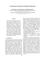
mutants that abolish Kid toxicity. According to this
model, Kid contains two symmetric and continuous
RNA-binding pockets, each involving residues of both
monomers (Fig. 1A). Residues E18 of one monomer
and R85 of the other are connected via a salt bridge.
Mutations in these residues subtly destabilize the struc-
ture of the toxin and abolish the toxicity of Kid [20].
Residues T46, S47, A55, F57, T69, V71 and R73 inter-
act with bases in the core sequence of the RNA sub-
strate (5¢-UAC-3¢) and contribute to the definition of
the specificity of the sequence recognized by the toxin
(Fig. 1B). Native MS showed that the toxin dimer
binds to a single RNA molecule [19], suggesting that
the second binding pocket is inactivated following
binding of the RNA substrate to the first. The model
proposes that residues D75, R73 and H17 are part of
the active site of the enzyme acting as a catalytic base,
A
B
C
Fig. 1. Graphic representation of Kid residues involved in RNA
binding specificity and cleavage. (A) Kid dimer with the residues
involved in RNA binding in blue. The analysed residues are indi-
cated. (B) Residues involved in the binding specificity. (C) Residues
involved in RNA cleavage. In (B) and (C) only the RNA bases of the
core sequence cleaved by the Kid toxin, UAC, are shown. Dotted
lines indicate the hydrogen bonds. Colour codes of the different
atoms are as follows: C, green; H, white; O, red and N, blue. Non-
analysed residues are shown in marine blue. The figure was
obtained using
PYMOL [36].
Analysis of Kid RNase model E. Diago-Navarro et al.
4974 FEBS Journal 276 (2009) 4973–4986 ª 2009 The Authors Journal compilation ª 2009 FEBS
catalytic acid and stabilizing residue, respectively
(Fig. 1C). Mutations in R73 and D75 that abolish Kid
toxicity have been reported previously [20]. Surpris-
ingly, R73 is not conserved among MazF and other
Kid homologues. The acidic residue at position 75 (D
or E), acting as a catalytic base, is present in MazF
and almost all other Kid-related toxins [21]. Interest-
ingly ChpBK, an homologous Kid toxin of the Escher-
ichia coli chromosome contains glutamine instead of
the acidic residue at this position and has reduced
endoribonuclease activity compared with MazF
[19,22]. A significant evaluation of the available model
on the interaction and cleavage of the RNA substrate
and the Kid toxin is of interest in itself because it is
the basis of important cellular roles of this toxin in
plasmid stabilization and the inhibition of cell growth;
it should also set an important point of reference for
comparisons with other toxins.
In this study, we evaluate the above model by test-
ing a limited, but significant, collection of specific
mutations in key residues of the protein and by analy-
sing in vitro their effects on RNA binding and RNA
cleavage using short RNA substrates and native MS
assays. Our analysis focuses on the protein residues
proposed to be involved in RNA binding and cleavage,
and strives to analyse the effect of mutations in these
residues on binding and cleavage at the 5¢-UAC-3 core
sequence using an in vitro approach. This core was
present at the highest frequency in RNA sequences
cleaved by PemK ⁄ Kid toxins in vitro and in vivo
[12,14,15]. Cleavage at this core occurred most fre-
quently between U and A. For our purpose, we
require short RNA substrates containing the above
core sequence. For the cleavage assays, we chose two
short RNAs, 5¢-AUACA-3¢ which, jointly with the
dinucleotide UpA, was the main substrate used to ana-
lyse the cleavage products of Kid, and 5¢-UUACU-3¢
which is a preferred target for Kid in vivo, as described
by Pimentel et al. [15], and which is also cleaved effi-
ciently by Kid in vitro [19]. Selection of these short
substrates allowed the use of MS in the cleavage
assays. RNA binding was assayed on 5¢-AdUACA-3¢,
the un-cleavable mimetic of 5¢-AUACA-3¢. This
mimetic RNA was used to obtain NMR data that sup-
ported the Kid–RNA structural model and it also
allowed us to establish the requirement for OH in the
2¢ position for RNA cleavage. The effects of the muta-
tions on toxicity and protein synthesis assays were also
tested. The results obtained are consistent with the
model’s predictions and show the important contribu-
tion of the T46 residue to RNA cleavage. These results
also show a good correlation between RNase activity,
protein synthesis inhibition and in vivo inhibition of
cell growth, underlining their relevance to our under-
standing of the basic activities of this toxin.
Results
Selection and isolation of Kid mutants in
residues involved in RNA binding and in RNA
cleavage
To evaluate the model’s predictions on residues
involved in RNA binding we selected and analysed
four Kid mutants: A55G, T46G, T69G and R85W.
A55, T46 and T69 establish hydrogen bonds (Fig. 1B,
dotted lines) and hydrophobic interactions with bases
of the core sequence 5¢-UAC-3¢ and they are proposed
to contribute to Kid–RNA binding specificity. Single
mutations in these residues could affect binding of the
toxin to the RNA substrate without inactivating its
RNase. However, because of the contribution made by
other residues to RNA binding specificity (see above),
single mutations in these residues may retain measur-
able RNA-binding potential. R85 does not interact
directly with bases at the core sequence 5¢-UAC-3¢.
However, it plays an important role in RNA binding
because it establishes a salt bridge with E18, connect-
ing the two monomers of the toxin, as required to
form the two RNA-binding pockets. KidR85W pre-
vents this salt bridge and locally distorts the structure
of the dimer [20]. Therefore, this mutation may have a
drastic effect on RNA binding which would explain its
highly reduced RNase activity [23].
As mentioned above, R73, D75 and H17 are pro-
posed to form part of the active centre of the toxin
(Fig. 1C). For a detailed analysis we selected the
mutants KidD75E, KidD75N, KidR73H and
KidH17P. These mutations should interfere with the
interactions required for catalysis and therefore have a
drastic effect on the RNase activity of the toxin and a
moderate or null effect on RNA binding.
Kid mutants suitable for the analysis should affect
specifically the RNA binding and ⁄ or cleavage activities
without altering other essential protein features and
functions, such as its structure, stability and potential
to interact with the antitoxin. The possible effects of
the mutations on the stability and structure of the pro-
tein were analysed by inmunoblotting and CD, respec-
tively. The potential of the Kid mutants to form a
functional complex with the Kis antitoxin was evalu-
ated by using native MS to test the formation of a
stable heterooctameric Kid
2
–Kis
2
–Kid
2
–Kis
2
complex
on the parD promoter [9]. We further analysed the
effects of the mutations on the co-regulatory activity
of the toxin, measuring their effect on the transcription
E. Diago-Navarro et al. Analysis of Kid RNase model
FEBS Journal 276 (2009) 4973–4986 ª 2009 The Authors Journal compilation ª 2009 FEBS 4975
of a parD–lacZ transcriptional fusion [24]. These
assays indicated that the different mutants maintain
the structural and functional features required to test
their specific involvement in RNA binding and ⁄ or
RNA cleavage activities (Figs S1–S3).
Mutations in residues proposed to be involved in
RNA interactions decrease RNA binding
The Kid mutants A55G, T69G, T46G, the double
mutants T46G ⁄ T69G and A55G ⁄ T69G, and R85W
affecting residues proposed to be involved in interac-
tions with the RNA substrate were evaluated in RNA
binding and cleavage assays.
To perform this analysis, we chose to use native MS
[25,26], a novel development in the field of MS using
relatively soft ionization of the sample by electrospray
ionization from solutions at physiological pH, which
enables the maintenance, detection and analysis of
macromolecular complexes. These protein complexes
are detected at different mass-to-charge ratios ( m ⁄ z),
separated by differences in their time-of-flight inside
the mass analyser. Here, we use this new powerful
technology to analyse complexes of the Kid toxin with
short RNA substrates, circumventing the inconve-
nience associated with more conventional methodolo-
gies (e.g. dissociation of the complexes when using
electrophoretic separation techniques). The MS analy-
sis is efficient and very sensitive, and it was particu-
larly useful for comparisons of the different mutational
variants of the same protein.
For RNA binding assays, a RNA–dU substrate that
could not be cleaved, 5¢-AdUACA-3¢, in which the
attacking OH of the ribose was replaced by a proton
H (deoxyribose), was used. This substrate was also
used to model the binding of Kid to the RNA, and
contains in its central core the bases at which cleavage
occurs in the target sequences identified previously
[12,14,15]. In all cases, analysis by native MS of sam-
ples containing equimolar concentrations of the toxin
(wild-type or mutants) and RNA binding substrate,
detected five peaks corresponding to different ioniza-
tion forms of the free dimeric toxin and also peaks
corresponding to the complex of the dimeric toxin and
a single RNA molecule (Fig. S4). Compared with Kid
wild-type protein, in which 18.4 ± 0.8% of the protein
was bound, a statistically significant decrease in the
relative binding was clear for KidA55G (11.9 ±
1.5%), KidT69G (12.3 ± 0.8%) and KidT46G
(13.4 ± 1.2%) (Fig. 2A). This indicates that A55, T69
and T46 residues make a significant contribution to
the RNA binding, but there are no significant differ-
ences between the binding strength of these mutated
proteins to the RNA substrate. For KidR85W, the
percentage of the protein–RNA complex with respect
to the free protein was drastically reduced
(6.8 ± 1.9%), indicating that the mutation efficiently
affected binding of the toxin to the RNA substrate.
MS analysis was also used to follow the activity of
Kid wild-type and mutant proteins on the cleavable
substrate 5¢-AUACA-3¢ used in the model [19], which
also contains the UAC core sequence. The progress of
the reaction over time was determined by measuring
the amount of uncleaved RNA remaining (Fig. 2B)
and the concomitant formation of RNA cleavage
products. Only products observed in all cases corre-
sponded to the expected species of a specific cleavage
(AU, 636.1 Da and ACA, 902.2 Da, data not shown),
thus indicating that the samples used were not contam-
inated with an unspecific RNase. Similar results were
obtained for the RNA 5¢-UUACU-3¢, but with this
substrate the assay required a 100-fold decrease in pro-
tein concentration, as reported previously [19]
(Fig. 2C). The expected cleavage products were found
in all reactions (Fig. S5), (UU, 614 Da and ACU,
880 Da), similarly indicating that samples were not
contaminated with a nonspecific RNase. The amount
of nonprocessed RNA obtained with KidA55G and
KidT69G decreased gradually over time, whereas the
RNA cleavage products increased concomitantly at the
same rate. The cleavage profiles obtained when
the 5¢-UUACU-3¢ substrate was used were quite similar
to those obtained with 5¢-AUACA-3¢ (Fig. 2B,C). This
indicates that these mutants retain substantial RNase
activity. However, in both cases, the levels of cleavage
obtained with KidA55G and KidT69G were lower
than those obtained with the wild-type protein, proba-
bly because of the effect of these mutations on RNA
binding. This interpretation is supported by the results
obtained with KidR85W: the interaction of KidR85W
and RNA was drastically reduced and this correlates
with the very low RNase activity of this mutant
(Fig. 2). Further analysis of this activity on longer
RNA substrates (CopT or CopA, which are RNA reg-
ulatory elements of R1 plasmid replication, and TAR,
a regulatory region of the RNA of the HIV virus)
show a highly reduced but detectable RNase activity in
this mutant [12] (data not shown) (see Discussion).
The T46G mutation also produced a drastic reduction
in the RNA cleavage on both short and full-length
RNA substrates, although substantial RNA binding
activity continued to be measured; possible alternative
explanations for this result are given in the Discussion.
The RNA binding and cleavage assays were also per-
formed with the double mutants KidA55G ⁄ T69G and
KidT46G ⁄ T69G affecting residues involved in specific
Analysis of Kid RNase model E. Diago-Navarro et al.
4976 FEBS Journal 276 (2009) 4973–4986 ª 2009 The Authors Journal compilation ª 2009 FEBS
interactions with the RNA. These double mutants, like
the Kid wild-type protein, interact efficiently with the
Kis antitoxin (data not shown) and form proper Kid–
Kis complexes (heterooctamers) at the promoter–oper-
ator region (see Fig. S2), showing that they maintain
the functional features required to test their specific
involvement in RNA binding and ⁄ or cleavage activi-
ties. We analysed the ability of these double mutants
to bind RNA, and the relative values found were
13.1 ± 0.8% for KidT46G ⁄ T69G and 13.9 ± 0.8%
for KidA55G ⁄ T69G, similar to values obtained with
the single mutants (13.4 ± 1.2% for T46G,
12.3 ± 0.8% for T69G and 11.9 ± 1.5% for A55G)
(Fig. 2A,D). All the data were statistically different
compared with the wild-type protein. Further differ-
ences were observed when the cleavage assay was per-
formed (Fig. 2B–F). The Kid protein containing the
double mutation A55G ⁄ T69G showed a further
decrease in the efficiency of RNA cleavage when com-
pared with Kid proteins containing the single muta-
tions. It was observed that this decrease was more
pronounced when the less-preferred 5¢ -AUACA-3¢
substrate was used; however, RNase activity was
clearly shown when the 5¢-UUACU-3¢ substrate was
used (Fig. 2F). The double mutant KidT46G ⁄ T69G,
like the KidT46G single mutant, prevented the cleav-
age of both short RNA substrates.
Mutations affecting catalytic residues of Kid
prevent RNA cleavage but not RNA binding
As indicated above, mutants KidR73H, KidD75E,
KidD75N and KidH17P affect residues proposed to be
involved directly in the cleavage of the RNA substrate.
The effects of these mutations on RNA-binding and
cleavage assays were evaluated.
A RNA binding assay of the different mutants was
performed using native MS, as indicated above. In all
cases, the relative binding percentages of KidD75E,
KidD75N, KidH17P and KidR73H (16.6 ± 1.1,
18.6 ± 1.1, 18.6 ± 1.0 and 17.5 ± 0.7, respectively)
were similar to that of the wild-type (18.4 ± 0.8%),
indicating that these mutations do not substantially
affect RNA binding (Fig. 3A). No statistically
significant differences from the wild-type protein were
found.
Fig. 2. Effect on RNA binding and cleavage of mutations in Kid residues, as measured by native MS (see Figs S4 and S5). RNA binding:
assays were performed with Kid wild-type, mutated proteins using a noncleavable mimetic RNA substrate (5¢-AdUACA-3¢). Protein and RNA
were added at 15 l
M. (A) and (D) show the percentage of protein bound to RNA relative to the total protein for Kid wild-type and Kid
mutants containing single or double mutations as indicated (rectangles). Bars indicate SD. RNA cleavage assays were performed using
proteins at 20 l
L and the cleavable RNA substrate, 5¢-AUACA-3¢,at50lM in (B) and (E), whereas in (C) and (F) the cleavable substrate
5¢-UUACU-3¢ was used at 50 l
M and the proteins were used at 0.2 lM. The amount of uncleaved RNA remaining at different times, with Kid
wild-type and mutant proteins is indicated. (B) and (C) show the line profiles obtained with single mutants, and (E) and (F) the profiles
obtained with the double mutants. SD for each value were calculated from three independent measures.
E. Diago-Navarro et al. Analysis of Kid RNase model
FEBS Journal 276 (2009) 4973–4986 ª 2009 The Authors Journal compilation ª 2009 FEBS 4977
The rates of cleavage of the cleavable RNA sub-
strate by the Kid wild-type and mutant proteins were
followed by MS, monitoring the amount of remaining
uncleaved RNA, 5¢-AUACU-3¢ and 5¢-UUACU-3¢,
over time (Fig. 3B,C). Compared with the wild-type
protein, a decrease in the uncleaved RNA over time
was not observed for all four mutants. A similar effect
was found with both substrates when the appropriate
protein concentration (0.2 lm for 5¢-UUACU-3¢ and
20 lm for 5¢-AUACU-3¢) was used. This indicates that
the mutations inactivate the RNase activity of the
toxin to a great extent. Analysis using longer RNA
substrates confirmed this inactivation (data not
shown).
On the whole, the results are consistent with the spe-
cific involvement of R73, D75 and H17 in the cleavage
reaction (see Discussion) and also indicate that this is
not because of the mutations having a significant effect
on the binding to the RNA substrate.
Protein synthesis and toxicity assays are
consistent with the above results
We tested the effects of the Kid mutations on protein
synthesis by monitoring Luciferase synthesis in E. coli
cell extracts (see Materials and methods). Protein syn-
thesis was inhibited by the wild-type Kid protein, the
KidT69G mutant and to a lesser extent by KidA55G
(Fig. 4). The double mutant KidA55G ⁄ T69G was also
able to inhibit protein synthesis but to a lesser extent
than the single mutants, even when the highest protein
concentration was used (0.6 lm). This is consistent
with the fact that these mutants, which partially affect
RNA binding, do not abolish the RNase activity of
the toxin. A different result was obtained with
Kid mutants KidR73H, KidD75E, KidD75N and
KidH17P, which affect residues in the catalytic centre.
These mutations abolished the potential of the toxin to
inhibit protein synthesis. The same result was obtained
for the KidR85W mutant protein (Fig. 4), which is
consistent with a drastic reduction in RNA binding
and RNase activity in this mutant (see Discussion).
KidT46G was not able to inhibit protein synthesis,
which is consistent with its failure to cleave RNA.
Similarly, the double mutant KidT46G ⁄ T69G was also
unable to inhibit protein synthesis.
Fig. 3. RNA binding and cleavage of Kid mutants affected in resi-
dues in the catalytic centre. (A) RNA binding: assays were carried
out by native MS. The uncleavable RNA (5¢-AdUACA-3¢) was incu-
bated for 2 min with Kid wild-type or mutated proteins. RNA and
proteins were added at 15 l
M and the ratios of RNA bound protein
to free protein obtained for the different mutants (rectangles) were
determined. Bars show the SD obtained for the wild-type or mutant
proteins from three independent assays. (B) RNA cleavage assays
were performed using proteins at 20 l
M when the cleavable sub-
strate 5¢-AUACA-3¢ was used at 50 l
M. (C) RNA cleavage assays
with 50 l
M of the cleavable substrate 5¢-UUACU-3¢ and 0.2 lM of
proteins. The amount of uncleaved RNA remaining at different
times after the addition of Kid wild-type or mutant proteins is
indicated. The profiles obtained for the different mutants are indi-
cated. SD for each value were calculated from three independent
measures.
Analysis of Kid RNase model E. Diago-Navarro et al.
4978 FEBS Journal 276 (2009) 4973–4986 ª 2009 The Authors Journal compilation ª 2009 FEBS
We analysed the effects of the above mutants on the
growth and viability of the host. For this purpose, the
different mutations were introduced by site-directed
mutagenesis into multicopy parD recombinant vectors
pBR1120 or pAB1120. These vectors carry an amber
mutation in the Kis antitoxin (kis74) and they were
established at 30 °C in OV2, a thermosensitive amber
suppressor (supFts) strain. In this background, a func-
tional antitoxin is synthesized at 30 °C, whereas at
42 °C an inactive antitoxin with the last 13 residues
removed is synthesized. Therefore, the effect of the
toxin on cell growth or cultivability can be monitored
at 42 °C. Analysis showed that at 30 °C, cultures
expressing the different Kid mutant proteins affecting
the proposed catalytic or RNA binding residues grew
with similar efficiency and viability. At 42 °C, cells
expressing the non-neutralized Kid proteins carrying
mutations in the catalytic residues grew normally
(Fig. 5). As expected, the growth of cells expressing
the wild-type toxin was clearly affected. T69G and
A55G mutations showed a similar inhibitory effect,
despite differences in their potential to inhibit protein
synthesis and, in addition, their inhibitory effects were
greater than that of the wild-type (see Discussion). A
different situation was found in cells carrying the
recombinant containing the R85W mutation. As
shown above, this mutation drastically affected Kid
RNA binding and, as previously reported [20], the
KidR85W toxin did not inhibit cell growth. Consistent
with the above results, KidT46G and KidT46G ⁄ T69G
did not affect cell growth or viability (Fig. 5). The
double mutant KidA55G ⁄ T69G showed a milder effect
on cell growth than either of the single mutants,
which is consistent with the RNA cleavage and protein
synthesis assays.
Discussion
In this study, we evaluated the roles assigned by the
available model to particular residues of Kid involved
in RNA binding or cleavage [19]. As mentioned above,
for the cleavage assays we chose two short RNAs:
5¢-AUACA-3¢, previously used to analyse the cleavage
products of Kid [19]; and 5¢-UUACU-3¢, a preferred
target of Kid in vivo and in vitro [15,19]. Selection of
these short substrates allowed us to use MS in the
Kid–RNA binding and cleavage assays. 5¢-AdUACA-3¢,
Fig. 5. Cell cultivability of strains containing different Kid mutants.
OV2 strain containing kid wild-type or the different kid mutants
were grown at 30 °C to mid-logarithmic phase (D
600
= 0.35) and
equal volumes of serial dilutions were spotted in plates containing
the appropriate antibiotic (tetracycline or kanamycine). Growth of
the spotted samples after 16 h of incubation at 30 or 42 °Cis
shown.
Fig. 4. Protein synthesis assays with the different mutants. Effect
of the Kid wild-type and mutant proteins (0.15, 0.3, 0.6 l
M in each
case) on the synthesis of a [
35
S]methionine-labelled Luciferase in
an in vitro transcription–translation assay. C+ shows the positive
controls with buffer, C) the negative controls with chloramphenicol
(1 lgÆlL
)1
), the remaining lanes show assays carried out in the
presence of different concentrations of Kid wild-type, KidA55G,
KidT69G, KidT46G, KidT46 ⁄ GT69G and KidA55G ⁄ T69G, KidD75E,
KidD75N, KidR73H, KidH17P and KidR85W proteins.
E. Diago-Navarro et al. Analysis of Kid RNase model
FEBS Journal 276 (2009) 4973–4986 ª 2009 The Authors Journal compilation ª 2009 FEBS 4979
the un-cleavable mimetic of 5 ¢ -AUACA-3¢ was used in
the binding assays. For the analysis, we selected four
single mutants of Kid, A55G, T69G, T46G and
R85W, and two double mutants, A55G ⁄ T69G and
T46G ⁄ T69G, which affect residues proposed to be
involved in RNA binding. Four other mutants, R73H,
D75E, D75N and H17P, which affect residues pro-
posed to form part of the catalytic centre of Kid were
also selected (Fig. 1). Because these mutations do not
substantially alter the stability or secondary structure
of the Kid toxin and maintain its capacity to interact
with the Kis antitoxin and form a functional repressor,
they seem appropriate for evaluation of their specific
effects on RNA binding and RNA cleavage.
A55 and T69 confer specificity to the interaction
with RNA because they establish hydrogen bonds with
bases at the RNA core sequence recognized by Kid
(Fig. 1B, dotted lines). They are located in flexible
regions of the toxin (Fig. 1A). Substitution of these
residues by glycine abolished interactions with the
bases without disturbing the structure of the flexible
region in which they are located. The fact that these
substitutions affect RNA binding in a clear way with-
out preventing cleavage of the RNA substrate is con-
sistent with the proposal that these residues play an
important and specific role in RNA binding. A
decrease in cleavage efficiency was observed, probably
as an indirect result of less efficient binding to the sub-
strate. This decrease was similar in both mutated pro-
teins. Consistent with the above analysis, it was found
that the mutations conserve the ability of the toxin to
inhibit protein synthesis and show expected effects on
cell growth and viability. KidA55G seems to inhibit
protein synthesis to a lesser extent than KidT69G, but
this is not reflected by differences in cell growth. In
addition, inhibition of cell growth is more pronounced
in both mutants than in the wild-type protein. Because
the system used to assay Kid toxicity depends on inac-
tivation of the Kis antitoxin at 42 °C, it cannot be dis-
counted that these differences are be caused by
unknown complexities related to this assay.
KidT46G shows an effect on RNA binding of Kid
similar to KidA55G and KidT69G, but unlike these
mutations it shows drastic inhibition of RNA cleavage.
Results obtained on the larger RNA substrates show
residual RNase activity that does not indicate changes
in cleavage specificity. Because the mutation should
extend to the adjacent S3–S4 loop (residues 47–57),
which is a dynamic region of the protein (M.B. Kam-
phuis, unpublished data), a plausible hypothesis is that
it may allow adjacent residues to interfere with others
on the active site. A possible alternative is that T46G
may interfere with correct binding of the RNA
substrate and that this could allow RNA binding but
prevent efficient RNA cleavage. T46 is highly con-
served in the alignment [21], which may suggest its
possible relevance in the specific recognition of the
substrate.
A drastic effect on RNA binding was found for
KidR85W. R85 stabilizes the RNA binding pocket by
forming a salt bridge with E18. R85W mutation abol-
ishes this salt bridge causing disruption of the binding
pocket [20], loss of the positive charge of R85 and full
exposure to the negative charge of E18 [20]. This, in
turn, may explain the very poor activity of this toxin
as an RNase. In addition, local distortion in the S1–S2
loop comprising residues 11–21 may also contribute to
this poor activity because this loop includes the H17
residue which is proposed to play a stabilizing role in
RNA cleavage. Previous RNase assays in solution with
larger RNA substrates (TAR, CopA and CopT) show
that, although with poor efficiency, the KidR85W
mutant can cleave RNA with the correct specificity;
this is consistent with the proposal that the mutation
does not completely prevent the RNase activity of Kid
or alter the cleavage specificity. As reported previously,
the R85W mutation impairs the toxicity of the Kid
protein. The decrease in RNase activity seen in pure
solutions was undetectable in whole-cell extracts of
E. coli [12], which is consistent with the effect of the
mutation on toxicity. The reasons for the differences
found in pure solutions and whole cells or in cell-free
extracts remain to be established.
Mutations R73H, D75N, D75E and H17P clearly
affect RNA cleavage without substantially altering
RNA binding. The relative positions and functions
that R73, D75 and H17 of Kid play to cleave the scis-
sile phosphate (catalytic acid, catalytic base and stabi-
lizing interaction) are equivalent to those of residues at
the active sites of RNaseA and RNase T1 [19]. The
mutations analysed should disrupt the critical interac-
tions of the three key residues. (a) R73H: arginine and
histidine are monocarboxylic acids with amine bases,
but the size and stereochemistry of the two lateral
chains are quite different, which prevents the effective
substitution of the two amine bases of arginine 73 by
the two amines of histidine. In addition to act as a
catalytic acid, R73 can play a second function in RNA
cleavage: reducing the pK
a
of the 2¢-OH group by
donating a charged hydrogen bond to the 2¢-O. This
can be accomplished by a single arginine, but not by
just one histidine. Note that although this residue was
proposed to contribute to the specificity of binding to
the core sequence [19], we could not measure an effect
of the mutation on RNA binding. This suggests
that the residue does not play a relevant role in this
Analysis of Kid RNase model E. Diago-Navarro et al.
4980 FEBS Journal 276 (2009) 4973–4986 ª 2009 The Authors Journal compilation ª 2009 FEBS
binding, or that the histidine amines can fulfil this
additional role of R73. (b) D75N: aspartic acid and
asparagine are, respectively, a dicarboxilic acid and its
amide. The stereochemistry of both residues might be
equivalent but the mutation changes the acidic charac-
ter of D75 which is required for its proposed role as
the catalytic base. (c) D75E: aspartic and glutamic
acids are dicarboxylic acids, but glutamic acid has an
additional carbon in the lateral chain. The clear effect
of this change in the RNase activity indicates that even
if the acidic character is conserved, the length of the
lateral chain is important to establish the necessary
catalytic interactions. Using longer and well-character-
ized substrates such as TAR (the regulatory region
of HIV), CopA and CopT (two RNAs involved in
copy number control of plasmid R1) we found that
this mutant has residual but specific RNase activity
(data not shown); this indicates that the acidic resi-
due may play a catalytic role, although far less effi-
ciently than D75. Thus the two substitutions in this
residue are consistent with the proposed role of D75
as the catalytic base. (d) H17P changes the pyrrolic
ring of histidine, which includes the amine that
establishes a hydrogen bond with the oxygen of the
scissile phosphate, for the heterocyclic ring of proline
containing three uncharged CH
2
residues; this sub-
stitution prevents the required hydrogen-bond for-
mation proposed by the model. These results are
consistent with the essential roles assigned to these
residues in the available model. In particular, the
two substitutions in D75 strongly support its role as
catalytic acid.
It should be taken into account that translation
factors or the translation process itself may influence
the mode of action or the accessibility to the target
of related RNase toxins. In the case of the YafQ
toxin, the target found in vivo is in inframe codons
of lysine, whereas in vitro the toxin cuts close to a
GG pair [27]. The translation process itself has been
shown to increase the accessibility to the targeted
sequences for the MazF toxin [28]. Finally, the releas-
ing factor RF1, which competes with the action of
the RelE toxin in vitro [29], is also involved in the
toxicity mediated by both the RelE and the Kid tox-
ins; this was revealed by the extra sensitivity of prfA
mutants to these toxins [30]. Further work is required
to determine the interactions involved in this extra
sensitivity.
From the work of Pimentel et al. [15], it seems quite
clear that preferential cleavage by Kid of the copB–
repA mRNA of plasmid RI at the 5¢-UUACU-3¢
sequence is very important to fine tuning the CopB ⁄
RepA ratio and the replication efficiency of the plas-
mid. Cleavage at these sequences in other mRNAs
may have an important role in the protein synthesis
and cell growth inhibition mediated by this toxin.
5¢-UUACU-3¢ is not the only sequence targeted in vivo
by the Kid ⁄ PemK toxin. Zhang et al. [14] reported the
cleavage of RNA by PemK in vivo at 5¢-CUACU-3¢
and 5¢-CUACG-3¢, both having the 5¢-UAC-3¢ core
sequence found in 5¢-UUACU-3¢. An interesting point
in this context is the possible functional relevance of
cleavage by this toxin at less favourable sites contain-
ing the core sequence. It remains to be evaluated if this
represents a way of regulating the action of the toxin.
The data reported by Zhang et al. that cleavage by
PemK can occur at the 5¢ or 3¢ A in the core sequence,
adds complexity to this repertory of sites and remains
to be explained at the mechanistic level.
To summarize, our results are consistent with the
functions assigned in the available model to R73,
D75 and H17 of Kid as catalytic residues involved in
RNA cleavage and the role of T46, A55, T69 and
R85 in toxin–RNA binding. In addition, they reveal
the unexpected importance of T46 in RNA cleavage.
The data are also consistent with similar modes of
action in Kid, RNase A and RNase T1, as proposed
previously [19], and give information on key Kid
toxin residues involved in its RNase activity. The
results further support the interrelations between the
toxicity of the Kid protein, its RNase activity and its
potential to inhibit protein synthesis. Because the
RNase activity of the protein is involved in plasmid
stability, we can predict that the mutations analysed
will also affect this toxin role. Our results offer clues
for comparison of the residues involved in the
specificity of RNA cleavage within the toxin family
and for the design of RNases based on the different
cleavage efficiencies of Kid.
Materials and methods
Bacterial strains
The bacteria used in this study were E. coli K12 strains:
OV2 (F, leu, thyA(deo), ara (am), lac-125 (am), galU42,
galE, trp (am), tsx (am), tyr (supF(ts)A81), ile, his), as a
host for the plasmids pAB1120 and pBR1120 derivatives;
TG1 (supE, D(lac-proB), thi1, hsdD5, F¢ (traD36, lacI
q
, lac-
ZM15, proAB
+
)), was used for protein over production;
MLM373 (D(lac, pro), supE,thi) [20] was used for b-galac-
tosidase assays.
Plasmids used and constructed
The plasmids used and constructed are listed in Table 1.
E. Diago-Navarro et al. Analysis of Kid RNase model
FEBS Journal 276 (2009) 4973–4986 ª 2009 The Authors Journal compilation ª 2009 FEBS 4981
Derivatives of pRG–his–KisKid, pAB24 and pBR1120
were constructed by site-directed mutagenesis using the
primers listed in Table 2 and QuikChange
Ò
Site-Directed
Mutagenesis or QuikChange
Ò
XL Site-Directed Mutagene-
sis Stratagene kits (La Jolla, CA, USA).
Proteins, DNA and RNA
Kid toxin, Kid mutants and His-tagged Kis were overex-
pressed from plasmids of the type pRG–his–KidKid. Purifi-
cation was performed with a protocol identical to that
Table 1. Plasmids used in this study.
Plasmid Description References
pAB1120 pAB112 (R1), parD (kis74amb,kid
+
), copB-,Km
R
[34]
pAB 24 pKN1562 y pBR322 (pMB9), parD
+
(kis, kid), Tc
R
[3]
pBR322 pMB9, Tc
R
,Ap
R
[35]
pAB17 pKN1562, kis17,Km
R
[3]
pRG–his–KisKid pRG-recA-Nhis, precA::his
6
:: parD
+
,Ap
R
R. Sabariegos-Jaren˜ o (unpublished data)
pRG–his–KisKidD75N pRG-recA-Nhis, precA::his
6
:: kis, kidD75N This study
pRG–his–KisKidD75E pRG-recA-Nhis, precA::his
6
:: kis, kidD75E This study
pRG–his–KisKidH17P pRG-recA-Nhis, precA::his
6
:: kis, kidH17P This study
pRG–his–KisKidR73H pRG-recA-Nhis, precA::his
6
:: kis, kidR73H This study
pRG–his–KisKidA55G pRG-recA-Nhis, precA::his
6
:: kis, kidA55G This study
pRG–his–KisKidT69G pRG-recA-Nhis, precA::his
6
:: kis, kid T69G This study
pRG–his–KisKidE5G pRG-recA-Nhis, precA::his
6
:: kis, kid E5G This study
pAB24–D75N pAB24 (kis, kidD75N) This study
pAB24–D75E pAB24 (kis, kidD75E) This study
pAB24–H17P pAB24 (kis, kidH17P) This study
pAB24–R73H pAB24 (kis kidR73H) This study
pAB24–A55G pAB24 (kis kidA55G) This study
pAB24–T69G pAB24 (kis kid T69G) This study
pAB24–E91K pAB24 (kis kidE91K) [24]
pAB24–R85W pAB24 (kis kid R85W) J. Lo
´
pez-Villarejo (unpublished data)
pMLM132 pparD::lacZ,Tc
R
[20]
pBR322–1120 pBR322, parD (kis74amb,kid
+
), Cm
R
S. Santos-Sierra (unpublished data)
pBR322–1120–D75E pBR322-1120, kis, kidD75E This study
pBR322–1120–H17P pBR322-1120, kis, kidH17P This study
pBR322–1120–R73H pBR322-1120, kis, kidR73H This study
pBR322–1120–T46G pBR322-1120, kis, kid T46G This study
pBR322–1120–A55G pBR322-1120, kis, kidA55G This study
pBR322–1120–T69G pBR322-1120, kis, kid T69G This study
pBR322–1120–T46G ⁄ T69G pBR322-1120, kis, kid T46GT69G This study
pBR322–1120–A55G ⁄ T69G pBR322-1120, kis, kidA55GT69G This study
pB24 pBR322-1120, kis, kidR85W [24]
pAB1120-D75N pAB1120, kis74amb, kid D75N [20]
Table 2. Primers used in this study.
Name Sequence (5¢-to3¢) Description
PD75E()) TTGTACGTTGCGAACAACCCCGGACAAT Change GAT–GAA in D75 (kid D75E)
PD75E(+) ATTGTCCGGGGTTGTTCGCAACGTACAA Change ATC–TTC in D75 (kid D75E)
PD75N()) TTGTACGTTGCAATCAACCCCGGACAAT Change GAT–AAT in D75 (kid D75N)
PD75N(+) ATTGTCCGGGGTTGATTGCAACGTACAA Change ATC–TTA in D75 (kid D75N)
PR73H()) ACCACAGGTGTTGTACATTGCGATCAACC Change CGT–CAT in R73 (kid R73H)
PR73H(+) GGTTGATCGCAATGTACAACACCTGTGGT Change ACG–ATG in R73 (kid R73H)
PH17P()) TCCTACCGCAGGTCCTGAGCAGCAGGGA Change CAT–CCT in H17 (kid H17P)
PH17P(+) TCCCTGCTGCTCAGGACCTGCGGTAGGA Change ATG–AGG in H17 (kid H17P)
PA55G()) TTTGCCCGCACTGGCGGCTTTGCGGTGTC Change GCC–GGC in A55 (kid A55G)
PA55G(+) GACACCGCAAAGCCGCCAGTGCGGGCAAA Change GGC–GCC in A55 (kid A55G)
PT69G()) TTGGCATACGTACCACAGGTGTTGTAC Change ACA–GGA in T69 (kid T69G)
PT69G(+) GTACAACACCTCCGGTACGTATGCCAA Change TGA–TCC in T69 (kid T69G)
Analysis of Kid RNase model E. Diago-Navarro et al.
4982 FEBS Journal 276 (2009) 4973–4986 ª 2009 The Authors Journal compilation ª 2009 FEBS
described previously [19,21]. The concentration of the pro-
teins was calculated spectroscopically, taking into account
their extinction coefficients.
For DNA binding assays a double-stranded DNA frag-
ment of 175 bp was used which includes the parD opera-
tor–promoter region; this fragment was obtained by PCR
amplification, as described previously [9], using as the
template a pUC18 recombinant containing the parD
sequences in a Sau3A fragment of mini R1 plasmid
pKN1562.
For MS studies, a 30-bp DNA fragment which contains
the region I of the parD promoter was used [9].
The single-stranded five-nucleotide RNA (5¢-UUACU-3¢,
5¢-AUACA-3¢), RNA-dU (5¢-AdUACA-3¢) oligonucleotides
were obtained from Eurogentec S.A. (Liege, Belgium). A
10 mm stock solution in H
2
O was prepared.
Western blot assays
TG1 strains containing the different pRG–his–KisKid
plasmids were grown in rich medium. At D
600
0.3 the
cultures were induced with nalidixic acid (25 lgÆmL
)1
).
After 4 h of induction, 2 mL of culture was collected,
centrifuged and resuspended in 200 lL of lysis buffer
(0.05 m Tris ⁄ HCl, pH 6.8, 10% w ⁄ v SDS, 0.01 m EDTA,
25% w ⁄ v glycerol, 0.5 g
)1
bromophenol blue, 5% v ⁄ v
b-mercaptoethanol). D
600
was adjusted to 0.1 and identi-
cal samples were incubated for 10 min at 100 °C, loaded
on a denaturing SDS ⁄ PAGE with 15% polyacrylamide,
and the proteins separated by electrophoresis. The pro-
teins were transferred onto a poly(vinylidene difluoride)
membrane using Trans-Blot Semi-Dry Transfer Cell appa-
ratus (Bio-Rad Laboratories, Hercules, CA, USA). After
blocking the membrane overnight with 10% non-fat dried
milk in TBST (137 mm NaCl, 20 mm Tris ⁄ HCl pH 7.5,
0.1% Tween), it was incubated for 60 min with anti-Kid,
anti-Kis or anti-DnaK sera in TBST. The membrane was
washed with TBST and incubated with an anti-rabbit
IgG bound to horseradish peroxidase (Amershan Bio-
sciences, GE Healthcare, Chalfont St Giles, UK) for
60 min followed by an additional washing step. The pro-
teins labelled with the antibody were revealed using an
ECL detection kit and detected by autoradiography
(AGFA Healthcare NV, Mortsel, Belgium). The mem-
brane was reprobed by using different primary sera (anti-
Kis, anti-Kid or anti-DnaK) after striping the previous
signal (striping buffer described in ECL Plus; Amersham)
and blocking the membrane as previously indicated.
b-Galactosidase activity assays
For this experiment, MLM373 strain bearing pAB24 deriv-
ative plasmids and pMLM132 reporter plasmid (Table 1)
were grown at 37 °C with shaking in LB medium contain-
ing tetracycline (10 lgÆlL
)1
) and chloramphenicol
(20 lgÆlL
)1
) until the cultures reached the mid-exponential
phase. Levels of b-galactosidase expression were monitored
as described previously [31]. Three independent values were
obtained for each strain.
Macromolecular mass spectrometry
MS studies were carried out in aqueous ammonium acetate
(100 mm, pH 7.0). Kid:Kis molar ratios were 2 : 1, 1 : 1
and 1 : 2. The lowest concentration was 10 lm. Samples
were incubated at 20 °C for 1 min. In Kid:Kis:DNA bind-
ing assays, a 30-bp fragment containing the perfect palin-
drome overlapping the )10 region of the promoter was
used. The molar ratios of Kid : Kis : DNA in the experi-
ment were 5 : 5 : 1. To monitor the binding of Kid to
RNA, proteins and RNA were used at 15 lm. For RNA
cleavage assays, proteins were used at 0.2 lm (for
5¢-UUACU-3¢)or20lm (for 5¢-AUACA-3¢) and RNAs at
50 lm. Nanoflow electrospray capillaries with an orifice of
5 lm were made of borosilicate glass capillaries (Kwik-
Fil, World Precision Instruments, Inc., Sarasota, FL, USA)
using a P-97 puller (Sutter Instrument Co., Novato, CA,
USA) and coated with a thin layer of gold ( 500 A
˚
) using
an Edwards ScanCoat Six Pirani 501 sputter coater
(Edwards High Vacuum International, Crawley, UK).
Native MS experiments were performed as described
previously [9] using a nanoflow electrospray ionization
orthogonal time-of-flight mass spectrometer (Micromass
LC-T; Waters, Manchester, UK) modified for high mass
operation and operating in positive ion mode [32]. To
monitor RNA cleavage by Kid protein a robotic chip-
based ESI source (Nanomate; Advion Biosciences, Ithaca,
NY, USA) was coupled to LC-T spectrometer. The ESI
source was programmed to aspirate 2 lL, as described pre-
viously [33]. The samples were measured with a scan time
of 2 s for a total of 10 min per sample. RNA binding
properties of the different mutants were monitored with an
LC-T spectrometer.
MS data analysis
MS data of the different mutants were semiquantified to
determine the relative binding percentage of the Kid dimer
protein to one molecule of RNA. Data were accumulated
over 2 min, averaged, smoothed and centred to obtain the
area values using the software program masslynx 4.0
(Waters). Total ion intensity for all the protein present was
calculated by summing the intensity of all ions belonging to
the Gaussian charge state envelope of the bound and
unbound protein under study; bound protein was calculated
by summing the intensity of the ions belonging to the
Gaussian charge state envelope of the bound protein. The
percentage of protein bound to RNA was the ratio between
the value of bound protein and the total protein present in
the sample. The relative percentage of binding was based
E. Diago-Navarro et al. Analysis of Kid RNase model
FEBS Journal 276 (2009) 4973–4986 ª 2009 The Authors Journal compilation ª 2009 FEBS 4983
on three independent measurements, using as a measure of
error their standard deviation.
Semiquantification of RNA products after cleavage by
Kid was performed in different experiments. The 10 min
data acquisitions were accumulated over 30 or 60 s, aver-
aged, smoothed and centred, to obtain area values using
the software program masslynx 4.0 (Waters). Total ion
intensity for each product was calculated by summing the
intensity of all ions belonging to the Gaussian charge state
envelope of the products under analysis and this value was
added to that obtained for the non-processed RNA to give
the intensity of the total RNA present in the measurement.
The amount of intact RNA and RNA products was based
on three independent measurements, using the standard
deviation as the errors bars.
Statistical analysis
All the data are represented by at least three independent
measurements. For the significance of the RNA binding data,
a Levene statistical was used for acceptance of variance
equality. One-way ANOVA and Bonferroni analyses were
used to evaluate the data to a 95% level of statistical signifi-
cance.
Protein synthesis assays
Reaction mixtures (10 lL) contained components from the
E. coli S30 Extract System for Circular DNA (Promega
Corporation, Madison, WI, USA): 4 lL S30 premix with-
out amino acids, 3 lL S30 extract, circular, 1 lL amino
acids minus methionine (1 mm), 3 lCi of [
35
S]methionine
and pBESTluc plasmid DNA (400 ng). The assays were
started by adding chloramphenicol 1 lgÆlL
)1
as a negative
control or 1 lL of purified Kid proteins diluted in 20 mm
Hepes, 100 mm KCl (6, 3 and 1.5 lm), followed by incuba-
tion for 60 min at 37 °C. The results were analysed by
SDS ⁄ PAGE (10%).
Toxicity assays
OV2 cells containing pBR1120 or pAB1120 derivative plas-
mids bearing the different mutations were grown at 30 °C
to the mid-exponential phase in LB medium containing tet-
racycline (10 lgÆlL
)1
) or kanamycin (50 lgÆlL
)1
). These
cultures were serially diluted and 7 lL of the undiluted cul-
tures and of each subsequent dilution were spotted (10
)1
steps from left to right) onto two plates of the same solidi-
fied medium. The plates were incubated at 30 °C (active
antitoxin Kis) or 42 °C (inactive antitoxin Kis74) overnight.
A semiquantitive assessment of the relative levels of toxicity
of the different mutants was derived by comparing the
number of colonies and growth of the spots at the different
levels of dilution.
CD spectroscopy
CD measurements and thermal denaturation were carried
out with 15 lm of Kid and Kid mutant proteins, as previ-
ously described [20]. Deconvolution analyses of the profiles
obtained were carried out using somcd (http://geneura.
ugr.es/cgi-bin/somcd/som.cgi?start=1).
Acknowledgements
RDO was supported by Project BFU2005-03911 from
the Spanish Ministry of Education and Science
(MEC, Spain), BFU 2008-01566 ⁄ BMC and CSD2008-
00013 from the Ministry of Science and Innovation
(MICIIN, Spain) and by a networking project of the
CM (COMBACT, Comunidad de Madrid, Spain).
EDN acknowledges the contribution of a predoctoral
fellowship (BFI05.35) from the Basque Country Gov-
ernment, Spain and of a short term EMBO fellowship
(ASTF No: 159-06) to visit and work at the Bio-
molecular Mass Spectrometry and Proteomics group
at Utrecht University, the Netherlands. The technical
assistance of Alicia Rodriguez-Bernabe
´
and discus-
sions with Marc Lemonnier, Ana Marı
´
a Hernandez-
Arriaga and Juan Lo
´
pez-Villarejo, are kindly
acknowledged. RB, AJRH, and MBK acknowledge
support from the Netherlands Organization for
Chemical Research (NWO ⁄ CW) and the Center for
Biomedical Genetics. RHH vdH was supported by a
VENI fellowship (700.54.402) from The Netherlands
Organization for Scientific Research (NWO). This
work in Utrecht was also supported by the Nether-
lands Proteomics Centre.
References
1 Ogura T & Hiraga S (1983) Mini-F plasmid genes that
couple host cell division to plasmid proliferation. Proc
Natl Acad Sci USA 80, 4784–4788.
2 Gerdes K, Rasmussen PB & Molin S (1986) Unique
type of plasmid maintenance function: postsegregational
killing of plasmid-free cells. Proc Natl Acad Sci USA
83, 3116–3120.
3 Bravo A, de Torrontegui G & Diaz R (1987) Identifica-
tion of components of a new stability system of plas-
mid R1, ParD, that is close to the origin of replication
of this plasmid. Mol Gen Genet 210, 101–110.
4 Pandey DG & Gerdes K (2005) Toxin–antitoxin loci
are highly abundant in free-living but lost from
host-associated prokaryotes. Nucleic Acids Res 55,
78–89.
5 Gerdes K & Wagner EG (2007) RNA antitoxins. Curr
Opin Microbiol 10, 117–124.
Analysis of Kid RNase model E. Diago-Navarro et al.
4984 FEBS Journal 276 (2009) 4973–4986 ª 2009 The Authors Journal compilation ª 2009 FEBS
6 Gerdes K, Christensen SK & Lobner-Olensen A (2005)
Prokaryotic toxin–antitoxin stress response loci. Nat
Rev Microbiol 3, 371–382.
7 Van Melderen L & Saavedra De Bast M (2009) Bacte-
rial toxin–antitoxin systems: more than selfish entities?
PLoS Genet 5, e1000437, doi:10.1371/journal.pgen.
1000437.
8 Ruiz-Echevarria MJ, Berzal-Herranz A, Gerdes K &
Diaz-Orejas R (1991) The kis and kid genes of the parD
maintenance system of plasmid R1 form an operon that
is autoregulated at the level of transcription by the
co-ordinated action of the Kis and Kid proteins. Mol
Microbiol 5, 2685–2693.
9 Monti MC, Hernandez-Arriaga AM, Kamphuis MB,
Lopez-Villarejo J, Heck AJ, Boelens R, Diaz-Orejas R
& Van den Heuvel RH (2007) Interactions of Kid–Kis
toxin–antitoxin complexes with the parD operator–
promoter region of plasmid R1 are piloted by the Kis
antitoxin and tuned by the stoichiometry of Kid–Kis
oligomers. Nucleic Acids Res 35, 1737–1749.
10 Ruiz-Echevarria MJ, de-la-Cueva G & Diaz-Orejas R
(1995) Translational coupling and limited degradation
of a polycistronic messenger modulate differential gene
expression in the parD stability system of plasmid R1.
Mol Gen Genet 248, 599–609.
11 Tsuchimoto S, Nishimura Y & Ohtsubo E (1992) The
stable maintenance system pem of plasmid R100: degra-
dation of PemI protein may allow PemK protein to
inhibit cell growth. J Bacteriol 174, 4205–4211.
12 Munoz-Gomez AJ, Lemonnier M, Santos-Sierra S,
Berzal-Herranz A & Diaz-Orejas R (2005) RNase ⁄ anti-
RNase activities of the bacterial parD toxin–antitoxin
system. J Bacteriol 187, 3151–3157.
13 Tsuchimoto S, Ohtsubo H & Ohtsubo E (1988) Two
genes, pemK and pemI, responsible for stable mainte-
nance of resistance plasmid R100. J Bacteriol 170,
1461–1466.
14 Zhang J, Zhang Y, Zhu L, Suzuki M & Inouye M
(2004) Interference of mRNA function by sequence-
specific endoribonuclease PemK. J Biol Chem 279,
20678–20684.
15 Pimentel B, Madine MA & de la Cueva-Mendez G
(2005) Kid cleaves specific mRNAs at UUACU sites to
rescue the copy number of plasmid R1. EMBO J 24,
3459–3469.
16 Ruiz-Echevarria MJ, de-la-Torre MA & Diaz-Orejas R
(1995) A mutation that decreases the efficiency of
plasmid R1 replication leads to the activation of parD,
a killer stability system of the plasmid. FEMS Microbiol
Lett 130, 129–135.
17 Steyaert J (1997) A decade of protein engineering on
ribonuclease T1 – atomic dissection of the enzyme–
substrate interactions. Eur J Biochem 247, 1–11.
18 Lacadena J, Martinez del Pozo A, Lacadena V,
Martinez-Ruiz A, Mancheno JM, Onaderra M &
Gavilanes JG (1998) The cytotoxin alpha-sarcin behaves
as a cyclizing ribonuclease. FEBS Lett 424, 46–48.
19 Kamphuis MB, Bonvin AM, Monti MC, Lemonnier M,
Munoz-Gomez A, Van den Heuvel RH, Diaz-Orejas R
& Boelens R (2006) Model for RNA binding and the
catalytic site of the RNase Kid of the bacterial parD
toxin–antitoxin system. J Mol Biol 357, 115–126.
20 Santos-Sierra S, Lemonnier M, Nunez B, Hargreaves
D, Rafferty J, Giraldo R, Andreu JM & Diaz-Orejas R
(2003) Non-cytotoxic variants of the Kid protein that
retain their auto-regulatory activity. Plasmid 50
,
120–130.
21 Hargreaves D, Santos-Sierra S, Giraldo R, Sabariegos-
Jareno R, de la Cueva-Mendez G, Boelens R, Diaz-Ore-
jas R & Rafferty JB (2002) Structural and functional
analysis of the kid toxin protein from E. coli plas-
mid R1. Structure 10, 1425–1433.
22 Masuda Y, Miyakawa K, Nishimura Y & Ohtsubo E
(1993) chpA and chpB, Escherichia coli chromosomal
homologs of the pem locus responsible for stable main-
tenance of plasmid R100. J Bacteriol 175, 6850–6856.
23 Munoz Gomez A (2004) Identificacio
´
n y caracterizacio
´
n
de la actividad RNasa de las toxinas bacterianas Kid y
ChpAK. PhD Thesis. Universidad Auto
´
noma de
Madrid, Madrid.
24 Lemonnier M, Santos-Sierra S, Pardo-Abarrio C &
Diaz-Orejas R (2004) Identification of residues of the
kid toxin involved in autoregulation of the parD
system. J Bacteriol 186, 240–243.
25 Heck AJ (2008) Native mass spectrometry: a bridge
between interactomics and structural biology. Nat
Methods 5, 927–933.
26 Sharon M & Robinson CV (2007) The role of mass
spectrometry in structure elucidation of dynamic protein
complexes. Annu Rev Biochem 76, 167–193.
27 Prysak MH, Mozdzierz CJ, Cook AM, Zhu L, Zhang
Y, Inouye M & Woychik NA (2009) Bacterial toxin
YafQ is an endoribonuclease that associates with the
ribosome and blocks translation elongation through
sequence-specific and frame-dependent mRNA cleavage.
Mol Microbiol 71, 1071–1087.
28 Christensen-Dalsgaard M & Gerdes K (2008) Transla-
tion affects YoeB and MazF messenger RNA interfer-
ase activities by different mechanisms. Nucleic Acids
Res 36, 6472–6481.
29 Pedersen K, Zavialov AV, Pavlov MY, Elf J, Gerdes K
& Ehrenberg M (2003) The bacterial toxin RelE
displays codon-specific cleavage of mRNAs in the
ribosomal A site. Cell 112 , 131–140.
30 Diago-Navarro E, Mora L, Buckingham RH, Diaz-
Orejas R & Lemonnier M (2009) Novel Escherichia coli
RF1 mutants with decreased translation termination
activity and increased sensitivity to the cytotoxic effect
of the bacterial toxins Kid and RelE. Mol Microbiol 71 ,
66–78.
E. Diago-Navarro et al. Analysis of Kid RNase model
FEBS Journal 276 (2009) 4973–4986 ª 2009 The Authors Journal compilation ª 2009 FEBS 4985
31 Miller J (1972) Experiments in Molecular Genetics. Cold
Spring Harbor Laboratory Press, Cold Spring Harbor,
NY.
32 van den Heuvel RH, van Duijn E, Mazon H, Synowsky
SA, Lorenzen K, Versluis C, Brouns SJ, Langridge D,
van der Oost J, Hoyes J et al. (2006) Improving the per-
formance of a quadrupole time-of-flight instrument for
macromolecular mass spectrometry. Anal Chem 78,
7473–7483.
33 van den Heuvel RH, Gato S, Versluis C, Gerbaux P,
Kleanthous C & Heck AJ (2005) Real-time monitoring
of enzymatic DNA hydrolysis by electrospray ionization
mass spectrometry. Nucleic Acids Res 33, e96.
34 Bravo A, Ortega S, de Torrontegui G & Diaz R (1988)
Killing of Escherichia coli cells modulated by compo-
nents of the stability system ParD of plasmid R1. Mol
Gen Genet 215, 146–151.
35 Bolivar F, Rodriguez RL, Greene PJ, Betlach MC,
Heyneker HL & Boyer HW (1977) Construction and
characterization of new cloning vehicles. II. A multipur-
pose cloning system. Gene 2, 95–113.
36 De Lano WL (2002) The PyMOL Molecular Graphics
System. DeLano Scientific, San Carlos, CA.
Supporting information
The following supplementary material is available:
Fig. S1. Stability of the different Kid mutants.
Fig. S2. Formation of the Kid–Kis–parD complexes by
Kid wild-type and mutants.
Fig. S3. Effect of the different Kid mutations shown in
S2 on the activity of the parD promoter monitored by
the synthesis of b-galactosidase.
Fig. S4. Interaction of dimers of Kid with a single
RNA molecule.
Fig. S5. RNA cleavage assays with Kid wild-type and
mutant proteins.
This supplementary material can be found in the
online article.
Please note: As a service to our authors and readers,
this journal provides supporting information supplied
by the authors. Such materials are peer-reviewed and
may be re-organized for online delivery, but are not
copy-edited or typeset. Technical support issues arising
from supporting information (other than missing files)
should be addressed to the authors.
Analysis of Kid RNase model E. Diago-Navarro et al.
4986 FEBS Journal 276 (2009) 4973–4986 ª 2009 The Authors Journal compilation ª 2009 FEBS