Báo cáo khoa học: The sulfur atoms of the substrate CoA and the catalytic cysteine are required for a productive mode of substrate binding in bacterial biosynthetic thiolase, a thioester-dependent enzyme doc
Bạn đang xem bản rút gọn của tài liệu. Xem và tải ngay bản đầy đủ của tài liệu tại đây (594.78 KB, 13 trang )
The sulfur atoms of the substrate CoA and the
catalytic cysteine are required for a productive mode
of substrate binding in bacterial biosynthetic thiolase,
a thioester-dependent enzyme
Gitte Merila
¨
inen
1,2
, Werner Schmitz
3
, Rik K. Wierenga
1,2
and Petri Kursula
1
1 Department of Biochemistry, University of Oulu, Finland
2 Biocenter Oulu, University of Oulu, Finland
3 Biozentrum der Universita
¨
t, Wu
¨
rzburg, Germany
The reaction mechanisms of many enzymes depend on
thioester chemistry. For example, enzymes involved in
lipid metabolism, functioning in both degradative
and synthetic pathways, use as substrates fatty acid
molecules, conjugated via a reactive thioester moiety
to the SH group of pantetheine. This pantetheine
moiety is part of either CoA or acyl carrier protein
(ACP). All members of the thiolase superfamily of
enzymes, including, for example, thiolases as well as
the related 3-ketoacyl-ACP-synthases (KAS) [1], have
a reactive cysteine in the active site, playing a key role
in the reaction cycle by accepting the fatty-acyl moiety
from either acyl-CoA (thiolases) or from acyl-ACP
(KAS).
The kinetic and structural properties of the bacterial
Zoogloea ramigera thiolase have been studied in detail
[2–11]. This biosynthetic thiolase is a condensing
enzyme that catalyses the formation of acetoace-
tyl(AcAc)-CoA from two molecules of acetyl(Ac)-CoA,
utilizing the unique chemistry of thioester compounds.
This reaction consists of two chemical conversions via
a ping-pong mechanism [12]: an acetyl transfer and a
Keywords
active site; calorimetry; coenzyme A;
thiolase; X-ray crystallography
Correspondence
P. Kursula, Department of Biochemistry,
University of Oulu, PO Box 3000,
FIN-90014, Oulu, Finland
Fax: +358 8 5531141
E-mail: petri.kursula@oulu.fi
(Received 1 July 2008, revised 2 September
2008, accepted 10 October 2008)
doi:10.1111/j.1742-4658.2008.06737.x
Thioesters are more reactive than oxoesters, and thioester chemistry is
important for the reaction mechanisms of many enzymes, including the
members of the thiolase superfamily, which play roles in both degradative
and biosynthetic pathways. In the reaction mechanism of the biosynthetic
thiolase, the thioester moieties of acetyl-CoA and the acetylated catalytic
cysteine react with each other, forming the product acetoacetyl-CoA.
Although a number of studies have been carried out to elucidate the thio-
lase reaction mechanism at the atomic level, relatively little is known about
the factors determining the affinity of thiolases towards their substrates.
We have carried out crystallographic studies on the biosynthetic thiolase
from Zoogloea ramigera complexed with CoA and three of its synthetic
analogues to compare the binding modes of these related compounds. The
results show that both the CoA terminal SH group and the side chain
SH group of the catalytic Cys89 are crucial for the correct positioning of
substrate in the thiolase catalytic pocket. Furthermore, calorimetric assays
indicate that the mutation of Cys89 into an alanine significantly decreases
the affinity of thiolase towards CoA. Thus, although the sulfur atom of the
thioester moiety is important for the reaction mechanism of thioester-
dependent enzymes, its specific properties can also affect the affinity and
competent mode of binding of the thioester substrates to these enzymes.
Abbreviations
Ac, acetyl; AcAc, acetoacetyl; ACP, acyl carrier protein; CT, cytosolic thiolase; ITC, isothermal titration calorimetry; KAS, 3-ketoacyl-ACP-
synthase; MPD, 2-methyl-2,4-pentanediol; PDB, Protein Data Bank; PP, pantetheine-11-pivalate.
6136 FEBS Journal 275 (2008) 6136–6148 ª 2008 The Authors Journal compilation ª 2008 FEBS
Claisen condensation (Fig. 1). During acetyl transfer,
the C1 atom of Ac-CoA reacts electrophilically with
the reactive SH of Cys89, forming a covalent acety-
lated intermediate [13]. In the subsequent Claisen con-
densation, the C2 atom of the second Ac-CoA attacks
the Ac-enzyme intermediate nucleophilically; the nucle-
ophile is generated by proton abstraction from the C2
of Ac-CoA (Fig. 1). It has been shown that two
oxyanion holes in the catalytic cavity play a key role
in this reaction mechanism [2]. Oxyanion hole I is
formed by a water molecule, referred to as Wat82, and
Ne2(His348). This oxyanion hole binds the Ac-CoA
thioester oxygen atom, facilitating the nucleophilic
attack of the C2 atom of Ac-CoA to the carbonyl
carbon atom of the acetyl moiety of the acetylated
enzyme. The electrophilic reactivity of the latter atom
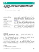
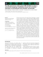
Fig. 1. The thiolase reaction mechanism and the compounds used. (A) The thiolase reaction. In the biosynthetic direction, the overall reac-
tion uses two molecules of Ac-CoA to generate AcAc-CoA and CoA. Cys89 is activated for nucleophilic attack by His348. (B) Comparison of
the covalent structures of CoA (top), SPP (middle) and OPP (bottom). SPP is different from CoA, such that the pantetheine moiety has an
ester linkage to a pivalate group instead of 3¢-phospho-ADP. In OPP, the reactive SH group of SPP is further replaced by an OH group. In
CoA, certain atoms of the pantetheine moiety are numbered.
G. Merila
¨
inen et al. Sulfur interactions at the thiolase active site
FEBS Journal 275 (2008) 6136–6148 ª 2008 The Authors Journal compilation ª 2008 FEBS 6137
is increased by oxyanion hole II, being formed by
N(Cys89) and N(Gly380) and binding the thioester
oxygen atom of the Cys89-bound acetyl group; oxyan-
ion hole II is similar to the ‘classical’ oxyanion hole
seen in, for example, serine proteases.
Although mutation of the reactive Cys89 to serine
still allows for transacetylation at low efficiency [6], the
exchange of the substrate thioester sulfur atom to an
oxygen apparently prevents the reaction from taking
place [4–7]. The compounds used in such studies have
consisted of thio- and oxoesters of a CoA analogue,
pantetheine-11-pivalate (PP) [3], which lacks the
3¢-phospho-ADP moiety of CoA, having a pivalate
group at the 11-hydroxyl moiety of pantetheine instead
(Fig. 1). The acetyl and acetoacetyl thioesters of PP
(Ac-SPP and AcAc-SPP) have been found to be
substrates of the bacterial biosynthetic thiolase [3–5].
Furthermore, 3-pentynoyl-SPP was used to identify
Cys378 as a catalytic residue [7,8].
The oxoesters of PP are nonreactive [5–7,14]. Lower
reactivity of the oxoester compared to the thioester is
also seen when comparing the kinetic properties of
crotonyl-CoA and crotonyl-oxyCoA: the hydration
rate of the oxoester by crotonase is 330-fold lower
[15]. Due to different resonance properties of the thio-
and oxoesters [16], the pK
a
of the a carbon of a thio-
ester is approximately 21, whereas the pK
a
for the
corresponding atom in an oxoester is 26 [17]. This will
make Claisen condensation more difficult with an oxo-
ester because the nucleophilic carbanion is harder to
generate (Fig. 1). Furthermore, the reactivity of the
carbonyl carbon of thioesters and oxoesters towards
nucleophilic attack is different [18,19], with oxoesters
being less reactive. Thus, from the chemical properties
of oxoesters, it can be postulated that they are less
reactive in both halves of the thiolase reaction than
thioesters. No previous studies have addressed the pos-
sibility that the poor reactivity of the esters of OPP in
the thiolase reaction could also be, at least partially,
related to the preferred nonproductive binding modes
of these oxoesters.
Members of the thiolase superfamily, being struc-
turally and mechanistically related [1], are of consider-
able interest in the field of biotechnology; the
applications utilizing their potential in biosynthetic
reactions are diverse [20–25]. The Z. ramigera thiolase
is a very close homologue of the human cytosolic
thiolase (CT) [11], which is a key enzyme in the cho-
lesterol synthesis pathway [26–28], and a detailed
understanding of the binding determinants of its
substrate could also help in the development of suit-
able drugs towards CT, with the aim of lowering high
cholesterol levels. Additionally, other human thiolases
have been found to be drug targets for the treatment
of heart failure [29–32].
In the present study, we used a combination of crys-
tallography and calorimetry to analyse the binding
determinants of CoA to the bacterial biosynthetic thio-
lase from Z. ramigera, concentrating specifically on the
SH groups of the substrate and the catalytic Cys89,
which is conserved throughout the thiolase superfam-
ily. The results obtained indicate that the sulfur atoms
of both the enzyme and the substrate are important
for the correct productive mode of binding of CoA in
the thiolase active site, improving our understanding
of substrate recognition by thioester-dependent
enzymes.
Results and Discussion
The biosynthetic thiolase from Z. ramigera is a tetra-
meric 160 kDa enzyme, consisting of four identical
subunits of 392 residues (Fig. 2). Three domains of
approximately equal lengths have been identified in the
thiolase fold. The core of the monomer is formed by
the N-terminal domain (with the catalytic cysteine)
and the C-terminal domain. The third domain, referred
to as the loop domain, protrudes out of the N-terminal
domain. The loop domain covers the catalytic site and
provides the binding site for the 3¢-phospho-ADP
moiety of CoA (Fig. 2).
In the present study, we aimed to analyse in detail,
using X-ray crystallography and isothermal titration
calorimetry, the factors that influence the productive
mode of binding of a CoA substrate to the active site
of biosynthetic thiolase. We solved five new liganded
crystal structures of Z. ramigera thiolase (Table 1; see
also Fig. S1) and compared these with the previously
available structures of liganded complexes of this and
other thiolases. The results obtained, as discussed in
detail below, indicate a crucial role for sulfur–sulfur
interactions in defining the catalytically competent
binding mode of CoA in the active site.
The pantetheine binding tunnel of biosynthetic
thiolase
When CoA binds to Z. ramigera biosynthetic thiolase,
its pantetheine moiety enters the narrow pantetheine
binding tunnel and its SH group closes off the catalytic
cavity, near the catalytically important residues Cys89,
His348 and Cys378. The residues forming the walls of
the tunnel include the side chains of Leu148, His156,
Met157, Ala234, Phe235, Ala243, Ala246, Ser247,
Gly248 and Leu249 of the loop domain, as well as
Ala318 and Phe319 from the C-terminal domain
Sulfur interactions at the thiolase active site G. Merila
¨
inen et al.
6138 FEBS Journal 275 (2008) 6136–6148 ª 2008 The Authors Journal compilation ª 2008 FEBS
b-strand Cb2, and Met134 of the neighboring subunit.
These residues encircle the pantetheine moiety. The
two peptide moieties of the pantetheine unit are both
similarly tightly wedged between Phe235 and Leu249
(the outermost peptide bond, near the bulk solvent)
and between Phe319 and Leu148 (the innermost pep-
tide bond, near the catalytic site).
The atoms shaping the binding environment of the
terminal sulfur atom of CoA are listed in Table 2 and
shown in Fig. 2. Apart from the sulfur-containing resi-
dues Cys89, Met157, Met288 and Cys378, these also
include side chain atoms of Ala318 and Phe319; the
latter two residues are a part of the highly conserved
NEAF sequence motif [1]. Within 4.8 A
˚
from the CoA
sulfur atom, there are also the catalytic water (Wat82)
and Ne2(His348) (Table 2) (i.e. the atoms forming
oxyanion hole I). Wat82 is hydrogen bonded to
Nd1(Asn316) and Wat49. Wat82 and Wat49 are pres-
ent in each of the five new structures (Fig. 3).
A prominent feature of the binding pocket for the
CoA sulfur atom is the presence of the four sulfur
atoms from Cys89, Met157, Met288 and Cys378. Due
to the high polarizability of sulfur atoms [33], it is
expected that sulfur–sulfur interactions will contribute
significantly to the van der Waals binding energy.
Cys89 and Cys378 are catalytic residues, but also
Met157 and Met288 are highly conserved in the thio-
lase family. An interesting exception is thiolase T2; in
this case, Met288 is replaced by a phenylalanine. This
is a unique feature of the T2 thiolase sequence, which
correlates with its unique substrate specificity: T2 is
able to use not only acetoacetyl-CoA, but also
the branched 2-methylacetoacetyl-CoA molecule as a
substrate [34].
AB
C
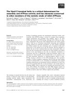
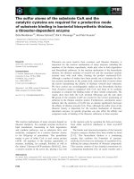
Fig. 2. The active site of thiolase. (A) The overall shape of the biosynthetic thiolase tetramer. The four individual active sites of the tetramer
are indicated by the bound CoA molecules. The catalytic site of the yellow domain is marked by an arrowhead. (B) The binding mode of CoA
to the thiolase monomer. The three domains are coloured yellow (N-terminal domain), light-green (loop-domain) and light gray (C-terminal
domain), respectively. The NEAF motif (including Phe319) is coloured red, the loop 231–240 (shifted in the OPP ⁄ Ac-OPP structures and con-
taining Phe235) is coloured purple and the catalytic Cys89 is coloured dark blue. His156 at the entrance of the pantetheine-binding cavity is
coloured orange. Note how the CoA interacts mainly with the loop domain and Cys89. (C) A detailed view of the surroundings of the termi-
nal moiety of CoA in the thiolase active site, as seen in the complex of unmodified thiolase with CoA (a similar view to that in B). Contacts
of the terminal sulfur are coloured green, hydrogen bonds are coloured red, and interactions between hydrophobic side chains and the planar
amide bonds of CoA are coloured yellow.
G. Merila
¨
inen et al. Sulfur interactions at the thiolase active site
FEBS Journal 275 (2008) 6136–6148 ª 2008 The Authors Journal compilation ª 2008 FEBS 6139
Only two hydrophilic side chains point into the
pantetheine binding tunnel: Ser247 and His156. Ser247
is known to adopt two different conformations; in the
unliganded form, it points away from the tunnel, and,
in the liganded conformation, it points into the tunnel,
interacting with the N4 atom of the CoA substrate
[2,10]. His156 has previously been observed in only
one conformation, having van der Waals contacts to
the pantetheine moiety of CoA at the outer edge of
the tunnel. Water molecules are also present in the
pantetheine binding tunnel, both in the unliganded and
liganded states [2,11]. There is one conserved water
near the bottom of the pantetheine binding tunnel
(Wat149), interacting with O(Gly248) and O(His348).
Table 2. Distances between the terminal sulfur or oxygen atom of the pantetheine moiety of the active site ligand and surrounding atoms.
As a reference, the structure of the unmodified Z. ramigera thiolase complexed with CoA (PDB entry 1DLV) has been used, considering all
atoms within 4.8 A
˚
of the CoA sulfur. The thiolases are from Z. ramigera, unless otherwise specified. In the oxidized structures, the active
site cysteine, Cys89 or its equivalent, has been oxidized to a sulfenic acid. In the acetylated structures, the active site cysteine has been
acetylated.
Thiolase Wild-type C89A Wild-type
Wild-type
oxidized
Wild-type
oxidized
Human CT
oxidized
Human T2
oxidized
Human T2
acetylated
Wild-type
acetylated
Ligand CoA CoA SPP OPP CoA CoA CoA CoA CoA
Sc-Cys89 3.9 – 4.1 5.2 3.8 4.2 4.3 3.8 4.3
Sc-Cys378 4.7 4.3 4.9 6.2 5.2 4.7 4.9 5.1 4.7
Sd-Met157 4.1 5.7 4.3 7.6 3.9 4.1 4.1 4.3 4.1
Cc-Met157 3.7 5.5 3.8 6.5 3.2 3.8 3.8 3.9 3.7
Cb-Met157 4.0 5.8 3.8 6.2 3.6 3.8 3.8 4.2 3.7
Sd-Met288 4.4 4.7 4.6 7.4 4.4 4.6 – – 4.5
Ce-Met288 3.7 4.3 3.4 6.2 3.7 3.6 – – 3.4
Ce2-Phe319 4.2 5.3 3.6 4.4 4.1 3.9 3.6 4.1 3.9
Cb-Ala318 4.2 3.8 3.9 3.2 4.5 3.8 3.8 4.0 4.0
Ne2-His348 4.6 3.3 4.9 3.5 4.9 4.9 5.2 5.0 4.7
Wat82 4.8 3.5 5.1 5.1 5.3 4.9 4.8 4.8 4.9
PDB entry 1DLV 2VTZ 2VU2 2VU1 2VU0 1WL4 2IBU 2F2S 1QFL
Table 1. Data processing and refinement statistics. The numbers in parentheses refer to the highest resolution shell.
Complex SPP OPP (oxidized Cys89)
CoA (oxidized
Cys89) CoA (C89A)
Ac-OPP
(oxidized Cys89)
Beamline EMBL ⁄ DESY X13 EMBL ⁄ DESY BW7B EMBL ⁄ DESY X11 EMBL ⁄ DESY X13 Rotating anode
Wavelength (A
˚
) 0.81 0.84 0.81 0.81 1.5418
Data processing
Resolution range (A
˚
) 20–2.65
(2.72–2.65)
20–1.51
(1.55–1.51)
20–1.87
(1.95–1.87)
20–2.30
(2.40–2.30)
20–2.07
(2.20–2.07)
<I ⁄ rI> 11.8 (3.5) 8.7 (2.1) 12.1 (4.4) 15.6 (4.8) 9.5 (4.5)
Completeness (%) 95.2 (98.4) 93.8 (70.1) 97.3 (86.3) 97.2 (86.1) 93.9 (83.6)
R
merge
(%) 9.9 (32.0) 9.1 (54.6) 7.1 (19.0) 6.8 (26.6) 10.0 (23.2)
Redundancy 3.2 (2.5) 3.5 (2.8) 3.1 (2.4) 3.5 (2.7) 2.8 (2.2)
Wilson B (A
˚
2
) 3621 253025
Space group P2
1
P2
1
P2
1
P2
1
P2
1
Unit cell parameters (A
˚
, °) 84.3, 79.2, 150.8
90, 92.9, 90
84.3, 78.7, 148.3
90, 92.9, 90
84.4, 79.1, 148.8
90, 92.7, 90
84.2, 79.6, 148.9
90, 92.1, 90
84.4, 79.0, 148.8
90, 93.0, 90
Refinement
R
cryst
(%) 23.1 21.6 19.4 22.2 16.1
R
free
(%) 28.6 24.9 22.9 26.2 21.2
rmsd Bond distances (A
˚
) 0.011 0.014 0.018 0.009 0.014
rmsd Bond angles (°) 1.2 1.4 1.6 1.1 1.4
rmsd B factors for bonded atoms
(main chain, side chain) (A
˚
2
)
1.0,2.0 1.5,1.7 1.4,2.7 0.7,1.3 1.9,2.9
PDB entry 2vu2 2vu1 2vu0 2vtz 1ou6
Sulfur interactions at the thiolase active site G. Merila
¨
inen et al.
6140 FEBS Journal 275 (2008) 6136–6148 ª 2008 The Authors Journal compilation ª 2008 FEBS
This water is present in all of the new structures
(Fig. 3).
The mode of binding of SPP closely resembles
that of CoA
The crystal structure of Z. ramigera thiolase was
solved in complex with SPP, a functional CoA ana-
logue. SPP and its thioesters are known to be func-
tional substrates for thiolase, but kinetic constants
have been reported only for AcAc-SPP [3]; for exam-
ple, K
m
and k
cat
are 73 lm and 469 ⁄ s for AcAc-SPP,
whereas they are 24 lm and 465 ⁄ s, respectively, for
AcAc-CoA. In the crystal structure, the binding modes
of the reactive sulfur moieties of SPP and CoA are
highly similar, explaining the reactive nature of SPP.
For example, the distance between the sulfur atoms of
SPP and the reactive Cys89 is 4.1 A
˚
, whereas the cor-
responding distance for the CoA complex is 3.9 A
˚
(Table 2).
The pantetheine moiety of SPP binds in the pant-
etheine binding tunnel in a similar (but not identical)
way to that seen for CoA (Fig. 3A). The pivalate head
group points outwards from the pantetheine binding
tunnel, overlapping with the pyrophosphate moiety of
CoA. It lies on a hydrophobic surface comprising
Leu249, Phe18 and Met134; the latter comes from the
tetramerization loop of an opposing subunit. These
results indicate that the 3¢-phospho-ADP group of
CoA is not crucial for the correct positioning of the
reactive end group of the substrate in the thiolase cata-
lytic cavity. However, it could be important for
increasing the affinity of binding, as well as the solub-
ility of the substrate.
Interestingly, the side chain of Ser247 points away
from SPP, as previously seen in unliganded thiolase; in
the case of a bound CoA ligand, it points towards the
ligand. This difference correlates with minor conforma-
tional differences between the pantetheine moieties of
CoA and SPP. The highly similar mode of binding of
the reactive moieties of CoA and SPP in the active site
of thiolase provides a structural basis for the observa-
tion that SPP compounds are functional substrates of
thiolase.
The binding mode of OPP is unproductive
The structure of Z. ramigera biosynthetic thiolase was
also solved in the presence of OPP and its acetylated
analogue. The complex with OPP was refined at a res-
olution of 1.51 A
˚
. The structure indicates a surprising
binding mode (Fig. 3A); OPP is bound further away
from the catalytic cavity than SPP and CoA.
A
B
C
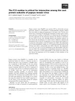
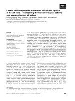
Fig. 3. Comparison of the active site ligand binding modes to bio-
synthetic thiolase. (A) The binding modes of SPP (magenta), OPP
(cyan), Ac-OPP (orange) and CoA (gray). A water molecule (I) in the
OPP complex is bound to oxyanion hole I. His156 has a double con-
formation in the Ac-OPP complex. (B) The binding mode of CoA to
thiolase harboring a modified active-site Cys89 is identical to that
seen for unmodified thiolase. The CoA complexes of unmodified
(gray), oxidized (yellow) and acetylated (green) Z. ramigera thiolase;
the human oxidized CT CoA complex (dark gray); and the human
acetyl-T2 CoA complex (pink) are shown. Oc(Ser247) points
towards the pantetheine moiety, except for the complex between
oxidized Z. ramigera thiolase and CoA, in which Ser247 has a dou-
ble conformation. (C) Superposition of CoA complexes of wild-type
(gray) and C89A (brown) thiolase. In the CoA C89A complex, there
are two extra water molecules in the active site cavity; one water
(II) is bound to oxyanion hole II and another nearby water molecule
(e) is hydrogen bonded to it.
G. Merila
¨
inen et al. Sulfur interactions at the thiolase active site
FEBS Journal 275 (2008) 6136–6148 ª 2008 The Authors Journal compilation ª 2008 FEBS 6141
The catalytic cavity lies at the bottom of the pant-
etheine-binding tunnel. The terminal oxygen of OPP is
5.2, 7.4, 6.2 and 7.6 A
˚
away from the sulfur atoms of
Cys89, Met288, and Cys378 and Met157, respectively
(Table 2). OPP has a terminal hydroxyl group, which
can form hydrogen bonds with water when in solution,
and with the protein once bound in the complex. From
the structure, it is apparent that the hydroxyl group of
OPP is in hydrogen bonding interaction with
Ne2(His348) (at 3.2 A
˚
) and O(Ala318) (at 3.3 A
˚
),
being inserted into a pocket between these atoms. This
pocket would be too tight for a sulfur atom, such as
those in CoA and SPP. This mode of binding places
the terminal oxygen atom further away from the center
of the catalytic cavity and, in this structure, oxyanion
hole I has a water molecule bound (Fig. 3).
In the CoA mode of binding [10], there is a water-
mediated hydrogen bond between Ne2(His156) and
O9(CoA), as well as a hydrogen bond between
Oc(Ser247) and N4(CoA), and the carbonyl O(Ser247)
is hydrogen bonded to N8(CoA). For OPP, these
hydrogen bonds are not observed. The side chain of
Ser247 points away from the bound OPP, and there
only exists a hydrogen bond between O(Ser247) and
N4(OPP) because the pantetheine group has moved
away from the bottom of the tunnel compared to SPP
and CoA (Fig. 3). Consequently, the terminal t-butyryl
moiety of OPP is in a different conformation than that
of SPP. Because this group is in a different conforma-
tion in SPP and OPP, it is not specifically recognized
by thiolase, which is as expected for a synthetic ana-
logue. The t-butyryl group of OPP is bound in a
pocket created by a rotation of the His156 side chain
around v1, away from its position observed in all
earlier structures. The residues forming the walls of
this hydrophobic pocket include Met143, Ile144,
Leu148, His156, Ala234, Phe235, Leu249 and Met134
(from the opposing subunit). The side chains of Ile144
and Met134 are also in a slightly different conforma-
tion compared to all previous structures. Moreover,
the entire loop containing residues 231–240 (Fig. 2)
moves away by approximately 1 A
˚
in the presence of
OPP; at the same time, the B factors for this loop indi-
cate a more rigid structure than in the presence of
CoA (data not shown). These changes are relatively
extensive compared to those seen upon CoA binding,
in which case it has been noted that the Sc atom of
Cys89 and the Oc atom of Ser247 are the only non-
hydrogen protein atoms that move detectably during
the thiolase reaction cycle [2,9,10].
OPP differs from SPP by only one atom, and the
replacement of the sulfur atom of SPP by an oxygen
changes the interaction such that OPP binds in a differ-
ent way to the thiolase active site. This mode of binding
of OPP to biosynthetic thiolase is unproductive, and
not competent for catalysis. Nonproductive binding is a
well-known phenomenon in enzymological studies [33].
Classic examples from structural studies include the
binding of (NAG)
3
to lysozyme [35] and that of the ind-
olyl acryloyl moiety in the active site of chymotrypsin
[36]. More recent examples concern crystallographic
binding studies of elastase [37], dihydrofolate reductase
[38] and methylmalonyl CoA decarboxylase [39].
We also solved the structure of the complex of thio-
lase with Ac-OPP; from the electron density map and
the subsequent structure comparison, it can be seen
that the binding mode is the same as that for OPP;
furthermore, the structural changes of the protein part
described for the OPP complex are seen as well in the
Ac-OPP complex. Due to low occupancy of the com-
pound in the structure, further detailed analysis of the
exact binding mode has not been performed, but the
mode of binding of the acetyl moiety is clearly differ-
ent from the mode of binding of the acetyl moiety of
Ac-CoA. Apparently, the common carbonyl oxygen
atom of the acetyl group of these two molecules does
not provide enough binding energy to favour a similar
mode of binding for Ac-OPP and Ac-CoA.
Binding mode of CoA to thiolases oxidized or
acetylated at Cys89
The structure of CoA-complexed Z. ramigera thiolase,
in which the catalytic Cys89 was oxidized, was also
determined. This was carried out to allow a detailed
comparison of the binding mode of CoA to thiolase
when the sulfur atom of Cys89 is modified covalently.
The oxidation of the catalytic cysteine to a cysteine
sulfenic acid has been observed in complexes of the
human cytosolic thiolase [11] and the human mito-
chondrial thiolase T2 [34]. The extra oxygen atom
points away from the ligand binding pocket into oxy-
anion hole II and, in these complexes, the mode of
binding of CoA is unaffected by the different oxidation
state of the cysteine sulfur (Fig. 3B). The same applies
to thiolases complexed with CoA in their acetylated
form, as seen in the structures of human T2 [Protein
Data Bank (PDB) entry 2F2S] and Z. ramigera thio-
lase [9]. For the Z. ramigera thiolase, the mode of
binding of CoA is unaffected by the oxidation (or acet-
ylation) state of the active site cysteine (Fig. 3B).
Indeed, the mode of binding of the CoA sulfur atom is
conserved also in the structures with acetyl-CoA com-
plexed with the acetylated active site [10]. In the OPP
complexes, Cys89 is oxidized to a cysteine sulfenic
acid, but the above results indicate that the different
Sulfur interactions at the thiolase active site G. Merila
¨
inen et al.
6142 FEBS Journal 275 (2008) 6136–6148 ª 2008 The Authors Journal compilation ª 2008 FEBS
modes of binding of OPP and Ac-OPP, when com-
pared with the corresponding SPP or CoA compounds,
cannot be attributed to the oxidation state of the cata-
lytic cysteine.
Binding mode of CoA to the C89A thiolase
variant
To better understand the importance of the sulfur–
sulfur interactions in the catalytic cavity of thiolase,
the CoA binding properties of its C89A variant were
also studied. In the crystal structure, CoA binding
essentially follows the wild-type mode, apart from the
reactive terminus of CoA. The terminal sulfur is
inserted deeper into the catalytic cavity by approxi-
mately 1.7 A
˚
, being sandwiched between His348 and
Cys378. In the wild-type complex, the reactive termi-
nus of CoA is apparently in a somewhat strained con-
formation (Fig. 3C). This indicates that the sulfur
atom of Cys89 is crucial for the correct positioning of
the reactive moiety of CoA. In addition, in the com-
plex of the C89A thiolase mutant with CoA, a water
molecule is placed in oxyanion hole II, formed by the
backbone nitrogens of residues Cys89 and Gly380, and
a second water is observed hydrogen bonded to
O(Gly147) (Fig. 3C). The tight fit of CoA into the
active site in this case is illustrated by the distances
from the S atom of CoA to Ne2(His348) (3.3 A
˚
),
Cb(Ala89) (3.6 A
˚
), a water bound to oxyanion hole II
(3.3 A
˚
) and water 82 (3.5 A
˚
). The distances to the
active site sulfurs are: Sc(Cys378) (4.3 A
˚
), Sd(Met288)
(4.7 A
˚
) and Sd(Met157) (5.7 A
˚
), reflecting the overall
weakening of the sulfur–sulfur van der Waals interac-
tions in the CoA binding mode of the C89A variant,
especially concerning Met157 (Table 2), in addition to
the absence of the sulfur–sulfur interaction between
CoA and Cys89.
Calorimetric analysis of the binding of CoA by
wild-type thiolase and the C89A variant
The detailed crystallographic binding studies described
above show that the sulfur–sulfur interactions in the
active site of thiolase are important for competent
binding of the substrate. Therefore, a calorimetric
study was carried out to further investigate the impor-
tance of sulfur–sulfur interactions for the affinity of
thiolase towards its substrate. Isothermal titration
calorimetry (ITC) was used to compare the affinity
of CoA to wild-type thiolase and its C89A variant.
The results obtained (Fig. 4 and Table 3) indicate that
the affinity is significantly higher for the wild-type
enzyme compared to the C89A variant, although only
one sulfur atom is missing in the mutant. The differ-
ence in affinity corresponds to a loss of free energy of
binding (DDG) of 0.8 kcalÆmol
)1
, whereas the loss of
enthalpy of binding (DDH) is 3.7 kcalÆmol
)1
. The key
difference in the crystal structures is the absence of the
sulfur atom of Cys89, generating small structural dif-
ferences (Fig. 3); the binding cavity for the reactive
moiety of the substrate is less compact in the C89A
mutant, which is consistent with the favorable differ-
ence binding entropy term [D(-TDS)]. The magnitude
of the unfavorable DDH term indicates that the sulfur–
AB
Fig. 4. Calorimetric analysis of CoA binding
by (A) wild-type and (B) C89A thiolase.
Curve fitting in both cases was carried out
by setting the binding stoichiometry to 1.
Note the different scales on the y-axis,
which are related to the binding enthalpy.
G. Merila
¨
inen et al. Sulfur interactions at the thiolase active site
FEBS Journal 275 (2008) 6136–6148 ª 2008 The Authors Journal compilation ª 2008 FEBS 6143
sulfur interactions provide a significant contribution to
the binding energy of CoA [33].
Concluding remarks
Cys89 is the catalytic residue in Z. ramigera thiolase,
and its mutation to alanine leads to a complete loss of
activity [2]. A corresponding catalytic cysteine residue
is conserved in all members of the thiolase superfamily.
In the present study, we have shown that, apart from
the lack of activity, the mutation C89A also lowers the
affinity of biosynthetic thiolase towards CoA signifi-
cantly. In complexes between biosynthetic thiolase and
CoA or SPP, a functional substrate analogue, the sulfur
atom of the substrate is always closely embraced by
four sulfur atoms from the enzyme. When OPP is used,
being identical to SPP except for the replacement of the
terminal sulfur atom by oxygen, the binding mode of
the ligand also changes, resulting in a nonproductive
binding mode. Our data indicate an important role for
the interactions between the CoA substrate sulfur
group and the thiolase active site in assuring an optimal
affinity, as well as a competent mode of binding.
There exists considerable interest in the properties
and engineering possibilities of enzymes in the thiolase
superfamily, both within biotechnology and pharma-
cology, due to their involvement in, for example, dif-
ferent natural product synthesis pathways, as well as
in lipid and cholesterol metabolism. The results
obtained in the present study indicate that the sulfur
atom of the thioester moiety is not only important
because of the high intrinsic reactivity of thioester sub-
strates, but also that it can play a key role in achieving
the proper affinity and competent mode of binding of
the substrates in the active site cavities of thioester-
dependent enzymes. This should be taken into consid-
eration when designing, for example, new substrates or
inhibitors for these enzymes.
Experimental procedures
Protein expression, purification and
crystallization
Z. ramigera thiolase and its C89A mutant were expressed
and purified as previously described [2,9]. Crystallization of
wild-type thiolase and its C89A variant were carried out at
22 °C using vapour diffusion in a mother liquour contain-
ing 1 m Li
2
SO
4
, 0.9 m (NH
4
)
2
SO
4
, 0.1 m sodium citrate
(pH 5), 1 mm EDTA, 1mM NaN
3
and 1 mm dithiothreitol.
Synthesis of OPP and SPP
All intermediates and products were purified using silica gel
chromatography, and their purity was checked by TLC.
The identity of the compounds was demonstrated by
1
H-NMR. All chemicals were obtained from Sigma-Aldrich
(Taufkirchen, Germany).
Synthesis of ethanolamino-tert.butyl-diphenyl-silane
A solution of 10 mmol tert.butyl-diphenyl-silyl-chloride in
20 mL of tetrahydrofurane was added dropwise with stirring
to 50 mmol ethanolamine in 30 mL of tetrahydrofurane at
0 °C. After stirring for 2 h at room temperature (RT), the
solution was evaporated. Yield: 8.7 mmol ethanolamino-
tert.butyl-diphenyl-silane.
Purification of pantothenoic acid
A solution of 20 mmol d(+)-calcium pantothenate in 5 mL
of 4 m HCl was extracted once with 40 mL of chloro-
form ⁄ methanol (2 : 1) and three times with 40 mL of chlo-
roform ⁄ methanol (9 : 1). The organic layers were
evaporated. Yield: 10 mmol pantothenoic acid.
Synthesis of tert.butyl-diphenyl-silyl-pantetheinate
Dicyclohexyl-carbodiimide (11 mmol) was added to
10 mmol pantothenoic acid in 50 mL of tetrahydrofurane.
The suspension was stirred until dicyclohexyl-carbodiimide
dissolved completely; 8.7 mmol ethanolamino-tert.butyl-
diphenyl-silane was then added. The solution was
stirred for 4 h at RT. Yield: 2.6 mmol tert.butyl-diphenyl-
silyl-pantetheinate.
1
H-NMR: C2: 3.48 p.p.m., s.; C11:
3.75 p.p.m., t.; phenyl: 7.65 p.p.m., d.
Synthesis of tert.butyl-silyl-OPP
A solution of 4 mmol pyridine was added to a solution of
2 mmol tert.butyl-diphenyl-silyl-pantetheinate in 50 mL of
dichlormethane; 2 mmol pivaloylchloride in 20 mL of dic-
hlormethane was then added dropwise with stirring. After
Table 3. Calorimetric analysis of CoA binding to the Z. ramigera biosynthetic thiolase at 25 °C. The values and error estimates are calculated
from separate measurements (three for the wild-type and two for C89A). K
a
and K
d
are the association and dissociation constants, respectively.
Sample DH (kcalÆmol
)1
) )TDS (kcalÆmol
)1
) DG (kcalÆmol
)1
) K
a
(M
)1
) K
d
(lM)
Wild-type )6.7 ± 0.9 1.1 ± 0.8 )5.6 ± 0.2 1.3 · 10
4
± 0.4 · 10
4
81 ± 29
C89A )3.0 ± 0.03 )1.8 ± 0.2 )4.8 ± 0.2 3.2 · 10
3
± 0.1 · 10
3
307 ± 64
Sulfur interactions at the thiolase active site G. Merila
¨
inen et al.
6144 FEBS Journal 275 (2008) 6136–6148 ª 2008 The Authors Journal compilation ª 2008 FEBS
stirring for 24 h at RT, the residue was dissolved in 20 mL
of acetoacetate and extracted with 25 mL each of 0.1 m
HCl, saturated CuSO
4
in water, and 2 m NaCl. Yield:
1.3 mmol tert.butyl-silyl-OPP.
1
H-NMR: C2: 3.67 p.p.m.,
s.; C11: 3.92 p.p.m., t.; phenyl: 7.82 p.p.m., d.; pivalate-
methyl: 1.26 p.p.m., s.
Synthesis of OPP
A solution of 1 mL of hydrogen fluoride was added to
1.3 mmol tert.butyl-silyl-O-pantetheine-11-pivalate in
30 mL of acetonitrile. After stirring for 30 min at RT, the
solution was evaporated. The residue was suspended in
20 mL of water and extracted three times with 20 mL of
ethylacetate. Yield: 1.2 mmol OPP.
1
H-NMR: C2:
3.78 p.p.m., s.; C11: 4.03 p.p.m., t.; pivalate-methyl: 1.43, s.
Synthesis of Ac-OPP
A solution of 1 mmol pyridine and 0.5 mmol acetyl chlo-
ride was added to a solution of 0.1 mmol OPP in 2 mL of
chloroform and stirred at RT for 15 min. The suspension
was extracted three times with 2 mL of brine. Yield:
89 lmol Ac-OPP.
1
H-NMR: C2: 3.59 p.p.m., s.; C11:
4.03 p.p.m., t.; pivalate-methyl: 1.23, s.; acetyl-methyl:
2.17 p.p.m., s.
Synthesis of SPP
Bis(N-pantothenylamidoethyl) disulfide was converted into
Bis(N-pantothenylamidoethyl-11-pivalate) disulfide accord-
ing to the synthesis of tert.butyl-silyl-OPP. The resulting
disulfide was cleaved by treatment with mercaptoethanol.
Crystal handling
Crystal structures were determined for the complexes of
thiolase with OPP, Ac-OPP and SPP. OPP, Ac-OPP and
SPP were poorly soluble in aqueous buffer solutions. In the
OPP soaking experiment, OPP could be dissolved in the
cryoprotectant solution, containing, in addition to the con-
stituents of the well solution, 12% 2-methyl-2,4-pentanediol
(MPD), 12% glycerol and 100 mm OPP. Prior to data
collection, the crystal was transferred to this cryosolution.
A second crystal was soaked similarly in a solution contain-
ing Ac-OPP instead of OPP. In both soaking experiments,
the crystals started suffering during the soak under all
tested conditions; thus, the soaking time was approximately
1 min.
SPP was dissolved in MPD, at an approximate concen-
tration of 100 mm. For soaking SPP into thiolase crystals,
the crystals were transferred to drops of mother liquor con-
taining one-fifth of the SPP stock solution. This soaking, in
approximately 20% MPD and 20 mm SPP in mother
liquor, was allowed to continue for 4 days prior to data
collection. This experiment was performed for both a wild-
type thiolase crystal and a C89A mutant crystal. Analysis
of the data collected from these crystals showed the mode
of binding of SPP to wild-type thiolase, but no binding was
observed when using the C89A crystals.
Two more structures were determined and analysed: the
structure of a complex of CoA with the C89A variant and
a complex of CoA bound in the active site with an oxidized
Cys89. The latter structure was obtained from a soaking
experiment of a wild-type thiolase crystal with 5 mm b-hy-
droxybutyryl-CoA for 1 min. During refinement, it was
seen that the structure contained only CoA and the active
site Cys89 was oxidized. The soaking of CoA into a C89A
thiolase crystal was performed at 5 mm CoA.
Data collection, structure solution and
refinement
Data were collected on beamlines BW7B, X11 and X13
at the EMBL-Hamburg Outstation ⁄ DESY (Hamburg,
Germany), except for the data from the Ac-OPP complex,
which were collected on a Nonius FR591 rotating anode
source (Bruker AXS, Delft, the Netherlands). Data process-
ing (Table 1) was carried out with xds [40] and xdsi [41].
Five percent of all reflections were used for calculating the
free R factor [42]. The structures were refined using
refmac5 [43]. tls parameters [44] were applied, and water
molecules were added using arp ⁄ warp [45]. CoA, OPP,
Ac-OPP and SPP were built when their electron densities
were strong and continuous. Model building and analysis
were performed using o [46] and coot [47]. The refinement
statistics are given in Table 1.
The coordinates and structure factors were deposited to
the PDB under the following accession codes: 2VU2 (thio-
lase-SPP complex), 2VU1 (thiolase-OPP complex), 2VU0
(oxidized thiolase-CoA complex), 2VTZ (C89A thiolase-
CoA complex) and 1OU6 (Ac-OPP complex).
Structure analysis
The A and B subunits of the biosynthetic thiolase tetra-
mer are always best defined in this crystal form of Z. ram-
igera thiolase due to the layer-like packing of the thiolase
tetramers in the crystal lattice, and the B subunit has been
used for previous analyses [2,9,10]. In the case of SPP and
OPP, however, the ligand is slightly better defined in the
A subunit, and this subunit has mainly been analysed in
the present study with respect to these ligands. All dis-
cussed features can, however, be seen in the B subunit.
Cys89 was oxidized in the structures of the complexes
with OPP and Ac-OPP, and it was built as a cysteine
sulfenic acid, with the oxygen atom pointing into oxyan-
ion hole II.
G. Merila
¨
inen et al. Sulfur interactions at the thiolase active site
FEBS Journal 275 (2008) 6136–6148 ª 2008 The Authors Journal compilation ª 2008 FEBS 6145
For the analysis and comparisons of the new structures,
six additional structures were used. The structures were
superimposed on each other using the ssm [48] approach
implemented in coot [47]. The sequence numbering always
refers to the Z. ramigera biosynthetic thiolase. The number-
ing of conserved active site water molecules (49, 82 and
149) in the text and figures corresponds to earlier analyses
of subunit B. In two of the structures used for the compari-
son, the active site cysteine was also oxidized: the CoA
complexes of human CT (PDB entry 1WL4) [11] and
human mitochondrial thiolase (T2; PDB entry 2IBU) [34].
In two other structures of CoA complexes, the active site
cysteine was acetylated: the Z. ramigera thiolase (PDB
entry 1QFL) [9] and human mitochondrial thiolase T2
(PDB entry 2F2S). The coordinates of the latter structure
were first refined further before being used for the compari-
son. Furthermore, the geometry of the Z. ramigera thiolase
complexes 1DM3 (acetylated cysteine with Ac-CoA bound)
[10] and 1DLV (wild-type with CoA bound) [10] were also
analysed.
Figures were made with ccp4mg [49], dino (http://
www.dino3d.org), povray () and
pymol ().
Isothermal titration calorimetry
The titration of 5 mm CoA into solutions of 50 lm wild-
type thiolase or 100 lm C89A mutated thiolase was made
with a VP-ITC Microcalorimeter (MicroCal, Northampton,
MA, USA). The poor solubility of OPP and SPP prevented
their use in the experiment. All ITC samples were prepared
in, or dialyzed against, 100 mm Tris–HCl (pH 7.5), 5%
glycerol, 1 mm dithiothreitol and degassed. The titration
was carried out at 25 °C. Blank experiments (without pro-
tein) were made to estimate the heat of dilution for CoA.
The binding isotherms obtained by integrating the injection
peaks were fitted to appropriate binding models (the stoi-
chiometry was set to 1 per subunit) by using the origin
software (MicroCal). The titration curve was fitted by the
nonlinear least-squares method, and the thermodynamic
parameters were determined. The reproducible binding
constants were derived from at least two independent
measurements.
Acknowledgements
The authors wish to acknowledge the excellent support
of the protein crystallography beamlines of the
EMBL-Hamburg Outstation ⁄ DESY. The skillful tech-
nical assistance of Ville Ratas and discussions with Dr
A. M. Lambeir (University of Antwerp, Belgium) and
Dr Inari Kursula (University of Oulu, Finland) are
gratefully acknowledged. This study was supported by
the Academy of Finland (grant 200966).
References
1 Haapalainen AM, Merila
¨
inen G & Wierenga RK (2006)
The thiolase superfamily: condensing enzymes with
diverse reaction specificities. Trends Biochem Sci 31,
64–71.
2 Kursula P, Ojala J, Lambeir AM & Wierenga RK
(2002) The catalytic cycle of biosynthetic thiolase: a
conformational journey of an acetyl group through four
binding modes and two oxyanion holes. Biochemistry
41, 15543–15556.
3 Davis JT, Moore RN, Imperiali B, Pratt AJ, Kobayashi
K, Masamune S, Sinskey AJ, Walsh CT, Fukui T &
Tomita K (1987) Biosynthetic thiolase from Zoogloea
ramigera. I. Preliminary characterization and analysis of
proton transfer reaction. J Biol Chem 262, 82–89.
4 Davis JT, Chen HH, Moore R, Nishitani Y, Masamune
S, Sinskey AJ & Walsh CT (1987) Biosynthetic thiolase
from Zoogloea ramigera. II. Inactivation with haloacetyl
CoA analogs. J Biol Chem 262, 90–96.
5 Masamune S, Walsh CT, Sinskey AJ & Peoples OP
(1989) Poly-(R)-3-hydroxybutyrate (PHB) biosynthesis:
mechanistic studies on the biological Claisen condensa-
tion catalyzed by b-ketoacyl thiolase. Pure Appl Chem
61, 303–312.
6 Thompson S, Mayerl F, Peoples OP, Masamune S,
Sinskey AJ & Walsh CT (1989) Mechanistic studies on
beta-ketoacyl thiolase from Zoogloea ramigera: identifi-
cation of the active-site nucleophile as Cys89, its muta-
tion to Ser89, and kinetic and thermodynamic
characterization of wild-type and mutant enzymes.
Biochemistry 28 , 5735–5742.
7 Palmer MA, Differding E, Gamboni R, Williams SF,
Peoples OP, Walsh CT, Sinskey AJ & Masamune S
(1991) Biosynthetic thiolase from Zoogloea ramigera.
Evidence for a mechanism involving Cys-378 as the
active site base. J Biol Chem 266, 8369–8375.
8 Williams SF, Palmer MA, Peoples OP, Walsh CT, Sins-
key AJ & Masamune S (1992) Biosynthetic thiolase
from Zoogloea ramigera. Mutagenesis of the putative
active-site base Cys-378 to Ser-378 changes the parti-
tioning of the acetyl S-enzyme intermediate. J Biol
Chem 267, 16041–16043.
9 Modis Y & Wierenga RK (1999) A biosynthetic thiolase
in complex with a reaction intermediate: the crystal
structure provides new insights into the catalytic mecha-
nism. Structure 7, 1279–1290.
10 Modis Y & Wierenga RK (2000) Crystallographic
analysis of the reaction pathway of Zoogloea ramigera
biosynthetic thiolase. J Mol Biol 297, 1171–1182.
11 Kursula P, Sikkila
¨
H, Fukao T, Kondo N & Wierenga
RK (2005) High resolution crystal structures of human
cytosolic thiolase (CT): a comparison of the active sites
of human CT, bacterial thiolase, and bacterial KAS I.
J Mol Biol 347, 189–201.
Sulfur interactions at the thiolase active site G. Merila
¨
inen et al.
6146 FEBS Journal 275 (2008) 6136–6148 ª 2008 The Authors Journal compilation ª 2008 FEBS
12 Cleland WW (1963) The kinetics of enzyme-catalyzed
reactions with two or more substrates or products. I.
Nomenclature and rate equations. Biochim Biophys Acta
67, 104–137.
13 Gehring U & Harris JI (1970) The active site cysteines
of thiolase. Eur J Biochem 16, 492–498.
14 Davis JT (1986) The Biosynthetic Claisen Condensation:
Mechanistic Enzymology of Biosynthetic Thiolase from
Zoogloea ramigera. PhD Thesis. Department of Chemis-
try, Massachusetts Institute of Technology, Cambridge,
MA.
15 Dai M, Feng Y & Tonge PJ (2001) Synthesis of croto-
nyl-oxyCoA: a mechanistic probe of the reaction cata-
lyzed by enoyl-CoA hydratase. J Am Chem Soc 123,
506–507.
16 Mathews CK, van Holde KE & Ahern KG (2000) Bio-
chemistry, 3rd edn. Addison Wesley Longman, San
Francisco, CA.
17 Richard JP & Amyes TL (2001) Proton transfer at car-
bon. Curr Opin Chem Biol 5, 626–633.
18 Yang W & Drueckhammer DG (2000) Computational
studies of the aminolysis of oxoesters and thioesters in
aqueous solution. Org Lett 2, 4133–4136.
19 Yang W & Drueckhammer DG (2001) Understanding
the relative acyl-transfer reactivity of oxoesters and
thioesters: computational analysis of transition state
delocalization effects. J Am Chem Soc 123, 11004–
11009.
20 Austin MB & Noel JP (2003) The chalcone synthase
superfamily of type III polyketide synthases. Nat Prod
Rep 20, 79–110.
21 Mickel SJ (2004) Toward a commercial synthesis of
(+)-discodermolide. Curr Opin Drug Discov Devel 7,
869–881.
22 Sutherlin A & Rodwell VW (2004) Multienzyme meva-
lonate pathway bioreactor. Biotechnol Bioeng 87, 546–
551.
23 Baltz RH (2006) Molecular engineering approaches to
peptide, polyketide and other antibiotics. Nat Biotechnol
24, 1533–1540.
24 Tokiwa Y & Ugwu CU (2007) Biotechnological produc-
tion of (R)-3-hydroxybutyric acid monomer. J Biotech-
nol 132, 264–272.
25 Jojima T, Inui M & Yukawa H (2008) Production of
isopropanol by metabolically engineered Escherichia
coli. Appl Microbiol Biotechnol 77, 1219–1224.
26 Fukao T (2002) Thiolases (acetyl-CoA transferases). In
Wiley Encyclopedia of Molecular Medicine (Kazazian
HH, Klein G, Moser HW, Orkin SH, Roizman B,
Thakker RV & Watkins H eds), pp. 3125–3129. John
Wiley and Sons, Hoboken, NJ.
27 Salam WH & Bloxham DP (1987) Hypolipidemic effect
of polymethylenemethane thiosulfonates: inhibitors of
acetoacetyl coenzyme A thiolase. J Pharmacol Exp Ther
241, 1099–1105.
28 Greenspan MD, Yudkovitz JB, Chen JS, Hanf DP,
Chang MN, Chiang PY, Chabala JC & Alberts AW
(1989) The inhibition of cytoplasmic acetoacetyl-CoA
thiolase by a triyne carbonate (L-660, 631). Biochem
Biophys Res Commun 163, 548–553.
29 Kantor PF, Lucien A, Kozak R & Lopaschuk GD
(2000) The antianginal drug trimetazidine shifts
cardiac energy metabolism from fatty acid oxidation
to glucose oxidation by inhibiting mitochondrial long-
chain 3-ketoacyl coenzyme A thiolase. Circ Res 86,
580–588.
30 Marzilli M (2003) Cardioprotective effects of trimetazi-
dine: a review. Curr Med Res Opin 19, 661–672.
31 Fragasso G, Spoladore R, Cuko A & Palloshi A (2007)
Modulation of fatty acids oxidation in heart failure by
selective pharmacological inhibition of 3-ketoacyl coen-
zyme-A thiolase. Curr Clin Pharmacol 2, 190–196.
32 Liu X, Wu L, Deng G, Li N, Chu X, Guo F & Li D
(2008) Characterization of mitochondrial trifunctional
protein and its inactivation study for medicine develop-
ment. Biochim Biophys Acta 1784, 1742–1749.
33 Fersht A (1999) Structure and Mechanism in Protein
Science: A Guide to Enzyme Catalysis and Protein Fold-
ing. WH Freeman and Co., New York, NY.
34 Haapalainen AM, Merila
¨
inen G, Pirila
¨
PL, Kondo N,
Fukao T & Wierenga RK (2007) Crystallographic and
kinetic studies of human mitochondrial acetoacetyl-
CoA thiolase: the importance of potassium and chloride
ions for its structure and function. Biochemistry 46,
4305–4321.
35 Phillips DC (1967) The hen egg-white lysozyme mole-
cule. Proc Natl Acad Sci USA 57, 483–495.
36 Henderson R (1970) Structure of crystalline alpha-
chymotrypsin. IV. The structure of indoleacryloyl-
alpha-chyotrypsin and its relevance to the hydrolytic
mechanism of the enzyme. J Mol Biol 54, 341–354.
37 Mattos C, Rasmussen B, Ding X, Petsko GA & Ringe
D (1994) Analogous inhibitors of elastase do not always
bind analogously. Nat Struct Biol 1, 55–58.
38 Oefner C, D’Arcy A & Winkler FK (1988) Crystal
structure of human dihydrofolate reductase complexed
with folate. Eur J Biochem 174, 377–385.
39 Benning MM, Haller T, Gerlt JA & Holden HM (2000)
New reactions in the crotonase superfamily: structure of
methylmalonyl CoA decarboxylase from Escherichia
coli. Biochemistry 39, 4630–4639.
40 Kabsch W (1993) Automatic processing of rotation
diffraction data from crystals of initially unknown
symmetry and cell constants. J Appl Cryst 26, 795–800.
41 Kursula P (2004) XDSi: a graphical interface for the
data processing program XDS. J Appl Cryst 37, 347–
348.
42 Brunger AT (1992) Free R value: a novel statistical
quantity for assessing the accuracy of crystal structures.
Nature 355, 472–475.
G. Merila
¨
inen et al. Sulfur interactions at the thiolase active site
FEBS Journal 275 (2008) 6136–6148 ª 2008 The Authors Journal compilation ª 2008 FEBS 6147
43 Murshudov GN, Vagin AA & Dodson EJ (1997)
Refinement of macromolecular structures by the maxi-
mum-likelihood method. Acta Cryst D53, 240–255.
44 Winn MD, Isupov MN & Murshudov GN (2001) Use
of TLS parameters to model anisotropic displacements
in macromolecular refinement. Acta Cryst D57, 122–133.
45 Perrakis A, Morris R & Lamzin VS (1999) Automated
protein model building combined with iterative struc-
ture refinement. Nat Struct Biol 6, 458–463.
46 Jones TA, Zou J, Cowan SW & Kjeldgaard M (1991)
Improved methods for building protein models in elec-
tron density maps and the location of errors in these
models. Acta Cryst A47, 110–119.
47 Emsley P & Cowtan K (2004) Coot: model-building tools
for molecular graphics. Acta Cryst D60, 2126–2132.
48 Krissinel E & Henrick K (2004) Secondary structure
matching (SSM), a new tool for fast protein structure
alignment in three dimensions. Acta Cryst D60, 2256–
2268.
49 Potterton L, McNicholas S, Krissinel E, Gruber J,
Cowtan K, Emsley P, Murshudov GN, Cohen S,
Perrakis A & Noble M (2004) Developments in the
CCP4 molecular-graphics project. Acta Cryst D60,
2288–2294.
Supporting information
The following supplementary material is available:
Fig. S1. Electron densities of ligands in the crystal
structures.
This supplementary material can be found in the
online version of this article.
Please note: Wiley-Blackwell is not responsible for
the content or functionality of any supplementary
materials supplied by the authors. Any queries (other
than missing material) should be directed to the corre-
sponding author for the article.
Sulfur interactions at the thiolase active site G. Merila
¨
inen et al.
6148 FEBS Journal 275 (2008) 6136–6148 ª 2008 The Authors Journal compilation ª 2008 FEBS