Báo cáo khoa học: Effects of salt on the kinetics and thermodynamic stability of endonuclease I from Vibrio salmonicida and Vibrio cholerae potx
Bạn đang xem bản rút gọn của tài liệu. Xem và tải ngay bản đầy đủ của tài liệu tại đây (463.7 KB, 13 trang )
Effects of salt on the kinetics and thermodynamic
stability of endonuclease I from Vibrio salmonicida
and Vibrio cholerae
Laila Niiranen
1
, Bjørn Altermark
2
, Bjørn O. Brandsdal
2
, Hanna-Kirsti S. Leiros
2
, Ronny Helland
2
,
Arne O. Smala
˚
s
2
and Nils P. Willassen
1
1 Department of Molecular Biotechnology, Institute of Medical Biology, Faculty of Medicine, University of Tromsø, Norway
2 Norwegian Structural Biology Centre (NorStruct), Department of Chemistry, Faculty of Science, University of Tromsø, Norway
Extracellular and periplasmic enzymes of marine
organisms are exposed to environments in which
large variations in temperature and salinity can
occur. Such conditions require the proteins to fold
effectively and maintain their stability in spite of the
stresses they face [1]. At the same time, enzymatic
activity is dependent on fine-tuned structural flexibi-
lity [2]. How do enzymes cope with these contradict-
ing demands?
The study of the structural and functional adapta-
tion of proteins from extremophilic organisms is an
active research area, and several interesting observa-
tions of the adaptive mechanisms have been made.
The extreme temperature stability of thermophilic
Keywords
endonuclease I; kinetics; salt adaptation;
thermodynamic stability; Vibrio
Correspondence
N. P. Willassen, Department of Molecular
Biotechnology, Institute of Medical Biology,
Faculty of Medicine, University of Tromsø,
N-9037 Tromsø, Norway
Fax: +47 776 453 50
Tel: +47 776 446 51
E-mail:
(Received 24 October 2007, revised 14
December 2007, accepted 1 February 2008)
doi:10.1111/j.1742-4658.2008.06317.x
Adaptation to extreme environments affects the stability and catalytic effi-
ciency of enzymes, often endowing them with great industrial potential.
We compared the environmental adaptation of the secreted endonuclease
I from the cold-adapted marine fish pathogen Vibrio salmonicida (VsEndA)
and the human pathogen Vibrio cholerae (VcEndA). Kinetic analysis
showed that VsEndA displayed unique halotolerance. It retained a consid-
erable amount of activity from low concentrations to at least 0.6 m NaCl,
and was adapted to work at higher salt concentrations than VcEndA by
maintaining a low K
m
value and increasing k
cat
. In differential scanning
calorimetry, salt stabilized both enzymes, but the effect on the calorimetric
enthalpy and cooperativity of unfolding was larger for VsEndA, indicating
salt dependence. Mutation of DNA binding site residues (VsEndA, Q69N
and K71N; VcEndA, N69Q and N71K) affected the kinetic parameters.
The VsEndA Q69N mutation also increased the T
m
value, whereas other
mutations affected mainly DH
cal
. The determined crystal structure of
VcEndA N69Q revealed the loss of one hydrogen bond present in native
VcEndA, but also the formation of a new hydrogen bond involving residue
69 that could possibly explain the similar T
m
values for native and N69Q-
mutated VcEndA. Structural analysis suggested that the stability, catalytic
efficiency and salt tolerance of EndA were controlled by small changes in
the hydrogen bonding networks and surface electrostatic potential. Our
results indicate that endonuclease I adaptation is closely coupled to the
conditions of the habitats of natural Vibrio, with VsEndA displaying a
remarkable salt tolerance unique amongst the endonucleases characterized
so far.
Abbreviations
DSC, differential scanning calorimetry; EndA, endonuclease I; VcEndA, Vibrio cholerae endonuclease I; VsEndA, Vibrio salmonicida
endonuclease I; Vvn, Vibrio vulnificus endonuclease I.
FEBS Journal 275 (2008) 1593–1605 ª 2008 The Authors Journal compilation ª 2008 FEBS 1593
proteins is thought to be a result of extensive intra-
molecular networks and compact packing that restrict
their flexibility [3,4]. Psychrophilic enzymes, in
contrast, are thermolabile and have been hypothesized
to use increased flexibility to cope with the increased
viscosity and decreased thermal vibrations at low
temperature [5,6]. They may also use local flexibility
to maintain a functional active site, whilst separate
more rigid domains confer stability to their structure
[7]. Compared with non-halophiles, halophilic proteins
have an excess of acidic amino acid residues that
create a negative surface potential and a protective
hydrated ion network [1,8]. The charged surface is,
however, destabilizing, especially at low salt concen-
trations [9], and most halophilic proteins are inacti-
vated at NaCl or KCl concentrations below 2 m [1].
The structural basis of salt-tolerant activity remains
to be elucidated, although electrostatic interactions
have been implicated [10]. The salinity of seawater is
significantly lower than that of extreme halophile
habitats, so that only milder forms of adaptation
may be necessary for periplasmic and extracellular
proteins of marine organisms.
The functional characterization of extremophilic
proteins has so far focused on the obvious, i.e. the
effects of temperature on thermophilic and psychro-
philic proteins, and salinity on halophilic proteins.
However, many of the environments in which extremo-
philes thrive are extreme with respect to more than just
one parameter. For example, in the field of psychro-
phile research, the majority of enzymes studied so far
have been extracellular and of marine origin [7], which
poses a problem when conclusions are to be drawn
about the mechanisms of cold adaptation. Are the
observed adjustments a result of true adaptation to
low temperature, or a combination of cold and salt
adaptation? Choosing non-marine (freshwater) psychro-
philes as study targets has been proposed as a
solution [7]. The interplay between the two types of
adaptation is, however, interesting in itself, and it is
possible to design experiments in a manner that facili-
tates the separation of the two effects. The first steps
towards this approach have been taken. In a compara-
tive study of a marine psychrophilic and an estuarine
mesophilic endonuclease I (EndA, EC 3.1.30) [11], the
different salt optima of the enzymes were taken into
consideration when the temperature-dependent enzy-
matic properties were characterized. In the discussion,
the authors stressed the importance of performing
measurements in buffers that were as physiological as
possible. Similar to psychrophilic EndA, marine car-
rageenase was found to display an activity optimum
around the salt concentration of seawater [12]. It
appears that the choice of buffer and the determina-
tion of the salt dependence of the activity are impor-
tant in comparative experiments on extracellular
enzymes.
EndA is a periplasmic or extracellular sugar non-
specific endonuclease. Its physiological function is not
known, but it has been proposed to be involved in the
prevention of the uptake of foreign DNA, the degrada-
tion of intestinal mucus to facilitate colonization, and
the provision of nucleotides for the cells [13]. Although
EndA has been isolated from many pathogenic bacte-
ria, it does not appear to be involved in virulence [13–
15]. It may, however, affect the bacterial survival rate
through the degradation of neutrophil extracellular
traps in mammals, and possibly also in fish [16,17].
The structures of three Vibrio endonucleases, V. sal-
monicida (VsEndA) [18], V. cholerae (VcEndA) [19]
and V. vulnificus (Vvn) [20], are available, and also the
structure for Vvn bound to 8 bp and 16 bp dsDNA
[20,21]. A reaction mechanism has been proposed
based on the protein–DNA complex structure [20].
Sequence identity between mature Vvn and VcEndA is
75% and between Vvn and VsEndA is 74%. The
structural fold and the active site containing the cata-
lytically important His80 are identical in all three
structures. Temperature adaptation has not been found
to affect the reaction mechanism of any homologous
enzymes studied so far [22]. The thermal adaptation of
VsEndA and VcEndA has been studied previously,
revealing that VsEndA has a higher k
cat
value at
5–37 °C and is less thermostable than VcEndA [11].
The shape-complementary surface of Vvn contacts
the DNA only at the backbone phosphate groups [20].
Comparisons of Vvn, VsEndA and VcEndA have
revealed that most of the charged residues in the bind-
ing cleft are conserved [18]. The exceptions to this are
two interesting regions with high-temperature B-factors
pointed out by Altermark et al. [18]. The first is loop
51–54 which contains two more positive charges in
VsEndA than in VcEndA, but is unlikely to contact
DNA. The second is residues 69 and 71 which partici-
pate in the formation of the substrate binding site.
These residues are Gln and Lys, respectively, in
VsEndA, but both are replaced by Asn in VcEndA.
Intuitively, such changes in charge and steric effects
may alter substrate binding and salt sensitivity.
In this study, the effect of NaCl concentration on
the kinetic constants and thermodynamic stability of
VsEndA and VcEndA was investigated. In addition,
the effects of reciprocal mutations of two non-con-
served DNA binding site residues (VsEndA, Q69N and
K71N; VcEndA, N69Q and N71K) on the kinetics,
thermostability and salt dependence of these enzymes
Effects of salt on Vibrio endonucleases L. Niiranen et al.
1594 FEBS Journal 275 (2008) 1593–1605 ª 2008 The Authors Journal compilation ª 2008 FEBS
were examined. Although salt stabilizes both native
enzymes equally, VsEndA is adapted to retain activity
at much higher salt concentrations than VcEndA. The
relationship between these observations and the struc-
ture determined for the VcEndA N69Q variant, as well
as the previously published native EndA structures, is
discussed. A thorough decomposition of the thermo-
dynamic data, together with mutational and structural
investigations, was used to gain an insight into halotol-
erant adaptation.
Results
Protein production and thermal stability
The VsEndA variants Q69N and K71N and the
VcEndA variants N69Q and N71K were expressed in
a soluble form at levels comparable with the native
endonucleases. The mutations did not change the puri-
fication properties of the enzymes.
The effect of NaCl on the thermal stability of
VsEndA and VcEndA was investigated by perform-
ing differential scanning calorimetry (DSC) scans in
the presence of different salt concentrations. The
thermal stabilities of the mutated enzymes were
determined at a single salt concentration (0.175 m
for VcEndA and 0.425 m for VsEndA variants) cho-
sen on the basis of the optimal activity conditions of
the native enzymes [11]. The denaturation peaks were
symmetrical, except for some exothermic distortion
of the thermograms after the denaturation peak,
especially in the VcEndA sample with 0.050 m NaCl.
Visible aggregation was present in most samples after
the thermal scan, and refolding experiments were not
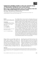
pursued. As shown in Fig. 1, NaCl stabilized both
VsEndA and VcEndA by increasing the T
m
value.
Salt also affected the shape of the thermograms,
making the denaturation peaks narrower and sharper
with increasing salt concentration. DSC of VsEndA
in 0.050 m NaCl was not performed because of sam-
ple instability.
The symmetrical shape of the thermograms suggests
that the transition proceeds via a single transition
state. The increases in T
m
from 0.175 to 1 m NaCl
were 10.1 and 9.0 °C for VsEndA and VcEndA,
respectively (Table 1). The DH
cal
values increased with
salt concentration, except for VcEndA above 0.425 m
NaCl, although DH
eff
also increased in this case.
VsEndA Q69N showed a higher T
m
value, but DH
cal
was unchanged. All other mutants showed T
m
values
comparable with the native enzyme, but a lower DH
cal
.
The accordance between DH
cal
and the model-depen-
dent van’t Hoff enthalpy (DH
eff
) was best at moderate
salt concentrations, decreasing at high extreme concen-
trations and for the mutants. The denaturation heat
capacity increment could not be determined because of
irreversibility of unfolding.
Kinetics
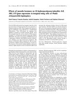
Kinetic measurements were made in 0–0.6 m NaCl.
Striking differences were observed in the K
m
and k
cat
values of the native endonucleases (Fig. 2). The K
m
value for VcEndA increased steeply at salt concentra-
tions above 0.25 m. An equivalent increase was seen
for VsEndA above 0.50 m NaCl. The k
cat
values also
showed the same salinity optima: 0.25 m for VcEndA
and 0.50 m for VsEndA. VsEndA was increasingly
more efficient than VcEndA in terms of k
cat
⁄ K
m
(Table 2) as the salt concentration increased. The
k
cat
⁄ K
m
salt optima were not very different for the two
Fig. 1. Denaturation heat capacity curves of the native and mutant
VsEndA (top) and VcEndA (bottom). Differential scanning calorime-
try profiles were recorded at a scan rate of 1 °CÆmin
)1
in a buffer
containing 0.175, 0.425 and 1.00
M NaCl for native VsEndA, and
also 0.050
M NaCl for native VcEndA. For VsEndA and VcEndA
mutants, 0.425 and 0.175
M NaCl, respectively, were used.
Thermograms were baseline-subtracted and normalized for protein
concentration.
L. Niiranen et al. Effects of salt on Vibrio endonucleases
FEBS Journal 275 (2008) 1593–1605 ª 2008 The Authors Journal compilation ª 2008 FEBS 1595
enzymes (0.175 and 0.1 m for VsEndA and VcEndA,
respectively), but the optimum was much broader for
VsEndA.
The reciprocal mutations of the two residues partici-
pating in creating the substrate binding site affected
both kinetic constants (Table 2). The variants dis-
played higher K
m
and k
cat
values, especially at high
salt concentrations, except for the VcEndA N71K
mutation which showed a decreased k
cat
value and a
minimal effect on K
m
. The catalytic efficiency of all
variants was decreased compared with the native
enzymes. Interestingly, the salt optimum of VcEndA
N69Q was shifted to zero salinity; both VsEndA vari-
ants were also more efficient than the native enzyme at
zero salinity.
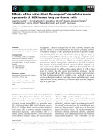
Structure of VcEndA N69Q
The crystal structure of VcEndA N69Q was deter-
mined to 1.7 A
˚
resolution, and data collection and
refinement statistics are presented in Table 3. The elec-
tron density was well defined for most of the protein
chain, and the mutated structure was similar to the
native VcEndA with an rmsd of 0.20 A
˚
for main chain
atoms. Differences were found for Asn71 and the
mutated residue 69. Electron density maps (Fig. 3)
showed that the orientation of these side chains was
different from the native structure. The side chain of
Gln69 in VcEndA N69Q was rotated away from resi-
due 72 and was unable to form the Asn69 OD1–
Arg72 N hydrogen bond, which has been suggested to
stabilize the 69–72 loop in VcEndA [18]. Instead,
Gln69 NE2 formed a hydrogen bond with Asn129
OD1 and a water-mediated hydrogen bond with
Glu125 O. Gln69 OE1 in VcEndA N69Q interacted
through water molecules to both the side chain of
Arg67 and to Glu125 O. The orientation of Arg67 was
slightly shifted relative to the native structure. Interest-
ingly, the side chain of Asn71 in the mutated structure
was also rotated, interacting only with a symmetry
related molecule with a hydrogen bond to Glu179 O
and a water-mediated bond to Gln180 O.
Electrostatic calculations
The electrostatic surface potentials of the enzyme vari-
ants were calculated at the optimal salinity of the
native enzymes (Fig. 4). The effects of the mutations
on the overall potentials were small, but some local
changes were observed. The mutations of VsEndA
appeared to result in a less positively charged surface
by increasing the exposure of a negatively charged
patch (VsEndA Q69N, Fig. 4B) or through the loss of
a positive charge (VsEndA K71N, Fig. 4C). VcEndA
N69Q mutation (Fig. 4E) led to the rotation of a
neighbouring positive charge, Arg67, whereas, in
Table 1. Thermodynamic parameters of the thermal unfolding of
VsEndA and VcEndA as a function of NaCl concentration deter-
mined by DSC.
NaCl
(
M)
T
m
(°C)
DH
cal
(kJÆmol
)1
)
DH
eff
(kJÆmol
)1
) DH
cal
⁄ DH
eff
VsEndA
a
Native 0.175 41.8 268 275 0.97
0.425 44.8 351 406 0.86
1.000 51.9 371
b
468
b
0.79
Q69N 0.425 49.0 359 445 0.81
K71N 0.425 45.3 194 275 0.71
VcEndA
a
Native 0.050 48.8 324
b
344
b
0.94
0.175 52.9 451 478 0.94
0.425 56.8 512 528 0.97
1.000 61.9 417 560 0.74
N69Q 0.175 52.6 321 415 0.77
N71K 0.175 53.7 305 385 0.79
a
Molecular masses: VsEndA, 25 005 Da; VcEndA, 24 732 Da;
VsEndA mutants, 24 645 Da; VcEndA mutants, 24 991 Da.
b
Mini-
mal values as a result of aggregation.
Fig. 2. Plot of the kinetic parameters K
m
(A) and k
cat
(B) for native
VsEndA (d) and VcEndA (s) in 0–0.6
M NaCl. The error bars repre-
sent maximum and minimum values.
Effects of salt on Vibrio endonucleases L. Niiranen et al.
1596 FEBS Journal 275 (2008) 1593–1605 ª 2008 The Authors Journal compilation ª 2008 FEBS
VcEndA N71K (Fig. 4F), an increased positive surface
potential was observed.
Discussion
For marine organisms and their extracellular proteins,
adaptation to environmental conditions can be
assumed to be somewhat more complex than simple
temperature or salt adaptation. Previous studies of the
two secreted endonucleases VsEndA (marine psychro-
philic) and VcEndA (estuarine mesophilic) have shown
that their activity is strongly dependent on tempera-
ture, but also on NaCl concentration [18]. We studied
how different salt concentrations and mutations affect
the stability and kinetic constants of VsEndA and
VcEndA, and found the effects to be striking, espe-
cially for VsEndA.
Thermal stability
At 175 mm NaCl, the native enzymes display a differ-
ence of 11.1 °CinT
m
. The difference in T
m
and the
calorimetric enthalpy is small compared with that
found for extremophilic DNA ligases [23]. This con-
firms our previous finding that, for a psychrophilic
enzyme, VsEndA has a relatively high temperature
optimum and kinetic stability [11]. The reason for the
small DT
m
may be linked to the charged residues, as
the hydrophobic cores of the two enzymes are similar.
The extra salt bridges in the C-terminus of VcEndA
and the smaller repulsion between the positively
charged residues are the likely cause for the increased
T
m
value compared with VsEndA.
At concentrations less than 1 m, salt interacts with
proteins in a non-specific manner by neutralizing
charges. The addition of salt may lead to a decrease in
intramolecular electrostatic repulsion, but an increase
in the hydrophobic effect [24,25]. Quantitative studies
of the effects of NaCl on protein thermostability are
scarce, but, in general, it has been found that there is a
direct relationship between salinity and the upshift in
the thermal unfolding temperature T
m
[26–28]. This
agrees with our finding of a nearly equal increase in
the T
m
values of the two enzymes when salt is added,
although the salt-induced increase in enthalpy is more
pronounced in VsEndA than in VcEndA.
A salt-induced increase in T
m
with a simultaneous
decrease in DH
cal
has been proposed to result from
stronger but less cooperative intramolecular interac-
tions [29]. In this context, cooperativity means that the
protein structure unfolds as a single unit (one single
transition), as opposed to several more or less inde-
pendent units (several transitions). The increase in T
m
and DH
cal
of EndA with increasing salt concentration
Table 2. Kinetic constants for native and mutant VsEndA and VcEndA at 0–0.6 M NaCl.
[NaCl] (
M) VsEndA VsEndA Q69N VsEndA K71N VcEndA VcEndA N69Q VcEndA N71K VsEndA ⁄ VcEndA
K
m
(nM) 0 64.5 ± 10.1 96.5 ± 15.8 67.5 ± 14.6 35.6 ± 4.8 100 ± 10 67.5 ± 7.3 1.8
0.100 32.0 ± 5.4 82.1 ± 10.7 82.5 ± 12.0 44.1 ± 4.5 283 ± 29 78.6 ± 8.8 0.72
0.175 34.6 ± 5.2 93.5 ± 13.3 92.9 ± 12.0 115 ± 14 650 ± 30 129 ± 14 0.30
0.250 49.5 ± 5.2 231 ± 35 99.3 ± 15.5 440 ± 21 2890 ± 380 567 ± 79 0.11
0.350 128 ± 13 377 ± 38 248 ± 18 2120 ± 230 9220 ± 1910 2170 ± 320 0.060
0.425 186 ± 10 1240 ± 120 975 ± 64 2720 ± 340 0.068
0.500 356 ± 16 2540 ± 400 1760 ± 280
0.600 1150 ± 100
k
cat
(s
)1
) 0 2.77 ± 0.11 4.82 ± 0.16 3.83 ± 0.12 2.40 ± 0.07 2.98 ± 0.08 1.62 ± 0.04 1.2
0.100 3.88 ± 0.15 7.64 ± 0.29 7.93 ± 0.34 4.05 ± 0.09 5.82 ± 0.22 2.88 ± 0.08 0.96
0.175 7.35 ± 0.25 12.4 ± 0.5 9.73 ± 0.33 5.75 ± 0.21 6.94 ± 0.15 4.18 ± 0.13 1.3
0.250 9.90 ± 0.25 23.8 ± 1.2 12.1 ± 0.5 6.39 ± 0.13 8.98 ± 0.88 4.68 ± 0.29 1.5
0.350 15.8 ± 0.5 29.4 ± 1.2 25.0 ± 0.6 4.43 ± 0.33 7.25 ± 1.33 4.86 ± 0.48 3.6
0.425 20.8 ± 0.4 43.7 ± 2.5 37.6 ± 1.3 1.95 ± 0.17 11
0.500 22.4 ± 0.4 53.3 ± 5.8 41.5 ± 4.2
0.600 18.6 ± 0.9
k
cat
⁄ K
m
(s
)1
ÆlM
)1
) 0 42.9 50.0 56.7 67.5 29.7 24.0 0.64
0.100 122 93.1 96.1 91.7 20.5 36.6 1.3
0.175 213 133 105 50.1 10.7 32.3 4.2
0.250 200 103 122 14.5 3.11 8.25 14
0.350 123 78.0 101 2.09 0.786 2.23 59
0.425 112 35.1 38.5 0.717 160
0.500 62.8 21.0 23.6
0.600 16.1
L. Niiranen et al. Effects of salt on Vibrio endonucleases
FEBS Journal 275 (2008) 1593–1605 ª 2008 The Authors Journal compilation ª 2008 FEBS 1597
could therefore be interpreted, conversely, as increased
cooperativity of unfolding and a more compact struc-
ture as a result of stronger intramolecular interactions.
These salt-induced effects on DH
cal
and DH
eff
are
stronger in VsEndA, possibly because of the increased
number of solvent-exposed charged and hydrophobic
residues relative to VcEndA, as found when viewing
the molecular surfaces and their amino acid properties.
This indicates a certain degree of salt dependence of
VsEndA stability, but is in disagreement with the
observation that the T
m
values for both enzymes are
equally affected by salt addition. It has been suggested
that salt stabilizes halophilic proteins to a greater
extent than non-halophilic proteins, and that halophilic
proteins are destabilized by low salt concentrations [9].
Both effects may originate from the characteristic high
negative surface potential of halophilic proteins and
increased solvent ion binding [8,30,31]. The equal
increase in T
m
of VsEndA and VcEndA may suggest
that VsEndA does not have any specific ion binding
sites on its surface relative to VcEndA, and is not halo-
philic. The more highly charged surface of VsEndA
may, however, constitute a more cooperative solvent
ion binding network, which makes it possible for the
enzyme to better tolerate fluctuations in salt concentra-
tion.
The observed general decrease in DH
cal
⁄ DH
eff
with
increasing salt concentration may imply that the theo-
retical model used is unable to tackle the increased
cooperativity of unfolding, but may also be explained
by an increase in the degree of irreversible unfolding
(Table 1). The reversibility of thermal unfolding of a
halophilic b-lactamase has been found to be inversely
dependent on salt concentration, and has been pro-
posed to be caused by the salting-out effect of NaCl
[28]. NaCl can neutralize the surface charges of
unfolded proteins and facilitate aggregation.
Kinetics
The release of coordinated ions and water molecules
from the solvation shells of enzymes and substrates
provides a positive entropic effect that drives substrate
binding. This effect is dependent on both temperature
and salt concentration [32,33]. At elevated salt concen-
trations or low temperatures, the gain in entropy on
release of ions is reduced and substrate binding is
therefore weaker [33]. This makes binding of highly
charged DNA very challenging for marine enzymes.
DNA binding to non-halophilic proteins has been
found to be inversely dependent on salt concentration
[32,34], whereas the binding efficiency of halophilic
proteins appears to actually increase with increasing
salt concentration [35,36]. A halophilic nuclease from
Micrococcus varians [37] with maximal activity in
3–4 m NaCl displays an excess of acidic residues char-
acteristic of many halophilic enzymes. It is possible
that this enzyme has a binding mechanism involving
counterion uptake, similar to that proposed for the
halophilic Pyrococcus woesei TATA-box binding pro-
tein [36]. Contrary to these halophilic proteins,
VsEndA displays an excess of basic residues contacting
the negatively charged substrate, and the K
m
value
increases with increasing salt concentration, although
this occurs at a much higher salinity than for VcEndA.
In our previous study of EndA temperature adapta-
tion, the more positively charged surface of VsEndA
was considered not to decrease the K
m
value relative
to VcEndA [11]. These measurements were made at
the respective optimal salt concentrations of the
enzymes, where the K
m
values were found to be of the
Table 3. X-ray data collection and crystallographic refinement sta-
tistics for the VcEndA N69Q structure.
Data collection
X-ray source In-house rotation anode
Space group P 2
1
2
1
2
1
Unit cell (A
˚
) a = 40.26, b = 64.41,
c = 75.64
Resolution (A
˚
) (highest bin) 25.00-1.70 (1.79-1.70)
Wavelength (A
˚
) 1.54180
No. unique reflections 22 098
Multiplicity 2.9 (2.8)
Completeness (%) 99.3 (99.1)
Mean (<I> ⁄ <rI>) 12.8 (2.2)
R-sym (%)
a
6.5 (35.6)
Wilson B-factor (A
˚
2
) 20.5
Refinement
PDB entry 2VND
Resolution (A
˚
) 15.00-1.70
R-factor (all reflections) (%) 19.7
R-free (%)
b
25.9
No. of atoms 1928
No. of water molecules 223
No. of other molecules 1 Mg
2+
,1Cl
)
rmsd bond lengths (A
˚
) 0.017
rmsd bond angles (deg) 1.520
Average B-factor (A
˚
2
)
All atoms 17.1
Protein 16.1
Water molecules ⁄ Mg
2+
⁄ Cl
)
24.4 ⁄ 22.7 ⁄ 11.1
Ramachandran plot
Most favoured regions (%) 93.9
Additionally allowed regions (%) 5.5
Generously allowed regions (%) 0.6
a
R-sym = (
P
h
P
I
| I
i
(h)–<I(h)> |) ⁄ (
P
h
P
I
I(h)), where I
i
(h) is the ith
measurement of reflection h and <I(h)> is the weighted mean of all
measurements of h.
b
5% of the reflections were used in the
R-free calculations.
Effects of salt on Vibrio endonucleases L. Niiranen et al.
1598 FEBS Journal 275 (2008) 1593–1605 ª 2008 The Authors Journal compilation ª 2008 FEBS
same magnitude. This can be explained by the similar
or slightly lower electrostatic surface potential of
VsEndA compared with VcEndA at the respective
optimal salinities (Fig. 4A,D). The results of the pres-
ent study show that the K
m
values are strongly affected
by the NaCl concentration, similar to the surface
charge of the enzyme. The higher positive charge of
VsEndA therefore decreases K
m
, but this is a method
of coping with the charge shielding of buffer solutes
rather than low temperatures. The higher charge may
allow VsEndA to retain sufficient charge, even at rela-
tively high salinity, to enable tight substrate binding,
contrary to VcEndA.
The salt adaptation of kinetic constants as striking
as that observed in the present study has not been pre-
sented previously. Only two comparative studies of the
salt-dependent kinetics of a non-halophilic and a salt-
adapted enzyme have been published to date. In the
comparison of halotolerant Dunaliella salina carbonic
anhydrases dCA I and dCA II and the human homo-
logue in 0–0.5 m NaCl, the largest differences were
found in the K
m
values [10,38]. Similar to our results,
the halotolerant enzymes retained a low K
m
, whereas
the K
m
value of the non-halophilic enzyme increased
considerably with the addition of salt. These results
imply that K
m
salt tolerance is a feature typical to
halotolerance. The role of k
cat
is less clear. Both
Bageshwar et al. [38] and Premkumar et al. [10] found
k
cat
to be increased only slightly by salt, whereas we
observed a large effect for k
cat
for both VsEndA and
VcEndA at high salinity. The higher catalytic rate may
reflect the dependence of k
cat
on the substrate binding
and dissociation rate constants, as found in the cold
adaptation of cod trypsin [39], or, in the case of
VsEndA, may be linked in some way to cold adapta-
tion, where an increase in k
cat
is a typical mechanism
[7]. A high k
cat
value may also be a feature of salt tol-
erance, but more studies on halotolerant enzymes are
required to verify this. The addition of salt may cause
the EndA substrate binding cleft to reach a more opti-
mal configuration for enzyme catalysis, thereby affect-
ing k
cat
. The DSC thermograms indicate that salt
constricts the structural fluctuations of the enzyme. At
a certain concentration, these fluctuations may become
optimal for enzymatic turnover, whereas, at salt con-
centrations above the optimum, the structure becomes
too rigid and will function less optimally. If the stabi-
lizing effect of salt is caused mainly by the weakening
Fig. 3. (A) Electron density (2F
o
– F
c
at 1r contoured in blue) and omit (F
o
– F
c
at 3r contoured in green) maps illustrating the orientation of
Asn71 and the N69Q mutation in the VcEndA N69Q structure. (B) Superposition of the VcEndA N69Q mutant (red), native VcEndA (blue)
and VsEndA (green) structures. (C) A partial sequence alignment of VsEndA and VcEndA. The asterisks indicate the non-conserved residues
selected for mutagenesis, and the plus sign denotes the catalytically important His80. Sequence numbering follows that of Vvn [20].
L. Niiranen et al. Effects of salt on Vibrio endonucleases
FEBS Journal 275 (2008) 1593–1605 ª 2008 The Authors Journal compilation ª 2008 FEBS 1599
of repulsive charges, it is reasonable to imagine that
VsEndA must be screened by a higher salt concentra-
tion than VcEndA to be able to function optimally.
Effects of mutations
The point mutations are not in the immediate vicinity
of the active site situated at the bottom of the posi-
tively charged pocket, but are still likely to affect the
shape, stability and charge of the DNA binding site
(Fig. 4). The Asn69 side chain in VcEndA forms a
hydrogen bond to Arg72 N, which may stabilize this
loop region relative to Vvn and VsEndA [19], whereas
the hydrogen bond observed in the VcEndA N69Q
structure (Gln69 to Asn129) stabilizes other regions.
The characterization of the VcEndA N69Q mutant
(Table 2) shows higher K
m
and k
cat
values compared
with native VcEndA. The lost hydrogen bond (from 69
to 72) in VcEndA N69Q may increase the flexibility of
the 69–72 loop, possibly explaining the decreased bind-
ing affinity and increased catalytic rate. In addition,
the shape of the DNA binding pocket in VcEndA
A
B
C
D
E
F
Fig. 4. Electrostatic surface potentials in
the DNA binding groove of VsEndA with a
modelled DNA (A), VsEndA Q69N (B) and
VsEndA K71N (C) all in 0.425
M NaCl, and
VcEndA (D), VcEndA N69Q (E) and VcEndA
N71K (F) all in 0.175
M NaCl. The black
arrows show the mutated residues. The sur-
face potential is coloured from )10 kT ⁄ q
(red) to 10 kT ⁄ q (blue).
Effects of salt on Vibrio endonucleases L. Niiranen et al.
1600 FEBS Journal 275 (2008) 1593–1605 ª 2008 The Authors Journal compilation ª 2008 FEBS
N69Q is slightly altered as both residues 71 and 69 are
moved in the crystal structure (Fig. 3), and the current
orientation of Gln69 is different from the Vvn–DNA
structure (Fig. 3B) and very close to a modelled DNA
backbone, possibly explaining the higher K
m
values
(Table 2). Gln69 in VsEndA is poorly defined in the
native crystal structure, and the characterization of the
VsEndA Q69N mutant (Table 2) reveals poorer DNA
binding and an increased k
cat
with a maximum at
0.5 m NaCl. The Gln69 side chain in the Vvn–DNA
structure (PDB 1OUP) is less than 3.2 A
˚
from the
DNA backbone, and mutation to the shorter Asn in
VsEndA Q69N may prevent the formation of favour-
able DNA–enzyme interactions, and lead to the higher
K
m
values observed. In addition, the Asn69 to Arg72
hydrogen bond lost in the VcEndA N69Q structure
may be formed in VsEndA Q69N, although this
should be verified by structural studies. This additional
hydrogen bond may explain the increased stability of
the VsEndA Q69N mutant, and the subsequent
decrease in flexibility may further impair substrate
binding and contribute to the higher K
m
values.
The introduction or removal of a positively charged
residue (N71K and K71N) has a large effect on the
electrostatic surface potential (Fig. 4). However, the
VcEndA N71K mutant has a binding affinity and
turnover comparable with the native VcEndA. Being
more distal from DNA, as observed in the Vvn–DNA
structure, residue 71 may have less influence on DNA
binding than residue 69. Interestingly, k
cat
starts to
decrease when the salt concentration exceeds 0.25 m
for both native VcEndA and VcEndA N69Q, but this
is not observed for the VcEndA N71K variant. The
VsEndA K71N mutant shows poorer DNA binding
and increased k
cat
compared with VsEndA, indicating
that the positive charge is more important for DNA
binding in VsEndA than in VcEndA.
No side chain contacts are seen for residue 71 in
the native structures or models of the mutants. As the
longer side chain of lysine has more rotamers, the
N71K substitution in VcEndA may stabilize the struc-
ture by increasing the rotational entropy, whilst retain-
ing the backbone interactions. An increase of 1 °Cis
observed for the T
m
value of this mutant. In VsEndA
K71N, both DH
cal
and the cooperativity of unfolding
are decreased, possibly indicating changes in the
hydrogen bonding networks. The increase in K
m
may
be the result of a slightly enlarged binding site or less
positive charge. Indeed, the changes seen in the electro-
static surface potential of each of the mutants (Fig. 4)
match surprisingly well with their kinetic results. Both
VsEndA mutants and the VcEndA N69Q mutant show
more dispersed or less positive charge, and, accord-
ingly, display higher and more salt-sensitive K
m
values.
VcEndA N71K does not display a lower K
m
value, but
one similar to the native enzyme, in spite of the acqui-
sition of an additional positive charge, possibly
because of other effects caused by the mutation.
Even minor changes in protein structure, such as
single amino acid replacements, can induce a signifi-
cant change in the cooperativity of unfolding, and be
detected as changes in the effective (van’t Hoff)
enthalpy [40]. In the DSC experiments, only the
VsEndA Q69N mutation had the expected effect,
increasing the stability via both T
m
and the cooperativ-
ity of unfolding, although the DH
cal
value was compa-
rable with that of the native enzyme. Whether or not
local changes are reflected in the global hydrogen
bonding networks, and how widespread are their
effects, cannot be discerned from the native and
mutant crystal structures. Effects on hydrogen bonding
networks may change the electron density distribution
around and in the active site and, together with small
conformational alterations, may speed up the rate-lim-
iting step of hydrolysis. Such long-range effects have
been proposed to be the cause of more mesophilic-like
kinetic behaviour in psychrophilic a-amylase, where
single amino acid mutations were introduced outside
the catalytic cleft [22,41]. The identity of the rate-limit-
ing step in the endonuclease reaction mechanism is not
known. However, the high catalytic efficiency of both
VsEndA and VcEndA (k
cat
⁄ K
m
in the region of
10
8
s
)1
Æm
)1
) shows that the reaction is nearly diffusion
controlled, suggesting that the rate-limiting step is
either substrate binding or dissociation. As all muta-
tions affect K
m
, especially at high salt concentrations,
the optimization and salt tolerance of binding interac-
tions are most probably hampered by the mutations
by electrostatic, steric or flexibility effects. The less
tight binding of DNA may enable the enzymes to
release the products more easily, thus leading to the
observed increase in the k
cat
values of three of the vari-
ants. Similarly, the seven-fold higher k
cat
value of the
hyperactive variant of Escherichia coli dihydrofolate
reductase, compared with the wild-type enzyme, has
been suggested to result from increased flexibility and
size of the substrate binding cleft, leading to an
increased product dissociation constant [42].
Conclusions
The experiments conducted in this study show that the
secreted endonuclease VsEndA from the marine psy-
chrophilic V. salmonicida is remarkably salt tolerant
and therefore unique amongst the endonucleases char-
acterized so far. Salt has striking effects on the kinetic
L. Niiranen et al. Effects of salt on Vibrio endonucleases
FEBS Journal 275 (2008) 1593–1605 ª 2008 The Authors Journal compilation ª 2008 FEBS 1601
constants of VsEndA, and the high positive charge of
VsEndA is considered to be essential in counteracting
the charge shielding of buffer solutes and maintaining
a low K
m
at high salinity. It is possible that K
m
salt
tolerance will emerge as a general feature for halotoler-
ant proteins. The role of the high k
cat
value observed
for VsEndA is less clear, and more studies on halotol-
erant enzymes are required to elucidate this further.
The salt-induced increase in enthalpy and cooperativity
of unfolding is more pronounced in VsEndA. This
effect indicates the formation of a more compact struc-
ture through the strengthening of intramolecular inter-
actions or the weakening of intramolecular repulsive
forces, and the salt dependence of VsEndA stability.
The higher positive electrostatic surface potential of
VsEndA compared with VcEndA plays a key role in
adaptation. On the whole, the characteristics of
VsEndA and VcEndA illustrate the fine-tuned adapta-
tion to their natural environments.
Materials and methods
Site-directed mutagenesis and plasmid
purification
Residue targets for mutagenesis were selected on the basis
of the sequence and structural alignments of Vvn, VsEndA
and VcEndA. The selected residues 69 and 71 were non-
conserved between VsEndA and VcEndA, located in the
DNA binding region and close to the active site. Site-direc-
ted mutagenesis was performed using a QuikChange Site-
Directed Mutagenesis Kit (Stratagene, Cedar Creek, TX,
USA), as described in the manual. The oligonucleotides
were synthesized by Sigma-Aldrich (St Louis, MO, USA).
Mutated plasmids were transformed into E. coli TOP10
cells (Invitrogen, Carlsbad, CA, USA), and plasmid extrac-
tion was performed using QIAprep minipreps (Qiagen,
Hilden, Germany) or the alkaline lysis method [43].
Expression and purification
The expression and purification of recombinant VsEndA
and VcEndA native enzymes and mutants were performed
as described previously [11] with a few modifications. Cells
were cultured in either shake-culture flasks or a Techfors S
fermenter (Infors, Bottmingen, Switzerland). The culture
temperature was kept at 37 °C until glucose was depleted,
after which the temperature was adjusted to 22 °C before
expression was induced. The cells were harvested when they
reached the stationary phase and were collected by centrifu-
gation. For periplasmic fractionation, the cells were resus-
pended in a 1 : 10 culture volume of fractionation buffer,
and incubated on ice for 1–1.5 h before the supernatant
was collected.
Enzyme assay
Enzyme activity measurements were assayed in triplicate at
23 °Cin75mm Tris ⁄ HCl, pH 8.0 and pH 8.5 (VcEndA and
VsEndA, respectively), 5 mm MgCl
2
and 0–0.6 m NaCl.
Eight different concentrations (12–1470 nm) of DNaseAlert
substrate (DNaseAlertÔ QC System Kit; Ambion, Austin,
TX, USA) were used for the kinetic measurements, and
200 nm substrate for the other activity measurements. The
total reaction volume was 100 lL and reactions were started
by the addition of 10 lL of enzyme diluted in reaction
buffer. Protein LoBind tubes from Eppendorf (Hamburg,
Germany) were used for enzyme dilutions because of the
sticky nature of the enzyme. The detailed assay procedure
is described elsewhere [11]. sigmaplot software (Systat
Software, San Jose, CA, USA) was used for data analysis,
and V
max
and K
m
values were calculated by fitting the
velocity data to the Michaelis–Menten equation.
Differential scanning calorimetry
Differential scanning calorimetry experiments were con-
ducted on a Nano-Differential Scanning Calorimeter III,
model CSC6300 (Calorimetry Sciences Corporation, Lin-
don, UT, USA). Preparations of the native enzymes were
first filtered with a 0.45 lm Spin-X centrifuge tube filter
(Corning, Corning, NY, USA), and then dialysed overnight
at 4 °C against 1 L of dialysis buffer (50 mm Hepes, 5 mm
MgCl
2
, pH 8.0) containing 0.050, 0.175, 0.425 or 1.00 m
NaCl. Slide-A-Lyzer dialysis discs from Pierce (Rockford,
IL, USA) with a 2 kDa cut-off were used. The protein con-
centration of the dialysed enzyme solution was determined
using BioRad Protein Assay Dye Reagent Concentrate
(BioRad, Hercules, CA, USA) with bovine serum albumin
(Sigma) as standard. The dialysates were used as blank ref-
erences in DSC runs. Reference buffers and samples were
carefully degassed before loading into the DSC cells. The
scans were performed at a constant pressure of 304 kPa in
the range 15–75 °C or 20–80 °C with a heating rate of
1 °CÆmin
)1
. Thermograms were analysed according to a
single non-two-state transition model in which T
m
, DH
cal
and DH
eff
were fitted independently using cpcalc software
(Calorimetry Sciences Corporation).
Crystallization, data collection and structure
determination
The mutant VcEndA N69Q was crystallized in similar con-
ditions as native VcEndA [19] using the hanging drop
vapour-diffusion technique at room temperature with
6.2 mgÆmL
)1
of protein in 50 mm Tris ⁄ HCl pH 8.0, 5 mm
MgCl
2
and 0.6 m NaCl. Drops were made by mixing 1 lL
of protein with 1 lL of reservoir solution consisting of
0.1 m sodium acetate, 0.3 m ammonium acetate, 10 mm
magnesium sulphate and 26% PEG8000. Crystals of about
Effects of salt on Vibrio endonucleases L. Niiranen et al.
1602 FEBS Journal 275 (2008) 1593–1605 ª 2008 The Authors Journal compilation ª 2008 FEBS
500 · 200 · 20 lm
3
were transferred to cryoprotectant
solution with 30% PEG8000, 15% glycerol and the other
reservoir additives, and flash-cooled in liquid nitrogen.
Data were collected at the in-house MicroMax-007 HF
rotating anode from Rigaku (Osaka, Japan) with an
R-AXIS IV detector, a 60 s exposure time per image and
0.5° oscillation, and a total of 78° of data were used in the
final data set. The data were integrated with the program
mosflm [44], scaled with scala, and the structure factors
obtained with truncate in the ccp4 program suite [45].
The structure was solved by molecular replacement using
the program molrep [46] in ccp4 and the structure of
native VcEndA (PDB code 2G7E) as a search model. The
structure was refined in refmac5 [47] interspersed with
rounds of manual model building in O [48] based on
r
A
-weighted 2F
o
– F
c
and F
o
– F
c
electron density maps.
The final model was validated using procheck [49].
Molecular modelling and electrostatic calculations
Continuum electrostatic calculations were carried out using
the delphi program package [50,51]. The parse3 set of
atomic radii [52], together with formal charges, was used in
all calculations. The electrostatics were determined using
the linear Poisson–Boltzmann equation and a three-dimen-
sional grid with a size of 165 · 165 · 165. Stepwise focus-
ing was used to increase the accuracy [53]. Initially, a rough
grid was calculated with Coulombic boundary conditions,
and the resulting grid was adopted as the boundary condi-
tion for one further focused calculation. The protein mole-
cules occupied 90% of the box in the final calculations. The
molecular surface was calculated using a solvent probe of
1.4 A
˚
. The solvent was described using a dielectric constant
of 80, whereas the protein was treated with a dielectric con-
stant of 4. The calculations were carried out using the
optimal salt concentrations of the native enzymes. The
structures used for the calculations were VcEndA (PDB
2G7F), VcEndA N69Q (this study, PDB 2VND) and
VsEndA (PDB 2PU3). The VcEndA N71K mutant was
generated from 2G7F, and the mutants of VsEndA were
generated from native VsEndA.
Acknowledgements
We thank Annfrid Sivertsen, Stefan Hauglid and
Bjarte Lund for technical assistance. This study was
supported by The National Programme for Research
in Functional Genomics in Norway (FUGE) in The
Research Council of Norway.
References
1 Madern D, Ebel C & Zaccai G (2000) Halophilic adap-
tation of enzymes. Extremophiles 4, 91–98.
2 Miller GP & Benkovic SJ (1998) Stretching exercises –
flexibility in dihydrofolate reductase catalysis. Chem
Biol 5, R105–R113.
3 Hough DW & Danson MJ (1999) Extremozymes. Curr
Opin Chem Biol 3, 39–46.
4 Li WF, Zhou XX & Lu P (2005) Structural features of
thermozymes. Biotechnol Adv 23, 271–281.
5 Zecchinon L, Claverie P, Collins T, D’Amico S, Delille
D, Feller G, Georlette D, Gratia E, Hoyoux A, Meuwis
MA et al. (2001) Did psychrophilic enzymes really win
the challenge? Extremophiles 5, 313–321.
6 Hochachka PW & Somero GN (1984) Biochemical
Adaptation. Princeton University Press, Princeton, NJ.
7 Siddiqui KS & Cavicchioli R (2006) Cold-adapted
enzymes. Annu Rev Biochem 75, 403–433.
8 Mevarech M, Frolow F & Gloss LM (2000) Halophilic
enzymes: proteins with a grain of salt. Biophys Chem
86, 155–164.
9 Elcock AH & McCammon JA (1998) Electrostatic con-
tributions to the stability of halophilic proteins. J Mol
Biol 280, 731–748.
10 Premkumar L, Greenblatt HM, Bageshwar UK, Sav-
chenko T, Gokhman I, Sussman JL & Zamir A (2005)
Three-dimensional structure of a halotolerant algal car-
bonic anhydrase predicts halotolerance of a mamma-
lian homolog. Proc Natl Acad Sci USA 102, 7493–
7498.
11 Altermark B, Niiranen L, Willassen NP, Smalas AO &
Moe E (2007) Comparative studies of endonuclease I
from cold-adapted Vibrio salmonicida and mesophilic
Vibrio cholerae. FEBS J 274, 252–263.
12 Ohta Y & Hatada Y (2006) A novel enzyme, lambda-
carrageenase, isolated from a deep-sea bacterium.
J Biochem (Tokyo) 140, 475–481.
13 Focareta T & Manning PA (1991) Distinguishing
between the extracellular DNases of Vibrio cholerae and
development of a transformation system. Mol Microbiol
5, 2547–2555.
14 Wu SI, Lo SK, Shao CP, Tsai HW & Hor LI (2001)
Cloning and characterization of a periplasmic nuclease
of Vibrio vulnificus and its role in preventing uptake of
foreign DNA. Appl Environ Microbiol 67, 82–88.
15 Moulard M, Condemine G & Robert-Baudouy J (1993)
Search for the function of the nuclease NucM of Erwi-
nia chrysanthemi. FEMS Microbiol Lett 112 , 99–103.
16 Buchanan JT, Simpson AJ, Aziz RK, Liu GY, Kristian
SA, Kotb M, Feramisco J & Nizet V (2006) DNase
expression allows the pathogen group A Streptococcus
to escape killing in neutrophil extracellular traps. Curr
Biol 16
, 396–400.
17 Palic D, Ostojic J, Andreasen CB & Roth JA (2006)
Fish cast NETs: neutrophil extracellular traps are
released from fish neutrophils. Dev Comp Immunol 31,
805–816.
L. Niiranen et al. Effects of salt on Vibrio endonucleases
FEBS Journal 275 (2008) 1593–1605 ª 2008 The Authors Journal compilation ª 2008 FEBS 1603
18 Altermark B, Helland R, Moe E, Willassen NP & Sma-
la
˚
s AO (2008) Structural adaptation of endonuclease I
from the cold-adapted and halophilic bacterium Vibrio
salmonicida. Acta Crystallogr D Biol Crystallogr,
doi:10.1107/S0907444908000097.
19 Altermark B, Smalas AO, Willassen NP & Helland R
(2006) The structure of Vibrio cholerae extracellular
endonuclease I reveals the presence of a buried chloride
ion. Acta Crystallogr D Biol Crystallogr 62, 1387–1391.
20 Li CL, Hor LI, Chang ZF, Tsai LC, Yang WZ & Yuan
HS (2003) DNA binding and cleavage by the periplas-
mic nuclease Vvn: a novel structure with a known active
site. EMBO J 22, 4014–4025.
21 Wang YT, Yang WJ, Li CL, Doudeva LG & Yuan HS
(2007) Structural basis for sequence-dependent DNA
cleavage by nonspecific endonucleases. Nucleic Acids
Res 35, 584–594.
22 D’Amico S, Gerday C & Feller G (2003) Temperature
adaptation of proteins: engineering mesophilic-like
activity and stability in a cold-adapted alpha-amylase.
J Mol Biol 332, 981–988.
23 Georlette D, Damien B, Blaise V, Depiereux E, Uver-
sky VN, Gerday C & Feller G (2003) Structural and
functional adaptations to extreme temperatures in psy-
chrophilic, mesophilic, and thermophilic DNA ligases.
J Biol Chem 278, 37015–37023.
24 Nishimura C, Uversky VN & Fink AL (2001) Effect of
salts on the stability and folding of staphylococcal
nuclease. Biochemistry 40, 2113–2128.
25 Lanyi JK (1974) Salt-dependent properties of proteins
from extremely halophilic bacteria. Bacteriol Rev 38,
272–290.
26 Dragan AI, Li Z, Makeyeva EN, Milgotina EI, Liu Y,
Crane-Robinson C & Privalov PL (2006) Forces driving
the binding of homeodomains to DNA. Biochemistry
45, 141–151.
27 Waldron TT, Schrift GL & Murphy KP (2005) The
salt-dependence of a protein–ligand interaction:
ion–protein binding energetics. J Mol Biol 346, 895–
905.
28 Tokunaga H, Arakawa T, Fukada H & Tokunaga M
(2006) Opposing effects of NaCl on reversibility and
thermal stability of halophilic beta-lactamase from a
moderate halophile, Chromohalobacter sp. 560. Biophys
Chem 119, 316–320.
29 Chan HK, Au-Yeung KL & Gonda I (1996) Effects of
additives on heat denaturation of rhDNase in solutions.
Pharm Res 13, 756–761.
30 Fukuchi S, Yoshimune K, Wakayama M, Moriguchi M
& Nishikawa K (2003) Unique amino acid composition
of proteins in halophilic bacteria. J Mol Biol 327, 347–
357.
31 Richard SB, Madern D, Garcin E & Zaccai G (2000)
Halophilic adaptation: novel solvent protein interac-
tions observed in the 2.9 and 2.6 A resolution structures
of the wild type and a mutant of malate dehydrogenase
from Haloarcula marismortui. Biochemistry 39, 992–
1000.
32 Ha JH, Capp MW, Hohenwalter MD, Baskerville M
& Record MT Jr (1992) Thermodynamic stoichiome-
tries of participation of water, cations and anions in
specific and non-specific binding of lac repressor to
DNA. Possible thermodynamic origins of the ‘‘gluta-
mate effect’’ on protein–DNA interactions. J Mol Biol
228, 252–264.
33 Fogolari F, Elcock AH, Esposito G, Viglino P, Briggs
JM & McCammon JA (1997) Electrostatic effects in
homeodomain–DNA interactions. J Mol Biol 267
,
368–381.
34 Record MT Jr, Zhang W & Anderson CF (1998) Anal-
ysis of effects of salts and uncharged solutes on protein
and nucleic acid equilibria and processes: a practical
guide to recognizing and interpreting polyelectrolyte
effects, Hofmeister effects, and osmotic effects of salts.
Adv Protein Chem 51, 281–353.
35 Britton KL, Baker PJ, Fisher M, Ruzheinikov S, Gil-
mour DJ, Bonete MJ, Ferrer J, Pire C, Esclapez J &
Rice DW (2006) Analysis of protein solvent interactions
in glucose dehydrogenase from the extreme halophile
Haloferax mediterranei. Proc Natl Acad Sci USA 103,
4846–4851.
36 Bergqvist S, Williams MA, O’Brien R & Ladbury JE
(2003) Halophilic adaptation of protein–DNA interac-
tions. Biochem Soc Trans 31, 677–680.
37 Kamekura M & Onishi H (1978) Properties of the
halophilic nuclease of a moderate halophile, Micro-
coccus varians subsp. halophilus. J Bacteriol 133, 59–
65.
38 Bageshwar UK, Premkumar L, Gokhman I, Savchenko
T, Sussman JL & Zamir A (2004) Natural protein engi-
neering: a uniquely salt-tolerant, but not halophilic,
alpha-type carbonic anhydrase from algae proliferating
in low- to hyper-saline environments. Protein Eng Des
Sel 17, 191–200.
39 Asgeirsson B & Cekan P (2006) Microscopic rate-con-
stants for substrate binding and acylation in cold-adap-
tation of trypsin I from Atlantic cod. FEBS Lett 580,
4639–4644.
40 Privalov GP & Privalov PL (2000) Problems and pros-
pects in microcalorimetry of biological macromolecules.
Methods Enzymol 323, 31–62.
41 D’Amico S, Gerday C & Feller G (2001) Structural
determinants of cold adaptation and stability in a large
protein. J Biol Chem 276, 25791–25796.
42 Iwakura M, Maki K, Takahashi H, Takenawa T,
Yokota A, Katayanagi K, Kamiyama T & Gekko K
(2006) Evolutional design of a hyperactive cysteine-
and methionine-free mutant of Escherichia coli
dihydrofolate reductase. J Biol Chem 281, 13234–
13246.
Effects of salt on Vibrio endonucleases L. Niiranen et al.
1604 FEBS Journal 275 (2008) 1593–1605 ª 2008 The Authors Journal compilation ª 2008 FEBS
43 Sambrook J, Fritsch EF & Maniatis T (1989) Molecular
Cloning: A Laboratory Manual. Cold Spring Harbor
Laboratory Press, Cold Spring Harbor, NY.
44 Powell HR (1999) The Rossmann Fourier autoindexing
algorithm in MOSFLM. Acta Crystallogr D Biol Crys-
tallogr 55, 1690–1695.
45 Collaborative Computational Project Number 4 (1994)
The CCP4 suite: programs for protein crystallography.
Acta Crystallogr D Biol Crystallogr 50 , 760–763.
46 Vagin A & Teplyakov A (1997) MOLREP: an auto-
mated program for molecular replacement. J Appl
Crystallogr 30, 1022–1025.
47 Murshudov GN, Vagin AA & Dodson EJ (1997)
Refinement of macromolecular structures by the maxi-
mum-likelihood method. Acta Crystallogr D Biol Crys-
tallogr 53, 240–255.
48 Jones TA, Zou JY, Cowan SW & Kjeldgaard M (1991)
Improved methods for building protein models in elec-
tron density maps and the location of errors in these
models. Acta Crystallogr A 47, 110–119.
49 Laskowski RA, Moss DS & Thornton JM (1993) Main-
chain bond lengths and bond angles in protein struc-
tures. J Mol Biol 231, 1049–1067.
50 Rocchia W, Alexov E & Honig B (2001) Extending the
applicability of the nonlinear Poisson–Boltzmann equa-
tion: multiple dielectric constants and multivalent ions.
J Phys Chem B 105, 6507–6514.
51 Rocchia W, Sridharan S, Nicholls A, Alexov E,
Chiabrera A & Honig B (2002) Rapid grid-based
construction of the molecular surface and the use of
induced surface charge to calculate reaction field
energies: applications to the molecular systems and
geometric objects. J Comput Chem 23, 128–137.
52 Sitkoff D, Sharp KA & Honig B (1994) Accurate
calculation of hydration free-energies using macroscopic
solvent models. J Phys Chem 98, 1978–1988.
53 Moreira IS, Fernandes PA & Ramos MJ (2005)
Accuracy of the numerical solution of the Poisson–
Boltzmann equation. J Mol Struct-Theochem 729,
11–18.
L. Niiranen et al. Effects of salt on Vibrio endonucleases
FEBS Journal 275 (2008) 1593–1605 ª 2008 The Authors Journal compilation ª 2008 FEBS 1605