Báo cáo khoa học: Structural studies of nucleoside analog and feedback inhibitor binding to Drosophila melanogaster multisubstrate deoxyribonucleoside kinase doc
Bạn đang xem bản rút gọn của tài liệu. Xem và tải ngay bản đầy đủ của tài liệu tại đây (609.23 KB, 10 trang )
Structural studies of nucleoside analog and feedback
inhibitor binding to Drosophila melanogaster
multisubstrate deoxyribonucleoside kinase
Nils E. Mikkelsen
1
, Birgitte Munch-Petersen
2
and Hans Eklund
1
1 Department of Molecular Biology, Swedish University of Agricultural Sciences, Biomedical Center, Uppsala, Sweden
2 Department of Science, Systems and Models, Roskilde University, Denmark
Cells need to keep a balanced pool of dNTPs to sus-
tain DNA synthesis and repair. The main source of
dNTPs comes from the de novo pathway where ribonu-
cleosides are converted to ribonucleotides by the
enzyme ribonucleotide reductase [1]. In resting cells,
where ribonucleotide reductase activity is low, there is
an alternative route for obtaining dNTPs, namely the
salvage pathway. Here, nucleosides that originate from
dead cells and food are salvaged from the extracellular
space and transported into the cell. Once inside, they
become phosphorylated by deoxyribonucleoside kinas-
es and are thus prevented from leaving the cell [2].
Mammalian cells have four different deoxynucleo-
side kinases with distinct, but overlapping, substrate
affinities. Thymidine kinase 1 (TK1) and deoxycytidine
kinase (dCK) are found in the cytosol, and thymidine
kinase 2 (TK2) and deoxyguanosine kinase (dGK) are
found in the mitochondria. TK1 has the most
restricted substrate specificity and phosphorylates only
deoxythymidine (dT) and deoxyuridine, whereas dCK
is somewhat more relaxed and phosphorylates both
pyrimidine and purine deoxynucleosides. The best sub-
strate for dCK is deoxycytidine (dC), but dCK also
phosphorylates deoxyadenosine and deoxyguanosine.
TK2, which phosphorylates the same substrates as
TK1, can also phosphorylate dC and other medically
interesting dT, deoxyuridine and dC analogs. dGK
only phosphorylates the purine deoxyribonucleosides
deoxyadenosine, deoxyguanosine and deoxyinosine.
In addition, many pharmacological nucleoside ana-
logs (NAs) that are used in both antiviral therapy and
cancer therapy need activation by deoxynucleoside
Keywords
cancer gene therapy; deoxyribonucleoside
kinase; nucleoside analogs; pyrimidines;
X-ray structures
Correspondence
H. Eklund, Department of Molecular
Biology, Swedish University of Agricultural
Sciences, Biomedical Center, S-751 24
Uppsala, Sweden
Fax: +46 18536971
Tel: +46 184714559
E-mail:
(Received 10 January 2008, revised 27
February 2008, accepted 3 March 2008)
doi:10.1111/j.1742-4658.2008.06369.x
The Drosophila melanogaster multisubstrate deoxyribonucleoside kinase
(dNK; EC 2.7.1.145) has a high turnover rate and a wide substrate range
that makes it a very good candidate for gene therapy. This concept is based
on introducing a suicide gene into malignant cells in order to activate a
prodrug that eventually may kill the cell. To be able to optimize the func-
tion of dNK, it is vital to have structural information of dNK complexes.
In this study we present crystal structures of dNK complexed with four dif-
ferent nucleoside analogs (floxuridine, brivudine, zidovudine and zalcita-
bine) and relate them to the binding of substrate and feedback inhibitors.
dCTP and dGTP bind with the base in the substrate site, similarly to the
binding of the feedback inhibitor dTTP. All nucleoside analogs investigated
bound in a manner similar to that of the pyrimidine substrates, with many
interactions in common. In contrast, the base of dGTP adopted a syn-
conformation to adapt to the available space of the active site.
Abbreviations
5FdU, floxuridine: 5-fluoro-2¢-deoxyuridine; AZT, zidovudine: 3¢-azidothymidine; BVDU, brivudin: (E)-bromvinyl-2¢-deoxyuridine; BVU, (E)-5-
(2-bromovinyl)-uracil; dC, deoxycytidine; dCK, cytosolic deoxycytidine kinase; ddC, zalcitabine: 2¢,3¢-dideoxycytidine; dGK, mitochondrial
deoxyguanosine kinase; dNK, Drosophila melanogaster deoxyribonucleoside kinase; dT, deoxythymidine; HSV-1, herpes simplex virus 1;
NA, nucleoside analog; TK, thymidine kinase; TK1, thymidine kinase 1; TK2, thymidine kinase 2; VZV, varicella zoster virus.
FEBS Journal 275 (2008) 2151–2160 ª 2008 The Authors Journal compilation ª 2008 FEBS 2151
kinase-catalyzed phosphorylation. In humans, the main
activators of the NAs are the deoxynucleoside kinases,
which phosphorylate the NAs, thereby trapping them
inside the cell. This is regarded as the rate-limiting step
and makes the deoxynucleoside kinases important
actors in combating malignant cells. One approach in
this battle is gene therapy, where a suicide gene is
introduced into a malignant cell followed by the addi-
tion of a NA specifically activated by the enzyme
encoded by this gene. The activated NA is then
expected to kill the malignant cell. This can occur
either by incorporation of the triphosphorylated form
of the NA into cellular DNA, causing chain break or
termination, or by other inhibitory effects that ulti-
mately inhibit viral replication or kill the recipient cell
[3] by inducing apoptosis [4]. Examples of NAs
targeted towards deoxynucleoside kinases are 1-b-
d-arabinofuranosylguanosine and 2-chloro-2¢-deoxyad-
enosine, which are phosphorylated by dCK and dGK,
respectively.
The Drosophila melanogaster multisubstrate deoxyri-
bonucleoside kinase (dNK; EC 2.7.1.145) can phos-
phorylate all natural substrates and a wide range of
medically important NAs with outstanding efficiency,
as shown in Table 1 [5–9]. This makes it a very prom-
ising candidate as a suicide gene in gene therapy and it
has also been shown to be transducible into human
cancer cell lines [10]. dNK mutants have given some
remarkable results by sensitizing different cancer cell
lines towards different NAs by more than 18 000-fold
compared with the parental cell line [9, W. Knecht
et al., unpublished data]. The possibility of tailoring
suicide genes with the end result being the almost com-
plete elimination of natural substrate affinities and
feedback inhibition, can therefore make the enzymes,
produced by these mutated genes, highly efficient acti-
vators for specific NAs. In this way, the lower amount
of NA needed may considerably reduce the toxic side
effects that often accompany this type of therapy.
The main drawback in gene therapy has been the
targeting and successful delivery of suicide genes into
the cells of interest. When this obstacle is overcome,
we will have an arsenal of very potent suicide genes
that are ready for use in anticancer therapies.
The 3D structure of dNK has previously been deter-
mined in complexes with substrates and a feedback
inhibitor [12,13]. It has a structure similar to that of
the human dGK and dCK and belongs to a structural
family that also contains some viral thymidine kinases
(TKs) [14]. These enzymes contain a P-loop and a LID
region that binds phosphates of the phosphate donor,
usually ATP (Fig. 1), and an LID region that closes
down on the phosphates of the phosphate donor
(Fig. 1).
In this article we describe the crystal structure of
dNK with four different NAs. In addition, we investi-
gated additional substrate and dNTP complexes. In
most cases, a truncated version of dNK lacking the
last 20 residues was used. This truncation mutant has
kinetic characteristics similar to those of the full-length
enzyme, but because the k
cat
is two- to threefold
higher, it is even faster [15].
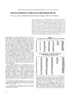
Table 1. Kinetic parameters for dNK with natural substrates and
NA from the crystal structures.
K
m
(lM)
V
max
(lmolÆmin
)1
Æmg
)1
)
k
cat
(s
)1
)
k
cat
⁄ K
m
(lM
)1
Æs
)1
)
dT
a
1.2 29.5 14.2 12
dC
a
2.3 34.2 16.5 7.2
dA
a
225 42.7 20.6 0.092
dG
a
665 31.3 19 0.029
5-FdU 1.0 29.8 14.2 14
BVDU
b
2.2 13.2 5.9 2.7
AZT
c
8.3 0.073 0.036 0.0043
ddC
c
1124 8.6 4.2 0.0037
a
Data are from [15].
b
Data are from [16].
c
Data are from [6].
LID
P-loop
ERS
α1
α2
α3
α4
α6
α5
α8
α7
β1
β2
β3
β4
β5
Fig. 1. 3D structure of dNK with dCTP bound as a feedback inhibi-
tor. The protein structure has a central parallel five-stranded
b sheet surrounded by helices. The LID region, P loop and ERS
motifs are in red.
Nucleoside analog deoxynucleoside kinase complexes N. E. Mikkelsen et al.
2152 FEBS Journal 275 (2008) 2151–2160 ª 2008 The Authors Journal compilation ª 2008 FEBS
Results and Discussion
Quality of the structures
dNK is an enzyme with flexible parts that had to be
stabilized to obtain well-diffracting crystals. The phos-
phate-binding regions have, in all structures deter-
mined to date, been stabilized by sulfate ions or by the
phosphates of a feedback inhibitor. Furthermore, the
C-terminus is flexible in all structures, such as in both
truncated proteins that we mainly used for crystalliza-
tion, as well as in the full-length enzyme (see below).
The best diffracting crystals have been obtained in
the presence of triphosphate inhibitors, where the
phosphate-interacting regions are stabilized, whereas
the binary complexes with NAs in the best cases dif-
fract slightly better than 3 A
˚
resolution. The structures
of dNK in complex with the substrates dC and dT
have previously been determined [12,13]. We have now
been able to determine the dC complex at a slightly
higher resolution (2.3 A
˚
), which is here used as a refer-
ence for the discussion of the NA complexes. Although
this complex was co-crystallized with the phosphate
donor product ADP, this nucleotide was not found at
the phosphate donor site. It had been outcompeted by
a sulfate ion, as in the other substrate complexes.
NA binding
We determined the structures of dNK with four
pyrimidine NAs: floxuridine (5FdU, 5-fluoro-2¢-deoxy-
uridine), zidovudine (AZT, 3¢-azido-2¢,3¢-dideoxythymi-
dine), zalcitabine (ddC, 2¢,3¢-dideoxycytidine) and
brivudin [BVDU, (E)-5-(2-bromovinyl)-2¢-deoxyuri-
dine]. The kinetic parameters for these are given in
Table 1. When discussing the binding and the effect of
the analogue on dNK, it is presumed, as previously
described [16], that the catalytic or preceding step is
rate determining, and that the size of the K
m
reflects
the nucleoside binding affinity. All refinement statistics
can be found in Table 2.
Floxuridine (5FdU) is an oncologic drug most often
used in the treatment of breast and colorectal cancer.
The nucleotide form of floxuridine (5FdUMP) irrevers-
ibly inhibits thymidylate synthase, which leads to a
strong reduction of thymine nucleotides in the cell and
this, in turn, inhibits DNA synthesis [17]. 5FdU is
phosphorylated efficiently by dNK with the same high
k
cat
⁄ K
m
of 2 · 10
7
m
)1
Æs
)1
as with thymidine, and
10-fold higher than with TK1 [14].
The crystal structure of dNK with 5FdU is very sim-
ilar to the previously solved substrate structures with
dT and dC [12,13]. It contains a sulfate ion bound in
the P loop, and the substrates are at nearly identical
positions in the active site. The interactions of the
deoxyribose and the base are identical to those of the
dT complex, except for the fluoride atom replacing
the methyl group on the base (Fig. 2A). In the dC
complex we find two water molecules occupying this
cleft, making an interacting bridge between OE2 on
Glu52 and N4 on the dC base, as shown in Fig. 3A.
In the 5FdU complex the fluoride occupies this space,
Table 2. Data collection and refinement statistics for the dNK ligand complexes.
Statistics dC (ADP) 5FdU ddC BVDU AZT dCTP dGTP dNKwt-dTTP
Space group P2
1
2
1
2P2
1
P2
1
P2
1
2
1
2P2
1
2
1
2P2
1
P2
1
2
1
2P2
1
2
1
2
Cell dimensions 120.6 70.4 70.5 137.5 140.0 67.9 119.7 119
62.5 70.7 70.8 112.8 111.9 119 65.1 64.9
68.2 225.4 226.0 69.7 71.1 70.5 69.2 69.1
Content au 1 dimer 4 dimers 4 dimers 2 dimers 2 dimers 2 dimers 1 dimer 1 dimer
Resolution (A
˚
) 50–2.3 30–3.0 50–2.9 30–2.9 20–2.8 50–2.2 50–2.5 45–2.2
Completness (%) 98.5 (91.2) 99.3 (99.3) 97.3 (97.1) 83.9 (87.4) 99.4 (99.8) 99.5 (99.5) 99.8 (99.7) 99.6 (99.6)
Rsym 0.075 (0.434) 0.084 (0.528) 0.116 (0.583) 0.094 (0.540) 0.114 (0.474) 0.071 (0.370) 0.096 (0.555) 0.069 (0.414)
Rmeas 0.089 (0.522) 0.103 (0.655) 0.136 (0.678) 0.116 (0.666) 0.134 (0.555) 0.088 (0.462) 0.103 (0.597) 0.082 (0.486)
Mn(I) ⁄ sd 13.1 (2.1) 11.3 (2.0) 13.0 (2.1) 9.7 (2.3) 9.3 (3.1) 11.3 (3.1) 17.4 (4.0) 16 (3.2)
Redundancy 3.4 (2.7) 2.9 (3.0) 3.7 (3.8) 2.8 (2.7) 3.6 (3.6) 2.8 (2.9) 7.1 (7.3) 3.4 (3.5)
Reflections 22045 46314 50991 19428 26596 53215 18113 26365
R factor (%) 23.4 25.6 24.8 24.2 23.5 19.5 20.7 21.4
Rfree (%) 27.3 28.1 28.7 28.6 27.1 24.9 26.7 25.9
rmsd bond lengths 0.009 0.013 0.015 0.013 0.012 0.012 0.011 0.010
rmsd bond angles 1.151 1.326 1.471 1.398 1.836 1.403 1.421 1.183
Mean B value (A
˚
2) 39.2 63.1 45.6 54.6 39.7 31.8 34.4 36.1
Beamline ID14-4 ID23-1 ID14-2 ID14-1 ID-14-1 ID-29 ID-29 ID-29
PDB-code 2vp5 2vp6 2vp9 2vqs 2jj8 2vp4 2vp2 2vp0
N. E. Mikkelsen et al. Nucleoside analog deoxynucleoside kinase complexes
FEBS Journal 275 (2008) 2151–2160 ª 2008 The Authors Journal compilation ª 2008 FEBS 2153
expelling the two water molecules in a manner similar
to that previously reported for dT and its methyl
group [13].
5FdU is phosphorylated efficiently by dNK with the
same K
m
and k
cat
values as with thymidine (Table 1).
This is in agreement with the high similarity observed
between the crystal structures obtained with dT and
5FdU.
Zalcitabine (ddC) is an NA used in the treatment of
HIV infections. The structure of the ddC complex
(Fig. 2B) shows that the analog binds similarly as the
natural pyrimidine substrates but lacks a hydrogen
bond because of the absence of the 3¢-OH. Two water
molecules bridge between Glu52 and N4 of the analog,
as seen in the dC complex.
The K
m
for ddC is almost 500-fold higher than for
dC, whereas the k
cat
is decreased only by 3.3-fold.
Thus, the catalytic step should be expected to be
R167
A
R169
E172
Y70
M69
M118
Q81
A110
M88
R105
E52
K33
T34
R167
R169
E172
Y70
M69
M118
Q81
A110
M88
R105
E52
K33
T34
B
Fig. 3. Initial difference density maps, contoured at 3r, for (A) dC
and one sulfate ion and for (B) AZT and two sulfate ions. All hydro-
gen bonds are shown as red dotted lines and water molecules are
shown as red balls.
E172
A
Y70
M69
M118
Q81
A110
M88
R105
E52
E172
Y70
M69
M118
Q81
A110
M88
R105
E52
M88
Y70
M69
M118
Q81
A110
S106
R105
E52
B
C
Fig. 2. Initial difference density maps, contoured at 3r, covering
the NAs (A) 5FdU, (B) ddC and (C) BVDU. Water molecules are
shown as red balls.
Nucleoside analog deoxynucleoside kinase complexes N. E. Mikkelsen et al.
2154 FEBS Journal 275 (2008) 2151–2160 ª 2008 The Authors Journal compilation ª 2008 FEBS
affected very little but the binding should be strongly
affected. The structure shows that ddC is in the proper
position for P transfer, but very poorly bound due to
the loss of the hydrogen bonds as a result of the miss-
ing 3¢-OH.
Brivudine (BVDU) is an NA used in the treatment
of herpes simplex virus type 1 (HSV-1) and varicella
zoster virus (VZV) infections. BVDU has also shown
potential as a cancer drug in gene therapy ⁄ chemother-
apy as a result of its cytostatic activity in cancer cells
transduced with viral TK genes. BVDU may also
enhance the potency of 5-fluorouracil in combined
chemotherapy, because BVDU becomes degraded by
thymidine phosphorylase to (E)-5-(2-bromovinyl)uracil
(BVU). This metabolite, in turn, inactivates dihydro-
pyrimidine dehydrogenase, which is the enzyme that
initiates the degradative pathway of 5-fluorouracil.
Balzarini et al. [18] have also shown some promising
results using BVDU as insecticide, where D. melanog-
aster and Spodoptera frugiperda embryonic cells
showed high sensitivity towards BVDU.
The dNK complexes with BVDU (Fig. 2C) and dT
have very similar overall structures. However, BVDU
is slightly displaced compared with dT to accommo-
date the bulky bromovinyl group in the deep cleft sur-
rounded by residues Ser109, Ala110, Val84, Trp57 and
Arg105. The LID is partly missing, and helix a3
(which interacts with the LID) is displaced similarly as
in the AZT complex (see below). There are no signifi-
cant conformational changes of the side chains in the
active site, as found in HSV-TK where Tyr132, the
equivalent to Met88 in dNK, is shifted to make room
for the more bulky groups of dT and BVDU. The
minor structural changes in the structure with BVDU
compared with dT are in agreement with the very simi-
lar kinetic values.
There are two previously determined structures, with
BVDU and brivudine monophosphate (BVDUMP) in
the HSV-1-TK + BVDU complex [19] and the
VZV + BVDUMP and ADP complex [20].
Zidovudine (AZT) is a potent inhibitor of HIV repli-
cation in vitro and at the time of publishing is still
included in the standard regimen for treatment of the
disease. AZT is also a substrate for dNK, although
with a k
cat
⁄ K
m
that is about 2800-fold lower than the
k
cat
⁄ K
m
for dT (Table 1).
We have determined a structure of dNK complexed
with AZT, and the difference density for the thymidine
part of AZT in the active site is well defined, as shown
in Fig. 3B. Surprisingly, there were two sulfate ions
present – one bound in the P loop, as observed in the
other substrate complexes, and the other located
between the first sulfate ion and the substrate. There
was no density for the N
3
azido group of AZT or the
part of the LID region ranging from Arg165 to Cys174.
This LID usually clamps down interacting with the sub-
strate and the sulfate ion bound in the P loop. The lack
of density here is probably caused by the N
3
group of
AZT, which protrudes into this loop region (Fig. 3B).
Superposition of the AZT complex with the dC
complex, clearly shows the steric impact that the N
3
group has on this section. The LID is totally distorted
and the interacting helix a3 (Fig. 4) on the opposite
side on top of the substrate is pushed back a little in a
rigid body-like movement, probably to accommodate
the azido group on AZT. This widening of the active
site probably also provides space for the second sulfate
ion to bind (Fig. 3B). There is also a small shift in the
P loop and the sulfate ion occupying this position,
which is displaced somewhat compared with the sul-
phate ion in the dC complex.
According to a k
cat
for AZT that is more than 400-
fold lower than with thymidine, and a K
m
that is
increased by eightfold, the catalytic step should be
effected considerably more than the binding. This is in
agreement with the N
3
group being somewhat of a
hindrance for proper binding but the LID being
completely distorted, making P transfer very difficult.
In yeast thymidylate kinase a similar shift in the
P loop was observed when the deoxythymidine mono-
phosphate (dTMP) complex was compared with
the AZT-monophosphate (AZTMP) complex. It was
Fig. 4. Superposition of dNK structures (tube representation) in
complex with AZT (red) and dC (grey) picturing the structural differ-
ences when the bulkier AZT (yellow) is bound in the active site
together with the two sulfate ions. Part of the LID is missing here
as there was no traceable density for this region.
N. E. Mikkelsen et al. Nucleoside analog deoxynucleoside kinase complexes
FEBS Journal 275 (2008) 2151–2160 ª 2008 The Authors Journal compilation ª 2008 FEBS 2155
speculated that the shift was probably a result of the
bulkier AZT and that this displacement of the loop was
the probable cause for the reduced catalytic activity of
the thymidylate kinase towards AZT [21]. The P loop is
involved in binding the phosphoryl donor and has evi-
dently moved to an unfavorable position, thereby affect-
ing the phosphoryl transfer negatively. Later work with
human thymidylate kinase [22,23] showed that mutants
with mutated amino acids in the LID region gained effi-
ciency in AZTMP phosphorylation. It was suggested
that the LID has to be in a closed conformation to be
able to phosphorylate the substrate efficiently.
Earlier work on dNK revealed that a N64D mutant
retained efficiency towards AZT, and structures of the
N64D mutant complexed with dT and dTTP were
investigated [8]. It was found that the increased effi-
ciency towards AZT was probably caused by a reduced
stability in the LID region, which made the enzyme
more relaxed towards the bulkier azido group.
Deoxynucleoside triphosphate complex
structures
Feedback inhibition of deoxynucleoside kinases is a
common way of regulating the nucleotide production
of these enzymes, and the end products of the pre-
ferred substrates are usually the best inhibitors [24].
Kim et al. [25] proposed that dCK was regulated by
the end product of the dCK metabolic pathway where
dCTP would act as a feedback inhibitor. They further
suggested that dCTP could function as a bisubstrate
analog where the triphosphate group would bind in
the phosphate donor site and the deoxycytidine base in
the phosphate acceptor site as a normal substrate. The
first structure of such a feedback-inhibited deoxyribo-
nucleoside kinase was human dGK, where it was
believed that the co-crystallized ATP was bound as a
feedback inhibitor, although the density suggested a
dATP [12]. Later work on human TK1 showed that
although this kinase was co-crystallized with different
substrates, there was always a dTTP bound as a feed-
back inhibitor [26]. The dTTP was bound so tightly
that even the purification process, which contained no
dTTP, did not release it. Similar observations were
reported for human TK2 where the feedback inhibitor
dTTP was strongly bound [27]. A re-investigation and
new refinement of the human dGK structure finally
convinced the authors that it actually was a dATP
molecule bound in dGK (pdb-code: 2ocp).
Earlier work of dNK complexed with dTTP had
demonstrated that the feedback inhibitor was indeed
bound as a bisubstrate inhibitor occupying both the
phosphate donor and acceptor sites. Here, a magne-
sium ion was bound to the phosphates [13]. The bind-
ing of the inhibitor induces a structural change where
the catalytically important residue Glu52 is shifted
along with the main chain to bind dTTP and coordi-
nate magnesium.
We have now determined two additional dNTP com-
plexes of dNK that bind like the feedback inhibitor
dTTP: one with dCTP at 2.2 A
˚
resolution and one with
dGTP at 2.5 A
˚
resolution (Fig. 5). The triphosphate
part of these dNTPs is nearly identical to the tripho-
sphate part of the dTTP structure and for dCTP the
base moiety superimposes perfectly with dC in the
dNK–dC complex. One difference, though, is that one
of the two water molecules bridging OE2 on Glu52 and
N4 on the dC base in the dNK–dC structure is now
absent. This is a result of the shift of the Glu52 to a
similar position as in the dTTP structure. There is no
R167
A
B
R169
E172
Y70
M69
M118
Q81
A110
R105
K33
T34
R167
R169
E172
Y70
M69
M118
Q81
A110
R105
K33
T34
Fig. 5. Initial difference density maps of (A) dCTP (2.2 A
˚
) and (B)
dGTP (2.5 A
˚
) and their binding in the dNK active site. All hydrogen
bonds are shown as red dotted lines and water molecules are
shown as red balls.
Nucleoside analog deoxynucleoside kinase complexes N. E. Mikkelsen et al.
2156 FEBS Journal 275 (2008) 2151–2160 ª 2008 The Authors Journal compilation ª 2008 FEBS
detectable magnesium coordinating Glu52, which in
this structure is tilted a little outwards compared with
Glu52 in the dNK–dTTP structure, as shown in Fig. 6.
In the structure of the dGTP complex, the guanosine
base occupies approximately the same geometrical space
as the base in the dCTP and dTTP ligands (Fig. 6). The
guanosine base is in the syn-conformation, in contrast
to the thymine and cytosine bases that are in the anti-
conformation in those complexes. There is a water mol-
ecule bridging ⁄ anchoring the N2 of the guanosine base
to Ser109 located at the bottom of this hydrophobic
cleft. Gln81 makes hydrogen bonds to N7 and O6 on
the side of the base acting as a clamp, but otherwise it is
supported by the same stacking interactions as
described previously in both the dC and dT structures.
Gln81 has been moved almost 1 A
˚
to be able to accom-
modate the slightly more bulky guanosine base, but
otherwise there are no significant changes to the overall
3D structure in the active site. This shows how flexible
dNK is in having room for many different substrates by
using mostly water molecules as bulk material to retain
stability around the bound ligand. There are two previ-
ously solved structures of a kinase with a guanosine
base in the active site, namely the HSV-TK complexed
with ganciclovir and penciclovir [19]. In those cases, the
base is in the anti-conformation.
Full-length dNK–dTTP complex
Most crystallographic studies on dNK have been
performed on a C-terminally truncated mutant that
has catalytic characteristics similar to those of the
wild-type enzyme [15] but was easier to crystallize.
However, we were finally able to crystallize the full-
length enzyme using the feedback inhibitor dTTP,
which made it possible to make comparisons with the
corresponding structure of the truncated enzyme. This
structure, determined at 2.2 A
˚
resolution, did not show
any additional traceable density compared with the
truncated dNK structures.
Several attempts have been made, to obtain a phos-
phate donor or a phosphate donor analog co-crystal-
lized together with a substrate, but with no success to
date. dNK that was crystallized with the substrate dC
and the phosphate donor product ADP or CDP
showed no density for either ADP or CDP. The pres-
ence of sulphate ions obviously hindered binding of
ADP or CDP. Preliminary studies of dNK complexed
with the substrate analogs AP
4
dT and AP
5
dT indicate
that it might be crucial to have the full-length enzyme
to accommodate sufficient binding for crystallization
of a complex with the phosphate donor to be able to
stabilize the structure of the last 32 amino acids suffi-
ciently to be visible in electron density maps.
Substrate specificity of dNK
Earlier crystallographic studies of substrates dT and dC
and on the structure of the feedback inhibitor complex
with dTTP, as well as mutation studies, have established
some of the basic rules for substrate specificity for this
enzyme [7,12,13]. Similar studies on human dGK and
dCK have confirmed and further complemented these
rules [28]. For dNK, the substrate site is formed by an
elongated cavity lined on the top and bottom of hydro-
phobic residues. Around this cavity, polar residues are
positioned to form specific interactions to the sugar and
the base of the substrate. The 3¢-oxygen of deoxyribose
is hydrogen bonded to Tyr70 and Glu172, and the
5¢-oxygen is hydrogen bonded to Glu52 and Arg105. A
key interaction shared by all the investigated NAs is the
binding to Gln81, which forms hydrogen bonds to the
nitrogen in position 3 and to the carbonyl or nitrogen at
position 4 of the pyrimidine ring.
In this study, we determined the structure of the com-
plexes of four pyrimidine analogs. It has so far not been
possible to obtain useful crystals with purine NAs. All
pyrimidine nucleotide analogs bind in similar modes in
spite of different substitutions. The interactions with
Gln81 are present in all analog complexes and the inter-
actions with the 5¢-position are preserved. The effect of
removing the 3¢-oxygen in ddC resulted in a weaker
interaction owing to the loss of hydrogen bonds. The
substitution of the 3¢-oxygen with an azide group in
AZT apparently destabilized part of the structure.
The only substitutions of the pyrimidine ring of the
analogs that we investigated were at the 5-position.
There is a pocket close to the 5-position that can
accommodate different substitutions. The largest one
E52
dGTP/dCTP/dTTP
Mg
Fig. 6. The three triphosphates dTTP (blue), dCTP (green) and
dGTP (yellow), superimposed together with Glu52 from each corre-
sponding structure. In the dCTP and dGTP structures Glu52 is suc-
cessively pointing outwards when compared with the dTTP
structure and in both dCTP and dGTP Glu52 makes contact with
Arg195 from the adjacent symmetry-related molecules. Magnesium
(grey) is only found in the dTTP structure.
N. E. Mikkelsen et al. Nucleoside analog deoxynucleoside kinase complexes
FEBS Journal 275 (2008) 2151–2160 ª 2008 The Authors Journal compilation ª 2008 FEBS 2157
that we analyzed was the bromovinyl group of BVDU
that fits snugly into this pocket. A larger substitution
would probably cause steric hindrance.
It has been shown, in kinetic measurements, that
dTTP is the only really efficient feedback inhibitor for
different substrates [29], which is analogous to dT
being the best substrate. In our structural studies, high
concentrations in the absence of substrate still allowed
binding of other dNTPs.
The study of the dNTPs enabled us, for the first
time, to obtain a complex with a purine bound at the
active site – the dGTP structure. To be able to bind to
this rather tight substrate site, the protein does not
adapt to the larger substrate by conformational
changes. Instead, the base adopts a syn-conformation
that differs from the anti-conformation in other sub-
strates, NAs and feedback inhibitors. Also in this case,
it is the pocket close to the 5-position in the pyrimi-
dines that accommodates the larger purine base. Gln81
forms hydrogen bonds to the base also in this case.
The position of the guanine is probably also present
in purine substrate complexes and may explain the
considerably larger K
m
values with these substrates.
Experimental procedures
Materials
Nucleosides and nucleotides were from Sigma (St Louis,
MO, USA).
Protein purification and kinetic studies
The D. melanogaster dNK was overexpressed in Escheri-
chia coli using the glutathione S-transferase (GST) gene
fusion expression system (Amersham Pharmacia Biotech,
Uppsala, Sweden). Filtered cell homogenate of induced
BL21 transformants was applied to a glutathione–Sepha-
rose column. The expressed protein was cleaved from gluta-
thione S-transferase by thrombin. Details of the expression,
purification and kinetic investigations of the recombinant
wild-type and truncated dNK have been described else-
where [6,15].
Crystallization
Crystals of all the dNK complexes were grown using the
vapor diffusion method with hanging drops. The solutions
(described below) were left to equilibrate at 14 °C and crys-
tals usually appeared after 1–2 days. After 2–3 weeks they
had typically grown to a suitable size and were flash frozen
in liquid nitrogen after a quick wash in a cryo-solution and
then stored in liquid nitrogen as described below.
dGTP
Hanging drops consisted of 2 lL of crystallization solution
containing 0.1 m Tris, pH 7.5, 0.2 m lithium citrate and 19%
poly(ethylene glycol) 3350 added to 2 lL of enzyme solution
containing 10 mgÆmL
)1
of protein and 5 mm dGTP. The
crystals were cryo-protected by a quick wash through the
crystallization solution containing 20% glycerol.
dCTP
Hanging drops consisted of 2 lL of crystallization solution
containing 0.1 m MES, pH 6.5, 0.2 m lithium citrate and
18% poly(ethylene glycol) 3350 added to 2 lL of enzyme
solution containing 10 mgÆmL
)1
of protein and 5 mm
dCTP. The crystals were cryo-protected by a quick wash
through crystallization solution containing 20% glycerol.
AZT
Hanging drops consisted of 2 lL of crystallization solution
containing 0.1 m MES, pH 6.5, 0.2 m Li
2
SO
4
and 26%
polyethylene glycol 2000 monomethylether added to 2L of
enzyme solution containing 30 mgÆmL
)1
of protein and
5mm AZT. The crystals were cryo-protected by a quick
wash through crystallization liquid containing 26%
mPEG2000.
ddC
Hanging drops consisted of 2 lL of crystallization solution
containing 0.1 m MES, pH 6.5, 0.2 m Li
2
SO
4
and 22%
mPEG2000 M added to 2 lL of enzyme solution contain-
ing 10 mgÆmL
)1
of protein and 5 mm ddC. The well solu-
tion consisted of 30% mPEG2000 and after 1 week the
coverslip with the hanging drop was further shifted to 35%
mPEG2000 for an additional week. The crystals were flash
frozen without further additions.
BVDU
Hanging drops consisted of 2 lL of crystallization solution
containing 0.1 m MES, pH 6.5, and 2.5 m Am
2
SO
4
added
to 2 lL of enzyme solution containing 20 mgÆmL
)1
of pro-
tein and 3.7 mm BVDU. The crystals were cryo-protected
by a quick wash through crystallization liquid containing
25% glycerol.
5FdU
Hanging drops consisted of 2 lL of crystallization solution
containing 0.1 m MES, pH 6.5, 0.2 m Li
2
SO
4
and 22%
mPEG2000 M added to 2 lL of enzyme solution contain-
ing 10 mgÆmL
)1
of protein and 5 mm 5FdU. The well
Nucleoside analog deoxynucleoside kinase complexes N. E. Mikkelsen et al.
2158 FEBS Journal 275 (2008) 2151–2160 ª 2008 The Authors Journal compilation ª 2008 FEBS
solution consisted of 30% mPEG2000 and after 1 week the
cover slip with the hanging drop was transferred to 35%
mPEG2000 for an additional week. The crystals were flash
frozen without further additions.
dC+ADP
Hanging drops consisted of 2 lL of crystallization solution
containing 0.2 m K
2
SO
4
, 20% poly(ethylene glycol) 3350,
pH 6.8 (Hampton Research PEG ⁄ Ion Screen condition
#34), added to 2 lL of enzyme solution containing
15 mgÆmL
)1
of protein, 5 mm dC and 5 mm ADP. After
1 week the cover slip with the hanging drop was shifted to
30% poly(ethylene glycol) 3350. The crystals were cryo-pro-
tected by a quick wash through a mixture of 80% crystalli-
zation solution, 10% ethylene glycol and 10% glycerol.
Full-length dNK+dTTP
Hanging drops consisted of 2 lL of crystallization solution
containing 0.1 m Tris, pH 7.5, 0.2 m potassium citrate,
12% polypropylene glycol P400 and 20% poly(ethylene gly-
col) 3350 added to 2 lL of enzyme solution containing
10 mgÆmL
)1
of protein and 5 mm dTTP. The crystals were
flash frozen without further additions.
Data collection
X-ray diffraction data were collected at 100 K at various
beamlines at ESRF Grenoble (Table 2). The data were
scaled and merged using the programs mosflm [30] and
scala [31]. Data collection statistics are shown in Table 2.
Structure determination and refinement
Structures with the same space group and similar cell
dimensions as previous complexes could often readily be
determined directly by a few rounds of rigid body refine-
ment. If this did not succeed, the structures were solved by
molecular replacement using the program phaser [32]. The
refined structure of the previously determined dNK–dC
dimer was used as a search model. After rigid-body and
restrained refinement in refmac5 [33], an initial electron
map was calculated. From this map most of the polypep-
tide chains could be built using the programs o [34] and
coot [35].
Acknowledgements
This work was supported by grants from the Swedish
Research Council (to H.E.), the Swedish Cancer Foun-
dation (to H.E.) and the Danish Research council (to
B.M.P) and the Novo Nordic Research Council (to
B.M.P.).
References
1 Thelander L & Reichard P (1979) Reduction of ribonu-
cleotides. Annu Rev Biochem 48, 133–158.
2 Arne
´
r ES & Eriksson S (1995) Mammalian deoxyribo-
nucleoside kinases. Pharmacol Ther 67, 155–186.
3 Niculescu-Duvaz I & Springer CJ (2005) Introduction
to the background, principles, and state of the art in
suicide gene therapy. Mol Biotechnol 30, 71–88.
4 Genini D, Adachi S, Chao Q, Rose DW, Carrera CJ,
Cottam HB, Carson DA & Leoni LM (2000) Deoxyad-
enosine analogs induce programmed cell death in
chronic lymphocytic leukemia cells by damaging the
DNA and by directly affecting the mitochondria. Blood
96, 3537–3543.
5 Munch-Petersen B, Piskur J & Søndergaard L (1998)
Four deoxynucleoside kinase activities from Drosophila
melanogaster are contained within a single monomeric
enzyme, a new multifunctional deoxynucleoside kinase.
J Biol Chem 273, 3926–3931.
6 Knecht W, Munch-Petersen B & Piskur J (2000) Identi-
fication of residues involved in the specificity and regu-
lation of the highly efficient multisubstrate
deoxyribonucleoside kinase from Drosophila melanogas-
ter. J Mol Biol 301, 827–837.
7 Knecht W, Sandrini MP, Johansson K, Eklund H,
Munch-Petersen B & Piskur J (2002) A few amino acid
substitutions can convert deoxyribonucleoside kinase
specificity from pyrimidines to purines. EMBO J 21,
1873–1880.
8 Welin M, Skovgaard T, Knecht W, Zhu C, Berenstein
D, Munch-Petersen B, Piskur J & Eklund H (2005)
Structural basis for the changed substrate specificity of
Drosophila melanogaster deoxyribonucleoside kinase
mutant N64D. Febs J 272, 3733–3742.
9 Knecht W, Rozpedowska E, Le Breton C, Willer M,
Gojkovic Z, Sandrini MP, Joergensen T, Hasholt L,
Munch-Petersen B & Piskur J (2007) Drosophila deoxy-
ribonucleoside kinase mutants with enhanced ability to
phosphorylate purine analogs. Gene Ther 14, 1278–
1286.
10 Zheng X, Johansson M & Karlsson A (2000) Retroviral
transduction of cancer cell lines with the gene encoding
Drosophila melanogaster multisubstrate deoxyribonu-
cleoside kinase. J Biol Chem 275, 39125–39129.
11 Reference withdrawn.
12 Johansson K, Ramaswamy S, Ljungcrantz C, Knecht
W, Piskur J, Munch-Petersen B, Eriksson S & Eklund
H (2001) Structural basis for substrate specificities of
cellular deoxyribonucleoside kinases. Nat Struct Biol 8,
616–620.
13 Mikkelsen NE, Johansson K, Karlsson A, Knecht W,
Andersen G, Piskur J, Munch-Petersen B & Eklund H
(2003) Structural basis for feedback inhibition of the
deoxyribonucleoside salvage pathway: studies of the
N. E. Mikkelsen et al. Nucleoside analog deoxynucleoside kinase complexes
FEBS Journal 275 (2008) 2151–2160 ª 2008 The Authors Journal compilation ª 2008 FEBS 2159
Drosophila deoxyribonucleoside kinase. Biochemistry
42, 5706–5712.
14 Eriksson S, Munch-Petersen B, Johansson K & Eklund
H (2002) Structure and function of cellular deoxyribo-
nucleoside kinases. Cell Mol Life Sci 59, 1327–1346.
15 Munch-Petersen B, Knecht W, Lenz C, Søndergaard L
& Piskur J (2000) Functional expression of a multisub-
strate deoxyribonucleoside kinase from Drosophila mela-
nogaster and its C-terminal deletion mutants. J Biol
Chem 275, 6673–6679.
16 Egeblad-Welin L, Sonntag Y, Eklund H & Munch-
Petersen B (2007) Functional studies of active-site
mutants from Drosophila melanogaster deoxyribonucleo-
side kinase. Investigations of the putative catalytic
glutamate-arginine pair and of residues responsible for
substrate specificity. FEBS J 274, 1542–1551.
17 Longley DB, Harkin DP & Johnston PG (2003) 5-fluo-
rouracil: mechanisms of action and clinical strategies.
Nat Rev Cancer 3, 330–338.
18 Balzarini J, Degreve B, Hatse S, De Clercq E, Breuer
M, Johansson M, Huybrechts R & Karlsson A (2000)
The multifunctional deoxynucleoside kinase of insect
cells is a target for the development of new insecticides.
Mol Pharmacol 57, 811–819.
19 Champness JN, Bennett MS, Wien F, Visse R, Sum-
mers WC, Herdewijn P, de Clerq E, Ostrowski T, Jar-
vest RL & Sanderson MR (1998) Exploring the active
site of herpes simplex virus type-1 thymidine kinase by
X-ray crystallography of complexes with aciclovir and
other ligands. Proteins 32, 350–361.
20 Bird LE, Ren J, Wright A, Leslie KD, Degreve B, Bal-
zarini J & Stammers DK (2003) Crystal structure of
varicella zoster virus thymidine kinase. J Biol Chem
278, 24680–24687.
21 Kenyon GL (1997) AZT monophosphate knocks thymi-
dylate kinase for a loop. Nat Struct Biol 4, 595–597.
22 Ostermann N, Lavie A, Padiyar S, Brundiers R, Veit T,
Reinstein J, Goody RS, Konrad M & Schlichting I
(2000) Potentiating AZT activation: structures of wild-
type and mutant human thymidylate kinase suggest rea-
sons for the mutants’ improved kinetics with the HIV
prodrug metabolite AZTMP. J Mol Biol 304, 43–53.
23 Ostermann N, Schlichting I, Brundiers R, Konrad M,
Reinstein J, Veit T, Goody RS & Lavie A (2000)
Insights into the phosphoryltransfer mechanism of
human thymidylate kinase gained from crystal struc-
tures of enzyme complexes along the reaction coordi-
nate. Structure 8, 629–642.
24 Park I & Ives DH (1995) Kinetic mechanism and end-
product regulation of deoxyguanosine kinase from beef
liver mitochondria. J Biochem (Tokyo) 117, 1058–1061.
25 Kim MY & Ives DH (1989) Human deoxycytidine
kinase: kinetic mechanism and end product regulation.
Biochemistry 28, 9043–9047.
26 Birringer MS, Claus MT, Folkers G, Kloer DP, Schulz
GE & Scapozza L (2005) Structure of a type II thymi-
dine kinase with bound dTTP. FEBS Lett 579, 1376–
1382.
27 Barroso JF, Carvalho RN & Flatmark T (2005) Kinetic
analysis and ligand-induced conformational changes in
dimeric and tetrameric forms of human thymidine
kinase 2. Biochemistry 44, 4886–4896.
28 Sabini E, Ort S, Monnerjahn C, Konrad M & Lavie A
(2003) Structure of human dCK suggests strategies to
improve anticancer and antiviral therapy. Nat Struct
Biol 10, 513–519.
29 Knecht W, Petersen GE, Munch-Petersen B & Piskur J
(2002) Deoxyribonucleoside kinases belonging to the
thymidine kinase 2 (TK2)-like group vary significantly
in substrate specificity, kinetics and feed-back regula-
tion. J Mol Biol 315, 529–540.
30 Leslie AGW (1992) Mosflm., Joint CCP4 and ESF-EA-
CMB Newsletter Protein Crystallography. Daresbury
Laboratory, Warrington, UK.
31 CCP4 (1994) The CCP4 suite: programs for protein
crystallography. Acta Crystallogr D Biol Crystallogr 50,
760–763.
32 McCoy AJ, Grosse-Kunstleve RW, Storoni LC &
Read RJ (2005) Likelihood-enhanced fast translation
functions. Acta Crystallogr D Biol Crystallogr 61, 458–
464.
33 Murshudov GN, Vagin AA & Dodson EJ (1997)
Refinement of macromolecular structures by the maxi-
mum-likelihood method. Acta Crystallogr D Biol Crys-
tallogr 53, 240–255.
34 Jones TA, Zou JY, Cowan SW & Kjeldgaard M (1991)
Improved methods for building protein models in elec-
tron density maps and the location of errors in these
models. Acta Crystallogr A 47, 110–119.
35 Emsley P & Cowtan K (2004) Coot: model-building
tools for molecular graphics. Acta Crystallogr D Biol
Crystallogr 60, 2126–2132.
Nucleoside analog deoxynucleoside kinase complexes N. E. Mikkelsen et al.
2160 FEBS Journal 275 (2008) 2151–2160 ª 2008 The Authors Journal compilation ª 2008 FEBS