Báo cáo khoa học: Tumor suppressor p16INK4a ) modulator of glycomic profile and galectin-1 expression to increase susceptibility to carbohydrate-dependent induction of anoikis in pancreatic carcinoma cells ppt
Bạn đang xem bản rút gọn của tài liệu. Xem và tải ngay bản đầy đủ của tài liệu tại đây (938.16 KB, 24 trang )
Tumor suppressor p16
INK4a
) modulator of glycomic profile
and galectin-1 expression to increase susceptibility to
carbohydrate-dependent induction of anoikis in pancreatic
carcinoma cells
Sabine Andre
´
1
, Hugo Sanchez-Ruderisch
2
, Hiroaki Nakagawa
3
, Malte Buchholz
4
,Ju
¨
rgen Kopitz
5
,
Pia Forberich
6
, Wolfgang Kemmner
6
, Corina Bo
¨
ck
1
, Kisaburo Deguchi
3
, Katharia M. Detjen
2
,
Bertram Wiedenmann
2
, Magnus von Knebel Doeberitz
5
, Thomas M. Gress
7
,
Shin-Ichiro Nishimura
3
, Stefan Rosewicz
2
and Hans-Joachim Gabius
1
1 Institute of Physiological Chemistry, Faculty of Veterinary Medicine, Ludwig-Maximilians-University Munich, Germany
2 Medizinische Klinik mit Schwerpunkt Hepatologie und Gastroenterologie, Charite
´
-Universita
¨
tsmedizin Berlin, Germany
3 Graduate School of Advanced Life Science, Frontier Research Center for Post-Genome Science and Technology, Hokkaido University,
Sapporo, Japan
4 Abteilung Innere Medizin I, Universita
¨
t Ulm, Germany
5 Institut fu
¨
r Angewandte Tumorbiologie, Klinikum der Ruprecht-Karls-Universita
¨
t, Heidelberg, Germany
6 Clinic of Surgery and Surgical Oncology, Robert Roessle Hospital, Charite
´
Campus Buch, Berlin, Germany
7 Department of Gastroenterology, Endocrinology and Metabolism, University Hospital Giessen and Marburg, Germany
Keywords
anoikis; fibronectin receptor; galectin;
glycosyltransferases; pancreas tumor
Correspondence
S. Andre
´
, Institute of Physiological
Chemistry, Faculty of Veterinary Medicine,
Ludwig-Maximilians-University Munich,
Veterina
¨
rstr. 13, 80539 Munich, Germany
Fax: +49 89 21802508
Tel: +49 89 21803279
E-mail:
(Received 21 February 2007, revised
23 March 2007, accepted 27 April 2007)
doi:10.1111/j.1742-4658.2007.05851.x
Expression of the tumor suppressor p16
INK4a
after stable transfection can
restore the susceptibility of epithelial tumor cells to anoikis. This property
is linked to increases in the expression and cell-surface presence of the
fibronectin receptor. Considering its glycan chains as pivotal signals, we
assumed an effect of p16
INK4a
on glycosylation. To test this hypothesis
for human Capan-1 pancreatic carcinoma cells, we combined microarray
for selected glycosyltransferase genes with 2D chromatographic glycan
profiling and plant lectin binding. Major differences between p16-positive
and control cells were detected. They concerned expression of b1,4-galacto-
syltransferases (down-regulation of b1,4-galactosyltransferases-I ⁄ V and
up-regulation of b1,4-galactosyltransferase-IV) as well as decreased
a2,3-sialylation of O-glycans and a2,6-sialylation of N-glycans. The changes
are compatible with increased b
1
-integrin maturation, subunit assembly and
binding activity of the a
5
b
1
-integrin. Of further functional relevance in line
with our hypothesis, we revealed differential reactivity towards endogenous
lectins, especially galectin-1. As a result of reduced sialylation, the cells’
capacity to bind galectin-1 was enhanced. In parallel, the level of transcrip-
tion of the galectin-1 gene increased conspicuously in p16
INK4a
-positive
cells, and even figured prominently in a microarray on 1996 tumor-associ-
ated genes and in proteomic analysis. The cells therefore gain optimal
responsiveness. The correlation between genetically modulated galectin-1
levels and anoikis rates in engineered transfectants inferred functional signi-
ficance. To connect these findings to the fibronectin receptor, galectin-1
was shown to be co-immunoprecipitated. We conclude that p16
INK4a
Abbreviations
b4GalT, b1,4-galactosyltransferase; GalNAc, N-acetylgalactosamine; GalNAcT, N-acetylgalactosaminyltransferase; GnT-V,
N-acetylglucosaminyltransferase V; LacNAc, N-acetyllactosamine; MAA, Maackia amurensis; ODS, octadecyl silane; PA, 2-aminopyridine;
pI, isoelectric point; pRb, retinoblastoma tumor-suppressor gene.
FEBS Journal 274 (2007) 3233–3256 ª 2007 The Authors Journal compilation ª 2007 FEBS 3233
Protein glycosylation has functional potential far
beyond the structural roles exerted on the protein back-
bone [1–3]. Endowed with a unique high-density coding
capacity, oligosaccharides are versatile biochemical sig-
nals in glycoprotein maturation and routing intracellu-
larly, as well as in diverse cell-surface activities such
as adhesion or trigger mechanisms for signaling to
regulate apoptosis ⁄ proliferation [4–7]. This paradigm
implies far-reaching functional consequences for the
carefully mapped aberrations of glycosylation upon
malignant transformation [8,9]. In global terms, struc-
tural studies on N-glycans from virally transformed
cells (polyoma, Rous sarcoma or hamster sarcoma
virus carrying the v-ras oncogene) have traced distinct,
nonrandom changes in the glycomic profile (i.e. an
increased degree of branching and chain extension)
[10–12]. Even more important, but still mostly descrip-
tive, oncogene presence has been shown to affect par-
ticular components of the glycosylation machinery, as
documented for H- and N-ras as well as the tyrosine
kinase oncogenes src and her-2 ⁄ neu [13–16]. Signaling
for the measured transcriptional regulation of N-acetyl-
glucosaminyltransferase V (GnT-V) and a2,6-sialyl-
transferase I (ST6Gal-I) is routed through Ras–Raf–
Ets or Ral guanine exchange factors, respectively
[15,16]. However, the conclusion to invariably link
the emergence of respective features, especially the
increased b1,6-branching of N-glycans, with the malig-
nant phenotype and therefore with an unfavorable
prognosis in tumor patients, is not justified. The oppos-
ite correlation was reported in tumor material from
nonsmall cell lung cancer, neuroblastoma and bladder
cancer [17–19]. If it were known which glycoproteins
are key targets, it could become possible to attribute
altered glycosylation to a distinct functionality.
In this respect, studies with 3T3 fibroblasts trans-
fected with the SV40 large T antigen gene, HD3 colon
epithelial cells expressing oncogenic ras and human
HT1080 fibrosarcoma cells overexpressing GnT-V
(mentioned above) have illustrated target selection
and, of special note, prominent appearance of the
b
1
-integrin or the fibronectin receptor (a
5
b
1
-integrin)
within this group [20–22]. N-Glycosylation of the
fibronectin receptor can be distributed over 14 poten-
tial sites in the a
5
-subunit and over 12 sites in the
b
1
-chain, covering at least 35 types of oligosaccharides
when analyzed for the protein from human placenta
[23]. The processing of these glycan chains is an
important part of integrin maturation, and the glyco-
sylation was shown to affect integrin association and
clustering, the capacity for fibronectin or lectin binding
and the interaction with the regulatory gangliosides
GT
1b
and GD
3
[21,24–29]. These collective insights
shape the hypothesis that remodeling the glycosylation
of the fibronectin receptor can act as a molecular
switch. If we could select a cell system in which this in-
tegrin plays a major role for the fate of the cells, then
it would be feasible to put our hypothesis to the
experimental test.
The recent finding that the tumor suppressor
p16
INK4a
restores susceptibility to anoikis induction in
human Capan-1 pancreatic carcinoma cells by increas-
ing a
5
b
1
-integrin expression and surface presentation
offers such a suitable test system [30]. We thus
assumed that the presence of the tumor suppressor –
beyond the transcriptional up-regulation of the a
5
-inte-
grin gene [30] – may engender biologically significant
influences on glycan synthesis and processing. Three
lines of evidence support the decision to test our hypo-
thesis in this system.
First, constitutive p16
INK4a
expression in human
A549 lung adenocarcinoma cells reduced global b1,4-
galactosyltransferase (b4GalT) activity on the cell sur-
face by 25%, mainly as a result of reduced expression
of the enzyme b4GalT-I [31]. Analysis of transforming
growth factor b
1
-induced rapid senescence of this
cell type by northern blots revealed two- to fivefold
increases in transcription for the b4GalT-II, -III, -V
and -VI genes and abolishment of transcription of the
b4GalT-IV gene [32].
Second, comparison of DNA microarray-based
expression profiles between specimens of pancreatic
cancer and normal tissue revealed differential gene
activities, especially up-regulation for b4GalT-V (factor
9.91) and b4GalT-I (factor 2.54) and an inverse regula-
tion for GnT-IVa and b (factors 3.23 versus )20.27)
[33].
Third, the glycosyltransferases b4GalT-V and GnT-V
were shown to be strongly expressed in a panel of eight
human cancer lines [34]. Transcriptional regulation of
this b4GalT is under the control of the transcription
factor Sp1 (which can activate genes for proteins with
pro-growth ⁄ survival properties) and probably Ets-1;
this property is shared with GnT-V [15,35,36]. Gene
orchestrates distinct aspects of glycosylation that are relevant for integrin
maturation and reactivity to an endogenous effector as well as the
effector’s expression. This mechanism establishes a new aspect of p16
INK4a
functionality.
New function of p16
INK4a
S. Andre
´
et al.
3234 FEBS Journal 274 (2007) 3233–3256 ª 2007 The Authors Journal compilation ª 2007 FEBS
expression of b4GalT-V – and also of b4GalT-I [37] –
can be enhanced by epidermal growth factor and dom-
inant active ras [38]. Intriguingly, both enzymes have a
bearing on a
5
b
1
-integrin features. b
1
-Integrin matur-
ation, and transcription of the a
5
-integrin gene, were
enhanced in human SHG44 glioma cells upon down-
regulating b4GalT-V expression [39], and the absence
of GnT-V in murine embryonic fibroblasts led to a pro-
tein kinase C-dependent stimulation of transcription for
the two integrin genes and of clustered cell-surface pres-
entation of the fibronectin receptor [40]. In contrast,
human H7721 hepatocarcinoma cells responded to
treatment with GnT-V-specific antisense cDNA with
attenuation of gene expression for both integrins [41].
Obviously, GnT-V-dependent effects in tumors and
cells thus appear to be more difficult to predict than the
consequences of b4GalT-V activity.
To delineate any p16
INK4a
effect on glycosylation at
different levels, we devised a so-far unique, three-step
strategy, starting with cDNA microarray analysis for
glycosyltransferases. Because the levels of mRNAs for
these enzymes may or may not directly translate
into the generation of respective oligosaccharides,
we performed global product analysis using 2D
chromatographic profiling. The documented evidence
on N-glycan alterations was the reason to focus on this
type of glycosylation. In order to find out the abun-
dance of accessible surface glycans, we mapped the
glycomic pattern of native cells using 24 plant lectins,
reactive with N- and ⁄ or O-glycans, and then with
human lectins. The p16
INK4a
-positive cells were much
more reactive with galectin-1 than control cells. Picking
up this trail, galectin-1 production was found to be sig-
nificantly up-regulated when analyzed by two separate
microarrays, proteomic profiling and flow cytofluoro-
metry, its association with a
5
b
1
-integrin was shown and
a positive correlation to anoikis rates was established.
The results presented thus reveal a connection between
a tumor suppressor and glycosylation, at the molecular
level probably between a
5
b
1
-integrin and galectin-1.
This interplay is, at least in part, responsible for restor-
ing susceptibility to anoikis in this cell system.
Results
Profiling of gene expression for
glycosyltransferases
In the first set of experiments, we addressed the question
of whether p16
INK4a
expression in Capan-1 pancreatic
carcinoma cells will have an influence on the expression
of glycosyltransferase genes. The sensitivity of detection
was refined to pick up minute signals from this class
of often rather low-abundant mRNA species. To
avoid missing important clues, we monitored enzymes
involved in N- and O-glycan as well as ganglioside
biosynthesis. Material from mock-treated and p16
INK4a
-
positive cells was processed under identical conditions,
and the ratio of measured signal intensities for cDNA
preparations from both types of clones was calculated
for each enzyme. In addition, the average signal inten-
sity for the p16
INK4a
-positive cells served as a relative
measure of the expression level. When setting a thresh-
old for a difference in ratio of ± 0.33, a total of 17
cases could be compiled; these are detailed in Table 1.
The overall mRNA supply for enzymatic capacity
to attach N-acetylgalactosamine (GalNAc) to serine ⁄
threonine residues of a target protein by a UDP-
GalNAc:polypeptide N-acetylgalactosaminyltransferase
(GalNAcT) of the two cell populations was measured
for the initiation step and ensuing cluster building
(here especi ally GalNAcTs-4 ⁄ -7). O n average, it appeared
to be rather similar. The same applied to three tested
cases for mucin-type O-glycan extension and its
a2,6-sialylation at the proximal GalNAc moiety by
ST6GalNAc-IV. However, an increased expression
level was measured in the cases of mRNA specific for
two sialyltransferases involved in the synthesis of
a-series gangliosides (Table 1). A high capacity for
the synthesis of a2,3 ⁄ a2,6-disialyl Le
a
⁄ Le
c
epitopes is
an attribute of nonmalignant epithelial cells, reducing
the presence of its sialyl Le
a
precursor. Although this
route of ganglioside synthesis could favor renormaliza-
tion, the tested gene expression profile for core mucin-
type O-glycosylation did not reveal any impact of
p16
INK4a
presence. In view of the current literature on
epithelial cancer or integrin glycosylation, it is reassur-
ing to add that a marked influence on GalNAcT activ-
ity could not really be counted upon. This situation is
different for the branching of N-glycans and elabor-
ation of their chain termini.
Turning therefore next to enzymes working on com-
plex-type N-glycan structures, no major alteration was
seen in the transcription of GnT-I, -III, -IVB and -V
genes. Because of its importance, the result on GnT-V
was deliberately ascertained by independent PCR
analysis. The same picture emerged in three other
groups of glycosyltransferases: b1,3-galactosyltrans-
ferases, b1,3-N-acetylglucosaminyltransferases, except
for the type II/V protein (Table 1), and most a-fuco-
syltransferases except for a decrease to a ratio of 0.41
(signal intensity: 1249) for enzyme VIII introducing
the core-fucose unit, as independently confirmed by
real-time PCR (data not shown). As outlined in the
Introduction, a different situation is anticipated for
b4GalTs, and, indeed, the constitutive presence of
S. Andre
´
et al. New function of p16
INK4a
FEBS Journal 274 (2007) 3233–3256 ª 2007 The Authors Journal compilation ª 2007 FEBS 3235
p16
INK4a
made its mark on this group. Overall, the
most conspicuous changes were detected in the expres-
sion levels of three b4GalT proteins, explicitly down-
regulation of gene expression for proteins I and V and
up-regulation of protein IV. Of note, transcriptional
regulation of b4GalTs-I ⁄ -V exhibits similarities noted
in the Introduction. In terms of signal intensity,
b4GalTs-I ⁄ -V were the dominant species. Set into rela-
tion with the rather low signal intensities for b1,3-ga-
lactosyltransferases, a preference for type-II termini of
N-glycans is inferred. Because the activity of b4GalT-
IV was increased, no drastic decrease in b1,4-galac-
tosylation of glycan chains should occur. Owing to
potent galactosylation of the O-glycan core 2 struc-
tures by b4GalT-IV, this synthetic route may be fav-
ored. A similar trend with up- and down-regulation
within one family was observed for N-glycan-specific
a2,3-sialyltransferases (ST3Gal-III versus -VI). While
the opposite direction of expression levels of these two
a2,3-sialyltransferase genes for enzymes of similar sub-
strate specificity for N-glycans may act in a compensa-
tory manner, the reduction of gene expression of GM
3
synthase (ST3Gal-V) may bear upon the capacity for
ganglioside synthesis (Table 1). Looking at O-glycan
a2,3-sialylation, gene expression for ST3Gal-II is
reduced in the p16
INK4a
-positive cells. In contrast, no
modulation is seen for a2,6-sialyltransferase at the
level of mRNA.
As with this case, it is essential to note, in general
terms, that the measured extent of gene expression
should not directly be extrapolated to enzyme and then
product presence in a linear manner. Of course, what
matters for the cellular fate is the manifestation of a
detected difference at the level of glycan production.
Naturally, post-transcriptional regulation and availabil-
ity of the activated substrates at the appropriate site
might also have their share in shifting glycan profiles.
Thus, we proceeded to the analysis of actual glycan
profiles via different approaches. Based on the presented
results on significant differences in the display of mRNA
levels of enzymes acting on N-glycans and the documen-
ted relevance of N-glycans for integrin maturation, we
first focused on N-glycans to spot any major differences
in their profile in situ. For this purpose, we performed
2D chromatographic mapping of neutral N-glycans
after labelling with 2-aminopyridine. A major differ-
ence in the branching pattern, especially affecting
b1,6-branching, will hereby be readily detectable. Also,
isomers can be separated and molar ratios determined.
Chromatographic profiling of N-glycans
The total population of N-glycans was obtained from
cellular extracts, and the reproducibility of results from
seven individual cell batches of the stably transfected
clones was ensured in the first set of experiments. Each
Table 1. Microarray data of mRNA expression for glycosyltransferases (± 0.33 from ratio of 1).
Accession Symbol
Ratio
p16 ⁄
mock
Signal
p16 Enzyme functionality
NM_003774-0 GALNT4 0.63 670 UDP-GalNAc:polypeptide N-acetylgalactosaminyltransferase 4 (GalNAcT-4)
NM_017423-0 GALNT7 1.50 2076 UDP-GalNAc:polypeptide N-acetylgalactosaminyltransferase 7 (GalNAcT-7)
NM_024642-0 GALNT12 0.55 750 UDP-GalNAc:polypeptide N-acetylgalactosaminyltransferase 12 (GalNAc-T-12)
NM_030965-0 ST6GALNAC5 1.67 1058 GD1a synthase (ST6GalNAc-V); a-series gangliosides
NM_013443-0 ST6GALNAC6 1.77 2122 GD1a synthase (ST6GalNAc-VI); a-series gangliosides, a3,6-disialyl Le
c
and Le
a
in a-series
gangliosides
NM_006577 B3GNT2 0.53 908 b3-N-acetylglucosaminyltransferase 2 (b3GnT-II); initiation and elongation of poly LacNAc
NM_032047 B3GNT5 1.65 654 b3-N-acetylglucosaminyltransferase 5 (b3GnT-V); initiation and elongation of poly LacNAc,
O-linked core 3, keratan sulfate, lactotriose
NM_001497 B4GALT1 0.33 1642 b4-galactosyltransferase 1 (b4GalT-I); branch extension of N- (b1,2-branch) and O-glycans
(mucin, especially core 4, O-fuc, O-man), lactosylceramide
NM_003780 B4GALT2 1.49 506 b4-galactosyltransferase 2 (b4GalT-II); branch extension of N- and O-glycans (see b4Gal-T-I)
NM_003778 B4GALT4 4.21 922 b4-galactosyltransferase 4 (b4GalT-IV); poly LacNAc extension on N-glycans (b1,6-branch)
and core 2 O-glycans, neolacto series glycolipids, 6¢-O-sulfated LacNAc
NM_004776 B4GALT5 0.29 3680 b4-galactosyltransferase 5 (b4GalT-V); branch extension of N- (b1,6-branch) and O-glycans
(mucin, O-fuc, O-man), lactosylceramide
NM_006927 ST3GAL2 0.59 848 CMP-sialic acid:Galb1,3GalNAc a3-sialyltransferase (ST3Gal-II)
NM_006279 ST3GAL3 0.34 1240 CMP-sialic acid:Galb1,3 ⁄ 4GlcNAcba3-sialyltransferase (ST3Gal-III)
NM_003896 ST3GAL5 0.14 1841 CMP-sialic acid:Galb1,4Glc-Cer a3-sialyltransferase (ST3Gal-V); GM
3
synthase
NM_006100 ST3GAL6 1.42 890 CMP-sialic acid:Galb1,4GlcNAcba3-sialyltransferase (ST3Gal-VI); synthesis of
6¢-O-sulfated sLe
x
New function of p16
INK4a
S. Andre
´
et al.
3236 FEBS Journal 274 (2007) 3233–3256 ª 2007 The Authors Journal compilation ª 2007 FEBS
individual peak from the first column was collected
separately, and all oligosaccharides with a molar ratio
above 1% were further analyzed, constituting a total
of 22 different types of neutral N-glycans. The elution
properties of 18 N-glycan species are documented to
underlie defined structures. As follows, we present the
effect of p16
INK4a
presence on the molar ratio and the
structures of the major N-glycans.
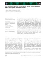
In global terms, the presence of p16
INK4a
appeared
to shift the balance from complex-type to oligomanno-
syl N-glycans, such as M6.1, M7.1 and M9.1 (Fig. 1).
Bi- and tetra-antennary N-glycans surpassed the 5%
threshold of molar ratio in the mock-treated cells.
Invariably, the presence of b1,6-branching was reduced
by p16
INK4a
expression (Fig. 1). Only the biantennary
complex-type N-glycan, with both a bisecting GlcNAc
residue and core fucosylation, and the two type-I-
branched triantennary N-glycans without core
fucosylation were slightly more abundant in the
p16
INK4a
-expressing cells than in the control. Of note,
two N-glycans of unknown structure either in the
region of mannose-rich compounds or between the tri-
mannosyl core and the biantennary structure were only
found for the p16
INK4a
-expressing cells, and no evi-
dence for the emergence of an abundant N-glycan with
poly N-acetyllactosamine extensions could be provided.
At this stage, it should be noted that work with whole
cell extracts allows us to reach a statement on total
glycan presence, at the level of sensitivity of this
method. Cytoplasmic N-glycans before and after mat-
uration, as well as the cell-surface profile, irrespective
of accessibility, will be simultaneously evaluated. How
the pattern of glycans presented on the cell surface
and accessible for binding partners looks will have to
be clarified by a different method. Binding studies with
glycan epitope-specific probes conducted on intact cells
are suited to address this question. For this purpose,
forming a panel of plant lectins is a validated
approach. Although they will not monitor any glycosy-
lation-dependent change of conformation or functional
status of a glycoprotein, the glycan(s) in these cases
directly acting on its (their) protein backbone, system-
atic application of these sensors for glycan structures is
a step to define potential in situ effectors on the glycan
side. In order to cover the main classes of glycan con-
stituents we selected 24 plant agglutinins. The list of
proteins and their sugar specificities are presented in
Table 2.
Profiling of cell-surface glycans by plant lectins
In the first step of these experiments, we established
the concentration and sugar dependence of lectin
binding, as illustrated in the supplementary material
(Fig. S1). These experiments were also instrumental in
determining a common concentration to detect relative
differences by comparative mapping and to avoid any
toxic effects of lectins. The experimental series was
systematically performed in parallel under identical
conditions for the two cell populations. Hereby, any
parameter change by prolonged or differential culture
periods was avoided. For convenient comparison, we
measured the percentage of positive cells and mean
fluorescence intensity in each panel, as listed in Fig. 2.
In full accordance with the results on the abundance
of mRNA for the GalNAcTs, the presence of GalNAc
residues, measured with five different lectins, was
rather similar except for VVA (Fig. 2). This lectin may
react preferentially with globo- and isoglobotetraosyl-
ceramides and not mucin-type O-glycans. An apparent
Fig. 1. Quantitative profiling of N-glycans. Complete representation of the N-glycan profiles of mock- and p16
INK4a
-transfected pancreatic car-
cinoma cells and their molar ratios determined by the 2D mapping technique. The N-glycan structure is given for each case, and an inset is
added for explanation.
S. Andre
´
et al. New function of p16
INK4a
FEBS Journal 274 (2007) 3233–3256 ª 2007 The Authors Journal compilation ª 2007 FEBS 3237
preference for the Thomsen-Friedenreich epitope anti-
gen was detected for p16
INK4a
-positive cells by PNA
and jacalin. This result may reflect either elevated core 1
synthesis or efficient core 1 masking by a2,3-sialylation
in the mock control, as suggested by the microarray
data. It was therefore essential to study this issue in
greater detail (please see the paragraph below). Using
the standard concentration Maackia amurensis-II
(MAA-II), the percentage of positive cells was
enhanced for mock-treated clones, possibly attributable
to different gene expression levels for the enzymes
responsible for the two sialylation steps.
N-Glycosylation, especially with core fucosylation,
appeared to be accessible on the cell surface to a
higher extent in the p16
INK4a
- versus mock-transfected
cell populations. Glycan profiling had revealed an
increase for biantennary glycans of this type containing
the additional bisecting GlcNAc unit, in contrast to an
otherwise decreased level of core fucosylation (Fig. 1),
in full accord with array data of a-fucosyltransferase
VIII. Testing two probes with similar specificities
(i.e. LCA ⁄ PSA) served as an internal control. Appar-
ently, the chromatographic profiling, on average,
detected more substituted N-glycan in mock controls
Table 2. Lectin panel for glycan profiling of cell surfaces listed in alphabetic order.
Latin name (common name) Acronym
Monosaccharide
specificity Potent oligosaccharide
a
Artocarpus integrifolia (jack fruit) Jacalin (JAC) Gal ⁄ GalNAc Galb3GalNAca
Arachis hypogaea (peanut) PNA Gal Galb3GalNAca
Canavalia ensiformis (jack bean) Con A Man ⁄ Glc GlcNAcb2Mana6(GlcNAcb2Mana3)Manb4GlcNAc
Datura stramonium (thorn apple) DSA GlcNAc (GlcNAc)
n
, Galb4GlcNAcb6(Galb4GlcNAcb2)Man (Galb4GlcNAc)
3
Dolichos biflorus (horse gram) DBA GalNAc GalNAca3GalNAca3Galb4Galb4Glc
Erythrina cristagalli (coral tree) ECA Gal Galb4GlcNAcb6(Galb4GlcNAcb2)Man
Galanthus nivalis (snowdrop) GNA Man Mana6(Mana3)ManaR
Glycine max (soybean) SBA GalNAc GalNAca3Galb6Glc
Griffonia simplicifolia I GSA I GalNAc GalNAca3Gal, GalNAca3GalNAcb3Gala4Galb4Glc
Griffonia simplicifolia II GSA II GlcNAc GlcNAcb4GlcNAc, N-glycans with terminal, nonreducing-end
GlcNAc
Lens culinaris (lentil) LCA Man ⁄ Glc N-glycan binding enhanced by core-fucosylation
Lycopersicon esculentum (tomato) LEA
b
core and stem regions of high-mannose-type N-glycans,
(GlcNAcb3Galb4GlcNAcb3Gal)
n
of complex-type N-glycans
Maackia amurensis I (leukoagglutinin) MAA I
b
Neu5Aca3Galb4GlcNAc ⁄ Glc
Maackia amurensis II (haemagglutinin) MAA II
b
Neu5Aca3Galb3(a6Neu5Ac)GalNAc
Phaseolus vulgaris erythroagglutinin
(kidney bean)
PHA-E
b
Bisected complex-type N-glycans: Galb4GlcNAcb2Mana6
(GlcNAcb2-Mana3)(GlcNAcb4)Manb4GlcNAc
Phaseolus vulgaris leukoagglutinin
(kidney bean)
PHA-L
b
Tetra- and triantennary N-glycans with b6-branching
Pisum sativum (garden pea) PSA Man ⁄ Glc N-glycan binding enhanced by core-fucosylation
Sambucus nigra (elderberry) SNA Gal ⁄ GalNAc Neu5Aca6Gal ⁄ GalNAc
Solanum tuberosum (potato) STA
b
(GlcNAc)
n
with preference for high-mannose-type N-glycans
Sophora japonica (pagoda tree) SJA GalNAc GalNAcb6Gal, Galb3GalNAc
Triticum vulgare (wheat germ) WGA GlcNAc ⁄ Neu5Ac (GlcNAc)
n
, Galb4GlcNAcb6Gal
Ulex europaeus I (gorse) UEA I Fuc Fuca2Galb4GlcNAcb6R
Vicia villosa (hairy vetch) VVA GalNAc GalNAca3(6)Gal, GalNAcb3Gal
Viscum album (mistletoe) VAA Gal Galb2(3)Gal, Gala3(4)Gal, Galb3(4)GlcNAc without ⁄ with
a2,6-sialylation, Fuca2Gal
a
Based on previously compiled information [121], extended and modified;
b
no monosaccharide known as ligand.
Fig. 2. Profiling of cell-surface glycans by plant lectins. Semilogarithmic representation of fluorescent surface staining by biotinylated plant
lectins (for an explanation of the acronyms and listing of oligosaccharide specificity, please see Table 2) of mock-transfected (gray line) and
p16
INK4a
-transfected (black line) Capan-1 pancreatic carcinoma cells determined in parallel assays. Quantitative data on the percentage of
positive cells and fluorescence intensity are given in each panel (first line: mock-treated cells; second line: p16
INK4a
-transfected cells). The
concentration of the biotinylated lectins was 0.5 lgÆmL
)1
except for SNA and DBA (1 lgÆmL
)1
), SJA and WGA (2 lgÆmL
)1
) and MAA-I
(5 lgÆmL
)1
).
New function of p16
INK4a
S. Andre
´
et al.
3238 FEBS Journal 274 (2007) 3233–3256 ª 2007 The Authors Journal compilation ª 2007 FEBS
S. Andre
´
et al. New function of p16
INK4a
FEBS Journal 274 (2007) 3233–3256 ª 2007 The Authors Journal compilation ª 2007 FEBS 3239
when compared with the relatively increased lectin
reactivity on the cell surface for p16
INK4a
-expressing
cells, indicating disparities in the levels of accessibility
and ligand preferences. The chromatographic profiling
and cell-surface detection of N-glycans with bisecting
GlcNAc by PHA-E could easily be reconciled, product
formation substantiating efficiency of only minute
quantities of detectable mRNA for GnT-III, whereas no
major accessibility difference could be discerned regard-
ing lectin binding of the b1,6-branch by PHA-L (Fig. 2).
Due to the potential interference by a2,6-sialylation,
this result, as noted for the Thomsen-Friedenreich
antigen epitope and a2,3-sialylation above, had to be
further scrutinized (please see below for the effect of
sialidase treatment). A clear difference in lectin binding
concerned the presence of accessible GlcNAc moieties
measured by applying DSA ⁄ WGA but not seen to this
extent with GSA-II and STA. Expression changes in
the b4GalT family may underlie this staining property.
LEA, a marker for extensions of N-glycan branches by
N-acetyllactosamine (LacNAc) units, failed to provide
clear evidence for marked cell-surface differences,
arguing in favour of the concept of compensatory
change within the tested b1,3-N-acetylglucosaminyl-
transferases and b4GalTs, as also seen in chromato-
graphic profiling. Because similar cell staining was
measured with GNA, the contribution of the dual
reactivity of LEA to high-mannose-type N-glycans will
probably have no influence on this result. Similarly,
the abundance of accessible poly N -acetyllactosamine
chains was rather similar, prompting a final check of
chain-end galactosylation.
The two respective probes (VAA and ECA) revealed
more intense staining of the p16
INK4a
-reconstituted
clones than of the mock control (Fig. 2; supplementary
Fig. S1). This result does not simply reflect the micro-
array data when adding up signal intensities for
b4GalT-specific cDNAs. As depicted above, sialylation
will make its presence felt in this approach. Because it
can mask terminal galactose residues for lectins, meas-
urement of its status was essential. a2,3-Sialylation of
N-glycans monitored comparatively with MAA-I at a
fairly high concentration showed a slight preference
for the p16
INK4a
-positive clone, indicating a compensa-
tory balance for ST3Gal-III ⁄ -IV ⁄ -VI gene expression
levels. By contrast, cell positivity expressed as cell
percentage was lowered in these cells when measuring
lectin-reactive N-glycan-specific a2,6-sialylation, despite
similar extents of ST6Gal-I gene expression (Fig. 2).
Ectopic ST6Gal-I expression in the p16
INK4a
-positive
cells did not change this parameter (data not shown).
As mentioned above, this observation on the noniden-
tical degree of sialylation makes it mandatory to deter-
mine comparatively the impact of sialylation on the
binding of plant lectins sensitive to its presence. In
addition to the inhibition control with haptenic sugar,
the pre-exposure of cells to neuraminidase is, at the
same time, a second control for ensuring carbohy-
drate-dependent binding of SNA. Standard conditions
for enzymatic treatment were established, minimizing
the influence on the cell phenotype. In line with the
inhibition studies, enzymatic pretreatment significantly
reduced (mock control) or almost completely abolished
(p16
INK4a
-positive cells) SNA binding (Fig. 3). The
same effect was observed for MAA-II used at a nearly
saturating concentration of 5 lgÆmL
)1
(Fig. 3). In
addition to its control character, these results support
the evidence for a quantitative difference in the
a2,6-sialylation status between the two cell popu-
lations. Should the level of mucin-type O-glycan
a2,3-sialylation also be lowered in the p16
INK4a
-posit-
ive cells, as suggested by the microarray data, then
neuraminidase activity should enhance PNA staining
of the control cells, with a minor influence on the
p16
INK4a
-expressing cells and on DBA staining. The
importance of this aspect has been pointed out above.
Fittingly, PNA, but not DBA, positivity was markedly
improved by the enzymatic removal of sialic acid resi-
dues from the cell surface for mock-transfected cells
but not for the p16
INK4a
-positive cells (Fig. 3). The
minor effect on the p16
INK4a
-positive cells probably
indicates the presence of mucin-type core 2 tetrasaccha-
rides, favored by an increased b4GalT-IV presence
and reduced O-glycan a2,3-sialylation. Thus, lectin-
accessible mucin-type O-glycosylation appeared to
be rather equally abundant, but the levels of its
a2,3-sialylation were definitely different. The apparent
preference for mock-treated cells to carry O-glycan core
1 a2,3-sialylation can be accounted for – at least in part
– by the microarray data.
Because the presence of a2,6-linked sialic acids can
impede PHA-L binding, the same procedure was also
carried out in this case. Using a lectin concentration of
1 lgÆmL
)1
, increased staining was seen in both cell
populations, and accessibility was still at an increased
level for the p16
INK4a
-positive cells (Fig. 3). To avoid
underestimation of the presence of b1,6-branched
N-glycans in the mock control, the remarkably differ-
ent levels of SNA binding after standard neuramini-
dase treatment must be recalled. In essence, data from
chromatographic mapping square well with the lectin
profiling. A major result emerging from these experi-
ments is that the extents of sialylation of N- and
mucin-type O-glycans in the two cell populations
(a2,6-substitution for N-glycans, a2,3-modification for
O-glycans) are different. In functional terms, these
New function of p16
INK4a
S. Andre
´
et al.
3240 FEBS Journal 274 (2007) 3233–3256 ª 2007 The Authors Journal compilation ª 2007 FEBS
processes may generate or mask sites for contact in situ
with endogenous lectins. As the results with the b-gal-
actoside-specific lectins VAA ⁄ ECA revealed, the levels
of accessible galactose residues were remarkably differ-
ent between the two cell populations. Monitoring of
cell-surface binding by plant lectins thus pinpointed
disparities in accessible glycans. These observations
directed our interest to the detection of endogenous
lectins. They might turn these newly defined properties
on the level of cell-surface glycosylation into effects, in
this case on the level of susceptibility to anoikis.
With focus on sialylation ⁄ galactosylation, the main
groups of human lectins that can read and translate
such differences are the C-type lectins, siglecs and
galectins [42]. The ensuing microarray monitoring of
expression of 42 C-type lectins and siglecs-2, -3, -5, -6,
-7 and -9 failed to provide positive data or, if positive,
a difference in signal intensities. When testing the third
mentioned lectin family, indications for transcription
of galectin genes were collected. As further ascertained
by systematic RT-PCR analysis within this lectin fam-
ily, transcription of genes for galectins-1, -3, -7 and -9,
respectively, was detected, the most pronounced signal
(i.e. 5369) seen in the case of galectin-1 in p16
INK4a
-
positive cells (data not shown). Having herewith
provided evidence for gene expression of members of
this galactoside-specific lectin family, we next probed
whether human adhesion ⁄ growth-regulatory galectins
can bind to the cells, as shown for the galactoside-
specific lectins ECA ⁄ VAA. By testing more proteins
than just galectin-1, the individual binding properties
of the structurally closely related members of this fam-
ily can also be profiled in one cell system, a compar-
ison so far not reported. For this purpose, we purified
the lectins and then biotinylated them under activity-
preserving conditions, ascertained a lack of harmful
effects on sugar binding by activity assays and deter-
mined the labeling efficiency with 2–8 modified resi-
dues per carbohydrate-recognition domain. As in the
binding studies with plant lectins, we routinely per-
formed experiments to assess concentration depend-
ence and inhibition of galectin binding by haptenic
sugar.
Profiling of galectin binding
The measurements with the two galactoside-specific
plant lectins and also with MAA-I (galectins tolerate
a2,3- but not terminal a2,6-sialylation) led to the
expectation that these human lectins may preferentially
bind to p16
INK4a
-expressing cells with their increased
presence of these epitopes. Indeed, the respective
studies confirmed this notion already in the first set of
experiments with galectin-1, when documenting the
dependence of cell staining on lectin concentration and
on glycan binding (supplementary Fig. S2, first and
second panels). This homodimeric family member is a
potent cross-linker for glycans on the cell surface.
Fig. 3. Effect of sialidase treatment on the cell-surface binding of plant lectins. Semilogarithmic representation of fluorescent surface staining
of mock-transfected (Mock) and p16
INK4a
-transfected (p16) Capan-1 pancreatic carcinoma cells without the incubation step using the biotinyl-
ated lectin (shaded) and after incubation with the labeled probe (0.5 lgÆmL
)1
of PNA, 1 lgÆmL
)1
of PHA-L, 5 lgÆmL
)1
of SNA and MAA-II, as
well as 10 lgÆmL
)1
of DBA), without (gray line) or after (black line) sialidase treatment. Quantitative data are presented as defined in the
legend to Fig. 2.
S. Andre
´
et al. New function of p16
INK4a
FEBS Journal 274 (2007) 3233–3256 ª 2007 The Authors Journal compilation ª 2007 FEBS 3241
Besides using haptenic sugar to relate lectin activity to
binding, we tested two mutants of human galectin-1.
Their carbohydrate-binding activity was impaired by
a crucial substitution (W68L, E71Q) in the carbo-
hydrate-recognition domain. The loss of binding to
the p16
INK4a
-transfected cells compared with the
His-tagged wild-type control protein served as an
independent validation of the inhibition control (sup-
plementary Fig. S2, third panel). The concentration-
and carbohydrate-dependent binding is also illustrated
for the chimera-type galectin-3. In this case, it is
obvious that the cells of the mock control are also
rather reactive, when considering cell-percentage posi-
tivity (supplementary Fig. S2). Given this indication
for intergalectin differences, we systematically assayed
a series of human galectins to define staining proper-
ties with these human effector proteins. As shown in
the supplementary material (Fig. S3), there is a clear
trend for galectin reactivity correlating with tumor
suppressor presence. The tandem-repeat-type galectin-8
reacted similarly to galectin-9 (data not shown). The
most prominent change for the combination of both
quantitative cell staining parameters was seen in the case
of galectin-1. Glycan-dependent galectin binding to
these cells is thus detectable, it is not a uniform
characteristic, and, finally and even more importantly,
the conspicuous difference of galectin-1 binding to the
two cell populations gives further study a clear
direction.
As a result of the blocking effect of terminal
a2,6-sialylation on galectin-1 binding, we assumed that
this type of sialylation will mask galectin-1-reactive
sites on the cells of the mock control. If therefore
exposed to a sialidase, these cells should become react-
ive, as shown for SNA or PNA binding in Fig. 3.
Indeed, reduction of sialylation under standard condi-
tions increased cell binding markedly for the mock
control, whereas the p16
INK4a
-transfected cells showed
only slightly improved binding properties (Fig. 4).
Galectin-1 specificity renders it very likely that remov-
ing the blocking a2,6-sialylation underlies this param-
eter change. That the same reactivity pattern was seen
for galectin-3 constitutes not only an inherent control.
As galectin-3 tolerates a2,6-sialylation in poly
N-acetyllactosamine chains already at the level of the
dimer, in stark contrast to galectin-1 [43], these results
signified no notable difference for the presence of such
chain extensions between the cell types, in full agree-
ment with LEA and microassay data. In view of an
effector function, the differential status of sialylation
thus appeared to influence the accessibility to ligand
sites effectively. This result might become functionally
relevant if a functional relationship between galectin-1
and the expression of the p16
INK4a
protein could be
delineated. In this sense, the p16
INK4a
-dependent
increase of the presentation of binding sites by reduced
a2,6-sialylation might even be associated with
enhanced galectin-1 expression, this regulatory event
accomplishing optimal sensitivity. We put this reason-
ing to the test in a stepwise manner by a gene array,
by northern blotting ⁄ nuclear run-off experiments, by
proteomic profiling and by flow cytofluorometry.
Identification of up-regulation of galectin-1
expression
Quantitative determination of the presence of galectin-
1-specific mRNA indicated a p16
INK4a
-associated
increase. In detail, we comparatively probed 1996
cDNAs in an array designed for pancreas tissue and
its cancer development and applied stringent criteria
for defining up-regulation. The threshold of 50%
increase was surpassed by 16 signals. Galectin-1 gene
transcription was the most prominent defined case
at a p16
INK4a
⁄ mock ratio of 3.01 (supplementary
Fig. 4. Effect of sialidase treatment on the cell-surface binding of
human galectins. Semilogarithmic representation of fluorescent
staining of mock-transfected (Mock) and p16
INK4a
-transfected (p16)
Capan-1 pancreatic carcinoma cells without the incubation step
using the biotinylated lectin (shaded) and after incubation with labe-
led galectins-1 and -3 (gal-1 and gal-3 used at 10 lgÆ mL
)1
) without
(gray line) or after (black line) sialidase treatment. Quantitative data
are presented as defined in the legend to Fig. 2.
New function of p16
INK4a
S. Andre
´
et al.
3242 FEBS Journal 274 (2007) 3233–3256 ª 2007 The Authors Journal compilation ª 2007 FEBS
Table S1). Northern blotting and nuclear run-off
experiments confirmed the array data and substan-
tiated an increase in de novo transcription (data not
shown). Extending our work from the mRNA level,
we next performed a proteomic analysis with the inten-
tion of establishing whether the galectin-1 protein is
produced at an amount reflecting gene expression.
With a total of 600–670 spots per gel and a reproduci-
bility of spot assignment between different gels of
89.4–99%, we detected one spot among the 48 signals
that showed a consistent increase in staining by 50%
in p16
INK4a
-positive cells, relative to control cells, with
mass ⁄ isoelectric point (pI) characteristics compatible
with galectin-1. As shown in Fig. 5, we confirmed this
hypothesis by western blotting and mass-spectrometric
fingerprinting. On the basis of staining of protein by a
dye or the western blotting procedure, galectin-1 pres-
ence as protein was found to be significantly up-regula-
ted (P ¼ 0.0154, P ¼ 0.0027). The advantage of the
proteomic profiling compared with western blotting
after 1D electrophoresis, shown separately in Fig. 6A
as a control, is the exclusion of formation of any
galectin-1 variants based on different pI values. After
synthesis, the protein underwent secretion, because we
detected its presence in the medium (data not shown).
As a consequence it may then associate with the cell
surface, prompting flow-cytofluorometric analysis.
Applying the antigalectin-1-specific immunoglobulin
for cell-surface detection, the difference in protein pro-
duction translated into increased surface presence in
the p16
INK4a
-positive cells (Fig. 5). We deliberately tes-
ted several cell batches and consistently measured an
enhanced cell-surface presentation in the p16
INK4a
-pos-
itive cells under standard culture conditions. When
determining the level of inhibition of galectin-1 binding
by the haptenic sugar, lactose, a notable difference
became apparent. It was comparatively lower in this
cell type than in the control cells, a measure for strong
affinity of the endogenous lectin to a set of particular
surface glycans. In fact, endogenous galectin-1 could
hardly be stripped off the cell surface, even in the pres-
ence of 200 mm lactose. In comparison, lectin binding
from the medium as source was much more sensitive
to inhibitor presence, as observed from loading the
cells with galectin-1 up to saturation (supplementary
Fig. S2), intimating visualization of the gradient of
decreasing affinity for binding multivalent ligands seen
in a recent model study [44]. To exclude that the
p16
INK4a
-dependent up-regulation of galectin-1 is a
singular event confined to Capan-1 cells only, we tes-
ted Dan-G pancreatic cancer cells without and with
p16
INK4a
expression. Of note, these two cell lines give
insight into the specificity of the effect as a result of
their differences in the status of the retinoblastoma
tumor-suppressor gene, pRb. Increased galectin-1
expression was determined by western blotting in both
clones of engineered transfectants with p16
INK4a
expres-
sion, despite maintained pRb status (data not shown). It
is thus tempting to propose a functional correlation
between the detected galectin-1 up-regulation, the
increased presentation of galectin-1-binding sites in
p16
INK4a
-expressing cells and acquisition of anoikis sus-
ceptibility associated with the fibronectin receptor.
Induction of anoikis by galectin-1
In order to test the hypothesis described above, we pur-
sued two independent approaches. First, we established
stable clones with reduced galectin-1 production by
transfection with a vector harboring full-length cDNA
for galectin-1 in the antisense orientation. After con-
firming the reduction of galectin-1 presence, to a level
characteristic of wild-type cells, by western blotting,
the cells of such a clone were subjected to monitoring
levels of anoikis. In line with our hypothesis, the extent
of anoikis was correlated to the level of galectin-1 pre-
sent (Fig. 6A). Second, we forced an increase of galec-
tin-1 production on wild-type cells weakly positive for
galectin-1 binding, using proliferating cell nuclear anti-
gen as an internal control. Anoikis induction was
enhanced, even showing a trend for dose dependence
among the tested clones (Fig. 6B). Corroborating this
biological effect on wild-type cells artificially overex-
pressing galectin-1 we could, in parallel, elicit anoikis
in regular wild-type cells by adding the lectin to med-
ium at a concentration of 125 lgÆmL
)1
(data not
shown). The presence of haptenic sugar interfered with
this process, revealing carbohydrate dependence, as did
the presence of the predominantly monomeric galectin-
3, revealing a requirement for cross-linking (data not
shown). Finally, to connect galectin-1 with the
p16
INK4a
-associated increase of cell-surface presence of
the fibronectin receptor, we reasoned that preparations
of the integrin, when immunoprecipitated from
p16
INK4a
-positive cells, should contain galectin-1.
Using cells grown adherent or in suspension, we tested
this assumption. Western blotting revealed that galec-
tin-1 was indeed co-immunoprecipitated with this
glycoprotein, the levels of galectin-1 presence being
consistently higher in p16
INK4a
-positive cells than in
control cells (Fig. 6C). Independently, the antibody
against the a
5
-subunit was effective at markedly redu-
cing the extent of binding of labeled galectin-1 to the
cell surface (data not shown). These results imply that
the a
5
b
1
-integrin is a binding partner of anoikis-indu-
cing galectin-1.
S. Andre
´
et al. New function of p16
INK4a
FEBS Journal 274 (2007) 3233–3256 ª 2007 The Authors Journal compilation ª 2007 FEBS 3243
Fig. 5. Enhanced galectin-1 expression in p16
INK4a
-reconstituted cells. Aliquots of total protein (200 lg) from mock-transfected (Mock) and
p16
INK4a
-transfected (p16) Capan-1 pancreatic carcinoma cells were subjected to 2D gel electrophoretic separation and silver staining.
The kDa-section where galectin-1 presence can be expected was marked (A, B) and the putative position of galectin-1 was labeled. Western
blot (WB) analysis with equal quantities of protein and 1 lgÆmL
)1
of galectin-1 antibody as probe revealed spots at this position after antigen
visualization by enhanced chemiluminescence (top panel). Mass-spectrometric fingerprinting after digestion of the protein of this spot by
trypsin ascertained identity to galectin-1, and quantification of the staining intensity in gel electrophoretic (2-DE) and WB analyses revealed
statistically significant up-regulation (middle panel). Cell-surface detection of galectin-1 in flow-cytofluorometric analysis was performed using
20 lgÆmL
)1
of polyclonal galectin-1 antibody as probe and fluorescent goat anti-rabbit IgG as the second-step reagent (bottom panel).
New function of p16
INK4a
S. Andre
´
et al.
3244 FEBS Journal 274 (2007) 3233–3256 ª 2007 The Authors Journal compilation ª 2007 FEBS
Discussion
Protein glycosylation affords a broad platform for
highly versatile modulation of diverse functional
aspects. The presence of distinct glycan determinants
in glycoproteins can underlie quantitative aspects of
intracellular routing and transport to the cell surface
as well as the regulation of activities of both protein
and glycan parts at the final destination. Deduced
from the fundamental concept of the sugar code, cells
derive much of their remarkable communication skills
from presenting an array of glycan signals [3,5,6,
45,46]. In this sense, glycan remodeling is gaining a
functional dimension in our understanding, and even
the introduction of at first sight minor substitutions,
such as a bisecting GlcNAc or core fucose residues,
has remarkable consequences for the shape and ligand
activity of the N-glycan [47–49]. It is thus intuitively
attractive to assume that changes in the glycomic
profile are nonrandom reprogramming events, acting
on the protein (e.g. conformation, protection from
proteolysis, aggregate formation or ligand binding)
and on the glycan (e.g. conformation and affinity to
lectins). Using the tumor suppressor, p16
INK4a
, the
fibronectin receptor and the Capan-1 cell line as
study objects, we herein have delineated a new route
towards re-establishing susceptibility for anoikis. The
presented results lend credit to a scenario of fine-
tuned and co-ordinated events translating into altera-
tions of protein–protein and protein–carbohydrate
recognition.
Towards this aim, we had designed a combined
approach using a cDNA microarray for glycosyltrans-
ferases, 2D chromatographic profiling and binding
studies with lectins on the cell surface. The array data
pinpoint several changes in expression of glycosyl-
transferase genes and hereby afforded first evidence
for functional links. To start with there is an
increased potential for the synthesis of a2,3 ⁄ a2,6-
disialylated Le
a
⁄ Le
c
-epitopes on gangliosides, an
attribute of nonmalignant epithelial cells and ligand
availability for siglec-7 [50,51]. Automatically, the
presence of a synthetic precursor (i.e. the sialyl Le
a
epitope, which is present in wild-type Capan-1 cells
and can act as mediator of tumor angiogenesis and
metastasis) will be diminished [51,52]. MAA-II stain-
ing at a probe concentration of 5 lgÆmL
)1
can be
interpreted to reflect the differential presence of the
respective disialylated core. Next, the previously
reported influence on b4GalT activity [31] was con-
firmed at the level of transcription and extended to
divergent regulation between b4GalTs-I ⁄ -V and
b4GalT-IV. Of interest, the noted down-regulation of
A
B
C
Fig. 6. Role of galectin-1 in p16
INK4a
-mediated anoikis induction.
Quantification of p16
INK4a
and galectin-1 presence in wild-type
(wt), p16
INK4a
-positive (p16) and p16
INK4a
⁄ antisense galectin-1
(gal-1AS) double-transfected cells by western blot analysis and
determination of anoikis rates of cells after 24 h in suspension in
at least three independent experiments (***, P < 0.01 for
p16
INK4a
cells versus wt; #, P < 0.05 for double transfectants ver-
sus p16
INK4a
cells; the study panel includes a mock control for
second transfection) (A). Quantification of galectin-1 presence in
wt, mock-treated and cells transfected with vector carrying galec-
tin-1-specific cDNA showing different levels of galectin-1 positi-
vity and determination of anoikis rates as given in panel A
(***, P < 0.01, *, P < 0.05 for galectin-1 transfectants versus wt
cells; sensitivity of detection was less than in panel A to avoid
overexposure of the lane for clone gal-1 ⁄ 1) (B). Western blot
detection of galectin-1 in preparations of immunoprecipitated fi-
bronectin receptor obtained from cells grown while adherent
(ad) or on polyhydroxyethylmethacrylate (PH) to keep them in
suspension (C).
S. Andre
´
et al. New function of p16
INK4a
FEBS Journal 274 (2007) 3233–3256 ª 2007 The Authors Journal compilation ª 2007 FEBS 3245
b4GalT-V can be linked to integrin maturation and
increased surface expression of a
5
b
1
-integrin. To
avoid severe effects caused by hypogalactosylation,
which would otherwise even lead to symptoms of
congenital disorder of glycosylation type IId [53,54],
compensation within this family is essential. The
negative impact on core 2 mucin-type O-glycan
galactosylation can in principle be attenuated by
b4GalTs-IV ⁄ -V [55,56], and it is b4GalT-IV whose
gene expression is substantially elevated. Its activity
produces short core 2 extensions, unless the synthetic
precursor core 1 is a2,3-sialylated [55–57]. Fittingly,
chromatographic profiling and LEA staining yielded
no evidence for a major change in poly N-acetyllacto-
samine presence, and gene expression of a respective
sialyltransferase decreased. As a consequence, con-
spicuous diminution was substantiated on the level of
the cell surface for a2,3-sialylation of mucin-type
O-glycans, especially when using PNA staining with-
out ⁄ with neuraminidase treatment. Because exposure
of human HT-29 colon cancer cells to GalNAc-a-O-
benzyl resulted in reduced a2,3-sialylation, which
engendered an impact on the apical delivery of glyco-
proteins [58], this parameter change may also have a
bearing on integrin routing.
As noted for a2,3-sialylation of mucin-type O-gly-
cosylation, cell-surface lectin staining likewise also
unraveled a decrease in a2,6-sialylation of N-glycans,
this one not predictable from our array data. Any
specific intermolecular mechanisms notwithstanding,
these combined events already bring about a major
parameter alteration. At the cell surface, the degree of
sialylation contributes markedly to charge distribu-
tion. Its change can in itself elicit charge-sensitive
processes, if, for example, electrostatic repulsion is
lessened. More specifically, a2,6-sialylation of the
a
5
b
1
-integrin is known to have a negative impact on
fibronectin binding to the extent of physiological rele-
vance that induction of myeloid differentiation by
phorbol ester targets ST6Gal-I expression [59]. Fur-
ther model studies on ST6Gal-I confirmed a more
general role of this sialylation mode in cell adhesion
and invasiveness [60–62]. In our experimental series
with plant lectins, it was essential to pay attention to
alterations in the degree of sialylation, also. The pres-
ence of a2,6-sialylation reduces the affinity of other-
wise suitable binding partners for DSA and PHA-L
so that this aspect needed to be further studied with
sialidase-treated cells [63–66]. Of note, the three-bond
system of the a2,6-linkage generates an unusually
high degree of intramolecular flexibility [67]. Flanked
by the controls with neuraminidase treatment, the dif-
ferent levels of sialylation were ascertained, whereas a
major influence of p16
INK4a
on b1,6-branching and
chain length was excluded. Results from all three
applied methods were in full agreement. The essential
prerequisite of a change in electrophoretic mobility of
a
5
b
1
-integrin from the two cell populations as an indi-
cator of altered glycosylation was also ascertained
(data not shown). As an excellent measure of the sen-
sitivity of the chromatographic profiling, a recent
report documented the ability of the technique to spot
quantitative differences reliably in the extent of b1,6-
branching between samples of normal bladder and
superficial cancer [19]. The array data are in full
accordance with this interpretation and, furthermore,
render any influence of the responsible enzyme GnT-V
on the malignant phenotype by a nonenzymatic mech-
anism as unlikely (i.e. its angiogenic property assigned
to the basic region between amino acids 254–269 and
responsible for mobilization of fibroblast growth fac-
tor-2 in situ) [68]. At this stage, and with focus on the
a
5
b
1
-integrin, we have thus detected at least two
changes with proven impact on its routing and bind-
ing activity.
The binding of b-galactoside-specific plant lectins
intimated a new role, attributing to the integrin’s gly-
cans the potential to become ligands for endogenous
lectins. This finding prompted studies with human lec-
tins, guided by array data with prominent positioning
of galectin-1 among 1996 cancer-related genes. Indeed,
this endogenous lectin appears to exploit the increased
level of a
5
b
1
-integrin expression with a2,6-hyposialyla-
tion of N-glycans. Regarding GnT-V activity, it is
fitting that galectin-1 does not discriminate between
type-I- and type-II-branched triantennary N-glycans
[69]. In addition to the N-glycans, mucin-type core 2
O-glycans, whose establishment and galactosylation is
strongly favored by the up-regulation of b4GalT-IV
[55], and the reduced core 1 a2,3-sialylation can con-
tribute to increased galectin-1 binding. The differential
degree of PNA positivity between p16
INK4a
-positive
and mock control cells after sialidase treatment argues
in favor of increased core 2 presentation associated
with the presence of p16
INK4a
. The transit from core 1
to core 2 structures can prove advantageous, because
model studies on asialofetuin with its three core 1
disaccharides have shown no binding of galectins to
these epitopes [44,70]. Moreover, local cluster forma-
tion to accomplish high-density presentation of galac-
tose residues will be beneficial for avidity, because
short core 2 structures appear to require chain exten-
sions to acquire the capacity of high-affinity ligands
[71]. As galectin-1 binding to activated T cells attests
[72], N- and O-glycans can both contribute to establish
such a suitable topology. The ensuing high-affinity
New function of p16
INK4a
S. Andre
´
et al.
3246 FEBS Journal 274 (2007) 3233–3256 ª 2007 The Authors Journal compilation ª 2007 FEBS
binding will then hardly be competed for by haptenic
sugar, which was, in fact, noted experimentally. The
high-affinity binding therefore can be considered to be
a cellular manifestation for results obtained in a recent
model study on galectin binding to multivalent ligands,
discovering negative co-operativity [44]. In other
words, the reduced level of a2,6-sialylation of N-gly-
cans in concert with the reduced a2,3-sialylation of
O-glycans, itself a factor promoting galectin-1 binding
to Lec2 CHO mutant cells defective in transport of
activated sialic acid into the Golgi [73], and the
presence of short-length core 2 structures presenting
clustered galactose residues, enhance the binding
avidity for an endogenous lectin. That topology of
ligand presentation is a salient factor for galectin
binding is highlighted by systematic interaction
analyses using natural glycoproteins and differential
ligand selection on the level of glycoproteins between
galectins despite their close sequence similarity [74–78].
That the lectin is an endogenous effector is documen-
ted by its increased production in p16
INK4a
-reconstitu-
ted cells and the functional assays.
In this combination, our data epitomize an elegant
orchestration of gene expression for glycosyltrans-
ferases and galectin-1 with functional consequence.
The general tissue-specific manner of expression of
glycosyltransferase genes in murine organs [79,80], as
well as the way that either tumor necrosis factor-a
alters this parameter in endothelial cells [81] or the
transcription factors Stat4 and T-bet prepare T cells
for selectin binding [82,83], are quoted at this point
to sensitize the reader to appraise the assumed wide
range of this fundamental concept. At the molecular
level, it is, in our case, not yet clear whether the
tumor suppressor acts on the expression of the quoted
genes directly or indirectly. With respect to tumor
cells, the case of the differentiation-dependent activa-
tion of a cell-surface ganglioside sialidase, which
engenders the inhibition of neuroblastoma prolifer-
ation in vitro by interaction between the product of
its activity (i.e. ganglioside GM
1
, and galectin-1 in
neuroblastoma cells), even points to a therapeutic per-
spective [84–88].
Scouring the records on galectin-1 teaches the lesson
that it is not only a negative growth regulator in
p16
INK4a
-positive cells and the mentioned SK-N-MC
neuroblastoma model. It is reactive, too, with other
carcinoma cells [28] and was related to differentiation
and apoptosis after its expression was induced by buty-
rate treatment in human LNCaP prostate cancer cells
[89]. Interestingly, its appearance followed p16
INK4a
induction by 5-azacytidine exposure of BL36 Burkitt
lymphoma cells [90]. Because the presence of p16
INK4a
did not activate transcription nonspecifically (please see
gene array and proteomics data as well as the illus-
trated proliferating cell nuclear antigen control) and
Dan-G pancreatic carcinoma cells, which retain high
expression of the pRb in contrast to Capan-1 cells [91],
respond to the presence of p16
INK4a
with the same
regulation of galectin-1, these data, albeit not allowing
full exclusion of a common effect by tumor suppres-
sion, at least indicate an association to p16
INK4a
pres-
ence irrespective of the activity of pRb. Based on the
illustrated western blotting, an occurrence of galectin-1
variants, as described to be produced under the influ-
ence of fosB gene products [92], could definitely be
ruled out. When proceeding to look at tumors in situ,it
might be expected that galectin-1 is not an abundant
gene product in pancreas cancer. Somewhat surpris-
ingly, galectin-1 was found to be up-regulated in cancer
tissue by expression profiling, an at-first puzzling result,
but its localization was confined to fibroblasts and
fibrotic tissue in and around tumors [33,93,94]. By the
way, the mode of localization of a galectin is a key
issue also for tumor cells, as galectin-1 co-operates clo-
sely with oncogenic H-ras intracellularly [95].
In summary, we have designed a combined strategy
to discover changes in glycomic profile and lectin
expression with general applicability. The predictive
value of individual data sets from the arrays was
remarkable. The presented results provide evidence for
a co-ordinated regulation of glycosylation to increase
galectin-1 binding and of expression of this endogenous
lectin in p16
INK4a
-positive Capan-1 pancreatic carci-
noma cells towards a common aim. The coregulation
of lectin ⁄ lectin ligand display is a biochemical means to
acquire susceptibility to anoikis at the level of cell
physiology. It is evocative of mechanisms of cellular
response to inflammation [6]. This co-ordinated regula-
tion of lectin ⁄ lectin ligand presentation is assumed to
be of more general relevance in tumor biology. To give
an inspiring example, independent studies on glioma
invasiveness have made a strong case for either
a2,6-sialylation or galectin-1 as key factors without so
far drawing a connection and envisioning a therapeutic
perspective [96–99]. When considering the next steps,
the results give our research a clear direction to (a) pin-
point the molecular cause for reduced sialylation, which
may for example reside in CMP-sialic acid transporter
deficiency, as seen for two such protein types in the
congenital disorders of glycosylation IIc and IIf
[100,101], (b) monitor other cell and suppressor types
to define the range and specificity of the detected effect,
(c) delineate the intranetwork functionality of galectins,
because galectin-3, a preferentially monomeric protein
[102] produced and secreted from Capan-1 wild-type
S. Andre
´
et al. New function of p16
INK4a
FEBS Journal 274 (2007) 3233–3256 ª 2007 The Authors Journal compilation ª 2007 FEBS 3247
cells and inactive to elicit anoikis, served as an endog-
enous inhibitor of galectin-1-dependent effects in neur-
oblastoma cells [86] and potent activator of K-ras [103],
as well as (d) dissect the same aspect of intranetwork
functionality in the cases of the b4GalTs and ST3Gals.
Experimental procedures
Reagents and cells
A panel of 22 biotinylated plant lectins, reactive with dis-
tinct building blocks of human glycans, was purchased from
Vector Laboratories (distributed by Alexis Germany, Gru
¨
n-
berg, Germany), supplemented by concanavalin A and
Viscum album agglutinin and rigorously checked for activity
by solid-phase and histochemical assays as positive controls,
as described previously [104,105]. The two mentioned plant
lectins and the panel of human galectins obtained by recom-
binant production were purified by affinity chromatography
on lactose- or mannose-bearing Sepharose 4B resins, pre-
pared with divinyl sulfone (Fluka, Munich, Germany) for
activation, and protein quality was routinely controlled by
1- and 2D gel electrophoresis, gel filtration and mass spectr-
ometry as well as hemagglutination, solid-phase and cell
assays [44,84,106–108]. The megaprimer PCR technique was
used to generate W68L ⁄ E71Q mutants of human galectin-1
as His-tagged proteins, which were purified by affinity chro-
matography on a Ni-CAM
TM
HC resin (Sigma, Munich,
Germany). Biotinylation of the natural and mutant proteins
was carried out with the N-hydroxysuccinimide ester deriv-
ative (Sigma) under activity-preserving conditions, product
quality was ascertained by solid-phase assays and the extent
of biotinylation quantitatively determined by measuring the
pI alterations in 2D gel electrophoresis and setting them in
relation to stepwise loss of free amino groups, as described
previously [109,110]. Cells of the human pancreatic carci-
noma line Capan-1 (HTB 79; American Type Culture
Collection, Rockville, MD, USA) stably transfected with
full-length cDNA for human p16
INK4a
inserted into a
pRC ⁄ CMV vector or with control vector (mock treatment)
were routinely grown as monolayers in RPMI 1640 medium
supplemented with 15% fetal bovine serum (Biochrom, Ber-
lin, Germany), 2 mml-glutamine, 100 UÆmL
)1
of penicillin
and 100 lgÆmL
)1
of streptomycin, as described previously
[30]. Cell clones from wild-type cells overexpressing galectin-
1, or from p16
INK4a
-positive cells harboring galectin-1-speci-
fic cDNA in an antisense orientation, were generated using
the pcDNA3.1 system and hygromycin at a concentration of
100 lgÆmL
)1
for selection, as described previously [104]. For
determination of anoikis, 2 · 10
5
cells per assay were cul-
tured in suspension on a surface coated with polyhydroxy-
ethylmethacrylate, and the cell cycle distribution, including
percentage of cells in the pre-G
1
fraction, was assessed using
the cellquest
TM
program [30].
Microarray and quantitative real-time PCR
analyses
RNA was extracted using the RNeasy Mini Kit (Qiagen,
Hilden, Germany), according to the manufacturer’s
instructions. The quantity and quality of the products were
determined by measurements in the NanoDrop ND-1000
UV ⁄ VIS spectrophotometer (Peqlab, Erlangen, Germany)
and the Bioanalyzer (Agilent, Waldbronn, Germany),
respectively, routinely resulting in RNA integrity number
values between 9.5 and 10. The SuperScript Plus Direct
cDNA Labeling System with Alexa Fluor aha-dUTPs (Invi-
trogen ⁄ Life Technologies, Karlsruhe, Germany) was used
for the production of cDNA harboring fluorescent tagging.
Its efficiency was routinely monitored by adsorption meas-
urements using a Nanodrop ND100 UV ⁄ VIS spectro-
photometer. Aliquots containing the labeled cDNA were
diluted to a volume of 200 lL in Microarray Hybridization
Solution, Version 2 (Amersham Biosciences, Braunschweig,
Germany) and hybridized onto the GlycoProfiler microar-
ray (Scienion, Berlin, Germany) using an a-Hyb hybridiza-
tion station (Miltenyi, Cologne, Germany) for 16 h at
42 °C. Subsequent washing steps were then performed using
2 · NaCl ⁄ Cit, 0.2% SDS as first, 1 · NaCl ⁄ Cit as second
and 0.05 · NaCl ⁄ Cit as third wash buffer. The glass slides
were dried in the centrifuge and scanned using the Agilent
DNA Microarray Scanner, and the obtained data were sub-
jected to analysis using the feature extraction 8.0 soft-
ware (Agilent). The GlycoProfiler microarray comprising
60-mer gene sequences for selected human glycosyltrans-
ferases and lectins, in addition to internal housekeeping
controls, is based on the quadruplicate spotting of each
gene sequence onto glass slides. Differential gene expression
was examined by competitive array hybridization of
fluorescently labeled probes (Alexa 555 versus Alexa 647)
prepared from p16
INK4a
- and mock-transfected cells,
respectively. Dye biases were removed by using the fea-
ture extraction 8.1 software (Agilent) with Rank Consis-
tent Probes as the dye-normalization probe selection
method and Linear and Lowess as the normalization cor-
rection method, hereby accounting for signal intensity-
dependent dye biases. The Linear and Lowess method
applies a linear normalization to the entire range of data,
then executes a nonlinear normalization to the linearized
data set. Errors were routinely computed using the setting
‘Most Conservative’, thereby considering a propagated
error model, which is advantageous for low-intensity fea-
tures, and a universal error model, instrumental for high-
intensity data. Two independent series with four individual
measurements in each series were carried out with standard
deviations not surpassing the 10–12% range. Quantitative
real-time PCR controls used M-MLV reverse-transcriptase-
driven cDNA synthesis. Expression levels were determined
in triplicate by assays-on-demand, delivered by Applied
New function of p16
INK4a
S. Andre
´
et al.
3248 FEBS Journal 274 (2007) 3233–3256 ª 2007 The Authors Journal compilation ª 2007 FEBS
Biosystems (Darmstadt, Germany), running the predevel-
oped b-actin assay 4310881E as a routine control. After
addition of TaqMan Universal ⁄ SYBR-Green PCR Master
Mix, the standard experiment was run in a 7000 Sequence
Detection System (Applied Biosystems). Thermal cycle
conditions were as follows: 95 °C for 10 min followed by
40 cycles of 95 °C for 15 s and then 60 °C for 1 min.
Data analysis was performed according to the DD thresh-
old cycle method, using threshold cycle values of a cDNA
stock prepared from cells of the colon carcinoma line
LS174T as a calibrator [111]. For an independent expres-
sion profiling of 1966 genes with a known or assumed role
in pancreas cancer development [for a complete listing,
please see />forschu ng/ag-gress/index.html (username: ag-gress, pass-
word genom)], a custom-made array was designed on
Hybond N
+
nylon membranes using a MicroGrid II arr-
ayer (Bio Robotics Ltd, Cambridge, UK). Hybridization
probes were generated from 30 lg of total RNA by oligo-
dT-primed reverse transcription using the Superscript II
enzyme (Gibco BRL, Karlsruhe, Germany) and [
32
P]dAT-
P[aP]. Probes were competed with 0.25 lgÆmL
)1
of soni-
fied cDNA from human placenta (Sigma) and COT-1
DNA (Gibco) for 1 h. Hybridizations were routinely per-
formed in triplicate, the processed nylon membranes were
scanned with a Storm 8600 Phosphoimager (Molecular
Dynamics GmbH, Krefeld, Germany) and image analysis
was performed with the arrayvision software (Imaging
Research Inc., St Catharines, Canada), including correc-
tion for local background and normalization to mean
signal intensities.
Glycan profiling by HPLC analysis
N-Glycans from the cells were obtained and labeled by
pyridylamination, as described previously [112,113]. In
detail, seven individual preparations of each cell type of
10 mg of total protein were carefully washed, heated to
90 °C for 15 min and then lyophilized. Then, each sample
was dissolved in 200 lLof50mm Tris ⁄ HCl buffer
(pH 8.0), containing 10 mm CaCl
2
, and digested with
100 lg each of trypsin (Sigma) and chymotrypsin (Calbio-
chem, Darmstadt, Germany) at 37 °C for 16 h. After again
heating at 90 °C for 10 min to inactivate the proteases,
samples were treated with 10 U of N-glycosidase F (recom-
binant; Roche Molecular Biochemicals, Mannheim,
Germany) at 37 °C for 16 h to release N-glycans from
glycoproteins. The remaining peptides were digested with
100 lg of pronase (Calbiochem) before glycan purification
by gel filtration chromatography. The resulting N-glycans
were fractionated using a Bio-Gel P-4 column (200–400
mesh; Bio-Rad, Munich, Germany) with H
2
O as the eluant
and thoroughly 2-aminopyridylated via reductive amination
with excess 2-aminopyridine (PA; Wako Pure Chemical,
Osaka, Japan) ⁄ HCl (pH 6.8) and sodium cyanoborohydride
(Sigma) at 90 °C for 1 h [114]. Derivatized N-glycans were
separated from reagents on a Sephadex G-15 column
(Amersham Biosciences) with 10 mm ammonium bicarbon-
ate solution as eluant. The fraction containing the glycans
was acid-hydrolyzed at 90 °C for 1 h with 0.01 m HCl
(pH 2.0) to remove sialic acids from the oligosaccharides
and subjected to the 2D mapping. Structural assignment
was based on the elution positions in the two modes of
HPLC analysis by comparing these results with a database
consisting of a total of 480 different types of N-glycans, as
described previously [115], and publicly accessible with
detailed structures and nomenclature (http://www.
glycoanalysis.info/index.html). An in-house control with
PA-derivatized N-glycans from natural sources (e.g. M5.1,
M6.1 and M9.1 from ribonuclease B) was included. In
technical detail, the mixtures of neutral PA-derivatized
N-glycans were first separated on an octadecyl silane
(ODS) reverse-phase column (ShimPack HRC-ODS,
6 · 150 mm; Shimadzu, Kyoto, Japan) by HPLC (D-7000
HPLC system equipped with an L-7485 fluorescence detec-
tor from Hitachi High-Technologies Co., Tokyo, Japan).
Elution was performed at a flow rate of 1.0 mLÆmin
)1
at
55 °C using a linear gradient elution system. Solvent A was
a10mm sodium phosphate buffer (pH 3.8) and solvent B a
10 mm sodium phosphate buffer (pH 3.8) containing 0.5%
(v ⁄ v) 1-butanol. The column was equilibrated with a mix-
ture of solvents A and B (80 : 20, v ⁄ v). After sample injec-
tion, elution was performed using a linear gradient to a
solvent ratio of 40 : 60 (A ⁄ B, v ⁄ v) in 80 min. Seven sam-
ples of each cell type revealed very similar patterns,
enabling pooling for further analysis. Material of each peak
separated on the ODS column was collected and then
applied to the amide-adsorption column (TSKgel Amide-
80, 4.6 · 250 mm). Elution from that column was per-
formed with a flow rate of 1.0 mLÆmin
)1
at 40 °C using a
gradient elution system. Solvents C and D were composed
of 3% acetic acid-triethylamine buffer (pH 7.3) and
acetonitrile at 35 : 65 and 50 : 50 (v ⁄ v), respectively. The
column was first equilibrated with solvent C, and elution
was performed using a linear gradient from a solvent ratio
of 100 : 0 to 40 : 60 (v ⁄ v) of solvents C and D in 30 min.
In both HPLC systems, the positions of elution of PA-deri-
vatized N-glycans were spotted by a fluorescence detector
using excitation and emission wavelengths of 320 and
400 nm, respectively. The molar ratio of PA oligosaccha-
rides was calculated based on peak areas of fluorescence
intensity.
Glycan profiling by cell-surface lectin staining
Quantitative determination of carbohydrate-dependent
lectin binding using streptavidin ⁄ R-phycoerythrin (1 : 40)
as a fluorescent marker (Sigma) was performed by flow cy-
tofluorometry in a FACScan instrument (Becton-Dickinson,
Heidelberg, Germany). Solutions with 4 · 10
5
cells per
S. Andre
´
et al. New function of p16
INK4a
FEBS Journal 274 (2007) 3233–3256 ª 2007 The Authors Journal compilation ª 2007 FEBS 3249
sample were carefully washed to remove any interfering
serum compounds, the extent of protein binding by nonspe-
cific interaction was reduced by an incubation step in Dul-
becco’s phosphate-buffered saline containing 100 lgÆmL
)1
of carbohydrate-free bovine serum albumin, and the biotin-
ylated lectins were incubated with the cells for 30 min at
4 °C to minimize endocytic uptake, as described in detail
previously [116–119]. Control experiments without the incu-
bation step with labeled lectin, or in the presence of hapten-
ic sugar to block carbohydrate-dependent binding, as well
as systematic titrations to define optimal concentrations,
were run for each cell batch in parallel. Sialidase treatment
was performed for 60 min at 37 °C with 0.2 UÆmL
)1
of
sialidase (from Clostridium perfringens; Roche).
Proteomic analysis and galectin-1 identification
Samples of 200 lg of extract protein were prepared by lysis
of frozen cells in 40 mm Tris ⁄ HCl containing 8 m urea, 4%
w ⁄ v 3-[[3-cholamidopropyl]-dimethylammonio]-1-propane-
sulfonate, 0.5% IPG buffer (pH 3–10; GE Healthcare
Europe GmbH, Freiburg, Germany) and 20 mm dithiothre-
itol. After incubation for 1 h at room temperature, the
solution was cleared by centrifugation for 5 min at
10 000 g with a 5415D centrifuge with F45-24-11 rotor
(Eppendorf, Hamburg, Germany). 2D gel electrophoresis
was carried out in a pI range of 3–10 in the IPGphor
TM
unit at 42 kVh, then the Hoefer SE 600 System was used
for separation for 1 h at 100 mA and 2 h at 200 mA per
four-gel set, as described previously [110]. Silver staining
and processing with the imagescanner (Labscan version
3.0; GE Healthcare Europe GmbH), using the Image-
MasterÒ 2d elite software 3.1 package, yielded quantitative
data for comparison of protein expression from six to seven
different gels per cell population, further subjected to statis-
tical analysis with the graphpad prism 3.0 package. Galec-
tin-1 identification was performed by western blotting using
a specific polyclonal IgG fraction (1 : 1000) rigorously
checked for the absence of cross-reactivity against other
human galectins and for sensitive detection by enhanced
chemiluminescence as well as by flow cytofluorometry on
native cells using the commercial fluorescein thiocarbamyl-
labeled conjugate of goat anti-rabbit IgG (Sigma), as des-
cribed previously [104]. This procedure was also used for
galectin-1 detection in preparations of the fibronectin recep-
tor immunoprecipitated from 800 lg of total extract protein
using a commercial antibody against the a
5
-integrin (Cym-
bus Biotechnology, Chandlers Ford, UK), as described pre-
viously [120]. Mass-spectrometric fingerprinting of tryptic
peptides was carried out after gel electrophoretic separation
in two dimensions and Coomassie blue staining. Following
excision of the gel piece with the spot and its washing first
with deionized water, then with acetonitrile in water (1 : 1,
v ⁄ v) and finally with acetonitrile, protein was digested
in situ using 100 ng of sequencing-grade trypsin (Promega,
Mannheim, Germany). Sample preparation for mass-spectr-
ometric analysis by cocrystallization with the matrix (a-cy-
ano-4-hydroycinnamic acid) followed and the mass profile
of the tryptic peptides was then recorded in the positive-
ion-reflector mode on a time-of-flight instrument (Bruker-
Daltronik, Bremen, Germany). Ion acceleration voltage was
set to 26 kV and reflector voltage to 30 kV. The spectrum
was obtained by averaging 300 individual laser shots. Data-
base search for assignment was performed against the
swissprot archive using the mascot search algorithm.
Acknowledgements
We are indebted to Dr R. Brossmer, Dr B. Friday, Dr
R. Hase, Professor S. Namirha and Dr A. Villalobo for
insightful discussions and critical reading of the manu-
script, to the reviewers of our manuscript for their valu-
able input, to B. Hofer, S. Kudo and L. Mantel for
excellent technical assistance, as well as to the Deutsche
Krebshilfe, Mizutani Foundation for Glycoscience,
National Project on Functional Glycoconjugate
Research Aimed at Developing for New Industry from
the Ministry of Education, Science, Sports and Culture
of Japan, Verein zur Fo
¨
rderung des biologisch-technol-
ogischen Fortschritts in der Medizin e. V and the Wil-
helm-Sander Stiftung for generous financial support.
References
1 Sharon N & Lis H (1997) Glycoproteins: structure and
function. In Glycosciences: Status and Perspectives
(Gabius H-J & Gabius S, eds), pp. 133–162. Chapman
& Hall, Weinheim, Germany.
2 Reuter G & Gabius H-J (1999) Eukaryotic glycosyla-
tion: whim of nature or multipurpose tool? Cell Mol
Life Sci 55, 368–422.
3 Lowe JB & Marth JD (2003) A genetic approach to
mammalian glycan function. Annu Rev Biochem 72,
643–691.
4 Laine R (1997) The information-storing potential of
the sugar code. In Glycosciences: Status and Perspec-
tives (Gabius H-J & Gabius S, eds), pp. 1–14. Chap-
man & Hall, Weinheim, Germany.
5 Hebert DN, Garman SC & Molinari M (2005)
The glycan code of the endoplasmic reticulum:
asparagine-linked carbohydrates as protein matura-
tion and quality-control tags. Trends Cell Biol 15,
364–370.
6 Gabius H-J (2006) Cell surface glycans: the why and
how of their functionality as biochemical signals in lec-
tin-mediated information transfer. Crit Rev Immunol
26, 43–80.
7 Villalobo A, Nogales-Gonza
´
les A & Gabius H-J (2006)
A guide to signalling pathways connecting protein–
New function of p16
INK4a
S. Andre
´
et al.
3250 FEBS Journal 274 (2007) 3233–3256 ª 2007 The Authors Journal compilation ª 2007 FEBS
glycan interaction with the emerging versatile effector
functionality of mammalian lectins. Trends Glycosci
Glycotechnol 18, 1–37.
8 Alhadeff JA (1989) Malignant cell glycoproteins and
glycolipids. Crit Rev Oncol Hematol 9, 37–107.
9 Hakomori S-i (1989) Aberrant glycosylation in tumors
and tumor-associated carbohydrate antigens. Adv
Cancer Res 52, 257–331.
10 Yamashita K, Ohkura T, TachibanaY, Takasaki S &
Kobata A (1984) Comparative study of the oligosac-
charides released from baby hamster kidney cells and
their polyoma transformant by hydrazinolysis. J Biol
Chem 259, 10834–10840.
11 Pierce M & Arango J (1986) Rous sarcoma virus-
transformed baby hamster kidney cells express higher
levels of asparagine-linked tri- and tetraantennary
glycopeptides containing [GlcNAc-b1,6Man-a1,6Man]
and poly-N-acetyllactosamine sequences than
baby hamster kidney cells. J Biol Chem 261, 10772–
10777.
12 Hubbard SC (1987) Differential effects of oncogenic
transformation on N-linked oligosaccharide processing
at individual glycosylation sites of viral glycoproteins.
J Biol Chem 262, 16403–16411.
13 Easton EW, Bolscher JGM & van den Eijnden DH
(1991) Enzymatic amplification involving glycosyltrans-
ferases forms the basis for the increased size of aspara-
gine-linked glycans at the surface of NIH 3T3 cells
expressing the N-ras proto-oncogene. J Biol Chem 266,
21674–21680.
14 Buckhaults P, Chen L, Fregien N & Pierce M (1997)
Transcriptional regulation of N-acetylglucosaminyl-
transferase V by the src oncogene. J Biol Chem 272,
19575–19581.
15 Chen L, Zhang W, Fregien N & Pierce M (1998) The
her-2 ⁄ neu oncogene stimulates the transcription of
N-acetylglucosaminyltransferase V and expression of
its cell surface oligosaccharide products. Oncogene 17,
2087–2093.
16 Dalziel M, Dall’Olio F, Mungul A, Piller V & Piller F
(2004) Ras oncogene induces b-galactoside a2,6-sialyl-
transferase (ST6Gal I) via a RalGEF-mediated signal
to its housekeeping promoter. Eur J Biochem 271,
3623–3634.
17 Dosaka-Akita H, Miyoshi E, Suzuki O, Itoh T, Katoh
H & Taniguchi N (2004) Expression of N-acetylgluco-
saminyltransferase V is associated with prognosis and
histology in non-small cell lung cancers. Clin Cancer
Res 10, 1773–1779.
18 Inamori K-i, Gu J, Ohira M, Kawasaki A, Nakamura
Y, Nakagawa T, Kondo A, Miyoshi E, Nakagawara A
& Taniguchi N (2006) High expression of N-acetylglu-
cosaminyltransferase V in favorable neuroblastomas:
involvement of its effect on apoptosis. FEBS Lett 580,
627–632.
19 Ishimura H, Takahashi T, Nakagawa H, Nishimura
S-I, Arai Y, Horikawa Y, Habuchi T, Miyoshi E,
Kyan A, Hagisawa S et al. (2006) N-Acetylglucosami-
nyltransferase V and b1–6 branching of N-linked
oligosaccharides are associated with good prognosis
of patients with bladder cancer. Clin Cancer Res 12,
2506–2511.
20 Asada M, Furukawa K, Segawa K, Endo T & Kobata
A (1997) Increased expression of highly branched
N-glycans at cell surface is correlated with the malig-
nant phenotypes of mouse tumor cells. Cancer Res
57,
1073–1080.
21 Guo HB, Lee I, Kamar M, Akiyama SK & Pierce M
(2002) Aberrant N-glycosylation of b
1
integrin causes
reduced a
5
b
1
integrin clustering and stimulates cell
migration. Cancer Res 62, 6837–6845.
22 Seales EC, Jurado GA, Singhal A & Bellis SL (2003)
Ras oncogene directs expression of a differentially sia-
lylated, functionally altered b
1
integrin. Oncogene 22,
7137–7145.
23 Nakagawa H, Zheng M, Hakomori S-i, Tsukamoto Y,
Kawamura Y & Takahashi N (1996) Detailed oligosac-
charide structures of human integrin a
5
b
1
analyzed by
a three-dimensional mapping technique. Eur J Biochem
237, 76–85.
24 Akiyama SK, Yamada SS & Yamada KM (1989) Ana-
lysis of the role of glycosylation of the human fibro-
nectin receptor. J Biol Chem 264, 18011–18018.
25 Zheng M, Fang H & Hakomori S-i (1994) Functional
role of N-glycosylation in a
5
b
1
integrin receptor.
De-N-glycosylation induces dissociation or altered
association of a
5
and b
1
subunits and concomitant loss
of fibronectin binding activity. J Biol Chem 269,
12325–12331.
26 Nadanaka S, Sato C, Kitajima K, Katagiri K, Irie S &
Yamagata T (2001) Occurrence of oligosialic acids on
integrin a
5
subunit and their involvement in cell
adhesion to fibronectin. J Biol Chem 276, 33657–
33664.
27 Wang X, Sun P, Al-Qamari A, Tai T, Kawashima I &
Paller AS (2001) Carbohydrate-carbohydrate binding
of ganglioside to integrin a
5
modulates a
5
b
1
function.
J Biol Chem 276, 8436–8444.
28 Fischer C, Sanchez-Ruderisch H, Welzel M, Wieden-
mann B, Sakai T, Andre
´
S, Gabius H-J, Khachigian L,
Detjen KM & Rosewicz S (2005) Galectin-1 interacts
with the a
5
b
1
fibronectin receptor to restrict carcinoma
cell growth via induction of p21 and p27. J Biol Chem
280, 37266–37277.
29 Ca
´
rcamo C, Pardo E, Oyanadel C, Bravo-Zehnder M,
Bull P, Ca
´
ceres M, Martı
´
nez J, Massardo L, Jacobelli
S, Gonza
´
lez A et al. (2006) Galectin-8 binds specific b
1
integrins and induces polarized spreading highlighted
by asymmetric lamellipodia in Jurkat T cells. Exp Cell
Res 312, 374–386.
S. Andre
´
et al. New function of p16
INK4a
FEBS Journal 274 (2007) 3233–3256 ª 2007 The Authors Journal compilation ª 2007 FEBS 3251
30 Plath T, Detjen K, Welzel M, von Marschall Z, Mur-
phy D, Schirner M, Wiedenmann B & Rosewicz S
(2000) A novel function for the tumor suppressor
p16 (INK4a): induction of anoikis via upregulation
of the a
5
b
1
fibronectin receptor. J Cell Biol 150,
1467–1478.
31 Zhang SW, Fu XY, Cao SL, Shen ZH & Gu JX
(1999) Down-regulation of b1,4-galactosyltransferase
gene expression by cell-cycle suppressor gene p16.
Biochim Biophys Acta 1444, 49–54.
32 Kawado T, Hayashi O, Sato T, Ito H, Hayakawa S,
Takayama E & Furukawa K (2004) Rapid cell senes-
cence-associated changes in galactosylation of N-linked
oligosaccharides in human lung adenocarcinoma A549
cells. Arch Biochem Biophys 426, 306–313.
33 Ide Y, Miyoshi E, Nakagawa T, Gu J, Tanemura M,
Nishida T, Ito T, Yamamoto H, Kozutsumi Y & Tani-
guchi N (2006) Aberrant expression of N-acetylgluco-
saminyltransferase-IVa and IVb (GnT-IVa and b) in
pancreatic cancer. Biochem Biophys Res Commun 341,
478–482.
34 Sato T, Shirane K, Kido M & Furukawa K (2000)
Correlated gene expression between b-1,4-galactosyl-
transferase V and N-acetylglucosaminyltransferase V in
human cancer cell lines. Biochem Biophys Res Commun
276, 1019–1023.
35 Ko JH, Miyoshi E, Noda K, Ekuni A, Kang R, Ikeda
Y & Taniguchi N (1999) Regulation of the GnT-V
promoter by transcription factor Ets-1 in various can-
cer cell lines. J Biol Chem 274, 22941–22948.
36 Sato T & Furukawa K (2004) Transcriptional regula-
tion of the human b1,4-galactosyltransferase V gene in
cancer cells: essential role of transcription factor Sp1.
J Biol Chem 279, 39574–39583.
37 Zhu X, Jiang J, Shen H, Wang H, Zong H, Li Z,
Yang Y, Niu Z, Liu W, Chen X et al. (2005) Elevated
b1,4-galactosyltransferase I in highly metastatic
human lung cancer cells. Identification of E1AF as
important transcription activator. J Biol Chem 280,
12503–12516.
38 Jiang J, Chen X, Shen J, Wei Y, Wu T, Yang Y,
Wang H, Zong H, Yang J, Zhang S et al. (2006) b1,4-
Galactosyltransferase V functions as a positive growth
regulator in glioma. J Biol Chem 281, 9482–9489.
39 Chen X, Jiang J, Yang J, Chen C, Sun M, Wei Y,
Guang X & Gu J (2006) Down-regulation of the
expression of b1,4-galactosyltransferase V promotes
integrin b
1
maturation. Biochem Biophys Res Commun
343, 910–916.
40 Guo H-B, Lee I, Bryan BT & Pierce M (2005) Deletion
of mouse embryo fibroblast N-acetylglucosaminyltrans-
ferase V stimulates a
5
b
1
integrin expression mediated
by the protein kinase C signaling pathway. J Biol
Chem 280, 8332–8342.
41 Guo P, Zhang Y, Zhao J-H, Wang L-y, Guo HB,
Zhang X-y, Chen H & -1 (2003) Regulation on the
expression and N-glycosylation of integrins by
N-acetylglucosaminyltransferase V. Biochem Biophys
Res Commun 310, 619–626.
42 Gabius H-J (1997) Animal lectins. Eur J Biochem 243,
543–576.
43 Ahmad N, Gabius H-J, Kaltner H, Andre
´
S, Kuwab-
ara I, Liu F-T, Oscarson S, Norberg T & Brewer CF
(2002) Thermodynamic binding studies of cell surface
carbohydrate epitopes to galectins-1, -3 and -7:
evidence for differential binding specificities. Can J
Chem 80, 1096–1104.
44 Dam TK, Gabius H-J, Andre
´
S, Kaltner H, Lensch M
& Brewer CF (2005) Galectins bind to the multivalent
glycoprotein asialofetuin with enhanced affinities and a
gradient of decreasing binding constants. Biochemistry
44, 12564–12571.
45 Gabius H-J (2001) Glycohistochemistry: the why and
how of detection and localization of endogenous lec-
tins. Anat Histol Embryol 30, 3–31.
46 Gabius H-J, Siebert H-C, Andre
´
S, Jime
´
nez-Barbero J
&Ru
¨
diger H (2004) Chemical biology of the sugar
code. ChemBioChem 5, 740–764.
47 Andre
´
S, Unverzagt C, Kojima S, Frank M, Seifert J,
Fink C, Kayser K, von der Lieth C-W & Gabius H-J
(2004) Determination of modulation of ligand proper-
ties of synthetic complex-type biantennary N-glycans
by introduction of bisecting GlcNAc in silico, in vitro
and in vivo. Eur J Biochem 271, 118–134.
48 Andre
´
S, Kojima S, Prahl I, Lensch M, Unverzagt C
& Gabius H-J (2005) Introduction of extended LEC14-
type branching into core-fucosylated biantennary
N-glycan. FEBS J 272, 1986–1998.
49 Park EI, Mi Y, Unverzagt C, Gabius H-J & Baenziger
JU (2005) The asialoglycoprotein receptor clears
glycoconjugates terminating with sialic acida2,6
GalNAc. Proc Natl Acad Sci USA 102, 17125–17129.
50 Itai S, Arii S, Tobe R, Kitahara A, Kim YC, Yamabe
H, Ohtsuki H, Kirihara Y, Shigeta K & Kannagi R
(1988) Significance of 2–3 and 2–6 sialylation of Lewis
a
antigen in pancreas cancer. Cancer 61, 775–787.
51 Miyazaki K, Ohmori K, Izawa M, Koike T, Kumamo-
to K, Furukawa K, Ando T, Kiso M, Yamaji T,
Hashimoto Y et al. (2004) Loss of disialyl Lewis
a
, the
ligand for lymphocyte inhibitory receptor sialic acid-
binding immunoglobulin-like lectin-7 (siglec-7), associ-
ated with increased sialyl Lewis
a
expression on human
colon cancers. Cancer Res 64, 4498–4505.
52 Peracaula R, Tabare
´
sG,Lo
´
pez-Ferrer A, Brossmer R,
de Bolo
´
s C & de Llorens R (2005) Role of sialyltrans-
ferases involved in the biosynthesis of Lewis antigens
in human pancreatic tumour cells. Glycoconjugate J 22,
135–144.
New function of p16
INK4a
S. Andre
´
et al.
3252 FEBS Journal 274 (2007) 3233–3256 ª 2007 The Authors Journal compilation ª 2007 FEBS
53 Hanßke B, Thiel C, Lu
¨
bke T, Hasilik M, Ho
¨
ning S,
Peters V, Heidemann PH, Hoffmann GF, Berger EG,
von Figura K et al. (2002) Deficiency of UDP-galac-
tose: N-acetylglucosamine b1,4-galactosyltransferase I
causes the congenital disorder of glycosylation type
IId. J Clin Invest 109, 725–733.
54 Kotani N, Asano M, Inoue N, Iwakura Y & Takasaki
S (2004) Polylactosamine synthesis and branch forma-
tion of N-glycans in b1,4-galactosyltransferase-1-defici-
ent mice. Arch Biochem Biophys 426, 258–265.
55 Ujita M, McAuliffe J, Schwientek T, Almeida R,
Hindsgaul O, Clausen H & Fukuda M (1998) Synthesis
of poly-N-acetyllactosamine in core 2 branched O-gly-
cans. The requirement of novel b1,4-galactosyltrans-
ferase IV and b1,3-N-acetylglucosaminyltransferase.
J Biol Chem 273, 34843–34849.
56 van Die I, van Tetering A, Schiphorst WECM, Sato T,
Furukawa K & van den Eijnden DH (1999) The
acceptor substrate specificity of human b4-galactosyl-
transferase V indicates its potential function in
O-glycosylation. FEBS Lett 450, 52–56.
57 Brockhausen I, Matta KL, Orr J, Schachter H, Koen-
derman AH & van den Eijnden DH (1986) Mucin
synthesis. Conversion of R1-b1–3Gal-R2 to R1-b1–3
(GlcNAc b1–6) Gal-R2 and of R1-b1–3GalNAc-R2 to
R1-b1–3 (GlcNAc b1–6) GalNAc-R2 by a b6-N-acetyl-
glucosaminyltransferase in pig gastric mucosa. Eur
J Biochem 157, 463–474.
58 Huet G, Hennebicq-Reig S, de Bolos C, Ulloa F,
Lesuffleur T, Barbat A, Carriere V, Kim I, Real FX,
Delannoy P et al. (1998) GalNAc-a-O-benzyl inhibits
NeuAca2–3 glycosylation and blocks the intracellular
transport of apical glycoproteins and mucus in differ-
entiated HT-29 cells. J Cell Biol 141, 1311–1322.
59 Semel AC, Seales EC, Singhal A, Eklund EA, Colley
KJ & Bellis SL (2002) Hyposialylation of integrins sti-
mulates the activity of myeloid fibronectin receptors.
J Biol Chem 277, 32830–32836.
60 Zhu Y, Srivatana U, Ullah A, Gagneja H, Berenson
CS & Lance P (2001) Suppression of a sialyltransferase
by antisense DNA reduces invasiveness of human
colon cancer cells in vitro. Biochim Biophys Acta 1536,
148–160.
61 Lin S, Kemmner W, Grigull S & Schlag PM (2002)
Cell surface a2,6 sialylation affects adhesion of breast
carcinoma cells. Exp Cell Res 276, 101–110.
62 Seales EC, Jurado GA, Brunson BA, Wakefield JK,
Frost AR & Bellis SL (2005) Hypersialylation of
b
1
integrins, observed in colon adenocarcinoma, may
contribute to cancer progression by up-regulating cell
motility. Cancer Res 65, 4645–4652.
63 Hammarstro
¨
m S, Hammarstro
¨
m M-L, Sundblad G,
Arnarp J & Lo
¨
nngren J (1982) Mitogenic leuko-
agglutinin from Phaseolus vulgaris binds to a
pentasaccharide unit in N-acetyllactosamine-type
glycoprotein glycans. Proc Natl Acad Sci USA 79,
1611–1615.
64 Green ED & Baenziger JU (1987) Oligosaccharide spe-
cificities of Phaseolus vulgaris leukoagglutinating and
erythroagglutinating phytohemagglutinins. Interactions
with N-glycanase-released oligosaccharides. J Biol
Chem 262, 12018–12029.
65 Yamashita K, Totani K, Ohkura T, Takasaki S, Gold-
stein IJ & Kobata A (1987) Carbohydrate binding
properties of complex-type oligosaccharides on immo-
bilized Datura stramonium lectin. J Biol Chem 262,
1602–1607.
66 Bierhuizen MFA, Edzes HT, Schiphorst WECM, van
den Eijnden DH & van DijkW (1987) Effect of a2–
6-linked sialic acid and a1–3-linked fucose on the inter-
action of N-linked glycopeptides and related oligosac-
charides with immobilized Phaseolus vulgaris
leukoagglutinating lectin (L-PHA). Glycoconjugate J 5,
85–97.
67 Siebert H-C, Rosen J, Seyrek K, Kaltner H, Andre
´
S,
Bovin NV, Nyholm PG, Sinowatz F & Gabius H-J
(2006) a2,3 ⁄ a2,6-Sialylation of N-glycans: non-syn-
onymous signals with marked developmental regula-
tion in bovine reproductive tracts. Biochimie 88, 399–
410.
68 Saito T, Miyoshi E, Sasai K, Nakano N, Eguchi H,
Honke K & Taniguchi N (2002) A secreted type of
b1,6-N-acetylglucosaminyltransferase V (GnT-V) indu-
ces tumor angiogenesis without mediation of glycosyla-
tion: a novel function of GnT-V distinct from the
original glycosyltransferase activity. J Biol Chem 277,
17002–17008.
69 Andre
´
S, Kojima S, Gundel G, Russwurm R, Schratt
X, Unverzagt C & Gabius H-J (2006) Branching mode
in complex-type triantennary N-glycans as regulatory
element of their ligand properties. Biochim Biophys
Acta 1760, 768–782.
70 Gupta D, Kaltner H, Dong X, Gabius H-J &
Brewer CF (1996) Comparative cross-linking activities
of lactose-specific plant and animal lectins and a
natural lactose-binding immunoglobulin G fraction
from human serum with asialofetuin. Glycobiology 6,
843–849.
71 Leppa
¨
nen A, Stowell S, Blixt O & Cummings RD
(2005) Dimeric galectin-1 binds with high affinity to
a2,3-sialylated and non-sialylated terminal N-acetyl-
lactosamine units on surface-bound extended glycans.
J Biol Chem 280, 5549–5562.
72 Amano M, Galvan M, He J & Baum LG (2003) The
ST6Gal I sialyltransferase selectively modifies N-gly-
cans on CD45 to negatively regulate galectin-1-induced
CD45 clustering, phosphatase modulation, and T cell
death. J Biol Chem 278, 7469–7475.
73 Patnaik SK, Potvin B, Carlsson S, Sturm D, Leffler H
& Stanley P (2006) Complex N-glycans are the major
S. Andre
´
et al. New function of p16
INK4a
FEBS Journal 274 (2007) 3233–3256 ª 2007 The Authors Journal compilation ª 2007 FEBS 3253
ligands for galectins-1, -3, and -8 on Chinese hamster
ovary cells. Glycobiology 16, 305–317.
74 Wu AM, Wu JH, Tsai M-S, Kaltner H & Gabius H-J
(2001) Carbohydrate specificity of a galectin from
chicken liver (CG-16). Biochem J 358, 529–538.
75 Siebert H-C, Andre
´
S, Lu S-Y, Frank M, Kaltner H,
van Kuik JA, Korchagina EY, Bovin NV, Tajkhorshid
E, Kaptein R et al. (2003) Unique conformer selection
of human growth-regulatory lectin galectin-1 for gan-
glioside GM
1
versus bacterial toxins. Biochemistry 42,
14762–14773.
76 Lo
´
pez-Lucendo MF, Solı
´
s D, Andre
´
S, Hirabayashi J,
Kasai K-i, Kaltner H, Gabius H-J & Romero A (2004)
Growth-regulatory human galectin-1: crystallographic
characterisation of the structural changes induced by
single-site mutations and their impact on the thermo-
dynamics of ligand binding. J Mol Biol 343, 957–970.
77 Sturm A, Lensch M, Andre
´
S, Kaltner H, Wieden-
mann B, Rosewicz S, Dignass AU & Gabius H-J
(2004) Human galectin-2: novel inducer of T cell
apoptosis with distinct profile of caspase activation.
J Immunol 173, 3825–3837.
78 Wu AM, Singh T, Wu JH, Lensch M, Andre
´
S&
Gabius H-J (2006) Interaction profile of galectin-5 with
free saccharides and mammalian glycoproteins:
probing its fine specificity and the effect of naturally
clustered ligand presentation. Glycobiology 16,
524–537.
79 Smith FI, Qu Q, Hong SJ, Kim KS, Gilmartin TJ &
Head SR (2005) Gene expression profiling of mouse
postnatal cerebellar development using oligonucleo-
tide microarrays designed to detect differences in
glycoconjugate expression. Gene Expr Patterns 5,
740–749.
80 Comelli EM, Head SR, Gilmartin T, Whisenant T,
Haslam SM, North SJ, Wong N-K, Kudo T, Narima-
tsu H, Esko JD et al. (2006) A focused microarray
approach to functional glycomics: transcriptional
regulation of the glycome. Glycobiology 16, 117–131.
81 Garcı
´
a-Vallejo JJ, van Dijk W, van Het-Hof B, van
Die I, Engelse MA, van Hinsbergh VWM & Gringhuis
SI (2006) Activation of human endothelial cells by
tumor necrosis factor-a results in profound changes in
the expression of glycosylation-related genes. J Cell
Physiol 206, 203–210.
82 White SJ, Underhill GH, Kaplan MH & Kansas GS
(2001) Differential requirements for Stat4 in expression
of glycosyltransferases responsible for selectin ligand
formation in Th1 cells. J Immunol 167, 628–631.
83 Underhill GH, Zisoulis DG, Kolli KP, Ellies LG,
Marth JD & Kansas GS (2005) A crucial role for
T-bet in selectin ligand expression in T helper 1 (Th1)
cells. Blood 106, 3867–3873.
84 Kopitz J, Andre
´
S, von Reitzenstein C, Versluis K,
Kaltner H, Pieters RJ, Wasano K, Kuwabara I, Liu
F-T, Cantz M et al. (2003) Homodimeric galectin-7
(p53-induced gene 1) is a negative growth regulator
for human neuroblastoma cells. Oncogene 22, 6277–
6288.
85 Kopitz J, von Reitzenstein C, Burchert M, Cantz M &
Gabius H-J (1998) Galectin-1 is a major receptor for
ganglioside GM
1
, a product of the growth-controlling
activity of a cell surface ganglioside sialidase, on
human neuroblastoma cells in culture. J Biol Chem
273, 11205–11211.
86 Kopitz J, von Reitzenstein C, Andre
´
S, Kaltner H, Uhl
J, Ehemann V, Cantz M & Gabius H-J (2001) Nega-
tive regulation of neuroblastoma cell growth by carbo-
hydrate-dependent surface binding of galectin-1 and
functional divergence from galectin-3. J Biol Chem
276, 35917–35923.
87 Andre
´
S, Kaltner H, Lensch M, Russwurm R, Siebert
H-C, Fallsehr C, Tajkhorshid E, Heck AJR, Knebel-
Doeberitz M, Gabius H-J et al. (2005) Determination
of structural and functional overlap ⁄ divergence of five
proto-type galectins by analysis of the growth–regula-
tory interaction with ganglioside GM
1
in silico and in
vitro on human neuroblastoma cells. Int J Cancer 114,
46–57.
88 Siebert H-C, Born K, Andre
´
S, Frank M, Kaltner H,
von der Lieth C-W, Heck AJR, Jime
´
nez-Barbero J,
Kopitz J & Gabius H-J (2006) Carbohydrate chain of
ganglioside GM
1
as a ligand: identification of the
binding strategies of three 15 mer peptides and their
divergence from the binding modes of growth-regula-
tory galectin-1 and cholera toxin. Chem Eur J 12,
388–402.
89 Ellerhorst J, Nguyen T, Cooper DNW, Estrov Y,
Lotan D & Lotan R (1999) Induction of differentiation
and apoptosis in the prostate cancer cell line LNCaP
by sodium butyrate and galectin-1. Int J Oncol 14,
225–232.
90 Poirier F, Bourin P, Bladier D, Joubert-Caron R &
Caron M (2001) Effect of 5-azacytidine and galectin-1
on growth and differentiation of the human B lym-
phoma line bl36. Cancer Cell Int 1,2.
91 Plath T, Peters M, Detjen K, Welzel M, von Marschall
Z, Radke C, Wiedenmann B & Rosewicz S (2002)
Overexpression of pRB in human pancreatic carcinoma
cells: function in chemotherapy-induced apoptosis.
J Natl Cancer Inst 94, 129–142.
92 Nishioka T, Sakumi K, Miura T, Tahara K, Horie H,
Kadoya T & Nakabeppu Y (2002) FosB gene products
trigger cell proliferation and morphological alteration
with an increased expression of a novel processed form
of galectin-1 in the rat 3Y1 embryo cell line. J Biochem
(Tokyo) 131, 653–661.
93 Berberat PO, Friess H, Wang L, Zhu Z, Bley T, Fri-
geri L, Zimmermann A & Buchler MW (2001) Com-
parative analysis of galectins in primary tumors and
New function of p16
INK4a
S. Andre
´
et al.
3254 FEBS Journal 274 (2007) 3233–3256 ª 2007 The Authors Journal compilation ª 2007 FEBS
tumor metastasis in human pancreatic cancer. J Histo-
chem Cytochem 49, 539–549.
94 Chen R, Pan S, Crispin DA & Brentnall TA (2006)
Gene expression and proteomic analysis of pancreatic
cancer: a recent update. Cancer Genomics Proteomics
3, 1–10.
95 Rotblat B, Niv H, Andre
´
S, Kaltner H, Gabius H-J &
Kloog Y (2004) Galectin-1 (L11A) predicted from a
computed galectin-1 farnesyl-binding pocket selectively
inhibits Ras-GTP. Cancer Res 64, 3112–3118.
96 Rorive S, Belot N, Decaestecker C, Lefranc F, Gor-
dower L, Micik S, Maurage C-A, Kaltner H, Ruchoux
MM, Danguy A et al. (2001) Galectin-1 is highly
expressed in human gliomas with relevance for modu-
lation of invasion of tumor astrocytes into the brain
parenchyma. Glia 33, 241–255.
97 Yamamoto H, Oviedo A, Sweeley C, Saito T & Mos-
kal JR (2001) a2,6-Sialylation of cell-surface N-glycans
inhibits glioma formation in vivo. Cancer Res 61,
6822–6829.
98 Camby I, Belot N, Lefranc F, Sadeghi N, de Launoit
Y, Kaltner H, Musette S, Darro F, Danguy A, Sal-
mon I et al. (2002) Galectin-1 modulates human
glioblastoma cell migration into the brain through
modifications to the actin cytoskeleton and levels of
expression of small GTPases. J Neuropathol Exp
Neurol 61, 585–596.
99 Dawson G, Moskal JR & Dawson SA (2004) Transfec-
tion of 2,6 and 2,3-sialyltransferase genes and GlcNAc-
transferase genes into human glioma cell line U-373
MG affects glycoconjugate expression and enhances
cell death. J Neurochem 89, 1436–1444.
100 Wild MK, Lu
¨
hn K, Marquardt T & Vestweber D
(2002) Leukocyte adhesion deficiency II: therapy and
genetic defect. Cells Tissues Organs 172, 161–173.
101 Martinez-Duncker I, Dupre
´
T, Piller V, Piller F, Can-
delier J-J, Trichet C, Tchernia G, Oriol R & Mollicone
R (2005) Genetic complementation reveals a novel
human congenital disorder of glycosylation of type II,
due to inactivation of the Golgi CMP-sialic acid trans-
porter. Blood 105, 2671–2676.
102 Ahmad N, Gabius H-J, Andre
´
S, Kaltner H, Sabesan
S, Roy R, Liu B, Macaluso F & Brewer CF (2004)
Galectin-3 precipitates as a pentamer with
synthetic multivalent carbohydrates and forms hetero-
geneous cross-linked complexes. J Biol Chem 279,
10841–10847.
103 Elad-Sfadia G, Haklai R, Balan E & Kloog Y (2004)
Galectin-3 augments K-Ras activation and triggers a
Ras signal that attenuates ERK but not phosphoinosi-
tide 3-kinase activity. J Biol Chem 279, 34922–34930.
104 Andre
´
S, Kaltner H, Furuike T, Nishimura S-I &
Gabius H-J (2004) Persubstituted cyclodextrin-based
glycoclusters as inhibitors of protein-carbohydrate
recognition using purified plant and mammalian lectins
and wild-type and lectin-gene-transfected tumor cells
as targets. Bioconjugate Chem 15, 87–98.
105 Manning JC, Seyrek K, Kaltner H, Andre
´
S, Sinowatz
F & Gabius H-J (2004) Glycomic profiling of develop-
mental changes in bovine testis by lectin histochemistry
and further analysis of the most prominent alteration
on the level of the glycoproteome by lectin blotting
and lectin affinity chromatography. Histol Histopathol
19, 1043–1060.
106 Gabius H-J (1990) Influence of type of linkage and
spacer on the interaction of b-galactoside-binding pro-
teins with immobilized affinity ligands. Anal Biochem
189, 91–94.
107 Gabius H-J, Darro F, Remmelink M, Andre
´
S, Kopitz
J, Danguy A, Gabius S, Salmon I & Kiss R (2001)
Evidence for stimulation of tumor proliferation in cell
lines and histotypic cultures by clinically relevant low
doses of the galactoside-binding mistletoe lectin, a
component of proprietary extracts. Cancer Invest 19,
114–126.
108 Lensch M, Lohr M, Russwurm R, Vidal M, Kaltner
H, Andre
´
S & Gabius H-J (2006) Unique sequence and
expression profiles of rat galectins-5 and -9 as a result
of species-specific gene divergence. Int J Biochem Cell
Biol 38, 1741–1758.
109 Gabius H-J, Wosgien B, Hendrys M & Bardosi A
(1991) Lectin localization in human nerve by biochemi-
cally defined lectin-binding glycoproteins, neoglycopro-
tein and lectin-specific antibody. Histochemistry 95,
269–277.
110 Purkra
´
bkova
´
T, Smetana K Jr, Dvor
ˇ
a
´
nkova
´
B,
Holı
´
kova
´
Z, Bo
¨
ck C, Lensch M, Andre
´
S, Pytlı
´
kR,
Liu F-T, Klı
´
ma J et al. (2003) New aspects of galec-
tin functionality in nuclei of cultured bone marrow
stromal and epidermal cells: biotinylated galectins as
tool to detect specific binding sites. Biol Cell 95,
535–545.
111 Livak KJ & Schmittgen TD (2001) Analysis of relative
gene expression data using real-time quantitative PCR
and the 2 (-DDC(T)) method. Methods 25, 402–408.
112 Tomiya N, Awaya J, Kurono M, Endo S, Arata Y &
Takahashi N (1988) Analyses of N-linked oligosacchar-
ides using a two-dimensional mapping technique. Anal
Biochem 171, 73–90.
113 Akama TO, Nakagawa H, Sugihara K, Narisawa S,
Ohyama C, Nishimura S, O’Brien DA, Moremen KW,
Millan JL & Fukuda MN (2002) Germ cell survival
through carbohydrate-mediated interaction with Sertoli
cells. Science 295, 124–127.
114 Yamamoto S, Hase S, Fukuda S, Sano O & Ikenaka T
(1989) Structures of the sugar chains of interferon-c
produced by human myelomonocyte cell line HBL-38.
J Biochem (Tokyo) 105, 547–555.
115 Takahashi N & Tomiya N (1992) Analysis of N-linked
oligosaccharides: application of glycoamidase A. In
S. Andre
´
et al. New function of p16
INK4a
FEBS Journal 274 (2007) 3233–3256 ª 2007 The Authors Journal compilation ª 2007 FEBS 3255
CRC Handbook of Endoglycosidases and Glycamidases
(Muramatsu T & Takahashi N, eds), pp. 199–332.
CRC Press, Boca Raton.
116 Andre
´
S, Kojima S, Yamazaki N, Fink C, Kaltner H,
Kayser K & Gabius H-J (1999) Galectins-1 and -3 and
their ligands in tumor biology. J Cancer Res Clin
Oncol 125, 461–474.
117 Andre
´
S, Liu B, Gabius H-J & Roy R (2003) First
demonstration of differential inhibition of lectin bind-
ing by synthetic tri- and tetravalent glycoclusters from
cross-coupling of rigidified 2-propynyl lactoside. Org
Biomol Chem 1, 3909–3916.
118 Andre
´
S, Pei Z, Siebert H-C, Ramstro
¨
m O & Gabius
H-J (2006) Glycodisulfides from dynamic
combinatorial libraries as O-glycoside mimetics for
plant and endogenous lectins: their reactivities in
solid-phase and cell assays and conformational analysis
by molecular dynamics simulations. Bioorg Med Chem
14, 6314–6326.
119 Andre
´
S, Maljaars CEP, Halkes KM, Gabius H-J &
Kamerling JP (2007) Discovery of galectin ligands in
fully randomized combinatorial one-bead-one-com-
pound (glyco) peptide libraries. Bioorg Med Chem Lett
17, 793–798.
120 Detjen KM, Brembeck FH, Welzel M, Kaiser A, Hal-
ler H, Wiedenmann B & Rosewicz S (2000) Activation
of protein kinase Ca inhibits growth of pancreatic
cancer cells via p21 (cip)-mediated G
1
arrest. J Cell Sci
113, 3025–3035.
121 Ru
¨
diger H & Gabius H-J (2001) Plant lectins: occur-
ence, biochemistry, functions and applications. Glyco-
conjugate J 18, 589–613.
Supplementary material
The following supplementary material is available
online:
Fig. S1. Controls for cell-surface staining with plant
lectins.
Fig. S2. Controls for cell-surface staining with human
galectins.
Fig. S3. Profiles of cell-surface binding of human
galectins.
Table S1. Microarray data of mRNA expression for
differentially regulated genes from a set of 1996
tumor-associated targets.
This material is available as part of the online article
from
Please note: Blackwell Publishing is not responsible
for the content or functionality of any supplementary
materials supplied by the authors. Any queries (other
than missing material) should be directed to the corres-
ponding author for the article.
New function of p16
INK4a
S. Andre
´
et al.
3256 FEBS Journal 274 (2007) 3233–3256 ª 2007 The Authors Journal compilation ª 2007 FEBS