Báo cáo khoa học: The common phospholipid-binding activity of the N-terminal domains of PEX1 and VCP/p97 doc
Bạn đang xem bản rút gọn của tài liệu. Xem và tải ngay bản đầy đủ của tài liệu tại đây (781.83 KB, 13 trang )
The common phospholipid-binding activity
of the N-terminal domains of PEX1 and VCP/p97
Kumiko Shiozawa
1
, Natsuko Goda
1
, Toshiyuki Shimizu
1
, Kenji Mizuguchi
2,3
, Naomi Kondo
4
,
Nobuyuki Shimozawa
4,5
, Masahiro Shirakawa
1,6
and Hidekazu Hiroaki
1
1 International Graduate School of Arts and Sciences, Yokohama City University, Tsurumi-ku, Yokohama, Kanagawa, Japan
2 Department of Biochemistry, University of Cambridge, UK
3 Department of Applied Mathematics and Theoretical Physics, Centre for Mathematical Sciences, University of Cambridge, UK
4 Department of Pediatrics, Gifu University School of Medicine, Japan
5 Division of Genomic Research, Life Science Research Center, Gifu University, Japan
6 Department of Molecular Engineering, Graduate School of Engineering, Kyoto University, Japan
The peroxisome is a single-membrane organelle
involved in various metabolic pathways [1]. Biogenesis
and maintenance of the peroxisome require at least 32
proteins, known as PEX gene products or peroxins [2].
Autosomal recessive mutations in any of 12 of the
PEX genes cause peroxisome biogenesis disorders, such
as Zellweger syndrome, neonatal adrenoleukodystro-
phy and infantile Refsum disease [3].
Peroxisomal membrane fusion and protein transloca-
tion are both ATP-dependent processes, but among
over 32 peroxins, only PEX1 and PEX6 are AAA-
ATPases. Dysfunction of the ATPase activity of either
of the two proteins results in peroxisome biogenesis
disorders [4–7]. The ATPase activities of PEX1 and
PEX6 are believed to be indispensable for normal
peroxisomal biogenesis, including the critical step of
Keywords
AAA-ATPase; N-terminal domain; PEX1;
phospholipid; valosine-containing protein
Correspondence
H. Hiroaki, Division of Molecular Biophysics,
Graduate School of Integrated Sciences,
Yokohama City University, 1-7-29,
Suehirocho, Tsurumi, Yokohama, Kanagawa,
Japan 230-0045
Fax: +81 45 508 7361
Tel: +81 45 508 7214
E-mail:
(Received 21 June 2006, revised 6 September
2006, accepted 11 september 2006)
doi:10.1111/j.1742-4658.2006.05494.x
PEX1 is a type II AAA-ATPase that is indispensable for biogenesis and
maintenance of the peroxisome, an organelle responsible for the primary
metabolism of lipids, such as b-oxidation and lipid biosynthesis. Recently,
we demonstrated a striking structural similarity between its N-terminal
domain and those of other membrane-related AAA-ATPases, such as valo-
sin-containing protein (p97). The N-terminal domain of valosine-containing
protein serves as an interface to its adaptor proteins p47 and Ufd1,
whereas the physiologic interaction partner of the N-terminal domain of
PEX1 remains unknown. Here we found that N-terminal domains isolated
from valosine-containing protein, as well as from PEX1, bind phosphoinos-
itides. The N-terminal domain of PEX1 appears to preferentially bind
phosphatidylinositol 3-monophosphate and phosphatidylinositol 4-mono-
phosphate, whereas the N-terminal domain of valosine-containing protein
displays broad and nonspecific lipid binding. Although N-ethylmaleimide-
sensitive fusion protein, CDC48 and Ufd1 have structures similar to that
of valosine-containing protein, they displayed lipid specificity similar to
that of the N-terminal domain of PEX1 in the assays. By mutational analy-
sis, we demonstrate that a conserved arginine surrounded by hydrophobic
residues is essential for lipid binding, despite very low sequence similarity
between PEX1 and valosine-containing protein.
Abbreviations
AAA, ATPase associated with a diversity of cellular activities; ER, endoplasmic reticulum; GST, glutathione-S-transferase; IPTG, isopropyl
thio-b-
D-galactoside; ND, N-terminal domain; NSF, N-ethylmaleimide-sensitive fusion protein; PA, phosphatidic acid; PC, phosphatidylcholine;
PE, phosphatidylethanolamine; PhA, phytic acid; PS, phosphatidylserine; PtdIns, phosphatidylinositol; QCM, quartz crystal microbalance;
SKD1, suppressor of K
+
transport growth defect 1; SNAP, soluble NSF attachment protein; VCP, valosine-containing protein.
FEBS Journal 273 (2006) 4959–4971 ª 2006 The Authors Journal compilation ª 2006 FEBS 4959
peroxisomal protein import and peroxisomal mem-
brane fusion [8,9,49]. The two proteins have been
shown to associate with each other both in vitro and
in vivo [5,10–12], and it has been found that ATP bind-
ing and hydrolysis are important for their interaction
in Saccharomyces cerevisiae [12]. Recently, it has been
reported that the PEX1–PEX6 complex is involved in
the dissociation of ubiquitinated PEX5 from the per-
oxisomal membrane [13]. PEX5 binds the peroxisomal
proteins (cargo proteins), which contain peroxisome
targeting signal 1 at the C-terminus in the cytosol, tar-
gets them to the protein complex machinery at the
peroxisomal membrane and releases them into the
peroxisomal lumen before returning to the cytosol.
Several groups have reported that PEX5 can return to
the cytosol in a ubiquitin-dependent manner [13–17],
and it is believed that the PEX1–PEX6 complex is
indispensable to this step [15].
AAA-ATPases are found in three domains of all liv-
ing organisms [18,19], and play an important role as
molecular chaperones, including in the dissociation of
protein complexes and protein translocation. PEX1
belongs to the class of type II AAA-ATPases, which
contain two copies of the AAA cassette. Extensive
structure–function studies of N-ethylmaleimide-sensi-
tive fusion protein (NSF) and valosine-containing pro-
tein (VCP) (p97), which also belong to this class, have
been reported [20–31,33–35]. These enzymes form a
hexameric ring and can act as protein unfoldases. Type
II AAA-ATPases are located in organelle membranes,
where they are involved in specific functions, such as
membrane fusion [20–22] and protein transport across
the membrane [23]. NSF, and its yeast ortholog Sec18,
are responsible for heterotypic membrane fusion
mainly in exocytic pathways [20,24,25]. a-soluble NSF
attachment protein (SNAP), b-SNAP and c-SNAP are
known to be the most important targets of NSF [21].
VCP and yeast CDC48 are involved in endoplasmic
reticulum (ER)-associated protein degradation
[23,26,27] as well as in remodeling of the Golgi and
nuclear membrane [20,28]. Although the specific target
of VCP unfoldase activity remains unclear, there are
several adaptor molecules, p47 [28], Ufd1 ⁄ Npl4 [27],
VCIP135 [29], Derlin-1 and VIMP [30,31], which may
determine VCP functions.
Previously, we have determined the crystal structure
of the N-terminal domain (ND) of mouse PEX1
(PEX1-ND) [32]. It bears a striking resemblance to
those of VCP (p97) [33], NSF [34,35], the archaeal
homolog VAT [36] and Sec18 [40], despite the low
level of sequence similarity. The domain architecture
of all five proteins contains an ND followed by the
tandem AAA domains D1 and D2. This architecture is
known as a ‘supradomain’, and is found in many cases
where two or three domains (in this case, ND, D1 and
D2) are persistently conserved in terms of their sequen-
tial order and biological context [37]. In the crystal
structure of PEX1-ND, the characteristic crevice, sim-
ilar to that of NSF, is conserved. NSF-ND is assumed
to be a binding site for a-SNAP [34]. In the case of
VCP-ND, a flat hydrophobic surface provides an inter-
face to the ubiquitin-like domain of p47 [38], and the
surface might be used for binding other adaptor pro-
teins, such as Npl4–Ufd1 complex and VCIP135. Inter-
estingly, the ND of Ufd1, which is similar to VCP-ND
as well as PEX1-ND, also shares a common hydropho-
bic interface for polyubiquitin binding [39]. Ufd1 is a
non-ATPase-type adaptor protein associated with VCP
and Npl4. This hydrophobic surface is not conserved
in PEX1-ND, and the molecular function of this pro-
tein remains to be resolved.
The structural similarity between VCP, NSF and
PEX1 suggested that, as VCP binds phospholipids
[22,58], PEX1 could have similar properties. We there-
fore investigated phospholipid binding and the binding
sites of PEX1 and VCP. Evolutionary trace analysis
revealed a conserved charged residue surrounded by
hydrophobic residues in the ND of PEX1 and VCP,
which may bind to the phospholipid. It is shown below
that both PEX1-ND and VCP-ND can bind phospho-
lipids in vitro with broad specificity for phosphatidyl-
inositol (PtdIns) monophosphate species. By analogy
to PEX1, we examined phospholipid binding and bind-
ing sites in the NDs of NSF, CDC48 and Ufd1. As
their surface properties differ from those of PEX1 and
VCP, no lipid-binding site was found by computa-
tional analysis, although experimentally, they were all
found to bind phospholipids.
Results
Evolutionary trace analysis of PEX1-ND
Based on the structure-restrained sequence alignment
of PEX1 orthologs with other AAA-ATPase NDs,
VCP is the closest neighbor of PEX1. To identify
potential protein–protein and protein–lipid interaction
surfaces, we have analyzed the available structural data
for PEX1 [32] and VCP [33] and searched for con-
served charged residues as well as hydrophobic resi-
dues exposed at the protein surface (Figs 1B and 2;
supplementary Fig. S1) [44,45]. R135 in PEX1 and
R144 in VCP are the only exposed charged residues,
conserved across the two protein families, whereas
K174 is relatively conserved among other PEX1 ortho-
logs (Fig. 1B). These positively charged residues are
Phospholipid-binding activity of adaptor domains K. Shiozawa et al.
4960 FEBS Journal 273 (2006) 4959–4971 ª 2006 The Authors Journal compilation ª 2006 FEBS
located at the end of the shallow groove between the
N-lobe and C-lobe of PEX1-ND. An exposed constel-
lation of hydrophobic residues is found along a longi-
tudinal line of the kidney-shaped PEX1-ND, which
surrounds the conserved basic residues. In addition,
the corresponding R144 in VCP-ND, equivalent to
R135 in PEX1, was found to be surrounded by
exposed hydrophobic residues (supplementary Fig. S1).
These residues are relatively well conserved among
VCP orthologs, although the shape of the hydrophobic
constellation differs (Fig. 2B).
In order to unravel the characteristics of these com-
mon charged and hydrophobic interfaces, protein
interface predictions were made based on optimal
docking areas (ODAs) in PEX1-ND and VCP-ND [46]
(supplementary Fig. S2). ODAs are a set of continuous
surface patches with optimal protein–protein docking
desolvation energy. A previous analysis correctly
located known protein–protein interfaces in 80% of
cases [46]. Indeed, the known p47 interaction surface
in VCP-ND was detected with a significant low-energy
value (< ) 10 kcal Æmol
)1
; shown in red in supplement-
ary Fig. S2). In contrast, the areas near R135 in
PEX1-ND and the corresponding region around R144
in VCP-ND displayed no significant ODAs despite
their hydrophobic nature, suggesting that this surface
may be used for interactions with nonprotein mole-
cules. This observation led to the hypothesis that
PEX1-ND and VCP-ND may directly associate with
phospholipids.
PEX1-ND as a phosphoinositide-binding domain
One of the common features of the type II AAA-ATP-
ases is their role in organellar membrane fusion. To
the best of our knowledge, there are no reports indica-
ting that PEX1-ND is responsible for recruitment of
PEX1 to the peroxisomal membrane. Several lines of
evidence indicate that the N-terminal region of PEX1
may directly associate with the membrane. The binding
of PEX1-ND to phospholipids as well as its specificity
have been investigated by incubating a purified gluta-
thione-S-transferase (GST)-tagged PEX1-ND on nitro-
cellulose membranes spotted with different lipid species
(PIP Strip). Binding of protein to the membrane was
quantified using anti-GST serum (Fig. 3A). Mouse
PEX1-ND (residues 3–180) clearly represented the
region that binds phospholipid. PEX1-ND bound most
strongly to PtdIns monophosphates (PtdIns3P,
PdsIns4P, and PtdIns5P) with approximately equal
affinity. PEX1-ND weakly bound to PtdIns bisphos-
phates [PtdIns(4,5)P
2
, PtdIns(3,4)P
2
and PtdIns(3,5)P
2
]
with lower or negligible affinity compared to PtdIns
monophosphate. Very weak binding of PEX1-ND to
PtdIns, phosphatidic acid (PA) and phosphatidylserine
(PS) was observed, while no binding to phosphatidyl-
choline (PC) and phosphatidylethanolamine (PE) could
be detected. GST alone did not bind to phospholipids
under these conditions (Fig. 3A, right). The same
experiment was carried out in the presence of 20 mm
phytic acid (inositol hexakisphosphate), which is a
competitive inhibitor of various phosphoinositide-bind-
ing domains with a broad specificity [47]. Nonspecific
electrostatic interactions between the characteristic
positively charged residues of PEX1-ND and the
phospholipid are likely to contribute significantly to
this binding. Indeed phytic acid inhibited the binding
of PEX1-ND to the phosphoinositides, thereby demon-
strating that lipid binding occurs specifically to the
inositol phosphate moiety (Fig. 3A, middle). In addi-
tion, we found that PtdIns binding to PEX1-ND is
Ca
2+
-independent (data not shown), in contrast to
the behavior of some other phosphoinositide-binding
domains, such as annexin and the C2 domain, whose
lipid binding is Ca
2+
-dependent [69].
The lipid-binding specificity of GST–PEX1-ND was
studied in detail using a liposome recruitment assay.
PC + PE (1 : 1)-based liposomes containing up to
5% PtdIns3P, PtdIns4 P, PtdIns5P or PtdIns were
prepared (Fig. 3B). The proteins that sedimented as
well as those that remained in solution were analyzed
by Coomassie brilliant blue-stained SDS ⁄ PAGE
and quantified. PEX1-ND bound most strongly to
PtdIns3P and PdsIns4P, and weekly to PtdIns5P. The
amount of PEX1-ND recruited to the liposomes
increased with the amount of PtdIns monophosphate
included in the liposome preparation. The apparent
binding constant K
d
for binding between PEX1-ND
and PtdIns3P was 1 lm, as judged from its saturation
curve. The inhibitory effect of phytic acid is also
demonstrated in Fig. 3B. We assumed that PEX1-ND
interacts with PtdIns4P more strongly and more spe-
cifically than with PtdIns3 P, as PtdIns4P binding is
maintained in the presence of excess phytic acids,
since phytic acid is not a perfect mimic of the head-
group of PtdIns4P. Although PEX1-ND is recruited
to the liposomes in the presence of a large excess of
PtsIns5P or PtdIns, there is no steep saturation curve,
suggesting that the binding is nonspecific.
The liposome interaction of PEX1-ND was further
confirmed by quartz crystal microbalance (QCM)
assays. Figure 3C shows some typical results for GST–
PEX1-ND immobilized on a sensor chip and titrated
by liposomes. The frequency change was only observed
when the liposome contained PtdIns4P, which is con-
sistent with the results above.
K. Shiozawa et al. Phospholipid-binding activity of adaptor domains
FEBS Journal 273 (2006) 4959–4971 ª 2006 The Authors Journal compilation ª 2006 FEBS 4961
A
B
Phospholipid-binding activity of adaptor domains K. Shiozawa et al.
4962 FEBS Journal 273 (2006) 4959–4971 ª 2006 The Authors Journal compilation ª 2006 FEBS
Determination of the residues responsible for
phospholipid binding in PEX1-ND
To determine the residues responsible for phosphoinos-
itide binding, two mutants of PEX1-ND, R135A and
K174A, were generated to eliminate the conserved pos-
itive residues, which may serve as acceptors of the
phosphate moiety of phosphoinositides. Both mutants
(R135A and K174A) were subjected to the PIP Strips
assay (Fig. 4A). Whereas K174A was found to bind to
PtdIns monophosphates with approximately the same
affinity and specificity as the wild-type protein, R135A
had effectively lost all phosphoinositide-binding activ-
ity. In order to estimate the binding ability, R135A
was subjected to the liposome recruitment assay
(Fig. 4B). R135A did not bind the PC + PE-based
liposome containing PtdIns4P.
Phosphoinositide-binding activity of ND
of mouse VCP
By analogy to PEX1, VCP may directly attach to the
membrane via the ND, despite the low level of
sequence similarity. As mentioned above, R144 in
VCP-ND, equivalent to R135 in PEX1-ND, is con-
served among VCP orthologs and surrounded by
exposed hydrophobic residues. A comparison of these
hydrophobic residues of VCP with the corresponding
residues of PEX1-ND is shown in Fig. 2B. In contrast,
some hydrophobic residues, colored in green, are only
conserved among VCP orthologs but not in PEX1.
To test possible binding of VCP-ND to phospholi-
pids through this surface, mouse VCP-ND was purified
as a GST fusion product and subjected to a lipid-bind-
ing assay using PIP Strips, and, as shown in Fig. 4C,
significant phospholipid binding was found. VCP-ND
binds PtdIns monophosphates (PtdIns3P, PtdIns4P
and PtdIns5P) as well as PtdIns bisphosphates, such as
PtdIns(3,4)P
2
(i.e. it has a lower specificity than PEX1-
ND). By analogy to PEX1-ND, the R144A mutant of
VCP was generated to confirm the residue responsible
for phosphoinositide binding. In the PIP Strips assays
(Fig. 4), R144A displayed no detectable binding to
PtdIns monophosphates, indicating the importance of
R144 for phospholipid binding.
Other NDs also bind phosphoinositides By analogy
to PEX1, other proteins containing NDs with the same
folding topology may attach to the membrane via this
domain, despite the low level of sequence conservation.
To test this hypothesis, we purified NDs derived from
mouse NSF, yeast CDC48, mouse Ufd1 and yeast
Ufd1 as GST fusion proteins, and subjected them to a
lipid-binding assay using PIP Strips. As shown in sup-
plementary Fig. S3, all GST fusion proteins of NDs
bound phospholipids to some extent. The specificity of
binding of these NDs to phosphoinositides was again
vague, as shown in supplementary Fig. S3. Most of
the NDs bound preferentially to PtdIns monophos-
phates, whereas some bound weakly to PtdIns bis-
phosphates. In addition, the phospholipid-binding
properties of NSF were sensitive to an excess of phytic
acid, suggesting that the interaction is specific for the
inositol phosphate molecules of phosphoinositides.
Discussion
Phospholipid-binding interface on PEX1-ND
Our results, which show that isolated PEX1-ND binds
phosphoinositides with broad specificity in vitro, are
consistent with the proposed function of PEX1 at the
peroxisomal membrane [10,48,49,62]. We have identi-
fied a specific surface region of PEX1-ND implicated in
phospholipid interactions. Although the surface charge
distribution of PEX1-ND is rather acidic, the protein
does not bind phospholipids containing basic head-
groups, such as PE and PC. From a multiple sequence
alignment analysis of PEX1 orthologs, we found a single
well-conserved basic residue (R135) located at one end
of a shallow groove, which is assumed to be a substrate-
binding site in NSF [34,50]. Furthermore, a constella-
tion of well-conserved hydrophobic residues, such as
V36, V49, W60, W116, L121, L130, L131, W146, V147,
L175, L176 and I177, is positioned vertically across the
surface of the groove, with R135 and K174 near one end
(Fig. 2A). The surfaces where the hydrophobic patch
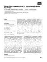
Fig. 1. Sequence comparison of the N-terminal domain (ND) of PEX1 with that of other related proteins. (A) Schematic representation of the
domain architecture of AAA-ATPases PEX6, PEX1, N-ethylmaleimide-sensitive fusion protein (NSF) and valosine-containing protein (VCP).
The ND AAA-ATPase domains (D1, D2) are shown. Physical interactions among the domains are indicated by arrows. (B) Multiple sequence
alignment of PEX1-ND and related NDs with secondary structure elements of PEX1-ND. For sequence names, see Experimental procedures.
The alignment was generated with
CLUSTALX [60] and manually edited. The secondary structure elements are shown at the top, with thick
line segments for the a-helices (a1–a4) and thin line segments for b-strands (b1–b14). The conserved residues of PEX1-ND found in the
putative phospholipid-binding site are shown on the second line. Conserved hydrophobic residues and basic residues of PEX1-ND are indica-
ted by closed circles and closed squares, respectively (see text). The conserved residues and class-specific residues of PEX1-ND and VCP-
ND are colored yellow and green, respectively. Well-conserved basic residues on the surfaces of PEX1-ND and VCP-ND are colored blue.
K. Shiozawa et al. Phospholipid-binding activity of adaptor domains
FEBS Journal 273 (2006) 4959–4971 ª 2006 The Authors Journal compilation ª 2006 FEBS 4963
and the conserved basic residues are found are flat. The
peroxisomal membrane is composed of approximately
10% PC, the head-group of which contains hydrophobic
methyl groups [51]. It is noteworthy that phospholipids
with hydrophilic head-groups such as phosphoinositides
may be sparsely distributed on the membrane, whereas
PC may form a hydrophobic cluster. Thus, the juxtapo-
sition of positively charged and hydrophobic residues
on a flat surface of PEX1-ND suggests an interaction
with such a membrane surface. The results obtained
with the R135A and K174A mutants clearly demon-
strate that this basic and hydrophobic surface is the
phospholipid-binding interface and that arginine 135 is
responsible for specific recognition of the phosphate
group. The importance of the conserved arginine residue
in the molecular recognition of phosphoinositides in
other phospholipid-binding domains, such as FYVE
[52] and PX [53], has been reported. We thus conclude
that PEX1-ND is one of the functional modules for
membrane binding.
Involvement of phosphoinositides in peroxisomal
biogenesis
The interaction between PEX1-ND and phospholipids
could be specific to an inositol phosphate moiety,
because lipid-binding activity is abolished in the pres-
ence of excess phytic acid as a competitor (Fig. 3A). In
terms of phospholipid specificity, there is only a limited
preference for PtdIns monophosphate species over other
phosphoinositides (Fig. 3A,B). Some of these nonspe-
cific lipid-binding domains are believed to act as
scaffolds for a specific membrane rather than in the
PtdIns-mediated signal transduction systems. There are
a few reports in the literature suggesting the involvement
of PtdIns-mediated signal transduction systems in
peroxisomal biogenesis, such as those involving phos-
phatidylinositol-3-kinase and phosphatidylinositol-
4-phosphate-5-kinase. Pexophagy, an event in the
autophagic degradation of excess of peroxisomes, may
be an exceptional phenomenon [54]. More recently,
peroxisome fusion, the critical early step in peroxisome
assembly, for which PEX1 and PEX6 are essential, has
been reported to require the phosphoinositides PtdIns4P
and PtdIns(4,5)P
2
, as well as a distinct set of peroxisom-
al membrane proteins that specifically bind to these two
lipids [62]. This supports our finding that PEX1-ND
interacts with phosphoinositides. Nevertheless, phos-
phoinositide-specific regulation of the peroxisome, akin
to receptor signaling systems or endocytosis, therefore
appears to be unlikely, because PtdIns4P is the most
abundant phosphoinositide in the cell. In contrast, the
number of nonspecific membrane-binding domains is
R135
V36
V49
W60
W146
L121
W116
I177
L176
L131
L175
K174
L130
L169
V147
N
C
I70
V108
Y110
Y143
A142
R144
F139
L140
V181
I182
F131
V154
F152
Y138
C
N
C
A
B
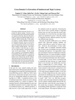
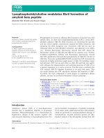
Fig. 2. Constellation of hydrophobic residues with conserved basic
residues on the surface of the N-terminal domains (NDs) of PEX1
and valosine-containing protein (VCP). (A) Conserved hydrophobic
residues within the PEX1 orthologs shown in Fig. 1B are colored
yellow. Conserved basic residues R135 and K174 are colored blue.
(B) Hydrophobic residues conserved among PEX1 and VCP (V108,
Y138, F139, F152, V154, I182) are colored yellow. Conserved
hydrophobic residues within VCP orthologs only (I70, Y110, F131,
A142, Y143, L140 and V181) are colored green. The conserved
basic residue R144 is colored blue. The inset is a ribbon diagram of
the molecule.
Phospholipid-binding activity of adaptor domains K. Shiozawa et al.
4964 FEBS Journal 273 (2006) 4959–4971 ª 2006 The Authors Journal compilation ª 2006 FEBS
often correlated with that of other domains responsible
for specific protein–protein interactions. For example,
more than 63% of pleckstrin homology-domain con-
taining proteins also contain one or more protein-inter-
acting domains such as SH3 [55]. This may enhance
specificity for a particular subcellular compartment.
PEX1-ND meets the criteria for such a protein, as it
forms a scaffold on the peroxisomal membrane through
specific interactions with phospholipids, whereas PEX6
interacts with either PEX26 (mammals) or PEX15
(yeast) on the membrane (Fig. 5) [56,57]. Some mem-
brane-binding proteins bind multiple lipids and ⁄ or pro-
teins simultaneously [63]. The binding of PEX1-ND to
PtdIns might also be attributed to coincident PtfIns
monophosphate binding.
Common phospholipid-binding activity
of VCP-ND and PEX1-ND
We have also demonstrated that the NDs of another
type II AAA-ATPase, VCP, can directly bind phos-
pholipids in vitro. Although this is the first report of
direct interaction between the isolated NDs and phos-
phoinositides, several lines of evidence concerning
VCP and NSF support our findings. Affinity purifica-
tion of NSF using immobilized phospholipids, such as
PtdIns(4,5)P
2
and PA, has been reported [67]. Purified
recombinant NSF was shown to promote fusion of
synthetic liposomes in the absence of a-SNAP and
SNAP receptors, although the required lipid content
was critical [41,42]. Involvement of PtdIns4P and
PtdIns(4,5)P
2
during Sec18-dependent homotypic vacu-
ole fusion in vitro has been reported [43]. All these
results suggest that NSF and Sec18 possess some abil-
ity to bind phospholipids. VCP, which promotes homo-
typic fusion of Golgi or nuclear membranes [20], in
concert with its adaptor protein p47 [28] and ⁄ or
VCIP135 [29], can also promote the fusion of PE-
based liposome vesicles in the absence of p47, with a
reduced but nevertheless substantial measurable effi-
ciency [58]. This suggests that a specific region of VCP
binds to PE-based liposomes. More recently, ATP-
mPEX1 ND (3-180)
A
GST only
20m
M PhA
B
Bound protein (%)
PI5PI3 PI4
Lipid content (%)
0
123450 1 2 3 4 5 0 1 2 3 4 5 012345
PI
30
25
20
15
10
5
0
C
(a)
(b)
0
-5
-10
-15
delta F / Hz
Time (s)
030-15 604515
1
2
3
4
5
6
7
8
9
10
11
12
13
14
15
16
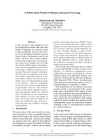
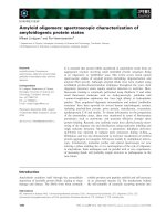
Fig. 3. The N-terminal domain (ND) of PEX1
is a phosphoinositide-binding domain. (A)
Phospholipid binding of PEX1-ND. The puri-
fied glutathione-S-transferase (GST)–PEX1-
ND fusion proteins were examined by PIP
Strip assay and detected by anti-GST serum.
The spots correspond to lyso-phosphatidic
acid (PA) (1), lyso-phosphatidylcholine (PC)
(2), phosphatidylinositol (PtdIns) (3),
PtdIns3P (4), PtdIns4P (5), PtdIns5P (6),
phosphatidylethanolamine (PE) (7), PC (8),
sphingosine 1-phosphate (9), PtdIns(3,4)P
2
(10), PtdIns(3,5)P
2
(11), PtdIns(4,5)P
2
(12),
PtdIns(3,4,5)P
3
(13), PA (14), PS (15), blank
(16). The negative control was GST only
(right panel). The middle panel corresponds
to the same experiments conducted in the
presence of 20 m
M phytic acid (PhA). (B)
Liposome-binding assay of GST–mPEX1-ND.
Fraction of protein bound to PC ⁄ PE (1 : 1)-
based liposomes was plotted against
increasing amounts of PtdIns3P (PI3),
PtdIns4P (PI4), PtdIns5P (PI5) and PtdIns
(PI) in the liposomes, in the absence (filled
bar) or presence (open bar) of 20 m
M PhA
as a competitor. Solid lines indicate standard
deviation of three experiments. (C) Quartz
crystal microbalance (QCM) assay of lipo-
somes, which were added onto GST–
mPEX1-ND immobilized on the QCM
electrode. A typical time-dependent drop
in frequency after injection of PC ⁄ PE
(1 : 1)-based liposome is shown. (a) 5%
PtdIns(4)P. (b) PC ⁄ PE only.
K. Shiozawa et al. Phospholipid-binding activity of adaptor domains
FEBS Journal 273 (2006) 4959–4971 ª 2006 The Authors Journal compilation ª 2006 FEBS 4965
hydrolysis-deficient mutants of VCP were shown to
accumulate on reticular membranes in vivo [22]. On the
other hand, colocalization of VCP and PtdIns-4-kinase
IIa on the buoyant subfraction of ER-derived mem-
brane was observed [66]. The ND of VCP, rather than
the D1 or D2 domains, was assumed to act as an
interface for the phospholipid membrane. The data
shown here may partly provide the molecular basis of
attachment of VCP to the ER-derived membrane. It is
noteworthy that other AAA-ATPase proteins, belong-
ing to the SF6 subfamily, possess transmembrane heli-
ces at their N-terminus [19].
Sequence similarities in D1–D2 domains as well as
the entire domain architectures of PEX1 and VCP sug-
gest that PEX1 can form a stable hexameric ring struc-
ture via the tandem D1–D2 AAA-ATPase domains
such as VCP. Although there are few data concerning
direct protein–lipid interaction involving AAA-ATP-
ases that would prove a common molecular function
of the ND, our results are largely consistent with pre-
vious observations. The apparent association of PEX1
and VCP with the subcellular membranes could, of
course, be attributed not only to protein–lipid interac-
tion, but also to protein–protein interactions via other
membrane-associated proteins. We do not rule out this
possibility, as simultaneous binding of a single ND to
both a lipid and a ligand may occur [63]. In addition,
binding of PtdIns to PEX1-ND may also coincide with
binding of PtdIns phosphates. Protein interface predic-
tions based on ODAs [46] (supplementary Fig. S2)
enabled detection of the known p47 interaction site of
VCP-ND, but the corresponding surface in PEX1-ND
produced no significant energy values. Therefore,
PEX1-ND is unlikely to bind the Ubx domain or
ubiquitin in a manner similar to that of the VCP-ND–
Ubx (p47) complex, consistent with our previous ana-
lyses [32]. Interestingly, a small patch of low-energy
surface was found in a-helix 3 of PEX1-ND, which is
highly conserved among its orthologs. This area might
be involved in protein–protein interaction. As this
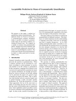
Fig. 5. Schematic representation of the binding of the N-terminal
domain (ND) of PEX1 to the peroxisomal membrane. PEX1 is pre-
dicted to interact with the PEX6 portion of AAA domains. PEX1-ND
binds to the peroxisomal membrane by recognizing specific lipids,
whereas PEX6 interacts with either PEX26 or PEX15 on the mem-
brane.
mPEX1 ND (3-180)
R135A K174A
GST only
A
Wild-type
B
Lipid content (%)
012345
PI(4)P
Bound protein (%)
30
25
20
15
10
5
0
1
2
3
4
5
6
7
8
9
10
11
12
13
14
15
16
GST onlyR144A Wild-type
mVCP ND (1-200)
Wild-type
C
1
2
3
4
5
6
7
8
9
10
11
12
13
14
15
16
20mM PhA
Fig. 4. Phospholipid binding of wild-type and R144A mutant of the
N-terminal domain (ND) of valosine-containing protein (VCP)-ND. (A)
Two mutants of the conserved basic residues, R135A and K174A,
were analyzed by PIP Strips assay. K174A retained the ability
to bind phosphatidylinositol (PtdIns) monophosphates, whereas
R135A did not. (B) The R135 mutant was subjected to the lipo-
some recruitment assay as in Fig. 3B. The fraction of protein bound
to PC ⁄ PE (1 : 1)-based liposomes containing increasing amounts of
PtdIns4P is plotted. Solid lines indicate the standard deviation of
three experiments. (C) PIP Strips assay for wild-type VCP-ND and
its R144A mutant, which does not bind PtdIns monophosphates.
Phospholipid-binding activity of adaptor domains K. Shiozawa et al.
4966 FEBS Journal 273 (2006) 4959–4971 ª 2006 The Authors Journal compilation ª 2006 FEBS
ODA area and the putative phospholipid-binding sur-
face do not overlap, simultaneous binding of a single
ND to both a membrane and a protein substrate might
occur. We are currently testing this hypothesis.
In the present study, we verified that the NDs of
PEX1, VCP, NSF and Ufd1 bind PtdIns. Ufd1-ND,
which has structural similarity with PEX1-ND, is the
only known example of a protein with an ND and no
AAA-ATPase domain. Furthermore, there are several
reports indicating that other AAA-ATPases bind phos-
phoinositides (e.g. in the ER and Golgi) [43,64,65,68].
Examples include phosphoinositide binding of SKD1,
which is a type I AAA-ATPase, whose ND (MIT
domain) is structurally different from that of PEX1
[68,70]. The binding mode and binding, however,
remain unknown and may differ from those of PEX1.
Experimental procedures
Expression and mutational study of PEX1-ND
The construction of a GST fusion protein containing the
mouse PEX1 gene (encoding residues 3–180) in pGEX-4T3-
PRESAT, according to the PRESAT vector methodology
[59], has been previously described [32]. Ala-substituted
mutants R135A and K174A were prepared with a Gene-
Editor site-directed mutagenesis system (Promega, Madison,
WI, USA), according to the manufacturer’s instructions.
The coding regions were sequenced after introduction of
the mutations. Mouse PEX1-ND and their mutants were
expressed as GST fusion proteins in LB medium containing
1% glucose (LBG) containing ampicillin (50 lgÆmL
)1
). The
cells were grown to a D
600
of 0.3, and heterologous gene
expression was induced by addition of 1 mm isopropyl
thio-b-d-galactoside (IPTG). The cells were collected
12–16 h after IPTG induction, washed, and disrupted by
sonication. Recombinant proteins were purified by a
single chromatography step using glutathione Sepharose
(Amersham Bioscience, Uppsala, Sweden). The GST fusion
proteins were dialyzed against buffer containing 150 mm
KCl and 50 mm Hepes (pH 7.5) prior to the phospholipid-
binding assays.
Expression and mutational study of VCP-ND
The plasmids for GST fusion proteins containing the mouse
VCP (residues 1–200) were constructed by a standard pro-
tocol using PCR, and subcloned into pGEX-4T3 (Amer-
sham Bioscience). Ala-substituted mutants R144A were
prepared with a Gene-Editor site-directed mutagenesis sys-
tem (Promega), according to the manufacturer’s instruc-
tions. The coding regions were sequenced after introduction
of the mutations. Mouse VCP and mutant were expressed
as GST fusion proteins in LBG containing ampicillin
(50 lgÆmL
)1
). The cells were grown to a D
600
of 0.5, and
heterologous gene expression was induced by addition of
1mm IPTG. The cells were collected after 3–5 h of IPTG
induction, pelleted, washed, and disrupted by sonication.
Recombinant proteins were purified by a single chromato-
graphy step using glutathione Sepharose. The GST fusion
proteins of the NDs were dialyzed against buffer containing
150 mm KCl and 50 mm Hepes (pH 7.5) prior to the
phospholipid-binding assays.
Expression of other NDs of NSF, CDC48 and Ufd1
A GST fusion expression construct containing the mouse
NSF gene (encoding residues 5–200) was constructed in a
manner similar to that for PEX1-ND. The plasmids for
GST fusion proteins containing yeast CDC48 (residues
1–210), mouse Ufd1 (residues 14–194) and yeast Ufd1 (resi-
dues 1–210) were constructed by a standard PCR protocol,
and subcloned into pGEX-4T3 (Amersham Bioscience).
The cells were grown to a D
600
of 0.5, and heterologous
gene expression was induced by addition of 1 mm IPTG.
The cells were collected after 3–5 h of IPTG induction, pel-
leted, washed, and disrupted by sonication. Recombinant
proteins were purified by a single chromatography step
using glutathione Sepharose. The GST fusion proteins of
the NDs, except mouse ⁄ yeast Ufd1, were dialyzed against
buffer containing 150 mm KCl and 50 mm Hepes (pH 7.5)
prior to the phospholipid-binding assays. Mouse and yeast
Ufd1 were dialyzed against buffer containing 85 mm KCl,
50 mm Hepes (pH 7.5) and 10% glycerol.
Phospholipid-binding assays
For assessment of phospholipid-binding properties, PIP
Strips (Echelon Bioscience Inc., Salt Lake City, UT, USA)
were blocked with binding buffer containing 150 mm NaCl,
10 mm Hepes (pH 7.4), supplemented with 3% fatty acid-
free BSA for 1 h at room temperature. The strips were then
incubated with purified GST fusion proteins at a concentra-
tion of 300 lgÆmL
)1
in blocking buffer at room temperature
for 3 h. After three washes in the binding buffer, PIP Strips
were incubated for 3 h at room temperature with anti-GST
(Nacalai Tesque, Kyoto, Japan) serum in the same buffer.
Secondary antibody incubation and 3,3¢,5,5¢-tetra-
methylbenzidine staining were performed to detect GST-
tagged proteins bound to the phospholipid spots on the
membrane.
Liposome recruitment assay
Liposome recruitment reactions were performed in 150 mm
KCl and 50 mm Hepes (pH 7.5). Liposomes were made
from a 1 : 1 mixture of PC and PE (Sigma Aldrich, Tokyo,
Japan) in the presence or absence of PtdIns3P, PtdIns4P,
K. Shiozawa et al. Phospholipid-binding activity of adaptor domains
FEBS Journal 273 (2006) 4959–4971 ª 2006 The Authors Journal compilation ª 2006 FEBS 4967
PtdIns5P, or PtdIns (5% weight ratio to the base liposome;
Sigma Aldrich). Purified GST fusion proteins (50 lgÆmL
)1
)
were incubated with the liposomes (1 mg lipidÆmL
)1
). After
5 min of incubation at room temperature, membranes were
recovered by centrifugation at 35 000 g at 4 °C with a
Beckman TL-100 ultracentrifuge, rotor type TLA-100
(Beckman Coulter, Fullerton, CA, USA). The reaction was
carried out on a 50 lL scale. Supernatants and pellets were
resuspended in SDS sample buffer and analyzed by
SDS ⁄ PAGE, followed by Coomassie brilliant blue staining.
The gel images were analyzed using the imagej 1.31v soft-
ware ( />QCM assay of in vitro protein–lipid interaction
The in vitro liposome binding of a mutant of PEX1-ND
was determined by frequency change in a 27-MHz QCM
using an AFFINIX-Q (QCM2000) instrument (Initiam Co.,
Tokyo, Japan). A GST fusion protein of wild-type PEX1-
ND was immobilized on a QCM gold electrode (diameter
4.5 mm, QCMST27) according to the manufacturer’s
instructions. A 2 lL aliquot of a 50 lgÆmL
)1
solution of
GST–PEX1-ND was used. The electrode was washed sev-
eral times in buffer containing 150 mm KCl and 50 mm
Hepes (pH 7.45), soaked in the same buffer (2 mL cuvette),
and then monitored continuously for QCM frequency chan-
ges at 25 °C. Once the frequency had stabilized, 8 lLof
the PC + PE (1 : 1) liposomes (1 mgÆmL
)1
) with or with-
out PtdIns4P (5% weight ratio to the base liposome) were
injected.
Evolutionary trace analysis
The four sequences of PEX1-ND orthologs and six
sequences of VCP-ND orthologs were obtained from NCBI;
they are PEX1_HUMAN (human PEX1; AAB99758),
PEX1_MOUSE (Mus musculus PEX1; XML131895),
PEX1_ARATH (Arabidopsis thaliana PEX1; AAG44817),
PEX1_YEAST (yeast PEX1; CAA82041), VCP_MOUSE
(Mus musculus VCP; NP_033529), VCP_HUMAN (human
CAH70993), VCP_XENOPUS (Xenopus VCP; AAH74716),
VCP_ZEBRAFISH (zebrafish VCP; NP_958889), CDC48_
YEAST (yeast cdc48; CAA98694) and VAT_THEAC
(Thermoplasma acidophilum VAT; AAC45089). The
multiple sequence alignment was produced using the pro-
gram clustalx [60] and then refined manually. The evolu-
tionary trace analysis [45] was then carried out on the
Evolutionary Trace Server (tracesuite II) [46]. Here, the
evolutionary trace method defined groups of proteins using
an evolutionary time cut-off in a phylogenetic tree, and
divided all residues of aligned sequences into three classes:
conserved (residues invariant throughout the groups), class-
specific (residues invariant within each group but that
change between groups), and neutral (all others) (supple-
mentary Fig. S1). Gaps were counted as an extra residue
type. The class-specific residues are the most interesting in
terms of the development of functional innovation. Trace
residues were mapped onto the structure by pymol [61]. The
surfaces of PEX1-ND and VCP-ND were further subjected
to ODA analysis by using program pydock (J. Fernandez-
Recio, unpublished results).
Acknowledgements
This work was supported by grants to MS and HH
from the Japanese Ministry of Education, Science,
Sports and Culture (Protein3000). We thank Dr Y.
Fujiki for a vector containing PEX1 cDNA. We thank
Dr M. H. J. Koch and Dr W. Schliebs for many valu-
able discussions and for critical reading of the manu-
script.
References
1 van den Bosch H, Schutgens RB, Wanders RJ & Tager
JM (1992) Biochemistry of peroxisomes. Annu Rev
Biochem 61, 157–197.
2 Eckert JH & Erdmann R (2003) Peroxisome biogenesis.
Rev Physiol Biochem Pharmacol 147, 75–121.
3 Wanders RJ & Waterham HR (2005) Peroxisomal dis-
orders I: biochemistry and genetics of peroxisome bio-
genesis disorders. Clin Genet 67, 107–133.
4 Imamura A, Tamura S, Shimozawa N, Suzuki Y,
Zhang Z, Tsukamoto T, Orii T, Kondo N, Osumi T &
Fujiki Y (1998) Temperature-sensitive mutation in
PEX1 moderates the phenotypes of peroxisome defi-
ciency disorders. Hum Mol Genet 7 , 2089–2094.
5 Geisbrecht BV, Collins CS, Reuber BE & Gould SJ
(1998) Disruption of a PEX1–PEX6 interaction is the
most common cause of the neurologic disorders Zellwe-
ger syndrome, neonatal adrenoleukodystrophy, and
infantile Refsum disease. Proc Natl Acad Sci USA 95,
8630–8635.
6 Tamura S, Okumoto K, Toyama R, Shimozawa N,
Tsukamoto T, Suzuki Y, Osumi T, Kondo N & Fujiki Y
(1998) Human PEX1 cloned by functional com-
plementation on a CHO cell mutant is responsible for
peroxisome-deficient Zellweger syndrome of
complementation group I. Proc Natl Acad Sci USA 95,
4350–4355.
7 Reuber BE, Germain-Lee E, Collins CS, Morrell JC,
Ameritunga R, Moser HW, Valle D & Gould SJ (1997)
Mutations in PEX1 are the most common cause of per-
oxisome biogenesis disorders. Nat Genet 17, 445–448.
8 Dodt G & Gould SJ (1996) Multiple PEX genes are
required for proper subcellular distribution and stability
of Pex5p, the PTS1 receptor: evidence that PTS1 protein
import is mediated by a cycling receptor. J Cell Biol
135, 1763–1774.
Phospholipid-binding activity of adaptor domains K. Shiozawa et al.
4968 FEBS Journal 273 (2006) 4959–4971 ª 2006 The Authors Journal compilation ª 2006 FEBS
9 Yahraus T, Braverman N, Dodt G, Kalish JE, Morrell
JC, Moser HW, Valle D & Gould SJ (1996) The peroxi-
some biogenesis disorder group 4 gene, PXAAA1,
encodes a cytoplasmic ATPase required for stability of
the PTS1 receptor. EMBO J 15, 2914–2923.
10 Kiel JA, Hilbrands RE, van der Klei IJ, Rasmussen
SW, Salomons FA, van der Heide M, Faber KN, Cregg
JM & Veenhuis M (1999) Hansenula polymorpha Pex1p
and Pex6p are peroxisome-associated AAA proteins that
functionally and physically interact. Yeast 15, 1059–
1078.
11 Tamura S, Shimozawa N, Suzuki Y, Tsukamoto T,
Osumi T & Fujiki Y (1998) A cytoplasmic AAA family
peroxin, Pex1p, interacts with Pex6p. Biochem Biophys
Res Commun 245, 883–886.
12 Birschmann I, Rosenkranz K, Erdmann R & Kunau
WH (2005) Structural and functional analysis of the
interaction of the AAA-peroxins Pex1p and Pex6p.
FEBS J 272, 47–58.
13 Platta HW, Grunau S, Rosenkranz K, Girzalsky W &
Erdmann R (2005) Functional role of the AAA peroxins
in dislocation of the cycling PTS1 receptor back to the
cytosol. Nat Cell Biol 7, 817–822.
14 Platta HW, Girzalsky W & Erdmann R (2004) Ubiquiti-
nation of the peroxisomal import receptor Pex5p. Bio-
chem J 384, 37–45.
15 Kiel JA, Emmrich K, Meyer HE & Kunau WH (2005)
Ubiquitination of the peroxisomal targeting signal type
1 receptor, Pex5p, suggests the presence of a quality
control mechanism during peroxisomal matrix protein
import. J Biol Chem 280, 1921–1930.
16 Kragt A, Voorn-Brouwer T, van den Berg M & Distel B
(2005) Endoplasmic reticulum-directed Pex3p routes to
peroxisomes and restores peroxisome formation in
Saccharomyces cerevisiae pex3 Delta strain. J Biol Chem
280, 7867–7874.
17 Kiel JA, Otzen M, Veenhuis M & van der Klei IJ
(2005) Obstruction of polyubiquitination affects PTS1
peroxisomal matrix protein. Biochim Biophys Acta 1745,
176–186.
18 Lupas AN & Martin J (2002) AAA proteins. Curr Opin
Struct Biol 12, 746–753.
19 Beyer A (1997) Sequence analysis of the AAA protein
family. Protein Sci 6, 2043–2058.
20 Rabouille C, Levine TP, Peters JM & Warren G (1995)
An NSF-like ATPase, p97, and NSF mediate cisternal
regrowth from mitotic Golgi fragments. Cell 82, 905–
914.
21 Clary DO, Griff IC & Rothman JE (1990) SNAPs, a
family of NSF attachment proteins involved in intracel-
lular membrane fusion in animals and yeast. Cell 61,
709–721.
22 Dalal S, Rosser MF, Cyr DM & Hanson PI (2004) Dis-
tinct roles for the AAA ATPases NSF and p97 in the
secretory pathway. Mol Biol Cell 15, 637–648.
23 Rabinovich E, Kerem A, Frohlich KU, Diamant N &
Bar-Nun S (2002) AAA-ATPase p97 ⁄ Cdc48p, a cytoso-
lic chaperone required for endoplasmic reticulum-associ-
ated protein degradation. Mol Cell Biol 22, 626–634.
24 Whiteheart SW & Kubalek EW (1995) SNAPs and
NSF: general members of the fusion apparatus. Trends
Cell Biol 5, 64–68.
25 Hay JC & Scheller RH (1997) SNAREs and NSF in tar-
geted membrane fusion. Curr Opin Cell Biol 9, 505–512.
26 Ye Y, Meyer HH & Rapoport TA (2003) Function of
the p97–Ufd1–Npl4 complex in retrotranslocation from
the ER to the cytosol: dual recognition of nonubiquiti-
nated polypeptide segments and polyubiquitin chains.
J Cell Biol 162
, 71–84.
27 Meyer HH, Shorter JG, Seemann J, Pappin D & War-
ren G (2000) A complex of mammalian ufd1 and npl4
links the AAA-ATPase, p97, to ubiquitin and nuclear
transport pathways. EMBO J 19, 2181–2192.
28 Kondo H, Rabouille C, Newman R, Levine TP, Pappin
D, Freemont P & Warren G (1997) p47 is a cofactor
for p97-mediated membrane fusion. Nature 388, 75–78.
29 Uchiyama K, Jokitalo E, Kano F, Murata M, Zhang
X, Canas B, Newman R, Rabouille C, Pappin D, Free-
mont P et al. (2002) VCIP135, a novel essential factor
for p97 ⁄ p47-mediated membrane fusion, is required for
Golgi and ER assembly in vivo. J Cell Biol 159, 855–
866.
30 Ye Y, Shibata Y, Yun C, Ron D & Rapoport TA
(2004) A membrane protein complex mediates retro-
translocation from the ER lumen into the cytosol.
Nature 429, 841–847.
31 Ye Y, Shibata Y, Kikkert M, van Voorden S, Wiertz
E & Rapoport TA (2004) Inaugural article: Recruit-
ment of the p97 ATPase and ubiquitin ligases to the
site of retrotranslocation at the endoplasmic reticulum
membrane. Proc Natl Acad Sci USA 102, 14132–
14138.
32 Shiozawa K, Maita N, Tomii K, Seto A, Goda N,
Akiyama Y, Shimizu T, Shirakawa M & Hiroaki H
(2004) Structure of the N-terminal domain of PEX1
AAA-ATPase: characterization of a putative adaptor-
binding domain. J Biol Chem 279, 50060–50069.
33 Zhang X, Shaw A, Bates PA, Newman RH, Gowen B,
Orlova E, Gorman MA, Kondo H, Dokurno P, Lally J
et al. (2000) Structure of the AAA ATPase p97. Mol
Cell 6, 1473–1484.
34 Yu RC, Jahn R & Brunger AT (1999) NSF N-terminal
domain crystal structure: models of NSF function. Mol
Cell 4, 97–107.
35 May AP, Misura KM, Whiteheart SW & Weis WI
(1999) Crystal structure of the amino-terminal domain
of N-ethylmaleimide-sensitive fusion protein. Nat Cell
Biol 1, 175–182.
36 Coles M, Diercks T, Liermann J, Groger A, Rockel B,
Baumeister W, Koretke KK, Lupas A, Peters J &
K. Shiozawa et al. Phospholipid-binding activity of adaptor domains
FEBS Journal 273 (2006) 4959–4971 ª 2006 The Authors Journal compilation ª 2006 FEBS 4969
Kessler H (1999) The solution structure of VAT-N
reveals a ‘missing link’ in the evolution of complex
enzymes from a simple betaalphabetabeta element. Curr
Biol 9, 1158–1168.
37 Vogel C, Berzuini C, Bashton M, Gough J & Teichmann
SA (2004) Supra-domains: evolutionary units larger than
single protein domains. J Mol Biol 336, 809–823.
38 Dreveny I, Kondo H, Uchiyama K, Shaw A, Zhang X
& Freemont PS (2004) Structural basis of the interac-
tion between the AAA ATPase p97 ⁄ VCP and its adap-
tor protein p47. EMBO J 23, 1030–1039.
39 Park S, Isaacson R, Kim HT, Silver PA & Wagner G
(2005) Ufd1 exhibits the AAA-ATPase fold with two
distinct ubiquitin interaction sites. Structure 13, 995–
1005.
40 Babor SM & Fass D (1999) Crystal structure of the
Sec18p N-terminal domain. Proc Natl Acad Sci USA
96, 14759–14764.
41 Brugger B, Nickel W, Weber T, Parlati F, McNew JA,
Rothman JE & Sollner T (2000) Putative fusogenic
activity of NSF is restricted to a lipid mixture whose
coalescence is also triggered by other factors. EMBO J
19, 1272–1278.
42 Otter-Nilsson M, Hendriks R, Pecheur-Huet EI, Hoek-
stra D & Nilsson T (1999) Cytosolic ATPases, p97 and
NSF, are sufficient to mediate rapid membrane fusion.
EMBO J 18, 2074–2083.
43 Mayer A, Scheglmann D, Dove S, Glatz A, Wickner W
& Haas A (2000) Phosphatidylinositol 4,5-bisphosphate
regulates two steps of homotypic vacuole fusion. Mol
Biol Cell 11, 807–817.
44 Lichtarge O, Bourne HR & Cohen FE (1996) An evolu-
tionary trace method defines binding surfaces common
to protein families. J Mol Biol 257, 342–358.
45 Innis CA, Shi J & Blundell TL (2000) Evolutionary
trace analysis of TGF-beta and related growth factors:
implications for site-directed mutagenesis. Protein Eng
13, 839–847.
46 Fernandez-Recio J, Totrov M, Skorodurnov C & Abag-
yan R (2005) Optimal docking area: a new method for
predicting protein–protein interaction sites. Proteins 58,
134–143.
47 Kojima T, Fukuda M, Watanabe Y, Hamazato F &
Mikoshiba K (1997) Characterization of the pleckstrin
homology domain of Btk as an inositol polyphosphate
and phosphoinositide binding domain. Biochem Biophys
Res Commun 236, 333–339.
48 Faber KN, Heyman JA & Subramani S (1998) Two
AAA family peroxins, PpPex1p and PpPex6p, interact
with each other in an ATP-dependent manner and are
associated with different subcellular membranous struc-
tures distinct from peroxisomes. Mol Cell Biol 18,
936–943.
49 Titorenko VI, Chan H & Rachubinski RA (2000) Fusion
of small peroxisomal vesicles in vitro reconstructs an
early step in the in vivo multistep peroxisome assembly
pathway of Yarrowia lipolytica. J Cell Biol 148, 29–44.
50 Horsnell WG, Steel GJ & Morgan A (2002) Analysis of
NSF mutants reveals residues involved in SNAP bind-
ing and ATPase stimulation. Biochemistry 41, 5230–
5235.
51 Hoepfner D, Schildknegt D, Braakman I, Philippsen P
& Tabak HF (2005) Contribution of the endoplasmic
reticulum to peroxisome formation. Cell 122, 85–95.
52 Kutateladze TG, Ogburn KD, Watson WT, de Beer T,
Emr SD, Burd CG & Overduin M (1999) Phosphatidyl-
inositol 3-phosphate recognition by the FYVE domain.
Mol Cell 3
, 805–811.
53 Ago T, Takeya R, Hiroaki H, Kuribayashi F, Ito T,
Kohda D & Sumimoto H (2001) The PX domain as
a novel phosphoinositide-binding module. Biochem
Biophys Res Commun 287, 733–738.
54 Kiel JA, Rechinger KB, van der Klei IJ, Salomons FA,
Titorenko VI & Veenhuis M (1999) The Hansenula
polymorpha PDD1 gene product, essential for the
selective degradation of peroxisomes, is a homologue
of Saccharomyces cerevisiae Vps34p. Yeast 15,
741–754.
55 Pawson T (1995) Protein modules and signalling net-
works. Nature 373, 573–580.
56 Matsumoto N, Tamura S & Fujiki Y (2003) The patho-
genic peroxin Pex26p recruits the Pex1p–Pex6p AAA
ATPase complexes to peroxisomes. Nat Cell Biol 5,
454–460.
57 Birschmann I, Stroobants AK, van den Berg M,
Schafer A, Rosenkranz K, Kunau WH & Tabak HF
(2003) Pex15p of Saccharomyces cerevisiae provides a
molecular basis for recruitment of the AAA peroxin
Pex6p to peroxisomal membranes. Mol Biol Cell 14,
2226–2236.
58 Pecheur EI, Martin I, Maier O, Bakowsky U, Ruyss-
chaert JM & Hoekstra D (2002) Phospholipid species
act as modulators in p97 ⁄ p47-mediated fusion of Golgi
membranes. Biochemistry 41, 9813–9823.
59 Goda N, Tenno T, Takasu H, Hiroaki H & Shirakawa
M (2004) The PRESAT-vector: asymmetric T-vector for
high-throughput screening of soluble protein domains
for structural proteomics. Protein Sci 13, 652–658.
60 Thompson JD, Gibson TJ, Plewniak F, Jeanmougin F
& Higgins DG (1997) The CLUSTAL_X windows inter-
face: flexible strategies for multiple sequence alignment
aided by quality analysis tools. Nucleic Acids Res 24,
4876–4882.
61 DeLano WL (2002) The PyMOL Molecular Graphics
System. DeLano Scientific, San Carlos, CA, USA
().
62 Boukh-Viner T, Guo T, Alexandrian A, Cerracchio A,
Gregg C, Haile S, Kyskan R, Milijevic S, Oren D, Solo-
mon J et al. (2005) Dynamic ergosterol- and ceramide-
rich domains in the peroxisomal membrane serve as an
Phospholipid-binding activity of adaptor domains K. Shiozawa et al.
4970 FEBS Journal 273 (2006) 4959–4971 ª 2006 The Authors Journal compilation ª 2006 FEBS
organizing platform for peroxisome fusion. J Cell Biol
168, 761–773.
63 Carlton JG & Cullen PJ (2005) Coincidence detection
in phosphoinositide signaling. Trends Cell Biol 15,
540–547.
64 Snider A, Gutsche I, Lin M & Baby S, Cox B, Butland
G, Greenblatt J, Emili A & Houry WA (2006) Forma-
tion of a distinctive complex between the inducible bac-
terial lysine decarboxylase and a novel AAA
+
ATPase.
J Biol Chem 281, 1532–1546.
65 Ikonomov OC, Sbrissa D, Yoshimori T, Cover TL &
Shisheva A (2002) PIKfyve kinase and SKD1 AAA
ATPase define distinct endocytic compartments. Only
PIKfyve expression inhibits the cell-vacuolating activity
of Helicobacter pylori VacA toxin. J Biol Chem 277,
46785–46790.
66 Waugh MG, Minogue S, Anderson JS, Balinger A,
Blumenkrantz D, Calnan DP, Cramer R & Hsuan JJ
(2003) Localization of a highly active pool of type II
phosphatidylinositol 4-kinase in a p97 ⁄ valosin-contain-
ing-protein-rich fraction of the endoplasmic reticulum.
Biochem J 373, 57–63.
67 Manifava M, Thuring JW, Lim ZY, Packman L, Hol-
mes AB & Ktistakis NT (2001) Differential binding of
traffic-related proteins to phosphatidic acid- or phos-
phatidylinositol (4,5)-bisphosphate-coupled affinity
reagents. J Biol Chem 276, 8987–8994.
68 Scott A, Chung HY, Gonciarz-Swiatek M, Hill GC,
Whitby FG, Gaspar J, Holton JM, Viswanathan R,
Ghaffarian S, Hill CP et al. (2005) Structural and
mechanistic studies of VPS4 proteins. EMBO J 24,
3658–3669.
69 Rizo J & Sudhof TC (1998) C2-domains, structure and
function of a universal Ca
2+
-binding domain. J Biol
Chem 273, 15879–15882.
70 Takasu H, Jee JG, Ohno A, Goda N, Fujiwara K,
Tochio H, Shirakawa M & Hiroaki H (2005) Structural
characterization of the MIT domain from human
Vps4b. Biochem Biophys Res Commun 334, 460–465.
Supplementary material
The following supplementary material is available
online:
Fig. S1. (A) Phylogenetic tree constructed from the
four sequences of PEX1 N-terminal domain (ND)
orthologs and six sequences of valosine-containing pro-
tein (VCP)-ND orthologs. (B) Location of trace resi-
dues in PEX1-ND and VCP-ND.
Fig. S2. Surface representations of N-terminal domains
(NDs) of PEX1 and valosine-containing protein
(VCP), colored according to the energy values of their
respective optimal docking areas (ODAs) [46].
Fig. S3. PIP Strip assay of the phospholipid binding
of the glutathione-S-transferase (GST) fusion protein
with the other N-terminal domains of type II AAA-
ATPases and the related adaptor proteins.
This material is available as part of the online article
from
K. Shiozawa et al. Phospholipid-binding activity of adaptor domains
FEBS Journal 273 (2006) 4959–4971 ª 2006 The Authors Journal compilation ª 2006 FEBS 4971