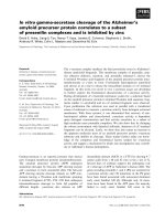
Báo cáo khoa học: In vitro embryonic developmental phosphorylation of the cellular nucleic acid binding protein by cAMP-dependent protein kinase, and its relevance for biochemical activities pdf
Bạn đang xem bản rút gọn của tài liệu. Xem và tải ngay bản đầy đủ của tài liệu tại đây (949.58 KB, 13 trang )
In vitro embryonic developmental phosphorylation of the
cellular nucleic acid binding protein by cAMP-dependent
protein kinase, and its relevance for biochemical activities
Vero
´
nica A. Lombardo, Pablo Armas, Andrea M. J. Weiner and Nora B. Calcaterra
Divisio
´
n Biologı
´
a del Desarrollo, IBR – CONICET, Area Biologı
´
a General, Facultad de Ciencias Bioquı
´
micas y Farmace
´
uticas,
Universidad Nacional de Rosario, Argentina
The zinc-finger cellular nucleic acid binding protein
(CNBP) shows striking sequence conservation among
vertebrates [1]. Recent works show that CNBP is
required for forebrain formation during vertebrate
organogenesis. Cnbp-null mutant mice are embryonic
lethal and show severe forebrain truncation and facial
abnormalities due to a lack of proper morphogenetic
movements of the anterior visceral endoderm during
Keywords
CNBP; Danio rerio; embryogenesis;
phosphorylation; PKA
Correspondence
N. B. Calcaterra, IBR – CONICET, A
´
rea
Biologı
´
a General, Dpto. de Ciencias
Biolo
´
gicas, Facultad de Ciencias Bioquı
´
micas
y Farmace
´
uticas, Universidad Nacional de
Rosario, Suipacha 531 (S2002LRK) Rosario,
Argentina
Tel ⁄ Fax: +54 341 4804601
E-mail: or
Database
CNBP
S158A
nucleotide sequence data is
available in the GenBank database under the
accession number DQ519386
(Received 9 October 2006, revised 6
November 2006, accepted 15 November
2006)
doi:10.1111/j.1742-4658.2006.05596.x
The zinc-finger cellular nucleic acid binding protein (CNBP) is a strikingly
conserved single-stranded nucleic acid binding protein essential for normal
forebrain formation during mouse and chick embryogenesis. CNBP cDNAs
from a number of vertebrates have been cloned and analysed. CNBP is
mainly conformed by seven retroviral Cys-Cys-His-Cys zinc-knuckles and a
glycine ⁄ arginine rich region box. CNBP amino acid sequences show a puta-
tive Pro-Glu-Ser-Thr site of proteolysis and several putative phosphoryla-
tion sites. In this study, we analysed CNBP phosphorylation by embryonic
kinases and its consequences on CNBP biochemical activities. We report
that CNBP is differentially phosphorylated by Danio rerio embryonic
extracts. In vitro CNBP phosphorylation is basal and constant at early
embryonic developmental stages, it begins to increase after mid-blastula
transition stage reaching the highest level at 48 hours postfertilization
stage, and decreases thereafter to basal levels at 5 days postfertilization.
The cAMP-dependent protein kinase (PKA) was identified as responsible
for phosphorylation on the unique CNBP conserved putative phosphoryla-
tion site. Site-directed mutagenesis replacing the PKA phospho-acceptor
amino acid residue impairs CNBP phosphorylation, suggesting that phos-
phorylation may not only exist in D. rerio but also in other vertebrates.
CNBP phosphorylation does not change single-stranded nucleic acid bind-
ing capability. Instead, it promotes in vitro the annealing of complementary
oligonucleotides representing the CT element (CCCTCCCC) from the
human cellular myelocytomatosis oncogene (c-myc) promoter, an element
responsible for c-myc enhancer transcription. Our results suggest that phos-
phorylation might be a conserved post-translational modification that
allows CNBP to perform a fine tune expression regulation of a group of
target genes, including c-myc, during vertebrate embryogenesis.
Abbreviations
CCHC, Cys-Cys-His-Cys; CK2, casein kinase 2; c-myc, cellular myelocytomatosis oncogene; CNBP, cellular nucleic acid binding protein;
Comp-CT, CT element complementary sequence; CT, CCCTCCCC containing sequence element; dpf, days postfertilization; GST, glutathione
S-transferase; hpf, hours postfertilization; hnRNP K, heterogenous ribonucleic protein K; PEST, Pro-Glu-Ser-Thr; PKA, cAMP-dependent
protein kinase; PKA-Ca, PKA subunit catalytic a; PKC, protein kinase C; PKI, protein kinase inhibitor; rp-mRNA, ribosomal protein mRNAs;
RGG, glycine ⁄ arginine rich region; 5¢ TOP, 5¢ terminal oligopyrimidine tract.
FEBS Journal 274 (2007) 485–497 ª 2006 The Authors Journal compilation ª 2006 FEBS 485
pregastrulation stage [2]. In chick embryos, Cnbp is
expressed in the equivalent tissues of the mouse
embryo and, furthermore, CNBP siRNA knockdown
produces forebrain truncation [3]. It was proposed
that the CNBP role during vertebrate organogenesis
is to regulate the forebrain formation by controlling
the expression of a number of rostral head trans-
cription factors, such as c-myc [2], BF-1, Six3, and
Hesx1 [3].
A vast range of cellular functions has been assigned
to CNBP. It was reported acting both as a negative
[4–6] and positive [7,8] transcriptional regulator. Apart
from this, CNBP showed interaction with several RNA
targets [9–12]. From these reports emerge two main
possible models where CNBP might act as single-stran-
ded nucleic acid binding protein: the first one involves
CNBP and heterogenous nuclear ribonucleic protein K
(hnRNP K) as transcriptional activators of the c-myc
gene via the CT promoter element (CCCTCCCC)
[7,13]; the second model proposes CNBP as a transla-
tional inhibitor of ribosomal proteins mRNAs (rp-
mRNAs) through binding to the 5¢ UTR containing a
5¢ terminal oligopyrimidine tract (5¢ TOP) motif [14].
CNBP cDNAs have been cloned from mammals
[7,12,15–17], chicken [18,19], amphibians [1,20,21] and
fish [10,22]. The protein shows a highly conserved struc-
tural organization and amino acid sequence sharing
the classical arrangement of seven tandem canonical
Cys-Cys-His-Cys (CCHC)-type zinc knuckle domains
(C-/-X-C-G-X
3
-H-X
4
-C, where / is an aromatic amino
acid and X is a variable amino acid) typical of retroviral
nucleocapsid proteins and the presence of an glycine ⁄
arginine rich region (RGG) box between the first
and second Zn knuckles. In silico analysis of CNBP
amino acid sequence reveals the presence of a putative
nuclear localization signal PKKEREQ, a putative Pro-
Glu-Ser-Thr (PEST) site of proteolysis, and several
putative phosphorylation sites [1,10,21,22] (Fig. 1).
The relationship between CNBP developmental
behaviour and phosphorylation has not been analysed
yet. To gain an understanding of the biochemical con-
sequences of CNBP phosphorylation, it was important
to analyse CNBP phosphorylation during embryonic
development and to identify kinases that catalyse
CNBP phosphorylation. We report here that CNBP
is in vitro phosphorylated by zebrafish embryonic
extracts from different developmental stages. The
cAMP-dependent protein kinase (PKA) was identified
as the predominant kinase that catalyses CNBP phos-
phorylation and, furthermore, it was found that CNBP
major phosphorylation occurred on the unique con-
served putative phosphorylation site. Finally, we
observed that CNBP phosphorylation promoted the
annealing of complementary oligonucleotides in vitro
but did not modify single-stranded nucleic acid binding
capability. Based on these results, we discuss the role
of phosphorylation-dephosphorylation events in the
biological function of CNBP.
Fig. 1. (A) In silico analysis of zebrafish CNBP amino acid sequence. The amino acid sequence of zebrafish CNBP was analysed using the
Prosite server. The amino acid residues involved in CCHC motifs are indicated in light grey boxes. Amino acids that participate in the coordi-
nation of the Zn atom are boxed in dark grey. The RGG box is boxed and the putative nuclear localization signal is underlined. The arrow
shows a putative site of proteolysis. (B) In silico analysis of putative phosphorylation sites. Zebrafish CNBP putative phosphorylation sites
were analysed by Prosite, NetPhos 2.0, ScanSite 2.0, GPS, and PredPhospho servers. Boxed amino acid residues were predicted by at least
three phosphorylation site servers.
In vitro developmental CNBP phosphorylation V. A. Lombardo et al.
486 FEBS Journal 274 (2007) 485–497 ª 2006 The Authors Journal compilation ª 2006 FEBS
Results and Discussion
CNBP shows several putative phosphorylation
sites
The amino acid sequence of zebrafish CNBP (Fig. 1A)
was analysed in silico searching for putative phos-
phorylation sites by using Prosite [23], NetPhos2.0
[24], ScanSite [25], GPS (group-based phosphorylation
scoring method) [26], and PredPhospho [27] servers.
The analysis yielded potential phosphorylation sites on
several amino acid residues (Fig. 1B). The amino acid
residues Ser4, Thr56, Ser70, and Ser158 were predicted
as putative phospho-acceptors by at least three phos-
phosite prediction servers (Fig. 1B), reinforcing the
hypothesis of CNBP phosphorylation.
Sequence alignment did not show major putative
phosphorylation site conservation among CNBP
sequences except for the site located immediately after
the seventh CCHC Zn knuckle motif (Table 1). The
conservation among CNBP vertebrates suggests that
phosphorylation in this site, which in zebrafish was
predicted by four of the five employed servers
(Fig. 1B), may play a relevant role in the regulation of
CNBP biochemical activities.
CNBP is phosphorylated in vitro by embryonic
kinases
To facilitate the biochemical characterization of
CNBP phosphorylation, the protein was expressed in
Escherichia coli as fusion proteins with N-terminal
hexa-histidine (His6-CNBP) and N-terminal glutathi-
one S-transferase (GST) (GST-CNBP) tags and used
for in vitro kinase activity assays. Both GST-CNBP
(Fig. 2A,B) as well as His6-CNBP (Fig. S1), were
phosphorylated by extracts prepared from 24 hours
postfertilization (hpf) embryos. This extract failed to
phosphorylate GST (Fig. 2A,B) demonstrating that
the kinase phospho-acceptor site is in CNBP itself.
From the analysis of gels, it was noticeable the exist-
ence of phospho-peptides with lower molecular masses
than the recombinant fusion protein GST-CNBP. This
phenomenon may be the consequence of CNBP pro-
teolysis by either bacterial or embryonic proteases, as
it was previously reported [11]. Indeed, the radioactive
polypeptides were recognized by a polyclonal antibody
raised against CNBP in western blot assays (Fig. S2).
Table 1. C-terminal amino acid region alignment of CNBPs from different organisms. The sequences shown correspond to the last 29 amino
acid residues from each CNBP protein. Numbers in Danio rerio sequence represent the amino acid position in the primary structure
shown in Fig. 1A. CCHC zinc knuckles are shaded gray. Consensus sequence for PKA is boxed. Amino acid residues predicted as phospho-
acceptors are shaded black.
Fig. 2. In vitro CNBP phosphorylation. In vitro phosphorylation
assays were performed using the recombinant CNBP (GST-CNBP)
and zebrafish embryonic extract prepared from embryos at 24 hpf.
Phosphorylated GST-CNBP was run in 12% (w ⁄ v) SDS ⁄ PAGE.
(A) Gel autoradiography. (B) Coomassie Blue stained gel. Arrow on
the right indicates GST-CNBP; asterisk on the right indicates GST
protein.
V. A. Lombardo et al. In vitro developmental CNBP phosphorylation
FEBS Journal 274 (2007) 485–497 ª 2006 The Authors Journal compilation ª 2006 FEBS 487
Taken together, these results confirm that CNBP is
phosphorylated by embryonic zebrafish kinases and
allow us to further characterize CNBP phosphoryla-
tion and to identify the candidate kinases.
CNBP is differentially phosphorylated in vitro
by protein extracts prepared from embryos at
different developmental stages
To further analyse CNBP phosphorylation, we carried
out in vitro recombinant CNBP phosphorylation using
extracts prepared from embryos at different develop-
mental stages. A basal and constant phosphorylation
level was consistently observed at early embryonic
developmental stages until the stage of 8-somite.
Beyond the 8-somite stage, CNBP phosphorylation
began to increase, reaching a maximum at 48 hpf
stage. Afterwards, phosphorylation level decreased
progressively (Fig. 3A,B) returning to a basal level at
5 days postfertilization (dpf) (Fig. 3C). Similar results
were obtained when His6-CNBP was analysed
(Fig. S3). The radioactivity ⁄ protein ratio raised a
maximum of approximately 10-fold at 48 hpf with
respect to the basal phosphorylation level (Fig. 3C).
These results confirm the existence of CNBP phos-
phorylation by embryonic kinases and, furthermore,
suggest a transient change in protein kinase activity
during zebrafish development.
It is worth mentioning that CNBP phosphorylation
began to increase at the 8-somite stage (Fig. 3C), the
developmental stage in which a CNBP subcellular
localization change became detectable in zebrafish as
well as in Bufo arenarum [1,10]. Therefore, it is
tempting to speculate that CNBP phosphorylation
would be the post-translational modification that
allows CNBP to move from the cytoplasm to the
nucleus during early development. The role of CNBP
phosphorylation in vivo, as well as the putative corre-
lation between the nucleo-cytoplasmic translocation
and the CNBP phosphorylation status remains to be
established. These issues are subject for further
research and are currently under investigation in our
laboratory.
CNBP is phosphorylated on Ser
⁄
Thr residues
In silico analysis showed that CNBP amino acid
sequences possess putative phosphorylation sites
mainly on Ser ⁄ Thr residues (Fig. 1). To confirm this,
GST-CNBP was in vitro phosphorylated and then sub-
jected to an alkaline treatment. This treatment
removes most of the phosphates bound to serine or
threonine but not to tyrosine residues from a phos-
pho-protein [28]. Radiolabelled GST-CNBP was run
on duplicated SDS ⁄ PAGE gels that were subsequently
autoradiographed (Fig. 4A). After that, one of the gels
was incubated in potassium hydroxide solution while
the other was used as control. Both gels were again
subjected to autoradiography (Fig. 4B) and finally
stained with Coomasie-blue (Fig. 4C). No radioactivity
was observed in the alkaline-treated gel while the pro-
tein was clearly detected by Coomasie-blue staining,
suggesting that CNBP phosphorylation occurs on Ser
and ⁄ or Thr residues. In agreement, specific phospho-
tyrosine monoclonal antibody failed to recognize
radioactively phosphorylated CNBP, but it detected
phospho-proteins from control extracts (Fig. 5). These
results allow us to rule out CNBP phosphorylation on
Fig. 3. Differential in vitro phosphorylation
of CNBP during zebrafish embryogenesis.
Embryonic extracts from different develop-
mental stages (8 cell to 120 hpf) were used
to perform in vitro CNBP phosphorylation.
Phosphorylated GST-CNBP was run in 12%
(w ⁄ v) SDS ⁄ PAGE. (A) Gel autoradiography.
(B) Coomassie Blue stained gel. (C) CNBP
phosphorylation level ⁄ fusion protein amount
ratio. (Means ± SEM, n ¼ 3).
In vitro developmental CNBP phosphorylation V. A. Lombardo et al.
488 FEBS Journal 274 (2007) 485–497 ª 2006 The Authors Journal compilation ª 2006 FEBS
Tyr residues and, furthermore, to confirm the data
obtained from phospho-site predictor servers.
CNBP is mainly phosphorylated by
cAMP-dependent protein kinase
To identify kinases capable of phosphorylating CNBP,
we carried out in vitro phosphorylation assays using
embryonic extracts previously incubated with specific
kinase inhibitors. Ser4 and Thr56 were predicted as
putative amino acid targets of casein kinase 2 (CK2)
and, furthermore, developmental CNBP phosphoryla-
tion profile encompassed profiles previously reported
for CK2 during zebrafish development [29]. Thus, we
analysed GST-CNBP phosphorylation in the presence
of heparin, a highly selective and potent inhibitor of
CK2 [30]. In several independent experiments, no signi-
ficant heparin phosphorylation inhibition was observed
(Fig. 6), suggesting that CNBP is not a CK2 substrate.
To confirm this, we used purified CK2 subunit alpha
(CK2a) enzyme for in vitro phosphorylation assays. In
all the conditions assayed, CK2a failed to phosphory-
late CNBP (Fig. S4). Taking into account that embryo-
nic development proceeds at high cell-division rates, we
analysed inhibition of cdc2, cdk2, erk1, and cdk5 kinas-
es by preincubation of S100 embryo extracts with the
cell cycle inhibitor olomoucine. Figure 6 shows that no
statistically significant inhibition was observed when ol-
omoucine was assayed, suggesting that CNBP is not a
substrate of cell cycle kinases. Finally, we studied the
effect of H-89 inhibitor on CNBP phosphorylation
because Ser158 was predicted as a cAMP-dependent
protein kinase (PKA) phosphorylation target. We
observed that CNBP phosphorylation was significantly
inhibited by H-89 (Fig. 6) and, furthermore, the inhibi-
tion was proportional to the amount of H-89 used
(Fig. S5). Because most protein kinase C (PKC) iso-
types may be inhibited by H-89, we analysed the effect
of the specific PKC inhibitor chelerytrine on CNBP
phosphorylation. We did not observe a significant
decrease in CNBP phosphorylation level, thus, indica-
ting that H-89 inhibition was predominantly on PKA
activity. In agreement, PKI, a specific PKA inhibitor,
significantly impaired CNBP phosphorylation (Fig. 6).
Finally, we carried out in vitro CNBP phosphorylation
using purified rat recombinant PKA catalytic subunit a
(PKA-Ca). CNBP was effectively phosphorylated
by PKA-Ca and, as expected, phosphorylation was
Fig. 5. Analysis of phospho-Tyr residues presence in phosphorylated CNBP. In vitro phosphorylation assays were performed using GST-
CNBP and zebrafish embryonic extracts prepared from different developmental stages as indicated. Proteins were run in 12% (w ⁄ v)
SDS ⁄ PAGE and blotted onto nitrocellulose membrane. (A) Proteins stained with Ponceau Red. (B) Autoradiography of the membrane.
(C) Tyrosine-phosphorylated proteins visualized by western blotting using monoclonal antibody raised against phosphotyrosine. Epidermal
growth factor-stimulated A431 cell lysate (Upstate Cell Signalling Solutions) was used as positive control. GST was used as negative control.
Arrow on the right indicates GST-CNBP; asterisk on the right indicates GST protein.
Fig. 4. Alkaline treatment of phosphorylated CNBP. In vitro phos-
phorylation assays were performed using GST-CNBP and zebrafish
embryonic extracts prepared from different developmental stages
as indicated. Proteins were run in 12% (w ⁄ v) SDS ⁄ PAGE. (A) Auto-
radiography of untreated gels. (B) Autoradiography of untreated
(left) and treated (right) gels. (C) Untreated (left) and treated (right)
Coomassie Blue stained gels.
V. A. Lombardo et al. In vitro developmental CNBP phosphorylation
FEBS Journal 274 (2007) 485–497 ª 2006 The Authors Journal compilation ª 2006 FEBS 489
inhibited by H-89 (see below). Taken together, our data
indicate that PKA is one of the kinases responsible for
CNBP phosphorylation.
PKA normally exists in the cytoplasm as an inactive
tretameric holoenzyme that is dissociated into two
catalytic and two regulatory subunits when cAMP is
generated as a consequence of adenylate cyclase activa-
tion. Alternatively, PKA activity may be induced by
c-myc, which provides an endogenous activation of the
cAMP signal transduction pathway independent of
extracellular signals [31]. A number of reports have
demonstrated that CNBP enhances c-myc transcription
[7,16]. If c-myc increases PKA activity and, conse-
quently, CNBP becomes phosphorylated, thus, CNBP
phosphorylation would function as a fine tuning for
c-myc transcriptional regulation.
Identification of CNBP phospho-acceptor residue
Zebrafish CNBP amino acid sequence shows a unique
putative PKA site spanning amino acids ARDCSIEAS
(154–162). In this site, the Ser158 is the hypothetical
phospho-acceptor residue (Table 1). To evaluate whe-
ther CNBP phosphorylation was on Ser158, we per-
formed site-directed mutagenesis on CNBP cDNA in
order to replace the Ser158 for an Ala residue. The
CNBP
S158A
mutant failed to be phosphorylated in vitro
by any of the S100 analysed embryonic extracts
(Fig. 7), even when using higher amounts of extracts
and longer reaction times than those used for wild-type
phosphorylation assays. To further analyse CNBP
S158A
phosphorylation, we subjected it to phosphorylation
by PKA-Ca. Actually, CNBP
S158A
was phosphorylated
by PKA-Ca, and
32
P incorporation was inhibited by
H-89 (Fig. 8A). However, radioactivity incorporation
in CNBP
S158A
was less than 20% with respect to the
radioactivity incorporated in the wild-type protein
(Fig. 8B). CNBP
S158A
phosphorylation by PKA might
be due to the high amount of PKA-Ca used in the
phosphorylation assay and, consequently, may not be
relevant. Alternatively, CNBP
S158A
phosphorylation
might occur by conformational changes in the mole-
cule due to the mutation that affect the accessibility of
other PKA phosphorylation site(s). Thus, these results
indicate that CNBP is mainly phosphorylated in
Ser158 residue and, moreover, that this phosphoryla-
tion is performed by PKA.
A comparison of CNBP primary structures from
a variety of species revealed that the amino acid
sequence for PKA phosphorylation is evolutionarily
conserved (Table 1). While Ser158 residue is found in
zebrafish, threonine residues occur in all known verteb-
rate sequences at equivalent positions. The residues
around this phosphorylation site also display a high
degree of conservation, which matches with the protein
kinase phosphorylation sequence for PKA site. This
Fig. 7. In vitro phosphorylation of wild-type CNBP and CNBP
S158A
by embryonic zebrafish extracts. Wild-type GST-CNBP (CNBP
WT
)
and mutant GST-CNBP
S158A
(CNBP
S158A
) were in vitro phosphoryl-
ated by embryonic extracts from different developmental stages
as indicated. Proteins (5 lg) were run in 12% (w ⁄ v) SDS ⁄ PAGE.
(A) Gel autoradiography. (B) Coomassie Blue stained gel.
Fig. 6. In vitro phosphorylation assays in the presence of different
specific kinase inhibitors. In vitro phosphorylation assays were per-
formed using GST-CNBP and zebrafish embryonic extract from
48 hpf stage in the presence of specific kinase inhibitors as indica-
ted. Phosphorylated GST-CNBP (5 lg) was run in 12% (w ⁄ v)
SDS ⁄ PAGE. (A) Gel autoradiography. (B) Silver stained gel.
(C) CNBP phosphorylation level ⁄ fusion protein amount ratio for
each treatment. (Means ± SEM, n ¼ 3). **Indicates P < 0.01 in
respect of controls (one way ANOVA, Tuckey test). Controls: kin-
ase buffer for heparin and PKI-(6-22)-amide; 4% (v ⁄ v) dimethylsulf-
oxide for olomoucine, chelerytrine and H-89. Inhibitors were
grouped according their respective controls.
In vitro developmental CNBP phosphorylation V. A. Lombardo et al.
490 FEBS Journal 274 (2007) 485–497 ª 2006 The Authors Journal compilation ª 2006 FEBS
fact suggests that CNBP phosphorylation may not
only exist in zebrafish, but also in other vertebrates
playing the same biological function.
Effect of CNBP phosphorylation on its
biochemical activities
CNBP was reported as single-stranded nucleic acid
binding protein able to recognize RNA as well as sin-
gle-stranded DNA molecules in electrophoretic mobil-
ity shift assays (EMSA) [10]. Here we studied the
biochemical consequences of CNBP phosphorylation
on nucleic acid binding by means of EMSAs following
reported protocols [7,10]. We assayed two ssDNA
probes representative of the informed CNBP single-
stranded nucleic acid targets, the L4-rp-mRNA
5¢ UTR [9] and the CT element complementary
sequence (Comp-CT) from the human c-myc promoter
[7]. EMSAs were performed using wild-type CNBP
and CNBP
S158A
treated with alkaline-phosphatase
(npGST-CNBP
WT
and npGST-CNBP
S158A
) or incuba-
ted with ATP and embryonic extracts as the in vitro
phosphorylation assay conditions (pGST-CNBP
WT
and
pGST-CNBP
S158A
). Regardless of the evaluated CNBP
form or the nature of the analysed probe, band-shift
intensity increased along with the protein increasing
amount. Nevertheless, no significant differences in
binding affinities were observed as a consequence of
phosphorylation (Fig. S6). This finding suggests that
in vitro phosphorylation does not modify the CNBP
binding capability for either L4 5¢ UTR or Comp-CT
nucleic acid probes. Moreover, the conservation of the
nucleic acid binding activity suggests that the replace-
ment of the Ser residue by an Ala does not signifi-
cantly modify the CNBP conformation.
The CCHC Zn finger domains found in CNBP are
structurally similar and functionally equivalent to
those present in retroviral proteins [32]. CCHC retro-
viral domains are responsible for nucleic acid structure
remodelling, a biochemical activity typically tested by
analysing the promotion of the annealing of comple-
mentary oligonucleotide sequences [33,34]. We wonder
if CNBP performs annealing promotion activity and,
furthermore, if phosphorylation modifies this bio-
chemical activity. To this purpose, we analysed CT
(sense) and [
32
P]-5¢ end-labelled Comp-CT (antisense)
oligonucleotides annealing profile in the presence of
phosphorylated and nonphosphorylated CNBP forms
(Fig. 9). Oligonucleotides were preincubated with the
different CNBP forms and then mixed to let the reac-
tions start. Aliquots of annealing reaction mixture
were taken at specific time points, and annealing reac-
tion was stopped as described in Experimental proce-
dures. Samples were run in native PAGEs, which were
followed by autoradiography. Radioactive bands were
quantified and annealing percentage was determined as
indicated in Experimental procedures. npGST-CNBP
WT
and npGST-CNBP
S158A
as well as pGST-CNBP
S158A
failed to promote complementary oligonucleotide
annealing. Conversely, the annealing activity signifi-
cantly increased in presence of pGST-CNBP
WT
. GST
per se failed to promote complementary oligonucleotide
annealing proving that this activity was dependent on
phosphorylated CNBP itself.
Our results show that CNBP phosphorylation does
not change single-stranded nucleic acid binding activity.
Instead, it promotes the annealing of complementary
DNA strands. Similar biochemical behaviour was
observed for hnRNP Al, a nucleo-cytoplasmic shuttling
Fig. 8. In vitro phosphorylation of CNBP
WT
and CNBP
S158A
by PKA-
Ca. Wild-type GST-CNBP (CNBP
WT
) and mutant GST-CNBP
S158A
(CNBP
S158A
) were in vitro phosphorylated by PKA-Ca. Proteins
(5 lg) were run in 12% (w ⁄ v) SDS ⁄ PAGE. (A) Autoradiography
(upper) and Coomassie Blue stained (lower) gels for wild-type (left)
and mutant (right) recombinant CNBPs. (B) Phosphorylation
level ⁄ protein amount ratio for wild-type and mutant CNBP subjec-
ted to the different treatments. Controls: buffer kinase, dimethyl-
sulfoxide (DMSO), and H-89 inhibition for PKA activity specificity.
V. A. Lombardo et al. In vitro developmental CNBP phosphorylation
FEBS Journal 274 (2007) 485–497 ª 2006 The Authors Journal compilation ª 2006 FEBS 491
protein that binds single-stranded nucleic acids and is
able to promote interstrand reannealing of DNA as
well as RNA [35,36]. hnRNP Al is phosphorylated
by CK2 and PKA. PKA phosphorylation suppresses
the ability of hnRNP Al to promote strand annealing
in vitro, without any detectable effect on its nucleic acid
binding capacity [37]. Taken together, it seems that
phosphorylation by PKA would be a post-translational
modification that affects the properties of a variety of
proteins with common biochemical activities, such as
the binding to single-stranded nucleic acids and the
remodelling of their secondary structures.
A model involving CNBP has been proposed for
5¢ TOP-mRNA translational control in which the
5¢ UTR is in equilibrium between open and closed con-
formations. A closed CNBP-bound form would result
in translation inhibition, while an open La autoantigen
(La)-bound form would, instead, allow translation. Cell
signals might influence the affinities of La or CNBP for
the 5¢ UTR, and the alternative binding of these pro-
teins may lead, either alone or together with additional
factors, to differential effects on the translation of
rp-mRNAs [14]. It was recently reported that hypo-
phosphorylation of human La protein increases binding
to and shifts the 5¢ TOP-mRNA encoding rpL37 off the
polysome distribution suggesting that increased levels
of nonphosphorylated La protein may have a negative
effect on rp-mRNA translation [38]. According with
our data, CNBP phosphorylation status would not be a
determinant for 5¢ TOP-mRNAs binding, but might
affect the stability of mRNA conformation through the
nucleic acid structure remodelling activity and, conse-
quently, might affect the translational control.
In the other reported model for CNBP function,
CNBP was found binding to the purine-rich strand of
the CT element from the human c-myc promoter,
opposite to hnRNP K that binds to the pyrimidine-
rich strand. These interactions determine the formation
of an open complex at the CT element composed of
unpaired strands, CNBP, hnRNP K, and additional
factors [7]. Modulation of the concentrations and ⁄ or
the biochemical activities of either CNBP or hnRNP
K could influence the ability of the other factors to
bind to the CT element and, thus, modulate CT ele-
ment activity. Here we report that in vitro phosphory-
lation does not modify Comp-CT binding capability.
Instead, it promotes the annealing of the CT and
Comp-CT sequences, an activity that may indicate the
ability of CNBP to remodel nucleic acid conformation.
Alteration of the CT element conformation might
either facilitate or block the action of mutually exclu-
sive transcriptional factors, thereby allowing a single
Fig. 9. Effect of CNBP phosphorylation on
nucleic acid annealing promotion activity.
(A) Annealing assay performed without
added protein or with pGST-CNBP
WT
, pGST-
CNBP
S158A
, np-GST-CNBP
WT
and np-GST-
CNBP
S158A
. GST was used as a control.
Protein concentrations used (0.3 l
M)
represent a protein ⁄ probe ratio of 30 : 1.
The last two lanes on the right correspond
to mobility controls: labelled Comp-CT alone
and a completely annealed reaction. Sam-
ples were taken at 0, 1, 3, 10, and 30 min.
Single-stranded (ss) and double-stranded
(ds) species are indicated. (B) Bar chart
representing annealing percentage (%)
obtained at 30 min (means ± SEM, n ¼ 3).
**Indicates P < 0.01 in respect of controls
(one way ANOVA, Tuckey test).
In vitro developmental CNBP phosphorylation V. A. Lombardo et al.
492 FEBS Journal 274 (2007) 485–497 ª 2006 The Authors Journal compilation ª 2006 FEBS
regulatory sequence to confer different properties upon
nearby promoters. Remodelling of nucleic acid confor-
mation has been documented as a possible mechanism
for transcriptional regulation [13]. The presence or
absence of a particular single-strand DNA binding
protein could promote or restrict the interaction of a
conventional transcription factor with the promoter.
Thus, single-strand loop formation directed by consti-
tutive and regulated sequence-specific DNA binding
proteins may provide an effective tool for customizing
the action of other factors and their upstream signal-
ling systems. In the case of c-myc gene, this mechanism
is proposed to proceed through the binding of trans
factors, such as CNBP and hnRNP K, to a single-
stranded DNA hinge on the cis CT element [7,39].
Thus, it is tempting to speculate that CNBP annealing
activity, as a consequence of phosphorylation, would
generate a chromatin remodelling that influences trans
factors binding, directly linking alterations in DNA
conformation and topology with potentially changes in
gene transcription efficiency.
The results presented here allow us to hypothesize
that CNBP phosphorylation might be a conserved
post-translational modification that enables CNBP to
perform a fine tune of the expression of a number of
target genes, including c-myc, during vertebrate embry-
ogenesis. The role of c-myc in embryogenesis has been
clearly demonstrated in mice [40] and Xenopus [41].
Myc family proteins have been extensively studied for
almost 25 years, and have been implicated in a pleth-
ora of essential cellular processes, including cell
growth, cell proliferation, apoptosis and cellular differ-
entiation [42,43]. Therefore, a central focus of the
c-myc field of research is to understand the key control
mechanisms responsible for the synthesis of this
important regulatory protein. Although at present we
cannot completely explain the participation of CNBP
phosphorylation in c-myc expression regulation, future
investigations may uncover the involved mechanisms.
Experimental procedures
Biological material
Adult zebrafish (Danio rerio) were raised and maintained
on a 14 ⁄ 10 h light ⁄ dark cycle at 28.5 °C and bred in mar-
bled tanks as described [44]. Embryos were staged accord-
ing to Kimmel [45].
S100 embryonic extract preparation
Zebrafish embryos from different developmental stages
were homogenized in four volumes of ice-cold buffer kinase
[50 mm Tris ⁄ HCl (pH 8), 10 m m MgCl
2
,1mm EGTA,
1mm phenylmethanesulfonyl fluoride, 1 mm dithiothreitol]
in a Potter-Elvehjem (Thomas, Philadelphia, PA, USA) at
0 °C. Homogenates were centrifuged twice for 15 min at
22 000 g at 4 °C and then for 60 min at 100 000 g at 4 °C.
Supernatant (S100) was used as cytosolic embryonic
extract. Protein concentration was estimated as described
previously [46].
Generation and expression of recombinant CNBP
protein forms
Site-directed CNBP
S158A
mutant (GenBank accession num-
ber DQ519386) was generated from the wild-type cDNA
(GenBank accession number AY228240) by PCR amplifica-
tion. Oligonucleotides used for mutant generation were:
forward 5¢-CGCGGATCCGCATGGACATGAGTACC
AG-3¢, and reverse 5¢-GGAATTCCTACGCAGACGCTT
CGATGGCGCAGTCTCTCGC-3¢. cDNAs coding for
CNBP wild-type and CNBP
S158A
mutant were sequenced
and subsequently cloned in pGEX-3X plasmid vector
(Amersham Biosciences, Piscataway, NJ, USA). Fusion
proteins were expressed in Escherichia coli DH5a and
purified by gluthathione sepharose (Amersham Biosciences)
affinity chromatography according to the manufacturer’s
instructions. For in vitro phosphorylation assays, proteins
were reloaded into the matrix and stored at 4 °Cin
NaCl ⁄ P
i
containing 1% (v ⁄ v) Triton X-100 until used.
Fusion protein molar concentrations were accurately esti-
mated by densitometic analyses of SDS ⁄ PAGE Coomassie
Blue stained gels.
In vitro phosphorylation assays
For in vitro kinase assays, 4–6 lg of GST-fusion proteins
bound to gluthathione sepharose beads were incubated with
100 lg of S100 embryonic extract in kinase buffer containing
100 lm ATP and 5 lCi [
32
P]ATP[cP] at 30 °C for 5 min.
Beads were subsequently washed three-fold with 10 volumes
of NaCl ⁄ P
i
, proteins were eluted, and run in 12% (w ⁄ v)
SDS ⁄ PAGE. Phosphorylation of CNBP was visualized by
using a GP-Storage Phosphor Screen and storm scanner and
software (Amersham Biosciences) and proteins were Coo-
massie Blue stained. Phosphorylation levels were normalized
with respect to the amount of protein loaded. For PKA sub-
unit catalytic a (PKA-Ca) in vitro phosphorylation, 4–6 lg
of GST-fusion proteins bound to gluthathione sepharose
beads were incubated with 0.3 mgÆmL
)1
PKA-Ca in kinase
buffer containing 50 lm ATP and 5 lCi [
32
P]ATP[cP].
Alkaline treatment
Alkaline treatment was performed essentially as described
[28]. Briefly, phosphorylated proteins were run in two
V. A. Lombardo et al. In vitro developmental CNBP phosphorylation
FEBS Journal 274 (2007) 485–497 ª 2006 The Authors Journal compilation ª 2006 FEBS 493
similar 12% (w ⁄ v) SDS ⁄ PAGE, exposed to a GP-Storage
Phosphor Screen and visualized using a storm scanner and
software (Amersham Biosciences). One of the gels was incu-
bated with 1 m KOH at 55 °C for 2 h. Then, both gels were
re-exposed and subsequently Coomasie Blue stained.
Western blotting
For western blot analysis, 4 lg of radioactively phosphor-
ylated GST-CNBP by different zebrafish embryonic
extracts as indicated, 4 lg of GST, and 4 lg of antigen
control (epidermal growth factor-stimulated A431 cell
lysate; Upstate Cell Signaling Solutions, Lake Placid,
NY, USA) were separated by 12% (w ⁄ v) SDS ⁄ PAGE
electrophoresis and blotted onto nitrocellulose membrane.
The monoclonal antiphosphotyrosine, clone 4G10 (Upstate
Cell Signalling Solutions) was used as primary antibody.
Immunoblots were performed according to the manu-
facturer’s instructions. Peroxidase-conjugated antimouse
secondary antibody (Amersham Bioscience) was used
and developed using chemiluminescence (ECL, Amersham
Bioscience) according to the manufacturer’s instructions.
Kinase inhibition assays
One hundred micrograms of S100 extract from 48 hpf
embryos were incubated during 15 min with different kinase
inhibitors at 30 °C and then used to analyse in vitro recom-
binant CNBP phosphorylation in kinase buffer containing
100 lm ATP and 5 lCi [
32
P]ATP[cP] at 30 °C for 5 min.
Kinase inhibitors analysed (Sigma Aldrich, St Louis, MO,
USA) were olomoucine (1 mm final concentration); H-89
(N-[2-(p-bromocinnamylamino)ethyl]-5-isoquinoline sulfona-
mide) (200 nm final concentration); chelerytrine (200 lm
final concentration), heparin (500 lgÆmL
)1
final concentra-
tion), and PKI (10 nm final concentration). Except for
heparin and PKI kinase phosphorylation inhibition assays,
4% (v ⁄ v) dimethylsulfoxide was added to kinase reaction
mixture as control.
Electrophoretic mobility shift assay (EMSA)
Binding reactions were performed in 20 mm Hepes, pH 8,
10 mm MgCl
2
,1mm EDTA, 1 mm dithiothreitol,
1 lgÆlL
)1
BSA, 0.01 lgÆlL
)1
of nonspecific competitor
DNA [poly(dI:dC)Æ(dC:dI) from Pierce Nucleic Acid Tech-
nologies, Milwaukee, NY, USA], 0.5 lgÆlL
)1
heparin and
10% (v ⁄ v) glycerol. Boiled [
32
P]-5¢ end-labelled probes were
added to a final concentration of 2nm, and accurate
molar amounts of fusion proteins were added as indicated.
Final reaction volume was 20 lL. Binding reactions were
incubated for 30 min at 37 °C and then loaded onto 8%
(w ⁄ v) polyacrylamide gels containing 5% (v ⁄ v) glycerol in
Tris-Borate-EDTA ·0.5. After electrophoresis, gels were
dried and autoradiography visualized by using GP-Storage
Phosphor Screen and storm scanner and software (Amer-
sham Biosciences). Single-stranded probes used were
L4-UTR representing the 5¢ UTR from Xenopus laevis L4
rp-mRNA (5¢-CCTTTTCTCTTCGTGGCCGCTGTGGAG
AAGCAGCGAGGAGATG-3¢), and Comp-CT represent-
ing the complementary (antisense) strand of the CT element
from the human c-myc promoter (see below).
Strand annealing assays
Oligonucleotide annealing assays were performed essentially
as previously described [47]. Probes for this assay were
oligonucleotides representing the CT element from the
human c-myc promoter: CT (sense): 5¢-CACCCTCCCCAC
CCTCCCCATAAGCGCCCCTCCCGGGTTCCCAAA-3¢;
and Comp-CT (antisense): 5¢-TTTGGGAACCCGGG
AGGGGCGCTTATGGGGAGGGTGGGGAGGGTG-3¢.
CT (10 nm) and [
32
P]-5¢ end-labelled Comp-CT (5 nm)
oligonucleotides were separately denatured by heating and
then independently preincubated at 37 °Cin50mm
Tris ⁄ HCl, pH 8.0, 0.1 mm dithiothreitol, 6 mm MgCl
2
,
80 mm KCl, and 0.1 mm ZnCl
2
in the absence or presence
of different GST-CNBP forms for 2 min, at the indicated
accurate molar amounts. To start the reactions, the prein-
cubated probes were mixed and aliquots were taken at spe-
cific time points as indicated. Annealing reaction was
stopped by adding stop solution [0.25% (w ⁄ v) bromphenol
blue, 0.25% (w ⁄ v) xilene cyanol, 20% (v ⁄ v) glycerol,
20 mm EDTA, 0.2% (w ⁄ v) SDS, and 0.4 mgÆmL
)1
yeast
tRNA]. Reactions were incubated in stop solution at 37 °C
for 1 min before being transferred to ice, and then subjec-
ted to 15% native PAGE in Tris-Borate-EDTA 1·. Gels
were dried and autoradiography visualized by using
GP-Storage Phosphor Screen and storm scanner and
software (Amersham Biosciences). Radioactive bands were
quantified and annealing percentage was determined by
dividing the amount of annealed oligonucleotide (ds) by the
total amount [annealed plus single-stranded (ss)] in each
lane, and multiplying by 100 [% annealing ¼ 100 · ds ⁄
(ds + ss)]. Mobility controls for the oligonucleotides were
the not annealed labelled Comp-CT probe alone, and the
completely annealed labelled Comp-CT probe heated and
slowly cooled in the presence of unlabelled CT probe.
Acknowledgements
We thank Dr J. Allende for the generous gift of zebra-
fish CK2 clone and Dr S. Moreno for the generous gift
of rat recombinant PKACa and PKI inhibitor. This
work was supported by ANPCyT PICT 01-8754,
CONICET PIP N° 03073, and Fundacio
´
n Josefina
Prats grants. AW, VL, and PA are Fellows of CONI-
CET; NBC is CONICET staff member.
In vitro developmental CNBP phosphorylation V. A. Lombardo et al.
494 FEBS Journal 274 (2007) 485–497 ª 2006 The Authors Journal compilation ª 2006 FEBS
References
1 Armas P, Cabada MO & Calcaterra NB (2001) Primary
structure and developmental expression of Bufo are-
narum cellular nucleic acid-binding protein: changes in
subcellular localization during early embryogenesis. Dev
Growth Differ 43, 13–23.
2 Chen W, Liang Y, Deng W, Shimizu K, Ashique AM,
Li E & Li YP (2003) The zinc-finger protein CNBP is
required for forebrain formation in the mouse. Develop-
ment 130, 1367–1379.
3 Abe Y, Chen W, Huang W, Nishino M & Li YP (2006)
CNBP regulates forebrain formation at organogenesis
stage in chick embryos. Dev Biol 295, 116–127.
4 Rajavashisth TB, Taylor AK, Andalibi A, Svenson KL
& Lusis AJ (1989) Identification of a zinc finger protein
that binds to the sterol regulatory element. Science 245,
640–643.
5 Liu M, Kumar KU, Pater MM & Pater A (1998) Identi-
fication and characterization of a JC virus pentanucleo-
tide repeat element binding protein: cellular nucleic acid
binding protein. Virus Res 58, 73–82.
6 Flink IL & Morkin E (1995) Alternatively processed
isoforms of cellular nucleic acid-binding protein interact
with a suppressor region of the human beta-myosin
heavy chain gene. J Biol Chem 270, 6959–6965.
7 Michelotti EF, Tomonaga T, Krutzsch H & Levens D
(1995) Cellular nucleic acid binding protein regulates
the CT element of the human c-myc protooncogene.
J Biol Chem 270, 9494–9499.
8 Konicek BW, Xia X, Rajavashisth T & Harrington MA
(1998) Regulation of mouse colony-stimulating factor-1
gene promoter activity by AP1 and cellular nucleic acid-
binding protein. DNA Cell Biol 17, 799–809.
9 Pellizzoni L, Lotti F, Maras B & Pierandrei-Amaldi P
(1997) Cellular nucleic acid binding protein binds a con-
served region of the 5¢ UTR of Xenopus laevis ribosomal
protein mRNAs. J Mol Biol 267, 264–275.
10 Armas P, Cachero S, Lombardo VA, Weiner A, Allende
ML & Calcaterra NB (2004) Zebrafish cellular nucleic
acid-binding protein: gene structure and developmental
behaviour. Gene 337, 151–161.
11 Calcaterra NB, Palatnik JF, Bustos DM, Arranz SE &
Cabada MO (1999) Identification of mRNA-binding
proteins during development: characterization of Bufo
arenarum cellular nucleic acid binding protein. Dev
Growth Differ 41, 183–191.
12 Yasuda J, Mashiyama S, Makino R, Ohyama S, Sekiya
T & Hayashi K (1995) Cloning and characterization of
rat cellular nucleic acid binding protein (CNBP) cDNA.
DNA Res 2, 45–49.
13 Tomonaga T, Michelotti GA, Libutti D, Uy A, Sauer B
& Levens D (1998) Unrestraining genetic processes with
a protein-DNA hinge. Mol Cell 1, 759–764.
14 Pellizzoni L, Lotti F, Rutjes SA & Pierandrei-Amaldi P
(1998) Involvement of the Xenopus laevis Ro60 auto-
antigen in the alternative interaction of La and CNBP
proteins with the 5 ¢ UTR of L4 ribosomal protein
mRNA. J Mol Biol 281, 593–608.
15 Flink IL & Morkin E (1995) Organization of the gene
encoding cellular nucleic acid-binding protein. Gene 163,
279–282.
16 Shimizu K, Chen W, Ashique AM, Moroi R & Li YP
(2003) Molecular cloning, developmental expression,
promoter analysis and functional characterization of the
mouse CNBP gene. Gene 307, 51–62.
17 Warden CH, Krisans SK, Purcell-Huynh D, Leete LM,
Daluiski A, Diep A, Taylor BA & Lusis AJ (1994)
Mouse cellular nucleic acid binding proteins: a highly
conserved family identified by genetic mapping and
sequencing. Genomics 24, 14–19.
18 Ruble DM & Foster DN (1998) Molecular cloning
and characterization of a highly conserved chicken
cellular nucleic acid binding protein cDNA. Gene 218,
95–101.
19 van Heumen WR, Claxton C & Pickles JO (1997)
Sequence and tissue distribution of chicken cellular
nucleic acid binding protein cDNA. Comp Biochem
Physiol B Biochem Mol Biol 118, 659–665.
20 De Dominicis A, Lotti F, Pierandrei-Amaldi P &
Cardinali B (2000) cDNA cloning and developmental
expression of cellular nucleic acid-binding protein
(CNBP) gene in Xenopus laevis. Gene 241, 35–43.
21 Flink IL, Blitz I & Morkin E (1998) Characterization
of cellular nucleic acid binding protein from Xenopus
laevis: expression in all three germ layers during early
development. Dev Dyn 211, 123–130.
22 Liu JX & Gui JF (2005) Expression pattern and devel-
opmental behaviour of cellular nucleic acid-binding pro-
tein (CNBP) during folliculogenesis and oogenesis in
fish. Gene 356, 181–192.
23 Gattiker A, Gasteiger E & Bairoch A (2002) ScanPro-
site: a reference implementation of a PROSITE scan-
ning tool. Appl Bioinformatics 1, 107–108.
24 Blom N, Gammeltoft S & Brunak S (1999) Sequence
and structure-based prediction of eukaryotic protein
phosphorylation sites. J Mol Biol 294, 1351–1362.
25 Obenauer JC, Cantley LC & Yaffe MB (2003) Scansite
2.0: Proteome-wide prediction of cell signaling interac-
tions using short sequence motifs. Nucleic Acids Res 31,
3635–3641.
26 Xue Y, Zhou F, Zhu M, Ahmed K, Chen G & Yao X
(2005) GPS: a comprehensive www server for phosphory-
lation sites prediction. Nucleic Acids Res 33, W184–
W187.
27 Kim JH, Lee J, Oh B, Kimm K & Koh I (2004) Predic-
tion of phosphorylation sites using SVMs. Bioinfor-
matics 20, 3179–3184.
V. A. Lombardo et al. In vitro developmental CNBP phosphorylation
FEBS Journal 274 (2007) 485–497 ª 2006 The Authors Journal compilation ª 2006 FEBS 495
28 Dasgupta JD & Garbers DL (1983) Tyrosine protein
kinase activity during embryogenesis. J Biol Chem 258,
6174–6178.
29 Antonelli M, Daniotti JL, Rojo D, Allende CC &
Allende JE (1996) Cloning, expression and properties of
the alpha¢ subunit of casein kinase 2 from zebrafish
(Danio rerio). Eur J Biochem 241, 272–279.
30 O’Farrell F, Loog M, Janson IM & Ek P (1999) Kinetic
study of the inhibition of CK2 by heparin fragments of
different length. Biochim Biophys Acta 1433, 68–75.
31 Wu KJ, Mattioli M, Morse HC III & Dalla-Favera R
(2002) c-MYC activates protein kinase A (PKA) by direct
transcriptional activation of the PKA catalytic subunit
beta (PKA-Cbeta) gene. Oncogene 21, 7872–7882.
32 McGrath CF, Buckman JS, Gagliardi TD, Bosche WJ,
Coren LV & Gorelick RJ (2003) Human cellular nucleic
acid-binding protein Zn
2+
fingers support replication of
human immunodeficiency virus type 1 when they are
substituted in the nucleocapsid protein. J Virol 77,
8524–8531.
33 Lapadat-Tapolsky M, Pernelle C, Borie C & Darlix JL
(1995) Analysis of the nucleic acid annealing activities
of nucleocapsid protein from HIV-1. Nucleic Acids Res
23, 2434–2441.
34 Rein A, Henderson LE & Levin JG (1998) Nucleic-acid-
chaperone activity of retroviral nucleocapsid proteins:
significance for viral replication. Trends Biochem Sci 23,
297–301.
35 Munroe SH & Dong XF (1992) Heterogeneous nuclear
ribonucleoprotein A1 catalyzes RNA.RNA annealing.
Proc Natl Acad Sci USA 89, 895–899.
36 Pontius BW & Berg P (1990) Renaturation of comple-
mentary DNA strands mediated by purified mammalian
heterogeneous nuclear ribonucleoprotein A1 protein:
implications for a mechanism for rapid molecular
assembly. Proc Natl Acad Sci USA 87, 8403–8407.
37 Cobianchi F, Calvio C, Stoppini M, Buvoli M & Riva S
(1993) Phosphorylation of human hnRNP protein A1
abrogates in vitro strand annealing activity. Nucleic
Acids Res 21, 949–955.
38 Schwartz EI, Intine RV & Maraia RJ (2004) CK2 is
responsible for phosphorylation of human La protein
serine-366 and can modulate rpL37 5¢-terminal oligo-
pyrimidine mRNA metabolism. Mol Cell Biol 24, 9580–
9591.
39 Michelotti GA, Michelotti EF, Pullner A, Duncan RC,
Eick D & Levens D (1996) Multiple single-stranded
cis elements are associated with activated chromatin of
the human c-myc gene in vivo. Mol Cell Biol 16, 2656–
2669.
40 Davis AC, Wims M, Spotts GD, Hann SR & Bradley A
(1993) A null c-myc mutation causes lethality before
10.5 days of gestation in homozygotes and reduced
fertility in heterozygous female mice. Genes Dev 7, 671–
682.
41 Bellmeyer A, Krase J, Lindgren J & LaBonne C (2003)
The protooncogene c-myc is an essential regulator of
neural crest formation in Xenopus. Dev Cell 4, 827–839.
42 Eisenman RN (2001) Deconstructing myc. Genes Dev
15, 2023–2030.
43 Grandori C, Cowley SM, James LP & Eisenman RN
(2000) The Myc ⁄ Max ⁄ Mad network and the transcrip-
tional control of cell behavior. Annu Rev Cell Dev Biol
16, 653–699.
44 Westerfield M (2005) The Zebrafish Book. Guide for
the Laboratory Use of Zebrafish (Danio Rerio) 3rd edn.
University of Oregon Press, Eugene, OR.
45 Kimmel CB, Ballard WW, Kimmel SR, Ullmann B &
Schilling TF (1995) Stages of embryonic development of
the zebrafish. Dev Dyn 203, 253–310.
46 Sedmak JJ & Grossberg SE (1977) A rapid, sensitive,
and versatile assay for protein using Coomassie brilliant
blue G250. Anal Biochem 79, 544–552.
47 Heath MJ, Derebail SS, Gorelick RJ & DeStefano JJ
(2003) Differing roles of the N- and C-terminal zinc
fingers in human immunodeficiency virus nucleocapsid
protein-enhanced nucleic acid annealing. J Biol Chem
278, 30755–30763.
Supplementary material
The following supplementary material is available
online:
Fig. S1. In vitro His
6
-CNBP phosphorylation. In vitro
phosphorylation assays were performed using the
recombinant zebrafish CNBP (His
6
-CNBP) and zebra-
fish embryonic extract prepared from embryos at
24 hpf. Proteins were run in 12% (w ⁄ v) SDS ⁄ PAGE.
(A) Gel autoradiography; (B) Coomassie Blue stained
gel. Arrow on the right indicates His
6
-CNBP; asterisk
on the right indicates GST protein.
Fig. S2. Inmunodetection of CNBP after in vitro phos-
phorylation assays. In vitro phosphorylation assays
were performed using GST-CNBP and zebrafish
embryo extracts prepared from different developmental
stages as indicated. Proteins were run in 12% (w ⁄ v)
SDS ⁄ PAGE. (A) Gel autoradiography; (B) Coomassie
Blue stained gel. (C) CNBP visualized by western blot-
ting using 1 : 1000 polyclonal antibody raised against
CNBP and DAB reaction. Arrow on the right indi-
cates GST-CNBP; asterisk on the right indicates GST
protein.
Fig. S3. Differential in vitro phosphorylation of His
6
-
CNBP during zebrafish embryogenesis. Embryonic
extracts from different development stages were used
to perform in vitro phosphorylation assays. Pro-
teins were run in 12% (w ⁄ v) SDS ⁄ PAGE. (A) Gel
autoradiography; (B) Coomassie Blue stained gel;
In vitro developmental CNBP phosphorylation V. A. Lombardo et al.
496 FEBS Journal 274 (2007) 485–497 ª 2006 The Authors Journal compilation ª 2006 FEBS
(C) Phosphorylation levels normalised with respect to
the amount of protein loaded.
Fig. S4. In vitro phosphorylation of CNBP by CK2a.
GST-CNBP and GST in vitro phosphorylation by
CK2a. Proteins were run in 12% (w ⁄ v) SDS ⁄ PAGE.
(A) Gel autoradiography; (B) Coomassie Blue stained
gel. Arrow on the right indicates GST-CNBP; asterisk
on the right indicates GST protein.
Fig. S5. In vitro phosphorylation assays in presence of
different concentrations of H-89. In vitro phosphoryla-
tion assays were performed using the fusion protein
GST-CNBP and zebrafish embryo extract from 48 hpf
stage in presence of different concentrations of H-89 as
indicated. Proteins were run in 12% (w ⁄ v) SDS ⁄ PAGE.
Gel was autoradiography and stained with Coomassie
Blue. Phosphorylation levels was normalised with
respect to the amount of protein loaded for each treat-
ment. Controls: 4% (v ⁄ v) dimethylsulfoxide.
Fig. S6. Effect of CNBP phosphorylation on nucleic
acid binding activity. (A) EMSAs using a labelled sin-
gle-stranded DNA probes containing the complement-
ary sequence of partial CT element from human c-myc
promoter (Comp-CT), or the complete sequence of
5¢ UTR from Xenopus laevis L4 rp-mRNA (L4-UTR).
Bands corresponding to free probes and shifts are indi-
cated. Assays were performed without added protein
or with pGST-CNBP
WT
, pGST-CNBP
S158A
, np-GST-
CNBP
WT
and np-GST-CNBP
S158A
. GST was used as a
control. (B) Bar charts representing the apparent K
d
values obtained from EMSAs (means ± SEM, n ¼ 3).
Band intensities from EMSAs were used as estimations
of relative concentrations of binding reaction species
for apparent K
d
calculation. No significant differences
(one way ANOVA) were found among the proteins
used.
This material is available as part of the online article
from
Please note: Blackwell Publishing is not responsible
for the content or functionality of any supplementary
materials supplied by the authors. Any queries (other
than missing material) should be directed to the corres-
ponding author for the article.
V. A. Lombardo et al. In vitro developmental CNBP phosphorylation
FEBS Journal 274 (2007) 485–497 ª 2006 The Authors Journal compilation ª 2006 FEBS 497