Báo cáo khoa học: Use of biomolecular templates for the fabrication of metal nanowires ppt
Bạn đang xem bản rút gọn của tài liệu. Xem và tải ngay bản đầy đủ của tài liệu tại đây (550.23 KB, 6 trang )
MINIREVIEW
Use of biomolecular templates for the fabrication
of metal nanowires
Ehud Gazit
Department of Molecular Microbiology and Biotechnology, George S. Wise Faculty of Life Sciences, Tel Aviv University, Israel
Bionanotechnology – the use of
biological tools for nanotechnology
Many functional biological assemblies represent genu-
ine nanotechnological systems and devices [1,2]. These
nano-objects are formed by the process of self-
assembly, facilitated by molecular recognition events
between building blocks, resulting in the formation of
functional devices. Even the simplest living organism
contains functional complex elements such as motors,
pumps, and cables, all functioning at the nano-scale
[3]. Much research is being devoted to the use of
nanotechnology tools for the advancement of biology
(nanobiotechnology) [4]. This is directly related to the
use of nanotechnology to address biological and med-
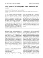
ical needs (Fig. 1). However, another very interesting
research direction involves the use of ordered biologi-
cal building blocks for the fabrication of various non-
biological nanostructures [5]. In recent years there has
been increasing interest in the utilization of biological
tools for nanotechnological applications that are not
related to biology such as micro-electronics and nano-
electronics, micro-fluidics and nano-fluidics, and
micro-electromechanical and nano-electromechanical
systems. This general field could be referred to as ‘bio-
nanotechnology’, the use of biology (or biological tools
and scaffolds) for nanotechnology. The present review
will focus on bionanotechnological applications for the
formation of metal and other inorganic wires. As will
be discussed next, biology may actually provide unique
tools for such fabrication at the nano-scale (Fig. 1).
The biological building blocks include proteins, pep-
tides, nucleic acids (DNA and RNA), bacteriophages
(viruses that infect bacteria), and plant viruses. These
biologically templated nanostructures may have appli-
cations in diverse fields that are very remote, such as
electronics, telecommunication, and materials engineer-
ing. In this minireview, I will limit the discussion to
the scheme in which the biological assemblies define
the 1D nature of the nanowire. However, it is worth
Keywords
bionanotechnology; electroless deposition;
fibrils; molecular recognition; self-assembly
Correspondence
E. Gazit, Department of Molecular,
Microbiology and Biotechnology,
George S. Wise Faculty of Life Sciences,
Tel Aviv University, Tel Aviv 69978, Israel
Fax: +972 3 640 5448
Tel: +972 3 640 9030
E-mail:
(Received 6 October 2006, accepted
3 November 2006)
doi:10.1111/j.1742-4658.2006.05605.x
The nano-scale spatial organization of metallic and other inorganic materi-
als into 1D objects is a key task in nanotechnology. Nano-scale fibers and
tubes are very useful templates for such organization because of their
inherent 1D organization. Fibrillar biological molecules and biomolecular
assemblies are excellent physical supports on which to organize the inor-
ganic material. Furthermore, these biological assemblies can facilitate high-
order organization and specific orientation of inorganic structures by their
utilization of highly specific biological recognition properties. In this mini-
review, I will describe the use of biomolecules and biomolecular assemblies,
including DNA, proteins, peptides, and even viral particles, which are
excellent templates for 1D organization of inorganic materials into wires.
This ranges from simple attempts at electroless deposition on inert biologi-
cal templates to the advanced use of structural motifs and specific protein–
DNA interactions for nano-bio-lithography as well as the fabrication of
multilayer organic and inorganic composites. The potential technological
applications of these hybrid biological–inorganic assemblies will be dis-
cussed.
FEBS Journal 274 (2007) 317–322 ª 2006 The Author Journal compilation ª 2006 FEBS 317
mentioning that other research directions involve bio-
logical modifications of nonbiological 1D objects such
as carbon nanotubes [6,7].
Use of DNA as a template for nanowire
formation
DNA molecules are very intriguing building blocks
for nanotechnological applications. Interestingly, more
than two decades ago, Seeman [8,9] showed that
specific recognition between complementary DNA
single-strands allowed them to be engineered to form
well-ordered structures at the nano-scale. The inherent
addressing capabilities, facilitated by specific inter-
actions between complementary single strands, are
manifested in specific recognition and self-assembly
processes. The formation of 2D arrays as well as 3D
nanocubes could be achieved by clever design of the
building blocks [8,9].
DNA is also a very interesting biomolecule for
nanotechnological applications from the material sci-
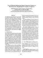
ence point of view. The diameter of ssDNA is less than
1 nm, and that of dsDNA is 2 nm (Fig. 2). Further-
more, DNA molecules are chemically very robust and
their frequent use in molecular biology applications
has significantly reduced the cost of large-scale chem-
ical DNA synthesis. Consequently, large amounts of
native and modified DNA molecules (for example, by
biotinylation or thiolation) can be rapidly synthesized
at a relatively low cost.
One of the early applications of DNA for the forma-
tion of nanowires, in 1998, involved the metallization
of dsDNA between two electrodes to form conductive
silver nanowire [10]. More specifically, the researchers
used complementary ssDNA to bridge a 12–lm gap
between two gold electrodes. The dsDNA formed was
then coated with silver by a deposition and enhance-
ment process to form 12–lm long, 100nm-wide con-
ductive silver wires. Other seminal work paved the way
to form a gold nanowire based on the use of a DNA
template [11]. This was achieved by the intercalation of
functionalized gold nanoparticles into dsDNA, fol-
lowed by covalent photochemical attachment of the
intercalator [11]. The use of metal-coated DNA mole-
cules was also demonstrated for DNA-assisted wiring
of gold electrodes on silicone wafers [12] and for the
specific metallization of a Y-shaped DNA that incor-
porated a central biotin moiety [13]. These patterned
and directed metallization schemes hold promise for
novel applications in the design and manufacture of
nanoelectronic devices in the future [12,13]. Although
lithography methods are constantly being improved,
template-assisted nanowire formation may be very use-
ful for making interconnections between lithographic-
ally defined elements [14].
Other research into much higher resolution pattern-
ing involves specific recognition between proteins and
defined DNA sequences by a process termed ‘mole-
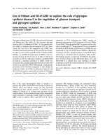
cular lithography’ [15] (Fig. 3). In this case RecA, a
sequence-specific DNA-binding protein, was allowed
ygolonhcetonaNygoloiB
ygolonhcetonanoiB
serutcurtsonande
lbmessa-fleS
slairetamderipsni-oiB
scinortcelera
l
u
celom-
o
iB
seilbmessa-oib
f
o
n
oit
a
z
il
l
ate
M
ygolonhcetoibonaN
pihc-A-nO-lleC
s
c
i
t
so
n
g
aidyar
r
a-naN
ygoloibnistodm
u
tnauQ
-
on
an
nog
n
i
re
e
nign
eeu
ss
i
T
setalpmet
Fig. 1. Interplay between biology and nano-
technology. Nanobiotechnology involves the
use of nanotechnological tools for various
biological and medical applications.
Bionanotechnology is the use of biological
and bio-inspired molecules and assemblies
for technological uses.
Biological templates for nanowire fabrication E. Gazit
318 FEBS Journal 274 (2007) 317–322 ª 2006 The Author Journal compilation ª 2006 FEBS
to bind a specific region on a DNA template before
the metallization process, thus serving as the equival-
ent of a ‘resist’ (Fig. 3). As the metallization process
proceeded, only noncovered parts of the DNA mole-
cule were coated, thus achieving nano-scale patterned
metallization of the DNA molecule [15]. RecA–DNA
interaction was also used to attach a genetically engin-
eered RecA containing a surface-associated cysteine
which allowed specific metal–thiol interactions [16].
Other DNA–protein complexes used for the formation
of ordered metallic assemblies at the nano-scale have
involved the strepavidin protein array of a 2D array of
biotinylated DNA, followed by metallization of the
array [17].
Use of the naturally occurring amyloid
fibrils for metal coating
Another use of DNA is to utilize protein and peptide fi-
bers [18–20]. Such nano-scale fibrils are formed by the
assembly of various building blocks and could be pro-
duced in large amounts by over-expression. Unlike
tnemaliFnitcA
mn7
lirbiFdiolymA
mn01-7
ebutonanTNDA
mn02
AND
mn2
egahpsuotnemaliF
m
n
6
Fig. 2. Molecular dimensions of 1D biological molecules and biomolecular assemblies for nanotechnological use. The biological molecules
and assemblies are schematically presented to provide an approximate indication of their dimensions. The DNA structure is formed by bio-
molecular assembly of double helix. All other structures are formed by self-assembly of the large number of nano building blocks.
Photo Lithography Molecular Lithography
Photoresist
SiO
2
Wafer
Mask
UV radiation
Photoresist removal
Etching
DNA
Recognition
sequences
DNA-binding
proteins
Metallization
Protein removal
Fig. 3. Use of DNA-binding proteins for ‘molecular lithography’. In photolithography, a photoresist layer is deposited on the silicone oxide
surface. The use of a mask allows differential treatment of the photoresist and the etching of specific parts of the layer. In molecular litho-
graphy, the specific DNA sequence is the equivalent of a mask, and the DNA-binding protein serves as the resist.
E. Gazit Biological templates for nanowire fabrication
FEBS Journal 274 (2007) 317–322 ª 2006 The Author Journal compilation ª 2006 FEBS 319
DNA structures, these are supramolecular assemblies
formed by the recognition and association of numerous
building blocks to create ordered structures. Compared
with DNA, protein allows much more chemical and bio-
logical flexibility as well as providing building blocks
with heterogeneity. As discussed above in the case of
DNA-based structures, genetically engineered DNA-
binding protein is used to achieve such variability.
The first reported attempt to use naturally occur-
ring fibers to make conductive nanowires involved the
use of amyloid fibrils as template [20]. Amyloid fibrils
are naturally occurring fibrillar assemblies with a
diameter of 7–10 nm and a length that can reach
several microns (Fig. 2). These assemblies are usually
associated with human disorders [18–20]. Yet the for-
mation of typical amyloid fibrils is observed in cases
involving bacterial biofilms and in yeast ‘prion-pro-
teins.’ In a pioneering study, yeast-derived amyloid
fibrils were found to be a useful protein template for
the formation of conductive metal wires [18]. Over-
expressed yeast amyloid proteins were genetically
engineered to contain a cysteine residue (as described
above for the RecA-mediated DNA metallization)
[18]. This additional thiol group served as a nuclea-
tion site for the metallization of the fibrils. The
researchers were able to demonstrate the formation of
conductive nanowires by directly measuring the cur-
rent carried by the modified fibrils across a nano-scale
gap between electrodes.
The novel concept of the use of amyloid fibrils for
nanowire formation may actually be utilized to make
wires by coating amyloid fibrils formed by simpler
building blocks. As it has been demonstrated that typ-
ical amyloid fibrils can be formed by peptides as short
as pentapeptides and tetrapeptides [21,22], and as the
molecular structure of amyloid assemblies has been
revealed by high-resolution methods [23–25], simpler
peptide building blocks could be used for future appli-
cations of amyloid fibrils for bionanotechnology.
Simpler building blocks could be synthesized in large
quantities by solid-phase techniques, as previously des-
cribed for DNA oligomers.
Use of cytoskeletal elements for the
assembly of nanowires
Another interesting use of naturally occurring fibers
for metal deposition is the use of cytoskeletal elements.
Various nano-scale fibers comprise part of the eukary-
otic cell skeleton including actin and tubulin as well as
intermediate filaments. Such fibers are ubiquitous in
the biological world, and homologous proteins, such
as the FtsZ protein, can also be found in bacteria.
The first use of cytoskeletal proteins for nanotech-
nology was the utilization of actin filaments as tem-
plates for nanowire formation [26]. Briefly, 7-nm actin
filaments were formed by self-assembly of the actin
protein, providing mechanical support for the cell
(Fig. 2). Preformed actin fibrils were covalently modi-
fied by the attachment of gold nanoparticles using an
amine-reactive agent (N-hydroxysuccinimide). This was
followed by disassembly using dialysis, repolymeriza-
tion of fibers, and an enhancement process, resulting
in the formation of a continuous gold nanowire.
The use of cytoskeletal elements adds another
dimension to the biological template of nanowires, as
these elements can be translated at the nano-scale
using biological nanomotors. The myosin nanomotor
can bind actin fibers and use ATP hydrolysis to gener-
ate force and can ‘walk’ along the filament. Thus, fur-
ther study of cytoskeletal modification may lead to
various nano-electromechanical system applications in
which mechanics, in addition to electrical conductivity,
is provided by the biological–inorganic complex.
Use of peptide nanostructures to form
conductive nanowires
Another key research direction for the fabrication of
biological fibrils involves the use of peptide and
hybrid–peptide building blocks for the assembly of
bio-inspired fibrillar assemblies. Such bio-inspired
assemblies were also used for the fabrication of metal-
lic nanowires. The simple peptide and peptide–hybrid
building blocks could be synthesized in large amounts
and readily modified.
Various classes of peptide nanotube had already
been used for the formation of 1D metal assemblies.
Glycylglycine bolaamphiphile peptide nanotubes are
examples of such bio-inspired peptide nanostructures
[27]. The functionalization of these peptide nanotubes
with histidine-rich peptide motifs enabled the forma-
tion of copper coating on the nanotube surface [27].
Other studies utilized aromatic dipeptide nanotubes
(Fig. 2). The preferential entrance of metal ions into
the lumen of aromatic dipeptide nanotubes allowed
the reduction of silver ions, with the formation of
silver-filled nanotubes [28]. After the peptide coat is
removed, silver nanowires 20 nm in diameter are
formed [28]. Another study used aromatic dipeptide
nanotubes to assemble platinum nanoparticles [29]. In
a follow-up study, silver-filled peptide nanotubes were
further coated with gold to achieve trilayer coaxial
nanocables [30].
Peptide–amphiphile nanofibers form part of another
class of peptide-based nanostructures. These fibers are
Biological templates for nanowire fabrication E. Gazit
320 FEBS Journal 274 (2007) 317–322 ª 2006 The Author Journal compilation ª 2006 FEBS
formed by the self-assembly of hydrophilic peptide
building blocks that are conjugated to a hydrophobic
aliphatic tail [31,32]. Amphiphile nanofibers were
shown to form 1D arrays of gold nanoparticles on the
surface of modified peptide fibers [31]. Such peptide–
amphiphile nanofibers were also modified using the
paramagnetic gadolinium(III) metal ion, forming inor-
ganically modified peptide fibers that could be used for
magnetic resonance imaging [32].
Use of bacteriophages and viruses for
nanowire assembly
Earlier in this minireview, I discussed the use of DNA
molecules or peptide and protein assemblies. Another
research direction in this organic–inorganic template-
assisted fabrication process is the use of much more
complex assemblies such as bacteriophages and viruses.
These viruses are self-assembled structures at the nano-
scale (Fig. 2). Viral structures are also very attractive
assemblies for fabricating 1D metallic objects. Both
viruses and bacteriophages have been used for this
purpose.
One of the first studies in bionanotechnology was the
metallization of tobacco mosaic virus particles [33,34].
This nano-scale biological entity is very effective as a
seamless template for the fabrication of various inor-
ganic materials. In the last few years, several protocols
for the deposition of various metals on the tobacco
mosaic virus surface have been developed [33,34].
Filamentous bacteriophages can provide an even bet-
ter molecular system for the formation of well-ordered
1D inorganic assemblies [35–38]. This is based on the
ability of bacteriophages to express various protein
motifs, including single-chain antibodies, on their sur-
face, a technique known as ‘phage display’. These are
proteins and peptides expressed on 6-nm elongated
fibrillar structures (Fig. 2). This technique, which is
widely used for selecting various peptide-binding
motifs, was later used for selecting peptide motifs that
can bind various inorganic metallic and semiconductive
nanoparticles [35–38]. This property was later used for
fabricating various metal and semiconductive nano-
wires by utilizing the bacteriophages. The bacterio-
phages used are engineered to express motifs that
interact with specific metal and semiconductive parti-
cles. These phages can then be aligned in such a way
that macroscopic metal or semiconductive wires are
formed. The application of these wires was recently
demonstrated for the fabrication of electrodes for thin
lithium-ion batteries [38]. The binding of gold to the
viruses followed by reduction of the cobalt ions resul-
ted in composite wires that contained both cobalt oxide
and gold, which serve as superb electrodes for batteries.
These wires have very good specific capacity, allowing
the production of batteries with high-energy density.
A very recent study used phage display technology
to select for single-chain antibodies (scFv) that specific-
ally discriminate between crystalline facets of a gallium
arsenide semiconductor [39]. The use of these recogni-
tion properties, combined with the metallization proto-
cols for bacteriophages, may allow further integration
of phage-based assemblies into electronic devices.
Conclusions
Ordered structures of biological molecules and assem-
blies at the nano-scale serve as excellent templates for
fabricating inorganic nanostructures. The structures
used range from single-stranded or double-stranded
nucleic acids and proteins to peptide assemblies and
even viral particles.
Acknowledgements
I thank the Israel Science Foundation (ISF) for their
support for this research.
References
1 Sarikaya M, Tamerler C, Jen AK, Schulten K & Baneyx
F (2003) Molecular biomimetics: nanotechnology
through biology. Nat Mater 2, 577–585.
2 Zhang S (2003) Fabrication of novel biomaterials
through molecular self-assembly. Nat Biotechnol 21,
1171–1178.
3 Drexler KE (1981) Molecular engineering: an approach
to the development of general capabilities for molecular
manipulation. Proc Natl Acad Sci USA 78, 5275–5278.
4 Wilkinson JM (2003) Nanotechnology applications in
medicine. Med Device Technol 14, 29–31.
5 Taton TA (2003) Bio-nanotechnology: two-way traffic.
Nat Mater 2, 73–74.
6 Katz E & Willner I (2004) Biomolecule-functionalized
carbon nanotubes: applications in nanobioelectronics.
Chemphyschem 20, 1084–1104.
7 Keren K, Berman RS, Buchstab E, Sivan U & Braun E
(2003) DNA-templated carbon nanotube field-effect
transistor. Science 302, 1380–1382.
8 Seeman NC (1998) DNA nanotechnology: novel DNA
constructions. Annu Rev Biophys Biomol Struct 27, 225–
248.
9 Seeman NC (2005) From genes to machines: DNA
nanomechanical devices. Trends Biochem Sci 30, 119–125.
10 Braun E, Eichen Y, Sivan U & Ben-Yoseph G (1998)
DNA-templated assembly and electrode attachment of a
conducting silver wire. Nature 391, 775–778.
E. Gazit Biological templates for nanowire fabrication
FEBS Journal 274 (2007) 317–322 ª 2006 The Author Journal compilation ª 2006 FEBS 321
11 Patolsky F, Weizmann Y, Lioubashevski O & Willner I
(2002) Au-nanoparticle nanowires based on DNA and
polylysine templates. Angew Chem Int Ed Engl 41,
2323–2327.
12 Griffin F, Ongaro A & Fitzmaurice D (2004) DNA-tem-
plated assembly of nanoscale wires and protein-functio-
nalized nanogap contacts. Analyst 129, 1171–1175.
13 Stanca SE, Eritjab R & Fitzmaurice D (2006) DNA-tem-
plated assembly of nanoscale architectures for next-gen-
eration electronic devices. Faraday Discuss 131, 155–165.
14 Shacham-Diamand Y, Inberg A, Sverdlov Y, Bogush V,
Croitoru N, Moscovich H & Freeman A (2003) Electro-
less processes for micro- and nanoelectronics. Electro-
chim Acta 48, 2987–2996.
15 Keren K, Krueger M, Gilad R, Ben-Yoseph G, Sivan U
& Braun E (2002) Sequence-specific molecular lithogra-
phy on single DNA molecules. Science 297, 72–75.
16 Nishinaka T, Takano A, Doi Y, Hashimoto M,
Nakamura A, Matsushita Y, Kumaki J & Yashima E
(2005) Conductive metal nanowires templated by the
nucleoprotein filaments, complex of DNA and RecA
protein. J Am Chem Soc 127, 8120–8125.
17 Yan H, Park SH, Finkelstein G, Reif JH & LaBean TH
(2003) DNA-templated self-assembly of protein arrays
and highly conductive nanowires. Science 301, 1882–
1884.
18 Scheibel T (2005) Protein fibers as performance pro-
teins: new technologies and applications. Curr Opin
Biotechnol 16, 427–433.
19 Scheibel T, Parthasarathy R, Sawicki G, Lin XM, Jae-
ger H & Lindquist SL (2003) Conducting nanowires
built by controlled self-assembly of amyloid fibers and
selective metal deposition. Proc Natl Acad Sci USA 100,
4527–4532.
20 Hamada D, Yanagihara I & Tsumoto K (2004) Engi-
neering amyloidogenicity towards the development of
nanofibrillar materials. Trends Biotechnol 22, 93–97.
21 Reches M, Porat Y & Gazit E (2002) Amyloid fibril for-
mation by pentapeptide and tetrapeptide fragments of
human calcitonin. J Biol Chem 277, 35475–35480.
22 Tjernberg L, Hosia W, Bark N, Thyberg J & Johansson
J (2002) Charge attraction and beta propensity are
necessary for amyloid fibril formation from tetrapep-
tides. J Biol Chem 277 , 43243–43246.
23 Makin OS, Atkins E, Sikorski P, Johansson J & Serpell
LC (2005) Molecular basis for amyloid fibril formation
and stability. Proc Natl Acad Sci USA 102, 315–320.
24 Nelson R, Sawaya MR, Balbirnie M, Madsen AO, Riekel
C, Grothe R & Eisenberg D (2005) Structure of the cross-
beta spine of amyloid-like fibrils. Nature 435, 773–778.
25 Inouye H, Sharma D, Goux WJ & Kirschner DA
(2006) Structure of core domain of fibril-forming
PHF ⁄ Tau fragments. Biophys J 90, 1774–1789.
26 Patolsky F, Weizmann Y & Willner I (2004) Actin-
based metallic nanowires as bio-nanotransporters. Nat
Mater 3, 692–695.
27 Banerjee IA, Yu L & Matsui H (2003) Cu nanocrystal
growth on peptide nanotubes by biomineralization: size
control of Cu nanocrystals by tuning peptide conforma-
tion. Proc Natl Acad Sci USA 100, 14678–14682.
28 Reches M & Gazit E (2003) Casting metal nanowires
within discrete self-assembled peptide nanotubes.
Science 300
, 625–627.
29 Song Y, Challa SR, Medforth CJ, Qiu Y, Watt RK,
Pena D, Miller JE, van Swol F & Shelnutt JA, (2004)
Synthesis of peptide-nanotube platinum-nanoparticle
composites. Chem Commun 1044–1045.
30 Carny O, Shalev DE & Gazit E (2006) Fabrication of
coaxial metal nanocables using a self-assembled peptide
nanotube scaffold. Nano Lett 6, 1594–1597.
31 Li LS & Stupp SI (2005) One-dimensional assembly of
lipophilic inorganic nanoparticles templated by peptide-
based nanofibers with binding functionalities. Angew
Chem Int Ed Engl 44, 1833–1836.
32 Bull SR, Guler MO, Bras RE, Meade TJ & Stupp SI
(2005) Self-assembled peptide amphiphile nanofibers
conjugated to MRI contrast agents. Nano Lett 5, 1–4.
33 Lee SY, Choi J, Royston E, Janes DB, Culver JN &
Harris MT (2006) Deposition of platinum clusters on
surface-modified Tobacco mosaic virus. J Nanosci Nano-
technol 6, 974–981.
34 Royston E, Lee SY, Culver JN & Harris MT (2006)
Characterization of silica-coated tobacco mosaic virus.
J Colloid Interface Sci 298, 706–712.
35 Mao C, Flynn CE, Hayhurst A, Sweeney R, Qi J,
Georgiou G, Iverson B & Belcher AM (2003) Viral
assembly of oriented quantum dot nanowires. Proc Natl
Acad Sci USA 100, 6946–6951.
36 Mao C, Solis DJ, Reiss BD, Kottmann ST, Sweeney
RY, Hayhurst A, Georgiou G, Iverson B & Belcher
AM (2004) Virus-based toolkit for the directed synthesis
of magnetic and semiconducting nanowires. Science 303,
213–217.
37 Chan P, Phan T, Kao MC, Dolan C & Tok JB (2006)
Generating short peptidic ligands for silver nanowires
from phage display random libraries. Bioorg Med Chem
Lett 16, 5261–5264.
38 Nam KT, Kim DW, Yoo PJ, Chiang CY, Meethong
N, Hammond PT, Chiang YM & Belcher AM (2006)
Virus-enabled synthesis and assembly of nanowires
for lithium ion battery electrodes. Science 312, 885–
888.
39 Artzy Schnirman A, Zahavi E, Yeger H, Rosenfeld R,
Benhar I, Reiter Y & Sivan U (2006) Antibody mole-
cules discriminate between crystalline facets of a gallium
arsenide semiconductor. Nano Lett 6, 1870–1874.
Biological templates for nanowire fabrication E. Gazit
322 FEBS Journal 274 (2007) 317–322 ª 2006 The Author Journal compilation ª 2006 FEBS