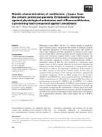
Báo cáo khóa học: Functional characterization of the evolutionarily divergent fern plastocyanin potx
Bạn đang xem bản rút gọn của tài liệu. Xem và tải ngay bản đầy đủ của tài liệu tại đây (398.41 KB, 8 trang )
Functional characterization of the evolutionarily divergent
fern plastocyanin
Jose
´
A. Navarro
1
, Christian E. Lowe
2
, Reinout Amons
3
, Takamitsu Kohzuma
4
, Gerard W. Canters
2
,
Miguel A. De la Rosa
1
, Marcellus Ubbink
2
and Manuel Herva
´
s
1
1
Instituto de Bioquı
´
mica Vegetal y Fotosı
´
ntesis, Centro de Investigaciones Cientı
´
ficas Isla de la Cartuja, Universidad de Sevilla y CSIC,
Spain;
2
Leiden Institute of Chemistry, Leiden University, the Netherlands;
3
Department of Molecular Cell Biology,
Leiden University Medical Center, the Netherlands;
4
Faculty of Science, Ibaraki University, Mito, Japan
Plastocyanin (Pc) is a soluble copper protein that transfers
electrons from cytochrome b
6
f to photosystem I (PSI), two
protein complexes that are localized in the thylakoid mem-
branes in chloroplasts. The surface e lectrostatic potential
distribution of P c p lays a key role in complex formation with
the membrane-bound partners. It is practically identical for
Pcs from plants and green algae, but is quite different for Pc
from ferns. Here we report on a laser flash kinetic analysis of
PSI reduction by Pc from various eukaryotic and prokary-
otic organisms. The reaction of fern Pc with fern PSI fits
a two-step kinetic model, consisting of complex formation
and electron transfer, whereas other plant systems exhibit
a mechanism that requires an additional intracomplex
rearrangement step. The fern Pc interacts in efficiently with
spinach PSI, showing no detectable complex formation. This
can b e explained by assuming that the unusual s urface
charge distribution of fern Pc impairs the interaction. Fern
PSI behaves in a similar way as spinach PSI in reaction with
other Pcs. The reactivity of fern Pc towards several soluble
c-type cytochromes, including cytochrome f, has been ana-
lysed by flavin-photosensitized laser flash photolysis, dem-
onstrating that the specific surface motifs for the interaction
with cytochrome f are conserved in fern Pc.
Keywords: Dryopteris;fern;Nephrolepsis; photosystem I ;
plastocyanin.
Plastocyanin (Pc) is a small copper-containing red ox protein
(molecular mass 10.5 kDa) that functions as a mobile
electron carrier between the two membrane-embedded
complexes cytochrome (Cyt) b
6
f and photosystem I (PSI)
in oxygenic photosynthesis (see [1,2] for reviews). Pc can be
acidic in higher plants and green algae (pI 4) , almost
neutral in cyanobacteria such as Synechocystis or Phormi-
dium (pI 6) or basic in other cyanobacteria such as
Anabaena (pI 9) [3]. Acidic Pc exhibits two negatively
charged surface regions formed by amino acids at positions
42–44 and 59–61, which are highly conserved. In neutral
and basic Pc, however, such acidic residues are replaced by
either neutral or positively charged amino acids [4,5].
At present, the high-resolution structures of Pcs from
many different organisms are available [4,6–8]. The com-
parison of all these crystal and solution structures indicates
that all Pcs possess an almost identical global fold, with the
polypeptide chain forming eight b strands connected by
seven loops along with a small a-helix [7,9]. The Ôeast sideÕ,
comprising charged residues 42–44 and 59–61, has been
proposed to be involved in electrostatic interactions with
both PSI and Cyt f, but the solvent-exposed edge of His87,
located in the hydrophobic patch at the so-called Ônorthern
sideÕ of the protein, constitutes the electron transfer pathway
to PSI and from Cyt f [1,10,11].
The crystal structure of a novel Pc from the fern
Dryopteris crassirhizoma has been solved [12]. This protein
presents an acidic p atch extend ed t owards the amino-
terminal end as well as other changes in the 42–45 positions,
resulting in very distinct electrostatic properties as compared
to typical eukaryotic Pcs, while maintaining the same global
structure. Thus, in Dryopteris Pc the acidic region is
relocated and surrounds the hydrophobic patch, the
large dipole moment protruding through the north side
surface [12].
Previous studies have shown that fern Pc shows similar
reactivity towards metal complexes and similar electron self-
exchange reaction rates to those of other plant Pcs [12,13],
and a study on electron transfer between zinc-substituted
Cyt c and fern Pc has shown that the reactivity towards this
nonphysiological redox partner differs significantly from
other plant Pcs [ 14]. However, no functional analysis has
been reported up to now on the reaction of fern Pc with its
physiological redox partners, Cyt b
6
f and P SI complexes.
The reaction mechanism of PSI reduction has been
analysed extensively in a wide variety of organisms from an
evolutionary point of view, thereby yielding a hierarchy of
kinetic models with a significant increase in efficiency
Correspondence to M. A. De la Rosa, Instituto de Bioquı
´
mica Vegetal
and Fotosı
´
ntesis, Centro de Investigaciones Cientı
´
ficas Isla de la
Cartuja, Universidad de Sevilla y CSIC, Ame
´
rico Vespucio s/n, 41092-
Sevilla, Spain. Fax: +34 954 460 065, Tel.: +34 954 489 506,
E-mail:
Abbreviations: Cyt, cytochrome; dRf, dRfH, deazariboflavin; k
2
, sec-
ond order rate constant; K
A
, equilibrium constant for complex for-
mation; k
et
, electron transfer rate constant; k
obs
, observed pseudo-first
order rate constant; k
sat
, first-order rate constant at saturating donor
concentration; Pc, plastocyanin; PSI, photosystem I; b-DM, b-dodecyl
maltoside.
(Received 25 May 2004, revised 5 July 2004, accepted 12 July 2004)
Eur. J. Biochem. 271, 3449–3456 (2004) Ó FEBS 2004 doi:10.1111/j.1432-1033.2004.04283.x
[1,3,15]. PSI reduction by the donor proteins Cyt c
6
and Pc,
isolated from different s ources, c an thus follow e ither a n
oriented collisional mechanism (type I), a mechanism
requiring complex formation (type II), or complex forma-
tion with rearrangement of the interface to properly orient
the redox centres to allow an efficient, fast electron transfer
(type III).
The type I and II models are found in some cyanobac-
teria, whereas the type III model is observed m ostly in
eukaryotic organisms, in which the intermediate complex is
first formed by electr ostatic attractions and the further
reorientation mainly involves hydrophobic interactions.
The kinetics of PSI reduction are typically monophasic for
the type I and II reaction mechanisms, but biphasic for the
type III model. The p roposal has b een made that t he
appearance in evolution of a fast kinetic phase in the Pc/PSI
system of higher plan ts would have involved structural
modifications in both the donor protein and PSI [5,15].
Ferns are a division of the seedless vascular plants that are
among the oldest terrestrial plant organisms known, and
taking into account the peculiar structure of its Pc, ferns are
an interesting case study to complete the evolutionary
analysis of the electron donation to PSI.
In this work, we have analysed the reaction mechanism of
electron t ransfer from Pc to PSI in ferns. The reactivity of
fern Pc with PSI isolated from different eukaryotic and
cyanobacterial sources has also been investigated in order to
extend the evolutionary insights into the process. In
addition, we have analysed the reduction of fern Pc by
different c-type Cyts ) including eukaryotic turnip Cyt f–
as a complementary way to explore the e lectron t ransfer
features and s urface electrostatics p roperties of s uch an
unusual Pc.
Experimental procedures
Proteins isolation and purification
Pc from Dryopteris was obtained by heterologous expres-
sion in Escherichia c oli. The construction of the synthetic
gene, e xpression c onditions, purification protocol and
characterization of the recombinant protein will be pub-
lished elsewhere. Mass and NMR of the pur ified recom-
binant Pc indicated that it was indistinguishable from the
native protein.
Nephrolepsis exaltata Pc was purified as follows: 200 g
fern were homogenized in 1 L 10 m
M
NaCl, 5 m
M
MgCl
2
,
10 m
M
Tris/HCl p H 8 s upplemented with a cocktail of
protease inhibitors in a Waring blender for 5 min at
medium speed. The resulting extract was fi ltered through
four layers of cheesecloth. The thylakoidal membranes were
sonicated in the presence of 0.1
M
NaCl to remove bound Pc
and then centrifuged at 12 000 g for 15 min. Solid ammo-
nium sulphate was added to the supernatant to 60%
saturation. Thereafter, the procedure was as described for
the purification of Synechocystis Cyt c
549
[16], except that
fern Pc was eluted from the DEAE-cellulose column with a
0–0.3
M
NaCl gradient and the protein was eluted directly
with the pH gradient in the chromatofocusing step, avo iding
the l ast salt gradient. About 3 mg of pure Pc with an
absorba nce ratio A
275
/A
590
of 1.27 were obtained. The pro-
tein concentration was determined spectrophotometrically
using an absorption coefficient of 4.7 m
M
)1
Æcm
)1
at 590 nm
for oxidized Pc [12]. Purification of other Pcs was carried
out as described elsewhere [17–19].
Paracoccus versutus Cyt c
550
was expressed using Para-
coccus denitrificans as a host [20]. Cultures were grown on
brain–heart infusion broth containing streptomycin
(50 lgÆmL
)1
) and spectinomycin (50 lgÆmL
)1
)ina5L
fermentor under vigorous agitation at 30 °Cfor20h
followedby4hat37°C. The protein was purified
according t o D iederix et al.[21].TurnipCytf and horse
heart Cyt c were purchased from Sigma and used without
further purification.
For reduction experiments, proteins were oxidized by
potassium ferricyanide and th en washed by several filtra-
tion–dilution cycles in an AMICON pressure cell.
Nephrolepsis PSI was prepared as follows: 200 g of fern
were homogenized in 1 L 10 m
M
NaCl, 5 m
M
MgCl
2
,
100 m
M
sodium ascorbate, 20 m
M
Tricine/KOH pH 7.5
buffer supplemented with a cocktail of protease inhibitors as
stated before. The resulting extract was filtered through four
layers of cheesecloth and the filtrate was centrifuged at
3000 g 1 min to remove debris. Thylakoid membranes were
collected by centrifugation at 25 000 g 20 min, resuspended
at 2 m g chlorophyll per mL in the homogenization buffer
without ascorbate plus 20% (v/v) glycerol, and frozen. PSI
particles f rom Neph rolepsis were obtained by b-dodecyl
maltoside (b-DM) solubilization as follows. Thylakoidal
membranes w ere diluted to 1 mg chlorophyll per mL with
buffer D (20 m
M
Mes pH 6.5, 10 m
M
CaCl
2
,10m
M
MgCl
2
,0.5
MD
-mannitol, 20% glycerol) and solubilized
for 30 min with 1.5% b-DM. The solution was centrifuged
5 min at 20 000 g, and the supernatant centrifuged 20 min
at 120 000 g to remove unsolubilized material. Th e resulting
supernatant was diluted two in three with buffer A (20 m
M
Mes pH 6.5, 10 m
M
CaCl
2
,10 m
M
MgCl
2
) and applied to a
discontinuous sucrose gradient (15, 20, 25, 40%) prepared
on buffer B (buffer A + 0.5
M
mannitol). After centrifuga-
tion for 20 h at 150 000 g, the lower half of the only green
band was collected, washed w ith 20 m
M
Tricine/KOH
buffer pH 7.5, with 0.03% b-DM, and concentrated in an
AMICON pressure cell. This fraction was a pplied to a
continuous sucrose gradient (17–30%) and centrifuged as
before. The lower half of the only green band was collected,
washed and concentrated as before, and stored at )80 °C.
The P700 content of PSI samples w as calculated from the
photoinduced absorbance changes at 820 nm using the
absorption coefficient of 6.5 m
M
)1
Æcm
)1
determined by
Mathis and Se
´
tif [22]. Chlorophyll concentration was
determined according to Arnon [23]. The chlorophyll/
P700 ratio of the resulting PSI preparation was 280 : 1.
Spinach, Synechocystis and Anabaena PSI were purified as
previously described [15].
Amino acid sequence determination of
Nephrolepsis
Pc
Sequencing of Nephrolepsis Pc (Fig. 1 ) was performed
with a Hewlett-Packard G1006A protein Sequencer
system, connected on-line to a Hewlett-Packard M odel
1100 HPLC system. As the amino terminus was
unblocked, first, the intact protein was sequenced as far
as possible. Pr otein sequencing of BrCN-generated p ep-
tides, and of peptides obtained by digestion with
3450 J. A. Navarro et al. (Eur. J. Biochem. 271) Ó FEBS 2004
endoproteinases Lys-C and Asp-N followed standard
procedures (see also [24]).
Blocking of amino groups with acetic anhydride and the
reduction and alkylation o f cysteine r esidues were p er-
formed according t o Amons [25,26]. The sequence starting
at C-87 was obtained by treating t he intact protein as
follows: (1) digestion with endo-Asp-N; (2) acetylation with
acetic anhydride; (3) oxidation with performic acid [27]; (4)
redigestion w ith endo-Asp-N, now cleaving at the c ysteic
acidresidueformedinstep3.
To isolate the small C-terminal BrCN peptide, the intact
protein was cleaved by BrCN, and the mixture applied to a
2 · 10-mm reversed-phase C18 column. The small peptide
was eluted at low acetonitrile concentration, and then
coupled to aminoaryl-poly(vinylide ne) difluoride (Millipore
Inc) with N-(3-dimethylaminopropyl)-N¢-ethylcarbonyl-
imide hydrochloride (EDC) according to t he manufactu rer’s
instructions.
Laser flash spectroscopy
Kinetics of flash-induced absorbance changes in PSI were
followed at 820 nm as described by Herva
´
s et al.[28].
Unless otherwise stated, the s tandard reaction mixture
contained, in a final volume of 0.2 mL, 20 m
M
buffer
(Tricine/KOH pH 7.5 or Mes pH 5), 10 m
M
MgCl
2
, 0.03%
b-DM, an amount of PSI-enriched particles equivalent to
0.75 mg of chlorophyll per mL ( 0.35 mgÆmL
)1
in case of
cyanobacterial PSI), 0.1 m
M
methyl viologen, 2 m
M
sodium
ascorbate and Pc at the indicated concentration. All
experiments were performed at 22 °Cina1mmpath-
length cuvette.
The optical set-up for kinetic experiments of inter-
molecular redox reactions between Cyts and Pcs has been
described previously [29]. The standard reaction mixture
contained, in a final volume of 1.2 mL, 5 m
M
potassium
phosphate pH 7.0, 2 m
M
EDTA, 100 l
M
5-dRf, and the
different p roteins at the indicated concentrations. L aser
flash experiments were performed anaerobically at room
temperature in a 1 cm path-length cuvette. Laser flash
photolysis of the 5-dRf/EDTA system generated 5-dRfH,
which in its turn reduced oxidized Cyt to yield the reduced
species [30]. Further exponential absorbance decreases were
concomitantly observed at 550 nm (554 nm for Cyt f)and
600 nm, which correspond to Pc reduction by Cyt. All of
the kinetic experiments were performed under pseudo first-
order c onditions, in w hich the c oncentration of p rotein
acceptor, either in the direct reduction by 5-dRfH, or in the
interprotein electron transfer, was in large excess over the
amount of dRfH or the donor protein, respectively.
Deazariboflavin w as a gift from G. Tollin (University of
Arizona, Tucson, USA).
In all c ases, kinetic data collection was as described
previously [15]. Oscilloscope traces were treated as sums of
several exponential components; exponential analyses were
performed using the Marquardt method with the software
devised by P. Se
´
tif (CEA, Saclay, France). The estimated
error in rate constants determination was 10%.
Results and Discussion
Fern Pc presents very distinct electrostatic properties as
compared to other eukaryotic Pcs, with the a cidic area
extending into the hydrophobic patch (Fig. 2). Electrostatic
forces play an important role in protein interactions and it is
thus of interest to analyse the functional behaviour of fern
Pc as compared to other plant Pcs. In this study, Pcs from
the fern genera Nephrolepsis and Dryopteris were used. The
amino acid sequence of N. exaltata Pc was determined and
is shown in Fig. 3. A microheterogeneity was found in the
sequence for three positions, 19 (Leu/Ile), 21 (Val/Ile) and
53 (Ser/Asn). At least the 19 and 2 1 positions are c oupled, so
proteins contain either Leu19 and Val21 or Ile19 and Ile21.
Fern Pc sequences seem to be extremely well conserved, as
illustrated by the alignment of the Pc sequen ces from
N. exalta ta, D. crassirhizoma [12] and Polystic hum longifrons
(Y. Nagai and F. Yoshizaki, Toho University, Japan,
unpublished results), shown in Fig. 3. The functional
analysis done in this work has shown that Nephrolepsis
and Dryopteris Pcs behave in an indistinguishable manner,
thus from this point fern Pc will refer to both proteins
indistinctively.
The reaction mechanism of electron transfer from fern Pc
to fern PSI has been analysed by laser-flash absorption
spectroscopy and compared w ith spinach Pc. The kinetic
traces of fern PSI reduction by fern Pc at pH 7.5 correspond
to monophasic kinetics, whereas those with spinach Pc are
better fitted to biphasic curves ( Fig. 4). T he amplitude of the
fast phase of the l atter represen ted up to 40% of the total
amplitude, with a rate constant (k
obs
) independent of Pc
concentration (data not shown). From the k
obs
values of this
fast phase, a first-order electron transfer rate constant (k
et
)
of 3 · 10
4
s
)1
can be directly estimated for the interaction
between spinach Pc and fern PSI. The k
obs
values with fern
Pc and those for the slower phase with spinach Pc exhibit
saturation profiles at increasing donor protein concentra-
tions (Fig. 5, upper panel), thereby suggesting that the two
metalloproteins are able to form transient complexes with
II
N
(a)
>>
(c)
>>
>>
(c)
>>
(c*)
(d)
(d*)
>>
(e)
+
AKVEVGDEVGNFKFYPDTLTVSAGEAVEFTLVGETGHNIVFDIPAGAPGTVASELKAaSMDe-DL
FYPDTITISAGE
TLVGETGHNIVFDIPAGAPGTVA
AASMDENDLL
FYPDTLTVSAGE
TFYCTPHk
DEPNFTAKVsT
-TPHK-AN-K
KGTLTVK
(b)
>>
-
DENDLLSEDEPNFTAKVSTpGTYtFY-TPHKsan
VSTPGT
Fig. 1. Outline of seque nce determinatio n of Nephrolepsis Pc. Sequence results for (a) the in tact protein, (b) the BrCN peptide starting after M-60, (c)
selected endo-Lys-C peptides, (c*) the Cys containing endo-Lys-C peptide, after reduction and alkylation, (d) selected endo-Asp-N peptides; (d*)
the peptide starting at C-87; and (e): the C-terminal BrCN-peptide. See also Experimental procedures.
Ó FEBS 2004 Function of fern plastocyanin (Eur. J. Biochem. 271) 3451
PSI. From the plots shown in Fig. 5, and applying the
formalism previously developed [31], it is possible (Table 1)
to estima te both K
A
(equilibrium c onstant for complex
formation) and k
sat
(first-order rate constant at saturating
Pc concentration). The fern Pc behaviour can be explained
as following the two-step type II mechanism with its own
PSI, invo lving complex formation and with k
sat
corres-
ponding to the further intracomplex electron transfer rate
[15]. Spinach Pc, however, i n which the k
obs
values f or the
first initial fast phase of electron transfer does not match the
values of k
sat
, follows with fern PSI the classical three-step
type III mechanism observed in other eukaryotic systems,
with an additional rearrangement of redox partners within
the intermediate complex prior to electron transfer [1,3,15].
From the data presented in Table 1 , it seems clear that
although fern PSI binds spinach and fern Pc with similar
efficiency, as shown by the similar K
A
values, the electron
transfer step is one order of magnitude higher with spinach
Pc (Table 1). Thus, whereas the kinetic constants presented
here for t he spinach P c/fern PSI s ystem are of the same
order of magnitude as those observed for the spinach Pc/PSI
system [15], the distance and/or orientation o f the redox
centers in the fern Pc/PSI complex seems to be nonoptimal
for electron transfer. When che cking fern PSI reactivity
towards cyanobacterial Pcs, linear plots against Pc concen-
tration were observed ( data not shown), indicating the
occurrence of a collisional type I mec hanism with very low
second-order rate constant (k
2
) values for PSI reduction
Nephrolepsis
I I N
AKVEVGDEVGNFKFYPDTLTVSAGEAVEFTLVGETGHNIVFDIPAGAPGTVASELKAASM 60
***************** *
*******************************
*******
AKVEVGDEVGNFKFYPDSITVSAGEAVEFTLVGETGHNIVFDIPAGAPGTVASELKAASM 60
******* ********** ****** *********************** **********
AKVEVGDDVGNFKFYPDSLTVSAGETVEFTLVGETGHNIVFDIPAGAPGPVASELKAASM 60
* * * * * * * ** * ** *** * * **
VEVLLGGGDGSLAFLPGDFSVASGEEIVFKNNAGFPHNVVFDEDEIPSGVDAAKI SM 57
DENDLLSEDEPNFTAKVSTPGTYTFYCTPHKSANMKGTLTVK 102
*********** * ****************************
DENDLLSEDEPSFKAKVSTPGTYTFYCTPHKSANMKGTLTVK 102
******************************************
DENDLLSEDEPSFKAKVSTPGTYTFYCTPHKSANMKGTLTVK 102
* *** * *** *** ** * * * **
SEEDLLNAPGETYKVTLTEKGTYKFYCSPHQGAGMVGKVTVN 99
D
ryopteris
Polystichum
Spinacea
Nephrolepsis
D
ryopteris
Polystichum
Spinacea
oo o
Fig. 3. Amino acid sequence alignment of
Nephrolepsis, Dryopteris, Polystichum and
Spinacea Pc. The alignment was made with
CLUSTALW
. Identical residues between the
sequences are marked with an asterisk. The
microheterogeneity found in Nephrolepsis Pc
is indicated by the residues above the main
sequence at positions 19, 21 and 53. Conser-
vation of these residues is indicated with ÔoÕ.
Fig. 4. Kine tic traces showing fern PSI reduction by fern and spinach
Pc. Absorbance changes were recorded at 820 nm with 100 l
M
Pc. The
kinetics were fi tted to either biphasic (spinach) or monophasic (fern)
curves. Other co nditions were as de scribedinExperimentalproce-
dures.
Fig. 2. Surface ele ctrost atic potential distr ibutio n of fern (PDB entry
1KDI) and spinach (PDB entry 1AG6) Pc. The molecules are similarly
oriented, with the lateral view showing the typical charged east patch
of eukaryotic Pcs (upper) and the top view, obtained by rotating 90°
around the horizontal x-axis as indicated, showing the stan dard
hydrophobic patch (lower). Negatively and positively charged regions
are shown in red and blue , respectively. Th e picture was gener ated with
the program
MOLMOL
[36] at an ionic strength of 50 m
M
.
3452 J. A. Navarro et al. (Eur. J. Biochem. 271) Ó FEBS 2004
(Table 1). This behaviour is very similar to that observed for
spinach PSI when reacting with cyanobacterial Pcs [15].
Taken together, all of these data clearly demonstrate that
fern PSI behaves as spinach PSI.
It has been reported previously that in fern Pc the red ox
potential exhibits a much less pronounced dependence on
pH than other eukaryotic Pcs [32]. This has been ascribed
to the absence of protonation of the His87 copper-ligand
at low pH, a fact that strongly contrasts with pK
a
values
of 5.5 for this residue in other eukaryotic Pcs [12,32].
Consequently we checked the reactivity of f ern PSI against
fern and spinach Pc at pH 5, a value close to the
physiological pH. As shown in Table 1, lowering pH only
has quantitatively minor effects i n the interaction between
fernPSIandspinachPc,asisthecaseforthespinachPc/
PSI system [15]. However, more drastic effects are
observed in the fern Pc/PSI system, for which linear
protein dependences are obtained (Fig. 6, u pper), indica-
ting the occurrence of a collision al t ype I mechanism. The
k
2
value at low pH ( 3 · 10
7
M
)1
Æs
)1
)calculatedfrom
this linear plot cannot be directly compared with the
kinetic values obtained at neutral pH, as different
mechanisms (i.e. type I I and type I) occur; however, the
k
obs
values obtained at pH 5 at high Pc concentration are
about 10 times higher than those observed at pH 7
Table 1. T ype of mechanism and kinetic constants for the reduction of
fern PSI by Pc from different organisms.
Pc pH Type K
A
(
M
)1
) k
sat
(s
)1
) k
et
(s
)1
) k
2
(
M
)1
Æs
)1
)
Fern 7.5 II 1.0 · 10
4
1.0 · 10
3
1.0 · 10
3a
–
Fern 5.0 I – – – 2.8 · 10
7
Spinach 7.5 III 2.5 · 10
4
1.5 · 10
3
2.7 · 10
4
–
Spinach 5.0 III 2.8 · 10
4
6.0 · 10
3
3.9 · 10
4
–
Synechocystis 7.5 I – – – 2.6 · 10
6
Anabaena 7.5 I – – – 1.3 · 10
6
a
For fern Pc at pH 7.5, the value for k
et
is inferred from that for
k
sat
.
Fig. 6. Dependence of the observed rate constant (k
obs
) on donor protein
concentration (upper) and ionic strength (lower) for fern PSI reduction by
fern Pc at pH 5.0 (s) and 7.5 (d). Lines represent theoretical fits as
described in the legend of Fig. 5.
Fig. 5. De pendence of the observed rate constant (k
obs
) on donor protein
concentration (upper) and ionic strength (lower) for f ern PSI reduction by
fern (s) and spinach (d)Pc.In case of spinach Pc, the k
obs
values
correspond to the slow phase. L ines represent theoretical fits as des-
cribed in the text (upper) and a ccording to the formalism developed by
Watkins et al.[37](lower).
Ó FEBS 2004 Function of fern plastocyanin (Eur. J. Biochem. 271) 3453
(Fig. 6, upper). T hus, it s eems that a t lower and more
physiological pH values, fern Pc reactivity against its own
PSI is significantly improved, thus approaching the
efficiency attained by other eukaryotic systems. It is
interesting to note that this pH effect is specific of the fern
Pc/PSI system, as fern Pc reactivity with spinach PSI
remains unchanged at low pH (data not shown).
The role of electrostatic interactions on fern PSI
reduction by fern and spinach Pcs was investigated at
varying NaCl concentrations (Fig. 5 , lower panel). In
plant Pc/PSI systems, it has been shown that salt initially
stimulates PSI reduction and further slows down the
reaction at high concentration, which has been explained
in terms of rearrangement of Pc within the complex
[15,33]. From the ionic strength profiles shown in Fig. 5, it
is clear that fern PSI does not show the bell-shaped
behaviour typical of other plant PSI, w hich is indeed
observed i n t he fern Pc/spin ach PSI system (data not
shown). However, the ionic strength dependence of k
obs
with fern PSI m akes evident the electrostatic nature of the
intermediate complexes with both Pcs, which are stabilized
by means of attractive electrostatic forces. The ionic
strength effect is more pronounced with spinach Pc than
with fern Pc, m ainly at l ow ionic strength ( Fig. 5, lower
panel), as expected from the differences in surface
electrostatic potential between both proteins. However,
at physiological ionic strength and pH, both proteins
present similar reactivity ( Fig. 6, lowe r panel).
Fern Pc reacti vity against P SI obt ained f rom o rgan-
isms with negative (spinach), basic (Anabaena)orneutral
(Synechocystis) P cs was also checked. Significant electron
transfer rates were only observed with spinach PSI, the
cross-reaction showing monophasic kinetics of PSI
reduction and linear plots against Pc concentration (data
not shown), indicating the absence of any kinetically
detectable Pc/PSI electron transfer complex. From these
data, a second-order rate c onstant for spinach PSI
reduction of 2.7 · 10
6
M
)1
Æs
)1
was obtained under stand-
ard conditions. This value is significantly lower that those
observed in homologous Pc/PSI systems from either
eukaryotic or prokaryotic sources [15,34,35]. These find-
ings indicate that the altered surface electrostatic poten-
tial of fern Pc drastically hinders its interaction with
spinach PSI.
In order to extend t he function al characterization of
fern Pc, we have also compared the reactivity of this
protein with that of other Pcs towards several soluble
c-type Cyts (including Cyt f)byusingthedRfH
•
radical
as a redox probe [30]. In all cases, the k
obs
values for the
electron transfer from Cyt to Pc depend linearly upon Pc
concentration. As an example, Fig. 7 (upper panel) shows
the linear protein concentration dependence observed for
the electron transfer from e ither turnip Cyt f or horse
Cyt c to fern Pc. F rom these plots, the k
2
values for the
Cyt/Pc intera ction can be estimated (Table 2). Effic ient
electron transfer is observed with any Cyt only when
eukaryotic Pc is used as an acceptor, whereas no relevant
reactivity is obtained with prokaryotic Pc (Table 2). This
is in agreement with t he electrostatic character of these
proteins: positively charged in the case of Cyts, n egatively
charged in eukaryotic Pcs, and neutral or basic in
prokaryotic Pcs [5,7].
The k
2
values presented in Table 2, obtained at 30 m
M
ionic strength, agree well with those previously reported for
spinach Pc r eduction by horse Cyt c, eukaryotic Cyt f or
positively ch arged bacterial Cyts at s imilar i onic strength
[31]. It is interesting to note that fern Pc reactivity against
turnip Cy t f is one order o f magnitude higher than with
other Cyts ( Fig. 7 a nd Table 2). Figure 7 (lower p anel)
shows that the rate constants for the electron transfer
between any Cyt and fern Pc decrease as the ionic
strength increases, indicating the existence of attractive
Fig. 7. Dependence of the observed rate constant (k
obs
) on donor protein
concentration (upper) and ionic strength (lower) for the reduction of fern
Pc by turnip Cyt f (s) and horse Cyt c (d). Lines in the lower panel
represent theoretical fits according to the formalism developed by
Watkins et al. [37]. Other conditions were as described in Experimental
procedures.
Table 2. B imolecular rate constants (k
2
,
M
)1
Æs
)1
) for the overall reaction
of reduction of different Pcs by c-type Cyts. n.d., Not determined.
Cytochrome
Plastocyanin
Fern Synechocystis Anabaena Poplar
Cyt c
550
2.4 · 10
7
2.5 · 10
6
<10
4
7.7 · 10
7
Horse Cyt c 4.1 · 10
7
<10
4
<10
4
4.6 · 10
7
Turnip Cyt f 7.5 · 10
8
n.d. n.d. 6.0 · 10
8a
a
Data from Meyer et al. [31].
3454 J. A. Navarro et al. (Eur. J. Biochem. 271) Ó FEBS 2004
protein–protein e lectrostatic forces related t o t he comple-
mentarity in electrostatic charges between Pc and Cyt.
Despite the peculiar surface charge distribution of fern Pc,
this finding suggests that t he specific interaction motifs
between this copper protein and its natural electron donor,
Cyt f, are conserved.
Concluding remarks
Fern Pc has electrostatic surface properties drastically
different from those of other eukaryo tic Pcs. The negat-
ively charged area around positions 42–45 in the acidic
patch is absent or strongly diminished, whereas new
charged groups form an arc around the edge of the
hydrophobic patch. From the data presented here we can
conclude that fern Pc conserves the main electrostatic
features of eukaryotic Pcs: its negatively charged character
allows this protein to e fficiently interact with p lant Cyt f
and PSI, but the unusual surface charge distribution in
fern Pc seems to impede f urther rearrangement of t he
complex to attain an optimized elec tron transfer rate. In
summary, fern Pc has followed a relatively independent
evolutionary pathway since ferns diverged from other
vascular plants, but keeping a charged area at the surface
level that is crucial to drive the electrostatic attractive
movements of the copper protein towards its membrane
partners. Within the more general context of protein
evolution, this finding reveals how important the surface
electrostatic features of molecules a re for their functional
interactions within the living cells.
Acknowledgements
Research work was supported by the Spanish Ministry of Science and
Technology (MCYT, Grant BMC2003-0458), and Andalusian Gov-
ernment (PAI, CVI-0198). C. E. Lowe acknowledge s the financial
support provided through the European Community’s Human Poten-
tial Programme under contracts FMRX -CT98-0218 (Ha emworks) and
HPRN-CT-1999-00095 (Transient). M. U bbink acknowledges financial
support from the Netherlands Organization for Scientific Research,
grant 700.52.425.
References
1. Hope, A .B. (2000) Electron tr ansfer amongst cytochrome f,
plastocyanin and photosystem I: kinetics and mechanisms. Bio-
chim. Biophys. Acta 1456, 5–26.
2. Herva
´
s, M., Navarro, J.A. & De la Rosa, M.A. (2003) Electron
transfer between membrane complexes and soluble proteins in
photosynthesis. Accounts Chem. Res. 36, 798–805.
3. De la Rosa, M.A., Navarro, J.A., Dı
´
az-Quintana, A., De la
Cerda, B., Molina-Heredia, F.P., Balm e, A., Murd och, P.S., Dı
´
az-
Moreno, I., Dura
´
n, R.V. & H erva
´
s, M. (2002) An evolutionary
analysis of the reaction mechanisms of photosystem I r eduction by
cytochrome c
6
and plastocyanin. Bioelectrochemistry 55, 41–45.
4. Gross, E.L. (1993) Plastocyanin: structure a nd fu nction . Pho-
tosynth. Res. 37, 103–116.
5. Navarro, J.A., Herva
´
s, M. & De la Rosa, M.A. (1997) Co-evo-
lution of cytochrome c
6
and plastocyanin, mobile proteins trans-
ferring electrons from cytochrome b
6
f to photosystem I. J. Biol.
Inorg. Chem. 2, 11–22.
6. Guss, J.M., Harrowell, P.R., Murata, M., Norris, V.A. &
Freeman, H.C. (1986) C rystal structure analyses o f reduced
(CuI) poplar plastocyanin at six pH values. J. Mol. Biol. 192,
361–387.
7. Sigfridsson, K. (1998) Plastocyanin, an electron-transfer protein.
Photosynth. Res. 57, 1–28.
8. Romero, A., De la Cerda, B., Varela, P.F., N avarro, J.A., Herva
´
s,
M. & De la Rosa, M.A. (1998) The 2.15 A
˚
crystal structure of a
triple mutant plastocyanin from the cyanobacterium Synechocys-
tis sp. PCC 6803. J. Mol. Biol. 275, 327–336.
9. Redinbo, M., Yeates, T.O. & Merchant, S. (1994) Plastocyanin:
structural and functional analysis. J. Bioenerg. Biomembr. 26,
49–66.
10. Ubbink, M., Ejdeback , M., Karlsson, B.G. & Bendall, D.S. (1998)
The structure of the complex of plastocyanin and cytochrome f,
determined by paramagnetic NMR and restrained rigid-body
molecular dynamics. Structure 6, 323–335.
11. Molina-Heredia, F.P., Herva
´
s, M., Navarro, J.A. & De la Rosa,
M.A. (2001) A single arginyl residue in plastocyanin and in
cytochrome c
6
from the cyanobacterium Anabaena sp. PCC 7119
is required for efficient reduction of photosystem I. J. Biol. Chem.
276, 601–605.
12. Kohzuma, T., Inoue, T., Yoshizaki, F., Sasakawa, Y., Onodera,
K., Nagatomo, S., Kitagawa, T., Uzawa, S., Isobe, Y., Sugimura,
Y., Gotowda, M. & Kai, Y. (1999) The structure and unusual pH
dependence of p lastocyanin from the fern Dryopteris c rassi-
rhizoma. The protonation of an active site histidine is hindered by
p–p interactions. J. Biol. Chem. 274, 11817–11823.
13. Sato, K., Kohzuma, T. & Dennison, C. (2003) Active-site struc-
ture and electron-transfer reactivity of plastocyanins. J. Am.
Chem. Soc. 125, 2101–2112.
14. Pletneva,E.V.,Fulton,D.B.,Kohzuma,T.&Kostic,N.M.(2000)
Protein docking and gated electron-transfer reactions between zinc
cytochrome c and the new plastocyan in from the fern Dryopteris
crassirhizoma. Direct kinetic evidence for multiple binary com-
plexes. J. Am. Chem. Soc. 122, 1034–1046.
15. Herva
´
s, M., Navarro, J.A., Dı
´
az,A.,Bottin,H.&DelaRosa,
M.A. (1995) Laser-flash kineti c analysis o f the fast electron
transfer from plastocyanin and cytochrome c
6
to photosystem I.
Experimental evidence on the evolution of the reaction mechan-
ism. Biochemistry 34, 11321–11326.
16. Navarro, J.A., Herva
´
s,M.,DelaCerda,B.&DelaRosa,M.A.
(1995) Purification and physicochemical properties of the lo w-
potential cytochrome c
549
from the cyanobacterium Synechocystis
sp. PCC 6803. Arch. Biochem. Biophys. 318 , 46–52.
17. Yocum, C.F. (1982) Purification of ferredoxin and plastocyanin.
In Methods i n Chloroplast Molecular Biology (Edelman, M .,
Hallick, R.B. & Chua, N H., eds), pp. 973–981. Elsevier Bio-
medical Press, Amsterdam.
18. Herva
´
s, M., Navarro, F., Navarro, J.A., Cha
´
vez, S., Dı
´
az, A.,
Florencio, F.J. & De la Rosa, M.A. (1993) Synechocystis
6803 plastocyanin isolated from both the cyanobacterium
and E.coli transformed c ells are identical. FEBS Lett. 319,
257–260.
19. Molina-Heredia, F.P., Herva
´
s, M., Navarro, J.A. & De la Rosa,
M.A. (1998) Cloning and correct expression in E. coli of the petE
and petJ genes respectively encoding plastocyanin and cytochrome
c
6
from the cyanobacterium Anabaena sp. PCC 7119. Biochem.
Biophys. Res. Commun. 243, 302–306.
20. Ubbink, M., Pfuhl, M., van der Oost, J., Berg, A. & Canters,
G.W. (1996) NMR assignments and relaxation studies of Thio-
bacillus versutus ferrocytochrome c-550 indicate th e p resence o f
a h ighly m obile 13-residues long C-terminal tail. Prot. Sci. 5,
2494–2505.
21. Diederix, R.E.M., Ubbink, M. & Canters, G.W. (2001) The per-
oxidase activity of cytochrome c-550 from Paracoccus versutus.
Eur. J. Biochem. 268, 4207–4216.
Ó FEBS 2004 Function of fern plastocyanin (Eur. J. Biochem. 271) 3455
22. Mathis,P.&Se
´
tif, P. (1981) Near infrared absorption spectra of
the chlorophyll a cations and triplet state in vitro and in vivo. Isr.
J. Chem. 21, 316–320.
23. Arnon, D.I. (1949) Copper enzymes i n isolated chloroplasts. Plant
Physiol. 24, 1–15.
24. Liang, P., Amons, R., MacRae, T.H. & Clegg, J.S. (1997) Puri-
fication, structure and in vitro molecular-chaperone activity of
Artemia p26, a small heat-shock/alpha-crystallin protein. Eur. J.
Biochem. 243, 225–232.
25. Amons, R. (1996) Use of acetic anhydride in automatic sequen-
cing. Abstracts of the 11th International Conference on Methods in
Protein Structure Analysis, Annecy, France.
26. Amons, R. (1997) Determination of cysteine residues in protein
sequence analysis at the picomole level. Anal. Biochem. 249,
111–114.
27. Darbre, A., ed. (1986) Practical Protein Chemistry – a Handbook.
John Wiley & Sons, NY, pp. 246–247.
28. Herva
´
s,M.,Myshkin,E.,Vintonenko, N., De la Rosa, M.A.,
Bullerja h n, G.S. & Navarro , J.A. ( 200 3) Mutagenesis of Pro-
chlorothrix plastocyanin reveals additional f eatures in Photo-
system I interactions. J. Biol. Chem. 278, 8179–8183.
29. Navarro, J.A., Herva
´
s,M.,Pueyo,J.J.,Medina,M.,Go
´
mez-
Moreno, C., De la Rosa, M.A. & Tollin, G . (1994) Laser
flash-induced p hotored uction of photosynthetic ferredoxins and
flavodoxin by 5-deazariboflavin and by a viologen analogue.
Photochem. Photobiol. 60, 231–236.
30. Tollin, G. (1995) Use of flavin photochemistry to probe intra-
protein and interprotein electron transfer mechanisms. J. Bioen-
erg. Biomembr. 27, 303–309.
31. Meyer, T.E., Zhao, Z.G., Cusanovich, M.A. & Tollin, G. (1993)
Transient kinetics of electron transfer from a variety of c-type
cytochromes to plastocyanin. Biochemistry 32, 4552–4559.
32. Dennison, C., Lawler, A.T. & Kohzuma, T. (2002) Unusual
properties of plastocyanin from the fern Dryopteris crassirhizoma.
Biochemistry 41, 552–560.
33. Sigfridsson, K. (1997) Ionic strength and pH depen dence of the
reaction between plastocyanin and Photosystem 1. Evidence of a
rate-limiting conformational change. Photosynth. Res. 54, 143–
153.
34. Bottin, H. & Mathis, P. (1985) Interaction of plastocyanin w ith the
photosystem I reaction center: a kinetic study by flash absorption
spectroscopy. Biochemistry 24, 6453–6460.
35. Molina-Heredia, F.P., Wastl, J., Navarro, J.A., Bendall, D.S.,
Herva
´
s, M., Howe, C.J. & D e la Ro sa, M.A. (2003) Phot o-
synthesis: a n ew function fo r an old cytochrome? Nature 424,
33–34.
36. Koradi, R., Billeter, M. & Wuthrich, K. (1996) MOLMOL: a
program for display and analysis of macromolecular structures.
J. Mol. Graph. 14, 51–55.
37. Watkins, J.A., Cusanovich, M.A.,Meyer,T.E.&Tollin,G.(1994)
A Ôparallel plateÕ electrostatic m odel for bimolecular rate constants
appliedtoelectrontransferproteins.Prot. Sci. 3, 2104–2114.
3456 J. A. Navarro et al. (Eur. J. Biochem. 271) Ó FEBS 2004