Báo cáo khóa học: S-(2,3-Dichlorotriazinyl)glutathione A new affinity label for probing the structure and function of glutathione transferases potx
Bạn đang xem bản rút gọn của tài liệu. Xem và tải ngay bản đầy đủ của tài liệu tại đây (429.97 KB, 9 trang )
S
-(2,3-Dichlorotriazinyl)glutathione
A new affinity label for probing the structure and function of glutathione transferases
Georgia A. Kotzia and Nikolaos E. Labrou
Laboratory of Enzyme Technology, Department of Agricultural Biotechnology, Agricultural University of Athens, Greece
S-(2,3-Dichlorotriazinyl)glutathione (SDTG) was synthes-
ized and shown to be a n effective alkylating affinity label
for recombinant maize glutathione S-transferase I (GST I).
Inactivation of GST I by SDTG at pH 6 .5 followed biphasic
pseudo-first-order saturation kinetics. The biphasic kinetics
can be described in terms o f a fast initial phase of inactiva-
tion followed by a slower phase, leading to 42 ± 3%
residual activity. The rate of inactivation for both phases
exhibits nonlinear d ependence o n SDTG c oncentration,
consistent with the formation of a r eversible complex with
the enzyme (K
d
107.9 ± 2.1 l
M
for the fast phase, and
224.5 ± 4.2 l
M
for the slow phase) before irreversible
modification with maximum rate constants of
0.049 ± 0.002 min
)1
and 0.0153 ± 0.001 min
)1
for the
fast and slow phases, respectively. Protection from inacti-
vation was afforded by substrate a nalogues, demonstrating
the specificity of the reaction. When the enzyme was inacti-
vated (42% residual activity), % 1 m ol SDTG per mol
dimeric enzyme was incorporated. Amino-acid analysis,
molecular modelling, and site-directed mutagenesis studies
suggested that the modifying residue is Met121, which is
located at the end of a-helix H¢¢¢
3
and forms part of the
xenobiotic-binding site. The results reveal an unexpected
structural communication between subunits, which consists
of mutually exclusive modification of Met residues across
enzyme subunits. T hus, modification of Met121 on one
subunit prevents modification of M et121 on the other sub-
unit. This communication is governed by Phe51, which is
located at t he dimer i nterface and forms part of the h ydro-
phobic lock-and-key intersubunit motif. The ability of
SDTG to inactivate other glutathione-binding enzymes a nd
GST isoenzymes was also investig ated, and it was concluded
that this new reagent may have general applicability as an
affinity reagent for other enzymes with glutathione-binding
sites.
Keywords: affinity labelling; chlorotriazine; h erbicides;
xenobiotics.
Glutathione transferases (GSTs; EC 2.5.1.18) comprise a
large family of glutathione (GSH)-binding enzymes which
catalyse the conjugation of GSH with a variety of hydro-
phobic electrophiles through the formation of a thioether
bond [1,2]. These enzymes offer protection against toxic
xenobiotics and byproducts of oxidative metabolism. In
addition to their catalytic activities, plant GSTs are also
involved in the response to different biotic and abiotic
stresses, and can be specifically induced in response to a
variety of stimuli, such as pathogens and chemicals [3–7].
The cytosolic GSTs are homodimers or heterodimers. Each
monomer has two domains, an a/b domain which includes
a1–a3, and a large a-helical domain comprising h elices
a4–a9. The former contains a GSH-binding site (G-site) on
top of the a domain. A hydrophobic pocket (H-site) lies
between the domains, in which a generally hydrophobic
substrate binds and reacts with GSH [8–16].
In plants, GSTs are grouped into five classes based on
their amino-acid sequences, namely Theta, Zeta, Phi, Tau
and O mega [3,4,9]. Whereas Zeta, Theta and Omega classes
of GSTs are found in plants and animals, the large Phi and
Tau classes a re unique to plants [9]. In maize ( Zea m ays L),
42 GST isoenzymes h ave been identified so f ar [12]. Some of
them and their subunits have been characterized in detail
[12–15]. The isoenzyme GST I (or ZmGSTF1, a ccording t o
the nomenclature of Edwards et al. [3]) has been the major
focus of interest as a model for herbicide detoxification.
Known to be the most abundant maize GST, it shows
constitutive expression in maize seedlings and is a homo-
dimer p rotein of 214 a mino acids [12].
Affinity labelling is a useful tool for t he ide ntification and
probing of specific catalytic and regulatory sites in purified
enzymes and proteins [17–20]. Affinity labelling e xperiments
complement the results from crystallography and provide
structural information on proteins in free solution. This
approach has been widely used to characterize GST
isoenzymes using electrophilic or photoactivated GSH
analogues, such as S-(4-succinimidyl)benzophenone
[21,22], S-(2-nitro-4-azidophenyl)glutathione [23], S-(4-bro-
mo-2,3-dioxobutyl)glutathione [24], S-azidophenacylgluta-
thione [25].
Detailed studies of GSTs are justified by their consid-
erable agronomic and therapeutic potential. F or example,
they are candidates for the development of transgenic
plants with increased resistance to biotic and abiotic stress
[26,27]. In addition, they are promising candidates for
Correspondence to N. E. Labro u, Laboratory of E n zyme Technology,
Department of Agricultural Biotechnology, Agricultural University of
Athens, I era Odos 75, GR-11855-Athens, Greece.
Tel./Fax: +30 2105294308, E-mail: lambro u @aua.gr
Abbreviations: CDNB, 1-chloro-2,4-dinitrobenzene; GSH, glutathi-
one; GST, glutathione S-transferase; G-site, glutathione-binding site;
H-site, xenobiotic-binding site; SDTG, S-(2,3-dichlorotri-
azinyl)glutathione.
Enzyme: Glutathione S-transferase (GST; EC 2.5. 1.18).
(Received 20 April 2004, r evised 8 July 2004, accepted 12 July 2004)
Eur. J. Biochem. 271, 3503–3511 (2004) Ó FEBS 2004 doi:10.1111/j.1432-1033.2004.04285.x
developing anticancer gene therapy drugs for protecting
normal cells from chemotherapeutics [28]. Recently,
GST I has b een successfully applied as a n a nalytical
enzyme for the determination of herbicides in solution
[29]. Therefore, detailed characterization of these en zymes
is of great importance. In this study, a new a lkylating
affinity label was designed and synthesized, and its
reaction with GST I investigated. A n unexpected mech-
anism of structural communication between the enzyme’s
subunits is revealed, w hich was obscure despite the
available kinetic [13] and crystallographic data [10,11].
The results may also be useful in the design of specially
engineered forms of GST I with potential application in
medicine and agrobiotechnology.
Experimental procedures
Materials
Crystalline BSA (fraction V ) was obtai ned from B oehringer,
Mannheim, Germany. Molecular biology reagents, kits,
and Pfu DNA polymerase were from Promega. C yanuric
chloride (1,3,5-sym-trichlorotriazine), GSH (99%),
1-chloro-2,4-dinitrobenzene (CDNB; 99%), glutathione
reductase from Saccharomyce s cerevisiae [300 unitsÆ(mg
protein)
)1
]and
L
-lactate dehydrogenase f rom bovine
heart [1000 unitsÆ(mg protein)
)1
]werefromSigma-
Aldrich C o.
Synthesis, purification and analysis of SDTG
SDTG (Fig . 1A) was s ynthesized by substituting the
chlorine ato m of cyanuric chloride with GSH as reported
by Katusz et al. [24] to produce S-(4-bromo-2,3-dioxobu-
tyl)glutathione, with t he follo wing modifications: cyanuric
chloride (1.6 mmol) was a dded to 30 mL c old (2 °C) water/
acetone (1 : 1, v/v). The pH was adjusted to 4.0. To the
above mixture was s lowly added aqueous GSH (1.6 mmol;
5 mL). The pH was maintained throughout the reaction at
4.0. After the reaction was complete (1–2 h; 5 °C), the
mixture was extracted five times with chloroform
(5 · 50 mL). The a queous phase was collected and concen-
trated on a r otary evaporator until a solid powder a ppeared.
The solid powder was stored desiccated at )20 °C. The
course of the reaction was followed and the p roducts were
analysed by ascending analytical TLC on silica g el 60 plates
with a fluorescent indicator, using t he solvent s ystem
propan-2-ol/acetic acid/water (4 : 1 : 1 , v /v/v). The product
contained primary amines (ninhydr in and 2 ,4,6-trinitroben-
zenesulfonic acid tests) and no free thiol groups (5,5-dithio-
bis-(2-nitrobenzoic) acid test).
SDTG was purified by HPLC on a C
18
reverse-phase S5
ODS2 Spherisorb silica c olumn (250 mm · 4.6 mm i nter-
nal diameter) using a water/acetonitrile linear gradient
containing trifluoroacetic acid (0.1%, v/v). T he starting
solvent system was 10% (v/v) acetonitrile and 90% (v/v)
water c ontaining trifluoroacetic acid (0.1%, v/v). The purity
of the product w as assessed by ascending analytical TLC on
silica gel 60 plates with a fluorescent indicator, using
propan-2-ol/acetic acid/water (4 : 1 : 1, v/v/v) a s the solvent
system, and by HPLC on a C
18
reverse-phase column. It
was found to be at least 98.4% pure.
SDTG was also analysed by positive ionization nano-
electrospray MS u sing the Q-Tof (Micromass UK Ltd,
Manchester, UK) mass spectrometer. A capillary voltage of
1000 V and a sampling cone voltage of 40 V were used.
Data were acquired o ver the m/z range 100–3000.
Chloride content was measured using the assay devel-
oped by Zall et al. [30], as modified by Hu and Colman
[31]. The absorption coefficient was measured in 50 m
M
potassium phosphate buffer, pH 7.0, on the basis of the
SDTG concentrations determined from the primary amine
content.
Determination of the stability of SDTG
The rate of d ecomposition of SDTG in a buffer identical
with that used in the inactivation studies (100 m
M
potas-
sium phosphate b uffer, p H 6.5) was determined by meas-
urement o f the time dependence of chloride release from the
molecule using the method of Zall et al. [30], as modified by
Hu & Colman [31].
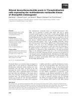
Fig. 1. Structure of SDTG (A) and time course of inactivation of
recombinant GST I by SDTG at pH 6.5 and 25 °C(B).En zyme
(2 units) was i ncubat ed in the absence (m)orpresenceof14.5l
M
SDTG (d), 36.36 l
M
SDTG (h), 72.7 l
M
SDTG (s), 145.5 l
M
SDTG (n) or 219.3 l
M
SDTG (j). At the times indicated, aliqu ots
were withdrawn and assa yed f or ac tivity.
3504 G. A. Kotzia and N. E. Labrou (Eur. J. Biochem. 271) Ó FEBS 2004
Expression and purification of maize GST I and other
enzymes
Maize G ST I w as cloned into a pQE70 expression vector to
yield the pQEGST expression plasmid as described by
Labrou et al. [14]. Expression and purification of wild-type
GST I were performed as described [14]. E xpression of
mutants was also performed as described by Labrou et al.
[14], but purification was achieved by using the a ffinity
adsorbent Cibacron blue 3GA–Sepharose, adsorbed at
0.1
M
potassium phosphate buffer, pH 7.0, and eluted with
0.1
M
potassium p hosphate buffer, pH 7.0, containing
5m
M
GSH. Recombinan t rat GST A 1-1 [32] and human
GST A1-1 [33] were expressed in Escherichia coli and
purified on a hexyl-GSH column as described previously
[34]. The expression vectors for rat GST A1-1 and human
GST A1-1 were much a ppreciated gifts from W. M. Atkins
(Department of Medicinal Chemistry, University of Wash-
ington, Seattle, WA, U SA). Glutathione synthase from
S. cerevisiae was purified to homogeneity as described
previously [35].
Site-directed mutagenesis
Site-directed mutagenesis was pe rformed a s described
by Weiner et al. [36]. The pairs of oligonucleotide primers
used in the PCRs were as follows: Phe51Ala mutation,
5¢-CGGAACCCC
GCAGGTCGAGTTTCC-3¢ and 5 ¢-GA
CGAGGTGCTCGGGGCTCTT-3¢; Met121Ala muta-
tion, 5 ¢-ATCAGTCCG
GCACTTGGGGGAACC-3¢ and
5¢-GAGGACGTCGAAGAGGATGGGTTACAG-3¢.
The mutatio n (codon underlined above) was confirmed
by DNA sequencing on Applied Biosystems Sequencer
373A with th e D yeDeoxy T erminator Cycle s equencing k it.
Assay of enzyme activity and protein
Enzyme assays were performed by monitoring the forma-
tion of the c onjugate of CDNB ( 1 m
M
)andGSH(2.5m
M
)
at 340 nm (e ¼ 9.6 m
M
)1
.cm
)1
)at30°C according to a
published method [13,14]. Observed reaction v elocities w ere
corrected for spontaneous reaction rates when necessary.
All initial velocities were determined in triplicate in buffers
equilibrated at c onstant t emperature. P rotein concentration
was determined by t he method of Bradford [37] using BSA
(fraction V ) as standard.
Enzyme inactivation studies
GST I was inactivated at 25 °C i n 1 mL incubation mixture
containing potass ium phosphate buffer, pH 6.5 ( 100 lmol),
SDTG (0–218.2 nmol) and enzyme (2 units, GST assay at
30 °C). T he rate of inactivation was followed by p eriodically
removing samples (20 lL) for assay of enzymatic activity
[17,20].
Rate constants for the reaction exhibiting biphasic
kinetics were calculated from log(% remaining activity)
vs. t ime (min), using the equation [19,20]:
Remaining a ctivity ¼ð1 À FÞe
Àk
fast
t
þ Fe
Àk
slow
t
where F represents the fractional residual activity of the
partial active enzyme intermediate, and k
fast
and k
slow
are
the rate constants for the slow a nd fast phase of the reaction.
Analysis was performed using the
GRAFIT
(Erithacus
Software L td, Horley, Surrey, UK) computer program.
K
d
was d etermined as described previously [19,20].
Studies of i nactivation of GST I b y SDTG in t he
presence of S-methyl-GSH and S-nitrobenzyl-GSH were
performed at 25 °C in 1 mL incubation mixture contain-
ing potassium phosphate buffer, pH 6.5 (100 lmol),
SDTG (218.2 nmol), S-methyl-GSH or S-nitrobenzyl-
GSH (0.5 lmol) and enzyme (2 units, GST assay a t
30 °C).
Inactivation of other enzy mes (S. cerevisiae glutathione
reductase, S . cerevisiae glutathione synthase, rat GST A1-1,
human GST A1-1 and bovine heart
L
-lactate dehydro-
genase) was performed (in the absence or presence of 1 m
M
S-methyl-GSH) in 1 mL incubation mixture containing
100 lmol potassium phosphate buffer, pH 6.5 (for rat GST
A1-1 and human GST A1-1) or 100 lmol potassium
phosphate buffer, pH 7.5 ( for glutathione reductase, gluta-
thione synthase and
L
-lactate deh ydrogenase), S DTG
(98.2 nmol) and enzyme (typically 2 units).
Kinetic analysis
Steady-state kinetic measurements of native and SDTG-
modified GST I were performed at 30 °Cin0.1
M
potassium phosphate buffer, pH 6.5. Initial velocities w ere
determined in the presence of 2.5 m
M
GSH; CDNB was
used in the concentration range 0.06–1.2 m
M
.Alternat-
ively, CDNB was used at a fixed concentration (1 m
M
),
and the GSH concentration varied in the range 0.15–
2.2 m
M
. Solutions of GSH and an alogues were f reshly
prepared each day and stored on ice under N
2
. All initial
velocities were determined in triplicate in buffers equili-
brated at constant temperature. The apparent kinetic
parameters k
cat
and K
m
were determined by fitting the
collected steady-state data to the Michaelis–Menten
equation by nonlinear regression analysis using t he
GRAFIT
computer program.
Stoichiometry of SDTG binding to GST I
GST I (100 lg) in 100 m
M
potassium phosphate buffer,
pH 6.5, was inactivated with 51.2 nmol SDTG at 25 °C.
The SDTG-modified e nzyme was separated from t he free
SDTG by ultrafiltration (in an Amicon stirred cell 8050
carrying a Diaflo YM10 ultrafiltration membrane; cut-off
10 kDa) after extensive washing with 100 m
M
potassium
phosphate buffer, pH 6.5. The s olution w ith S DTG–GST I
covalent complex was then lyophilized and stored at
)20 °C. The lyop hilized SDTG-modified e nzyme was
dissolved in 8
M
urea and i ncubated w ith N-ethylmaleimide
to block free -SH groups, and then with Woodward’s
reagent K (5 m
M
) or 2 ,4,6-trinitrobenzenesulfonic acid
(5 m
M
) for the determination of total carboxyl and primary
amino groups, respectively. The same treatment was also
applied to the unmodified GST I, as a control. Total
carboxy and primary amino groups in the modified and
unmodified enzyme were d etermined at 340 nm as described
previously [38,39].
Amino-acid analysis of native and SDTG-modified
GST I was performed by the method of Davey & Ersser [40].
Ó FEBS 2004 Affinity labelling of maize GST I (Eur. J. Biochem. 271) 3505
UV spectroscopic studies
Far-UV spectra were measured with a Perkin–Elmer
Lamda 16 recording spectrophotometer at 25 °C. The
enzyme (typically 0.02–0.05 mgÆmL
)1
) was dialysed against
0.01
M
potassium phosphate buffer, pH 7.0, and its UV
spectra were recorded between 250 and 190 nm.
Molecular modelling
Structure manipulations and analysis were performed using
the
PYMOL
software [41].
Results and Discussion
Synthesis of SDTG
SDTG was synthesized by nucleophile displacement of a
chlorine atom of the 1,3,5-sym -trichlorotriazine ring by the
-SH group of GSH. The 1,3,5-triazine scaffold is of special
interest because of i ts synthetic accessibility, i.e . one can t ake
advantage of the temperature-dependent successive dis-
placement of the chloride atoms b y different nucleophiles
[18,42,43]. O ther advantages o f t riazine-based a ffinity labels
are their high stability in neutral buffer solutions and the
presence of electron-rich nitrogen sites on the triazine ring,
which increase the capability of forming additional hydro-
gen bonds with amino-acid residues within protein bin ding
sites [42,43]. We have previously described the use of s uch
triazine-based probes to stoichiometrically label t he active
site of oxaloacetate decarboxylase [18] and Clostridium
histolyticum clostripain [43].
The overall yield in the synthesis of SDTG f rom GSH
was % 55%. SDTG was p urified by HPLC on a C
18
reverse-
phase S5 ODS2 Spherisorb silica column. The product was
eluted at 8.8 min. SDTG purity w as assessed as described in
Experimental procedures. The product showed a single spot
with R
f
¼ 0.55, a UV absorption spectrum w ith a peak at
230 nm, and w as negative in the 5,5-dithio-bis-(2-nitro-
benzoic) acid test for f ree -SH grou ps and positive in the
ninhydrin and 2,4,6-trinitrobenzenesulfonic acid test for
primary a mine. The chloride content w as found to be 2 mol
per mol SDTG. P urified SDTG was a lso analysed b y positive
ionization nano-electrospray MS. Evidence for one major
ion at m/z 457.4 was found, indicating a molecular mass of
456.3 Da, which corresponds well to the mass of SDTG
(455.28 Da).
Kinetics of reaction of SDTG with GST I
When maize GST I was incubated with SDTG at pH 6.5
and 25 °C, it was progressively inactivated (Fig. 1B),
whereas, in the absence of SDTG, virtually no change in
activity was observed. This inactivation was irreversible,
and activity was not restored by extensive dialysis or gel
filtration on Sephadex G-25. The pH used for inactivation
(pH6.5)wasthesameasthatnecessaryforhighGST
activity. This probably affords an enzyme conformation
similar to that adopted during catalysis, t hus creating more
favourable conditions for ligand binding. The kinetics of
inactivation were biphasic (Fig. 1B), wit h t he r apid reaction
occurring immediately on e xposure of the enzyme to SDTG
and the slow inactivation continuing to yield enzyme with a
final residual activity of 42 ± 3%. A t all concentrations of
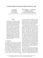
SDTG used, biphasic kinetics were observed. The rate of
inactivation for the fast and slow phases was dependent on
SDTG c oncentration, as illustrated in F ig. 2. F or both
phases, a plot of 1/k
obs
vs. 1/[SDTG] yields a straight line.
This indicates that t he reaction obeys pseudo-first-order
saturation kinetics and is consistent with reversible binding
of reagent before covalent modification according to the
following equation [17–20]:
E þ SDTG Ð
k
d
E:SDTG À!
k
3
E-SDTG
whereErepresentsthefreeenzyme,E:SDTGisthe
reversible complex, and E-SDTG is the covalent product.
The steady-state rate equation f or the interaction is [2 3–25]:
1=k
obs
¼ 1=k
3
þ k
d
=ðk
3
½SDTGÞ
where k
obs
is the rate of enzyme inactivation for a given
concentration of S DTG, k
3
isthemaximalrateofinacti-
vation (min
)1
), and K
d
is the apparent dissociation constant
of the E :SDTG complex. F rom the data shown in F ig. 2, K
d
values of 107.9 ± 2.1 l
M
and 224.5 ± 4.2 l
M
,forthefast
and slow reactions, respectively, were estimated. Apparent
maximal rate constants were determined to be 0.049 ±
0.002 min
)1
for the fast reaction, and 0.0153 ± 0.001 min
)1
for t he slower reaction.
The stability of SDTG against hydrolysis was demon-
strated b y m easuring the rate o f c hloride released f rom the
molecule in conditions identical with those used i n t he inac-
tivation e xperiments. The results showed t hat t he fir st-order
rate constant for SDTG hydrolysis was 1.2 · 10
)5
min
)1
.
This corresponds to 0.07% and 0.03% of the rate ob served
for the slow and fast phase, respectively. This suggests that
Fig. 2. Dependence of the pseudo-first-order rate constant for the fast
(j) and slow phase (r) of inactivation on the concentration of SDTG,
expressed a s a doub le-reciprocal plot. GS T I (2 units) was i ncubated at
pH 6.5 and 25 °C with various concentrations of SDTG (14.5–
219.3 l
M
), and the rate constants were calculated as d escribed in the
text. The slope and intercept of the double -reciprocal plot were cal-
culated by u nweight ed linear r egression analysis .
3506 G. A. Kotzia and N. E. Labrou (Eur. J. Biochem. 271) Ó FEBS 2004
the slow phase of inactivation observed is not due to the
decomposition o f SDTG but is the direct result of S DTG–
enzyme inte raction [31].
To determine the stoichiometry of i ncorporation of
SDTG, modified and unmodified G ST I were treated with
Woodward’s reagent K and 2,4,6-trinitrobenzenesulfonic
acid, and the amount of covalently bound SDTG was
determined by subtraction o f the total number of c arboxy
and amino groups in the modified and un modified enzyme.
The results of this experiment are shown in Table 1. As
indicated by the data, 1 mol SDTG is bound per mol wild-
type enzyme at 42% inactivation.
The specificity of a protein chemical modification
reaction can be indicated by the ability of natural
ligands or active-site-directed reagent to protect against
inactivation [17–24]. The effect of GSH a nalogues S-
methyl-GSH and S-nitrobenzyl-GSH on the reaction of
SDTG with GST I was investigated. S-Methyl GSH
and S-nitrobenzyl GSH protect GST I against inacti-
vation by SDTG. T he protective effect afforded by S-
nitrobenzyl GSH was more significant than that
afforded by S-methyl GSH, at comparable concentra-
tions, which is in agreement with their relative affinity
constants.
Kinetic analysis of the modified enzyme (42% remaining
activity) showed that the e nzyme exhibits kinetic properties
that are different from that of the unmodified enzyme. The
results are shown in T able 2. The modified e nzyme exhibits
about threefold reduced affinity for GSH and a bout
twofold increased affinity for CDNB. A final activity of
less than 50% (e.g. 42%) accords with the incorporation of
SDTG into one subunit, producing a change in the
unmodified subunit which alters its activity to a small
degree (% 7%).
Identification of GST I residue modified by SDTG
To identify which residue in GST I became modified by
SDTG, we used amino-acid analysis, molecular modelling
and s ite-directed mutagenesis. Direct amino-acid sequence
determination o f the modified peptide was not possible
because of its instability during E dman degradation reac-
tions. The results from a typical amino-acid analysis
indicated that the modified enzyme exhibits loss of 1 mol
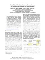
Met per mol enzyme. From analysis of t he crystal structure
of the enzyme in complex with S-atrazine–GSH conjugate
[11], it is evident that Met121 is within or close to the
binding site and accessible for covalent modification
(Fig. 3). It is located at the end of a-helix H¢¢¢
3
and forms
part of the xenobiotic-binding site [11]. A lthough the
thioether bond of methionine is usually considered to be
of low reactivity, a number of pieces of experimental
evidence from affinity labelling experiments, suggest that in
Table 1. Stoichiometry of SDTG bin ding to GST I. Total carboxy and
primary am ino groups for the mo dified and un modified enzyme were
determined by the Woodward’s Reagent K and 2,4,6-trinitro-
benzenesulfonic a cid assays.
Unmodified
GST I
SDTG-modified
GST I
SDTG-modified
Phe51Ala mutant
Carboxy groups 26.8 ± 0.3 29.2 ± 0.2 31.2 ± 0.3
Primary amino
groups
14.9 ± 0.1 16.2 ± 0.2 17.3 ± 0.3
Table 2. Steady-state kinetic parameters of unmodified and SDTG-
modified GST I for the CDNB conjugation reaction at pH 6.5 and
30 °C.
GST I
K
m
(m
M
)
k
cat
· 10
)2
(s
)1
)
GSH CDNB
Unmodified 1.1 ± 0.20 1.60 ± 0.10 29.3
SDTG-modified 2.9 ± 0.15 0.78 ± 0.02 11.2
Fig. 3. Structural representation depicting important residues of maize
GST I. (A) Bound S-atrazine–GSH conjugate is shown in red. Met121
is dr awn in a spacefill representation. (B) Possible mode of commu-
nication between subunits. Bound S-atrazine–GSH conjugate is shown
in red. Met121 is drawn in a sp acefill representation and P he51 is
shown in b lue .
Ó FEBS 2004 Affinity labelling of maize GST I (Eur. J. Biochem. 271) 3507
several enzymes may act as a r eactive nucleophile. For
example, a methionine residue is modified in isocitrate
dehydrogenase [44], in human uterine progesterone receptor
[45], and
D
-amino acid oxidase [46,47] by reaction with
iodoacetate, 16a-(bromoacetoxy)progesterone and O-(2 ,4-
dinitrophenyl)hydroxylamine, respectively.
To provide further experimental evidence and establish
the i nvolvement of Met121 in the reaction with SDTG, site-
directed mutagenesis experiments were carried out. Met121
was m utated to Ala, and the resulting mutant was subjected
to inactivation studies. T he Met121Ala mutant w as resistant
to inactivation by SDTG (90 l
M
)atpH6.5and25°C,
compared with the wild-type enzyme. Comparison of the
far-UV difference spe ctra of native and mutated enzyme
indicated the absence of any structural perturbation caused
by the mutation (Fig. 4). This rules out the possibility that
the resistance of the Met121Ala mutant to inactivation is
due to conformational changes in its structure. These
observations imply that SDTG binds at one site at all stages
of the reaction. The best explanation for these results may
be that the reaction of SDTG at the binding site of one
subunit changes the conformation of the o ther subunit,
thereby completely abolishing reaction of SDTG with the
second subunit.
Analysis of the crystal structu re of t he enzyme in co mplex
with the S-atrazine–GSH conjugate [11] provides a struc-
tural explanation for the intrasubunit c ommunication
observed on reaction of Met121 with SDTG. Although
the H -sites of neighbouring subunits are distant (Fig. 3B), a
plausible mode o f communication between them exists.
Structural examin ation r eveals that the key residue bridging
the dimer interface, Phe51, may have an important role in
intrasubunit communication. This residue forms the lock-
and-key motif responsible for a highly conserved hydro-
phobic interaction in the subunit i nterface. This residue
makes contact with a hydrophobic patch on the alternate
subunit, comprising, in part, Trp97, Val96, Val100 and
Gln104. As the interface contacts on the a lternate subunit
are largely found in a single kinked a-helix H¢¢
3
(Fig. 3B),
the s ignal may be transmitted via the helix to H-site residues
such as Met121, Ile118, Leu122 and Phe114, which are
located at the end of t his helix. Conformational changes in
these residues would then change the affinity for CDNB
binding, which is supported by the finding that the K
m
of the
modified enzyme for CDNB is lower (see Table 2), and
abolish reaction of SDTG with Met121 at the second
subunit. Thus, the observed intrasubunit communication is
probably directed via Phe51 of the monomer–monomer
contact region, to a-helix H¢¢¢
3
of the a djacent subunit which
contains Met121.
To confirm the key role of Phe51 in this hypothesis,
site-directed mutagenesis was used. T he mutant Phe51Ala
was expressed, p urified, and subjected t o i nactivation
studies (Fig. 5). Upon reaction with SDTG at pH 6.5 and
25 °C, the mutant was progressively inactivated to a final
residual activity of about 1.9% (Fig. 5). Comparison of
the far-UV difference spectra of n ative and mutated
enzyme (data not shown) indicates the absence of any
structural perturbation caused by the mutation. Amino-
acid analysis of the SDTG-modified Phe51Ala mutant
and determination of its total amino and carboxy c ontent
suggests that the modified residue is also methionine, and
% 2 mol SDTG per mol enzyme was incorporated
(Table 1). This provides strong evidenc e that the inability
of SDTG to attack the other subunit in native GST I is
the indirect result of the interaction between the two
enzyme subunits, and that this subunit interaction is
absent in the Phe51Ala mutant.
The hydrophobic lock-and-key intersubunit motif
involving Phe51 is the major structural feature conserved
Fig. 4. Far-UV difference spectroscopy of the wild-type GST I
(a, 0.05 mgÆmL
-1
) and mutant Met121Ala (b, 0.0375 mgÆmL
-1
). Spectra
were measured at 25 °Cin0.01
M
potassium phosphate buffer at
pH 7.0.
Fig. 5. Time course of inactivation of wild-type GST I and mutant
Phe51Ala by SDTG. Wild-type (r)andmutantPhe51Ala(j)were
incubated i n the presence of 72.7 and 92 l
M
SDTG, respectively, at
pH 6.5 and 25 °C. At th e times indicated, aliquo ts were with drawn
and assayed f or e nzymatic a ctivity.
3508 G. A. Kotzia and N. E. Labrou (Eur. J. Biochem. 271) Ó FEBS 2004
at the dimer interface of GST I. Similar l ock-and-key motifs
have also been observed for the classes Alpha, Mu and Pi
GSTs [48–50]. T he conserved h ydrophobic i nteraction
formed by the side chain of the Phe51 residue, which
protrudes from the loop in domain I of one monomer into
the h ydrophobic pocket of domain I I o f t he other mono-
mer, physically anchors the two subunits together at either
end o f the interface.
Explanation of the biphasic kinetics
An average incorporation of 0.5 mol reagent per mol
enzyme subunit indicates that reaction o f SDTG with one
Met121 prevents the reaction of the Met121 of the s econd
subunit. The biphasic kinetics observed may be explained
by assuming that the two subunits, o r at least the
conformation of the Met121 side chains in each subunit,
are not equivalen t regarding the reaction with SDTG, and
exhibit differen t reactivity. The existence of such a
nonsymmetrical arrangement of G ST I subunits has been
observed in the crystal structures [10,11]. The two subunits
of GST I complexed with various product analogues
show some structural differences betw een them, suggesting
that the two substrate-binding sites in the enzyme dimer
may not act independently [10,11]. Furthermore, other
important factors must be considered with regard to the
dynamics of this enzyme. A plot of the crystallographic B-
factors along the polypeptide chain can give an indication
of the relative flexibility of the different portions of the
protein (Fig. 6). GST I displays a well-defined flexibility
pattern. Several regions with high mobility can be
identified. The plot shows significant d ifferences in several
regions between chains A and B, including a-he lix H¢¢¢
3
(residues 188–122). A large difference is cent red on
Met121. In particular, the mean B-factors of Met121 at
the A and B chains are 26.67 A
˚
2
and 49.26 A
˚
2
,andthe
B-factors of S atoms are 38.14 A
˚
2
and 79.30 A
˚
2
, respect-
ively. It is therefore reasonable to propose that conform-
ational changes and changes in dynamics may also
contribute to the observed biphasic kinetics.
Results f rom steady-state kinetics using CDNB and 1,2-
dichloro-4-nitrobenzene as e lectro philic substrates for GSTs
from several classes are consistent with the idea of two
noncooperative binding sites. However, the large bulky
aflatoxin–GSH conjugate [51] and the product analogue
glutathionyl S-[4-(succinimidyl)benzophenone] [22] have
been shown to bind to mouse Alpha class 2-2 and rat liver
GST enzymes with a stoichiometry of 1 mol per mol
enzyme dimer, and b inding of this ligand completely
abolished the catalytic activity of both enzyme subunits.
In addition, binding studies of GSH to the human P1-1
enzyme have shown that binding displays positive cooper-
ativity above 35 °C, whereas n egative cooperativity occurs
below 25 °C [52]. These results suggest t hat t he two binding
sites may not be independent and further support the
Ôcooperative self-preservationÕ mechanism proposed by
Ricci et al. [54] for the human P1-1 enzyme. According to
this mechanism, a c ooperativity is utilized by the e nzyme to
provide s elf-preservation against inhibitors or physical
factors t hat threaten i ts catalytic e fficiency. T his mechanism
is based on a structural intersubunit communication by
which one subunit, as a consequence of an inactivating
modification, triggers a defence arrangement in the other
subunit to prevent modification [54]. In the present study,
we observe th at the modification of one enzyme subunit of
the GST I homodimer prevents modification of the other
subunit, which suggests t hat the two e nzyme a ctive sites are
co-ordinated.
Reaction of SDTG with other GSH-binding enzymes
To demonstrate the wide applicab ility o f S DTG as an
affinity label for other GSH-binding enzymes such as
S. cerevisiae glutathione reductase, glutathione synthase, rat
GST A1-1 a nd hum an GST A 1-1, inactivation studies were
carried out. T he pseu do-first-order rates of inactivation
observed at a SDTG concentration of 98.2 l
M
and in the
presence and a bsence of 1 m
M
S-methyl-GSH a re summar-
ized in Table 3. All enzymes were susceptible to inactivation
by SDTG. The protective effect of S-methyl-GSH suggests
that the r eaction is s pecific. It i s interesting to note that the
human and r at GST A1-1 isoenzymes obeyed b iphasic
kinetics, with residual activity after labelling of 48% and
33%, respectively, which confirms the conclusions o n maize
Fig. 6. Structural flexibility of GST I. A plot of the crystallographic
B-factors along the p olypeptide chains A and B obtained from the
crystal structure of GST I i n complex with S-atrazine–GSH conjugate
(PDB code 1bye [11]). The plot was produced by the
WHAT IF
software
package [53]. The height at e ac h residue position indicates the average
B-factor of al l atoms in the residue.
Table 3. Observed rates of inactivation (k
obs
) of GSH-binding enzymes
and bovine heart
L
-lactate dehydrogenase b y SDTG in t he presence and
absence of 1 m
M
S-methyl-GSH. NI, No inactivation.
Enzyme
k
obs
· 10
)3
(min
)1
)
(in the absence of
S-methyl-GSH)
% Protection from
inactivation
(in the presence of
S-methyl-GSH)
Rat GST A1-1 1.12 ± 0.1
a
85.5
Human GST A1-1 2.83 ± 0.1
a
84.3
S. cerevisiae glutathione
reductase
3.5 ± 0.2 86.5
S. cerevisiae glutathione
synthase
1.9 ± 0.1 85.4
Bovine heart
L
-lactate
dehydrogenase
NI –
a
Fast phase inactivation rate.
Ó FEBS 2004 Affinity labelling of maize GST I (Eur. J. Biochem. 271) 3509
GST I. In a ddition, the ability o f SDTG t o inactivate a non-
GSH-dependent enzyme such as bovine heart
L
-lactate
dehydrogenase was investigated. SDTG did not inactivate
L
-lactate dehydrogenase and did not show any inhibitory
effect on its catalytic reaction. This finding strengthens the
view that the SDTG acts a s a true affinity label for the GSH-
binding site and i ndicates t hat t his new reagent may have
wider applicability as an affinity label for other enzymes
with GSH-binding sites.
References
1. Sheehan, D., Mead e, G., Foley, V.M. & Dowd, C.A. (2001)
Structure, function and evolution of glutathione transferases:
implications for classification of non-mamm alian membe rs of an
ancient enzyme s uperfamily. Biochem. J. 360, 1–16.
2. Wilce, M.C.J . & Parker, M.W. (1994) Structure and fu nction of
glutathione S-transferases. Biochim. Biophys. Acta 1205, 1 –18.
3. Edwards, R., D ixon, D .P. & Walnot, V. (2000) Plant g lutathione
S-transferases: enzymes with multiple functions in s ickness and in
health. Trends Pharmacol S ci 5, 193–198.
4. Marrs, K.A. (1996) The functions and regulation of glutathione
S-transferases in plants. Annu. Rev. Plant Physiol. Plant Mol. Biol.
47, 127–158.
5. Noctor, G. & Foyer, C.H. (1998) Ascorbate and glutathione:
keeping active oxygen under control. Annu. Rev. Physiol. Plant
Mol. Biol. 49 , 2 49–279.
6. Smith, A.P., Nourizadeh, S.D., Pe er, W.A., Xu, J., B andyop ad-
hyay, A ., Murphy, A.S. & Goldsbrough, P.B. (2003) Arabidopsis
AtGSTF2 is regulated by ethylene and auxin, and encodes a
glutathione S-transferase that interacts with flav onoids. Plant J .
36, 433–442.
7.Dixon,D.P.,Cummins,I.,Cole,D.J.&Edwards,R.(1998)
Glutathione-mediated detoxification systems in plants. Curr. Opin.
Plant Biol. 1, 258–266.
8. Armstrong, R.N. (1997) Structure, catalytic mechanism, and
evolution of the glu tathione transferases. Chem. Res. Toxicol. 10,
2–18.
9. Dixon, D., Laptthorn, A. & Edwards, R. (2002) Pla nt glutathione
transferases. Genome Biol. 3, 1–10.
10. Neuefeind, T., Huber, R., Dasenbrock, H., Prade, L. & Bieseler,
B. (1997) Crystal structure of herbicide-detoxifying maize glu-
tathione S-transferase-I in complex with lactoylglutathione: evi-
dence f or an induced-fit mech anism. J. Mol. Biol. 274, 446–453.
11. Prade, L., Huber, R. & Bieseler, B. (1998) Structures of herbicides
in complex with t heir detoxifying enzyme glutathione S-transfer-
ase: explanations for the selectivity of the enzyme in plants.
Structure 6, 1445–1452.
12. McGonigle,B.,Keeler,S.J.,Lau,S.M.,Koeppe,M.K.&O’Keefe,
D.P. (2000) A genomics approach to the com prehensive analysis
of the glutathione S-transferase gene family in soybean and maize.
Plant Physiol. 124, 1 105–1120.
13. Labrou, N.E., Mello, L.V. & Clonis, Y .D. (2001) Functional and
structural roles of the glutathione -binding residues in maiz e ( Zea
mays) glutathione S-t ransferase I. Bioc hem. J. 358 , 101 –110.
14. Labrou, N.E., Mello, L.V. & Clonis, Y.D. (2001) The conserved
Asn49 of maize glutathione S-transferase I m odulates substrate
binding, catalysis and intersubunit communication . Eur. J. Bio-
chem. 26 8, 3950–3957.
15. Labrou, N.E., Rigden, D.J. & Clonis, Y.D. (2004)
Engineering the pH-dependence of kinetic parameters of maiz e
glutathione S-transferase I by site-directed mutagenesis. Biomol.
Eng. 21, 61–66.
16. Thom, R., Cummins, I., Dixon, D.P., Edwards, R., Cole, D.J. &
Lapthorn, A.J. (2002) Structure of a tau class glutathione
S-transferase from wheat active in herbicide detoxification.
Biochemistry 41, 7008–70020.
17. Labrou, N.E., Rigden, D.J. & Clonis, Y.D. (2000) Characteriza-
tion of NAD
+
binding site of Candida boidinii formate dehy-
drogenase b y affinity l abelling and site-directed mutagenesis. Eur.
J. Biochem. 267, 6657–6664.
18. Labrou, N.E. (1999) Afinity labeling of oxaloacetate decarboxy-
lase by novel d ichlorotriazine linked a-ketoacids. J. Pro tein Chem.
18, 729–733.
19. Bailey, J. & Colman, R.F. (1985) Affinity labeling of NADP-
specific isocitrate dehydrogenase by a new fluorescent nucleotide
analogue 2- [(4-bromo-2.3-dioxobutyl) t hio]-1,N6-ethanoad eno-
sine 2¢,5¢-biphosphate. Bi ochemistry 35, 2 658–2667.
20. King, M.M. & Colman, R. (1983) Affinity labeling of nicotina-
mide adenine dinucleotide dependent isocitrate dehydrogenase by
the 2¢,3¢-dialdehyde derivative of a denosine 5¢-diphosphate. Evi-
dence for the formation of an unusual reaction product. Bio-
chemistry 22, 1656 –1665.
21. Wang, J., Bauman, S. & Colman, R.F. (1998) Photoaffinity
labeling of rat liver glutathione S -transferase, 4–4, by glutathionyl
S-[4-(succinim idyl)-ben zoph enone]. Biochemistry 37, 15671–15679.
22. Wang,J.,Bauman,S.&Colman,R.F.(2000)Probingsubunit
interactions in Alpha c lass rat liver glutathione S-transferase with
the p hotoaffinity label glu tathionyl S-[4-(succinimid yl)-benzophe-
none]. J. Biol. Chem. 27 5 , 5493–5503.
23. Cooke, R.J., Bjornestedt, R., Douglas, K.T., M cKie, J .H., King,
M.D., Coles, B., Ketterer, B. & Mannervik, B. (1994) Photo-
affinity labelling of the active site of the rat glutathione transferases
3–3 and 1–1 and human glutathione transferase A1–1. Biochem. J.
302, 383–390.
24. Katusz,R.M.,Bono,B.&Colman,R.F.(1991)S-(4-Bromo-2,3-
dioxobutyl)glutathione: a new a ffinity label f or the 4–4 isoenzyme
of rat liver glutathion e S-transferase. Biochemistry 30, 11230–
11238.
25. Hoesch, R.M. & Boyer, T.D. (1 989) Localization of a portion of
the a ctive site of two rat liver glutathione S-transferases using a
photoaffinity l ab el. J. Biol. Chem . 264 , 1 7712–17717.
26. Milligan, A.S., Daly, A., Parry, M.A.J., Lazzeri, P.A. & Jepson, I.
(2001) The expression of a maize glutathione S-transferase in
transgenic w heat confers herb icide tolerance, both in planta and
in vivo. Mol. Breed. 7, 3 01–305.
27. Roxas, V.P., Smith, R.K., Allen, E.R. & Allen, R.D. (1997)
Overexpression of glutathione S-transferase/glutathione peroxi-
dase enhances the growth of transgenic tobacco seedlings during
stress. Nat. Biotechnol. 15, 988 –991.
28. Yue, H., Corley, N. & Shah, P. (2003) Human glutathione-S-
transferase. United States Patent No. 6 506 571.
29. Andreou, V. & Clonis, Y.D. (2002) Novel fiber-optic biosensor
based on immobilized glutathione S-transferase and sol–gel en-
trapped bromcresol g reen for t he determination o f atrazine. Anal.
Chim. Acta 460 , 1 51–161.
30. Zall, D.M., Fisher, D. & Garner, M.Q . (1956) P hotometric
determin at ion of chlo rid es in wat er. Anal. Chem. 28, 1665–1668.
31. Hu, L. & Colman, R .F. (1995) Monobromobimane as an a ffinity
label of the xenobiotic binding site of rat glutathione S-transferase
3–3. J. Bio l . C he m. 270 , 2 1875–21883.
32. Ibarra, C.A., Chowdhury, P., Petrich, J.W. & Atkins, W.M.
(2003) The anomalous pKa of Tyr-9 in glutathione S-transferase
A1–1 ca talyze s p rod uct release. J. Biol. Chem. 278, 19257–19265.
33. Lyon, R.P. & Atkins, W.M. (2002) K inetic characterization of
native and cysteine 112-modified glutathione S-transferase A1–1:
reassessment of nonsubstrate ligand binding. Bioch em ist ry. 41,
10920–10927.
34. Simons, P.C. & Vander Jagt, D.L. (1977) Purification of glu-
tathione S-transferases from human liver by glutathione-affinity
chromatography. Anal . B iochem. 82, 334–341.
3510 G. A. Kotzia and N. E. Labrou (Eur. J. Biochem. 271) Ó FEBS 2004
35. Dennda, G. & Kula, M R. (1986) Purification and evaluation of
the glutathione-synthesizing e nzymes from Candida boidinii for
cell-free synthesis of glutathione. J. Biotechnol. 3, 143–158.
36. Weiner,M.P.,Costa,G.L.,Schoettlin,W.,Cline,J.,Mathur,E.&
Bauer, J.C. (1994) Site-directed mutagenesis o f double-stranded
DNA by t he polymerase chai n r eaction. Gene 151 , 1 19–123.
37. Bradford, M.A. (1976) A rapid and sensitive method for the
quantitation of microgram quantities of protein utilizing the
principle o f p rotein -dye b indin g. Anal. Biochem. 72, 248–254.
38. Sinha, U. & Brewer, J.M. (1985) A spectrophotometric method
for q u antitation of carboxyl group modification of p roteins using
Woodward’s Reagent K . Anal . Biochem. 151, 3 27–333.
39. Snyder, S.L. & Sobocinski, P.Z. (1975) An improved 2,4,6-trini-
trobenzenesulfonic a cid m etho d f or the determination of amines.
Anal. Bio chem. 64, 284–288.
40. Davey, J.F. & Ersser, R.S. (1990) Amino acid analysis of phy-
siological flu ids by h igh-performance liquid chromatography with
phenylisothiocyanate derivatization and comparison with ion-
exchange chromatography. J. Chromatogr. 528, 9–2 3.
41. DeLano, W.L. (2004) The PyMOL molecular graphics s ystem on
word wide web. http: //www.pymol.org.
42. Labrou,N.E.&Clonis,Y.D.(2002)Immobilisedsyntheticdyesin
affinity chromatography. In Biochromatography: Theory and
Practice (Vijayalakshmi, M.A., ed.), pp. 235–251. Taylor &
Francis Publishers, London.
43.Labrou,N.E.&Rigden,D.J.(2004)Thestructure–function
relationship in the c lostripain family of peptidases. Eur. J. Bio-
chem. 27 1, 983–992.
44. Colman, R. (1968) Effect of modification of a methionyl residue
on the kinetic and molecular properties of isocitrate dehydro-
genase. J. Bio l. C hem. 24 3 , 2454–2464.
45. Holmes, S.D. & Smith, R.G. (1983) Identification of histidine and
methionine residues in the active site of the human u terine pro-
gesterone receptor with the affinity labels 11a- and 16a-(bromo-
acetoxy) progesterone. Bi ochemistry 22 , 1729–1734.
46. D’Silva, C., Williams, C.H. & Massey, V. (1986) Electrophilic
amination o f a single methionine residu e located at the a ctive site
of
D
-amino acid oxidase b y O-(2,4-dinitrophenyl) hydroxylamine.
Biochemistry 25, 5602–5608.
47. D’Silva, C., W illiams, C.H. & Massey, V. (1987) Identification of
methionine-110 as the residue covalently modified in the electro-
philic inactivation of
D
-amino acid oxidase by O-(2,4-dinitro-
phenyl) h yd roxy lamine. Bioc hemistry 26 , 1717–1722.
48. Hegazy, U.M., Mannervik, B. & Stenberg, G. (2004) Functional
role of the l ock and key motif at the subun it interface of glu-
tathione transferase P 1–1. J. Biol. C hem. 279 , 9586–9596.
49. Sayed, Y., Wllance, L.A. & Dirr, H .W. (2000) The hydrophobic
lock-and-key intersubunit motif of glutathione transferase A1–1:
implications for catalysis, ligandin function and stability. FEBS
Lett. 465, 169–172.
50. Vargo, M.A., Nguyen, L. & Colman, R.F . (2004) Subunit inter-
face residues of g lu tathione S-transferase A1–1 that a re important
in the m onomer–dimer equili brium. Biochemistry 43, 3327–3335.
51. McHugh, T.E., Atkins, W.M., Racha, J.K., Kunze, K.L. &
Eaton, D.L. (1996) Binding of the aflatoxin-glutathione conjugate
to mouse g lutathione S-transferase A3–3 is saturated a t o nly one
ligand per dimer. J. Biol. Chem. 271 , 27470–27474.
52. Caccuri, A.M., Antonini, G., Ascenzi, P ., Nicotra, M., Nuccetelli,
M.,Mazzetti,A.P.,Federici,G.,LoBello,M.&Ricci,G.(1999)
Temperature adaptation of glutathione S-transferase P1–1. A case
for homotropic regulation of substrate binding. J. Biol. Chem. 274,
19276–19280.
53. Vriend, G . (1990)
WHAT IF
: a molecular modeling a nd drug design
program. J. Mol. Gr aph. 8, 52–56.
54. Ricci, G., Caccuri, A.M., Lo Bello, M., Parker, M.W., Nuccetel li,
M., Turella, P., Stella, L., Di Iorio, E.E. & Federici, G. (2003)
Glutathione transferase P1–1: self-preservation of an anti-cancer
enzyme. Biochem. J. 376, 71–76.
Ó FEBS 2004 Affinity labelling of maize GST I (Eur. J. Biochem. 271) 3511