Báo cáo khoa học: Membrane distribution of epidermal growth factor receptors in cells expressing different gangliosides doc
Bạn đang xem bản rút gọn của tài liệu. Xem và tải ngay bản đầy đủ của tài liệu tại đây (371.8 KB, 10 trang )
Membrane distribution of epidermal growth factor receptors in cells
expressing different gangliosides
Adolfo R. Zurita
1
, Pilar M. Crespo
1
, Nicola
´
s P. Koritschoner
2
and Jose
´
L. Daniotti
1
1
Centro de Investigaciones en Quı
´
mica Biolo
´
gica de Co
´
rdoba, CIQUIBIC (UNC-CONICET), Departamento de Quı
´
mica Biolo
´
gica,
Facultad de Ciencias Quı
´
micas, Universidad Nacional de Co
´
rdoba, Argentina;
2
Departamento de Bioquı
´
mica Clı
´
nica, Facultad de
Ciencias Quı
´
micas, Universidad Nacional de Co
´
rdoba, Argentina
Gangliosides have been found to reside in glycosphingolipid-
enriched microdomains (GEM) of the plasma membrane
and to be involved in the regulation of epidermal growth
factor receptor (EGFr or ErbB1) activity. To gain further
insight into the mechanisms involved in EGFr modulation
by gangliosides, we investigated the distribution of EGFr
family members in the plasma membrane of CHO-K1 cells,
which were genetically modified to express different gan-
glioside molecules or depleted of glycolipids. Our data
demonstrate that at least four different sets of endogenously
expressed gangliosides, including GD3, did not have a sig-
nificant effect on EGFr distribution in the plasma mem-
brane. In addition, using confocal microscopy analysis we
clearly demonstrated that the EGFr co-localizes only to a
minor extent with GD3. We also explored the endogenous
expression, in wild-type CHO-K1 cells, of the orphan
receptor ErbB2 (which is the preferred heteroassociation
partner of all other ErbB proteins) and the effect of GD3
expression on its membrane distribution. Our results showed
that CHO-K1 cells endogenously express ErbB2 and that
expression of the GD3 affected, to some extent, the
membrane distribution of endogenous ErbB2. Finally, our
findings support the notion that most EGFr are excluded
from GEM, while an important fraction of ErbB2 is found
to be associated with these microdomains in membranes
from CHO-K1 cells.
Keywords: gangliosides; EGFr; ErbB2; membrane lipid
domains; CHO-K1 cells.
1
Gangliosides – glycolipids containing sialic acid – are
ubiquitous components of mammalian cell membranes.
They are involved in the regulation of cell proliferation
and differentiation [1]. They have also been implicated in
tumour growth and the formation of metastases. All
tumours exhibit aberrant ganglioside expression. This
includes overexpression of normal ganglioside constituents
and expression of gangliosides not found in normal adult
tissue [2].
On the basis that the bulk of gangliosides present in the
cell are plasma membrane bound, it has been speculated
that they participate in cell-surface events such as modula-
tion of tyrosine kinase growth factor receptors. In this
sense, it has been demonstrated, mainly by the exogenous
addition of gangliosides or by changes of the endogenous
content, that gangliosides regulate the activities of the TrkA
receptor [3,4], the insulin receptor [5,6], the epidermal
growth factor receptor (EGFr, ErbB1) [7] and the platelet-
derived growth factor (PDGF) receptor [8,9].
In previous studies, we described the modulation of
EGFr phosphorylation by endogenously expressed ganglio-
sides [10]. In particular, we observed an inhibition of EGFr
autophosphorylation when CHO-K1 cells (GM3
+
)were
induced to express the disialoganglioside, GD3, by stable
transfection of CMP-NeuAc:GM3 sialyltransferase (Sial-
T2). The effect of GD3 on EGFr function could not be
attributed to an alteration in the affinity of EGFr to EGF
(because the K
d
values were essentially the same as in the
wild-type cells) or to a decrease of GM3 content by
conversion into GD3 ganglioside.
Gangliosides are constituents of the glycosphingolipid-
enriched microdomains [GEM; also called DRMs (deter-
gent-resistant membranes) or rafts], dynamic assemblies of
cholesterol, saturated phospholipids, and sphingolipids,
which are characterized by a light buoyant density and
resistance to solubilization by Triton X-100 at 4 °C [11–14].
Initial evidence showed the association of gangliosides to
GEM present in the plasma membrane of CHO-K1 cells
lines expressing different gangliosides [15,16]. These data
Correspondence to J. L. Daniotti, CIQUIBIC (UNC-CONICET),
Departamento de Quı
´
mica Biolo
´
gica, Facultad de Ciencias Quı
´
micas,
Universidad Nacional de Co
´
rdoba, Ciudad Universitaria,
5000 Co
´
rdoba, Argentina. Fax: + 54 351 4334074,
Tel.: + 54 351 4334171, E-mail:
Abbreviations:BS
3
, bis(sulfosuccinimidil)suberato; Cer, ceramide;
CFP, cyan fluorescence protein; EGF, epidermal growth factor;
EGFr, EGF receptor; ErbB1, epidermal growth factor receptor 1;
ErbB2, epidermal growth factor receptor 2; GEM, glycosphingolipid-
enriched microdomain(s); GFP, green fluorescent protein; Gal,
galactose; GalNAc-T, UDP-GalNAc:LacCer/G3/GD3 N-acetyl-
galactosaminyltransferase; GlcCer, glucosylceramide; GPI, glycosyl-
phosphatidylinositol; HPTLC, high-performance thin layer
chromatography; LacCer, lactosylceramide; PPPP,
D
,
L
-threo-1-
phenyl-2-hexadecanoylamino-3-pyrrolidino-1-propanol-HCl;
Sial-T2, CMP-NeuAc:GM3 sialyltransferase; VSVG, vesicular
stomatitis virus glycoprotein; YFP, yellow fluorescence protein.
(Received 3 March 2004, revised 13 April 2004,
accepted 16 April 2004)
Eur. J. Biochem. 271, 2428–2437 (2004) Ó FEBS 2004 doi:10.1111/j.1432-1033.2004.04165.x
revealed that an important fraction of plasma membrane,
GM3, and most GD3 and GT3, resided in GEM, while
more complex gangliosides, such as GM1, GM2 and GD1a,
were almost excluded from GEM.
To gain insight into the mechanisms involved in EGFr
modulation by gangliosides (particularly GD3), we inves-
tigated the distribution of human EGFr in the plasma
membrane of CHO-K1 cells that were genetically modified
to express different ganglioside molecules or depleted of
glycolipids by inhibiting glucosylceramide synthase activity.
Our data demonstrated that at least four different sets of
endogenously expressed gangliosides, including GD3, did
not have a significant effect on EGFr distribution in the
plasma membrane. In addition, using confocal microscopy
analysis we clearly demonstrated that EGFr co-localizes
only to a minor extent with GD3.
We also explored the endogenous expression, in wild-type
CHO-K1 cells, of the orphan receptor epidermal growth
factor receptor 2 (ErbB2), which is the preferred hetero-
association partner of all other ErbB proteins, and the effect
of GD3 expression on ErbB2 membrane distribution. Our
results show that CHO-K1 cells endogenously express the
receptor ErbB2 and that expression of the GD3 affected, to
some extent, the membrane distribution of endogenous
ErbB2. Finally, our data support the notion that most
EGFr is excluded from GEM, while an important fraction
of ErbB2 is found to be associated with these microdomains
in membranes from CHO-K1 cells.
Materials and methods
Cell lines and DNA transfections
The following CHO-K1 cell clones, expressing different
ganglioside glycosyltransferases, were used: wild-type CHO-
K1 cells (ATCC); clone 2 (formerly clone 18 [10]), a stable
Sial-T2 transfectant expressing the ganglioside GD3 [17];
clone 3 (formerly clone 7 [10]), a stable UDP-GalNAc:Lac-
Cer/G3/GD3 N-acetyl-galactosaminyltransferase (Gal-
NAc-T) transfectant mostly expressing gangliosides GM3
andGM2and,toalesserextent,GM1[18].
Inhibition of glycolipid synthesis with
D
,
L
-threo-1-phenyl-
2-hexadecanoylamino-3-pyrrolidino-1-propanol-HCl
(PPPP; Matreya Inc.
2
, Pleasant Gap, PA, USA) was carried
out as previously described [18]. Wild-type CHO-K1 cells
were treated for 5 days with 2 l
M
PPPP. Inhibition of
glycolipid synthesis was monitored through the analysis of
cellular ganglioside content by HPTLC (high-performance
thin layer chromatography).
Cells were maintained at 37 °C/5% CO
2
in DMEM
(Dulbecco’s modified Eagle’s minimal essential medium)
supplemented with 10% (v/v) fetal bovine serum and
antibiotics. Lipofectamine (Gibco-BRL) was used to tran-
siently transfect CHO-K1 cells with the following constructs
(1 lg per dish) carrying cDNAs coding for the total
sequence of the yellow fluorescence protein (YFP) fused
to a glycosylphosphatidylinositol (GPI)-attachment signal
(GPI-YFP), or for the vesicular stomatitis virus glycopro-
tein (VSVG) fused to the cyan fluorescence protein
(VSVG-CFP), or human EGFr [10]. Fifteen hours after
transfection, cells were washed with cold phosphate-
buffered saline (NaCl/P
i
) and harvested with 10 m
M
Tris/HCl (pH 7.2), containing 0.25
M
sucrose, for Western
blot assays, or treated with lysis buffer as indicated below.
Membrane extraction using Triton X-100
Cells were washed with cold NaCl/P
i
and harvested by
scraping. Samples were treated with 0.5 mL of lysis buffer
containing 150 m
M
NaCl, 5 m
M
EDTA, 1% Triton X-100,
0.1
M
Na
2
CO
3
,5lgÆmL
)1
aprotinin, 0.5 lgÆmL
)1
leupep-
tin, 0.7 lgÆmL
)1
pepstatin and 25 m
M
Tris/HCl, pH 7.5
(TNE lysis buffer) at 4 °C for 1 h, and then centrifuged for
1 h at 100 000 g at 4 °C. The supernatant (soluble fraction)
was removed, and the pellet (insoluble fraction) was
resuspended in 0.5 mL of lysis buffer. Proteins from soluble
and insoluble fractions were precipitated with chloroform/
methanol (1 : 4, v/v) and subjected to SDS/PAGE and
Western blot.
Sucrose density-gradient separation
Cells were lysed in 0.2 mL of TNE lysis buffer containing
various concentrations (0.25, 0.5 or 1%) of Triton-X-100,
andlefttostandat4°C for 1 h. Lysates were centrifuged
(10 h, 150 000 g,4°C) on continuous sucrose gradients
(5–30%) in TNE buffer without Triton X-100. Twelve
fractions were collected from the bottom of the tube using a
fraction collector. Proteins in each fraction were precipitated
with 10% (v/v) trichloroacetic acid, resuspended and
subjected to SDS/PAGE and Western blot.
Chemical cross-linking
The procedure of cross-linking was essentially applied as
described previously [19]. Cells were washed twice with cold
NaCl/P
i
andincubatedat4°C with 0.5 m
M
bis(sulfosuc-
cinimidil)suberato (BS
3
; Pierce Chemical Co.) for 45 min.
Cross-linking was quenched by the addition of 50 m
M
glycine for 15 min at 4 °C. Cells were washed with NaCl/
P
i
, collected and pelleted by centrifugation for 5 min at
9000 g.
Proteins from pellets were resolved by electrophoresis
through SDS/PAGE gels under reducing conditions.
Experiments of cross-linking with BS
3
were also carried
out after stimulation with 100 n
M
EGF at 4 °Cfor1h.
Subsequent to extensive washing with NaCl/P
i
, the cross-
linking procedure was followed as indicated above [20,21].
Western blot
Electrophoresis and transfer was carried out as previously
described [10]. Membranes were blocked with 5% nonfat
dry milk or with 2.5% BSA/2.5% polyvinyl pyrrolidone 40,
in TBS (200 m
M
NaCl, 100 m
M
Tris/HCl pH 7.5), depend-
ing on the antibody. Anti-(green fluorescent protein) (GFP)
polyclonal Ig (Roche Molecular Biochemicals), anti-ErbB2
(Dako), anti-EGFr and anti-p53 (all from Santa Cruz
Biotechnology) were used at a dilution of 1 : 800, 1 : 500,
1 : 200 and 1 : 500, respectively. Anti-(phospho-EGFr) Ig
(Cell Signaling Technology, Inc.
3
, Beverly, MA, USA),
recognizing phosphorylated tyrosines (Tyr845, 992 and
1068), was used at a dilution of 1 : 1000, following the
instructions of the manufacturer. Bands were detected by
Ó FEBS 2004 EGF receptors distribution in cell membranes (Eur. J. Biochem. 271) 2429
protein A or appropriate secondary antibodies coupled to
horseradish peroxidase combined with the chemilumines-
cence detection kit (Western lightning; PerkinElmer Life
Sciences) and Kodak Biomax MS films. The molecular
weights were calculated based on calibrated standards
(Gibco-BRL) run in every gel. The relative contribution of
individual bands was calculated using the computer soft-
ware
SCION IMAGE
on scanned films of low-exposure images.
Immunofluorescence microscopy
CellsgrownincoverslipswerewashedinNaCl/P
i
,fixedin
acetone at )20 °C for 7 min, and incubated in NaCl/P
i
containing 3% (w/v) BSA
4
for 1 h at 37 °Ctoblock
nonspecific binding sites. Coverslips were then incubated
overnight at 4 °C with primary antibodies, washed five
times with NaCl/P
i
buffer, and exposed to secondary
antibodies for 2 h at 37 °C. The primary antibodies were
mouse monoclonal anti-GD3 (IgG3), clone R24 (a gift of
K. Lloyd, Memorial Sloan Kettering Cancer Research
Center,NewYork,NY,USA),diluted1:200andrabbit
polyclonal anti-EGFr (Santa Cruz Biotechnology), diluted
1 : 150. Secondary antibodies were Alexa 488-conjugated
goat anti-mouse (Santa Cruz Biotechnology), diluted
1 : 1000, or rhodamine-conjugated donkey anti-rabbit
(Jackson ImmunoResearch), diluted 1 : 500. After final
washes with NaCl/P
i
, cells were mounted in mounting fluid
(Light Diagnostics
5
, Temecula, CA, USA). To explore GD3
and EGFr expression in nonpermeabilized cells, R24
antibody (mouse IgG3), and a specific anti-EGFr antibody
(mouse IgG2b, R1 antibody from Santa Cruz Biotechno-
logy), recognizing its extracellular domain, were used. Cells
from clone 2 (GD3
+
), transiently expressing human EGFr,
were fixed with 3% formaldehyde for 30 min at 4 °C. R24
antibody was used at a dilution of 1 : 50, while R1 antibody
was used at a dilution of 1 : 100. After overnight incubation
at 4 °C and extensive washing with NaCl/P
i
, cells were
incubated with goat polyclonal anti-(mouse IgG3) (Sigma-
Aldrich), at a dilution of 1 : 200, for 90 min at 37 °C.
Finally, coverslips were incubated with Alexa 546-conju-
gated donkey anti-goat Ig (Molecular Probes) and fluoresc-
ein-conjugated rat anti-IgG2b (Pharmingen) at a dilution
of 1 : 800 or 1 : 700, respectively, for 90 min at 37 °C.
Appropriate controls were included to guarantee the
specificity of all antibodies used.
Confocal images were collected using a Zeiss LSM5
Pascal laser-scanning confocal microscope equipped with an
argon/helium/neon laser and an X63 1.4 NA oil-immersion
objective (Zeiss Plan-Apochromat). Single confocal sections
of 0.3 lm were taken parallel to the coverslip (xy-sections).
Images were acquired using a Zeiss CCD camera and
processed with the
LSM
software and
ADOBE PHOTOSHOP
.
Results
CHO-K1 cell lines
To closely simulate endogenous shifts in ganglioside
expression, we have recently established CHO-K1 cell
clones that are able to change the glycolipid composition
of the plasma membrane by altering the ganglioside
biosynthetic activity of the cell, while maintaining the
normal process of intracellular transport and membrane
insertion [10,15,22]. Using the panel of genetically engin-
eered CHO-K1 cell clones, we explored the modulation of
EGFr phosphorylation in the different glycolipid environ-
ments [10].
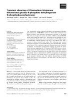
A scheme of glycolipid biosynthesis is shown in Fig. 1. It
is appreciable how the pathways of ganglioside synthesis are
branched by transfection of Sial-T2 (GD3 synthase, clone 2)
or GalNAc-T (GM2 synthase, clone 3) to the wild-type
CHO-K1 cells (CHO-K1 wt). Wild-type CHO-K1 cells
predominantly express the ganglioside GM3, while those
Fig. 1. Glycolipid labelling of CHO-K1 cell clones. Aschematicrep-
resentation of the pathway of glycolipid biosynthesis is shown at the
top of the figure. It is appreciable how the pathways of ganglioside
synthesis are branched following transfection of CMP-NeuAc:GM3
sialyltransferase (Sial-T2) (GD3 synthase, clone 2) or UDP-Gal-
NAc:LacCer/G3/GD3 N-acetylgalactosaminyltransferase (GalNAc-
T) (GM2 synthase, clone 3) to wild-type CHO-K1 cells (CHO-K1 wt)
expressing only the ganglioside, GM3. Also indicated in the scheme is
the enzymatic reaction affected by the glycolipid inhibitor,
D
,
L
-threo-1-
phenyl-2-hexadecanoylamino-3-pyrrolidino-1-propanol-HCl (PPPP).
Wild-type CHO-K1 cells (CHO-K1 wt), cells from clones 2 and 3, and
PPPP-treated wild type CHO-K1 cells (CHO-K1/PPPP) were meta-
bolically labelled with 2 lCi of [
14
C]Gal for 12 h. Lipids were purified
and chromatographed by HPTLC, as previously described [10]. The
positions of co-chromatographed glycolipid standards are indicated.
2430 A. R. Zurita et al. (Eur. J. Biochem. 271) Ó FEBS 2004
stably expressing the Sial-T2 cDNA (clone 2) [17] synthesize
mostly GD3 and GT3, accumulate LacCer and show very
little accumulation of GM3 (Fig. 1). CHO-K1 cells stably
expressing the GalNAc-T cDNA (clone 3) [18], synthesize
the a-series ganglioside GM2 and, to a lesser extent, GM1
because of the constitutive endogenous expression in
these cells of the enzyme involved in the synthesis of GM1
(Fig. 1) [23].
To reduce the content of all glycosphingolipid classes,
wild-type CHO-K1 cells were treated with PPPP, a
competitive inhibitor of ceramide glucosyltransferase and
hence of the synthesis of complex glycolipids (Fig. 1, CHO-
K1/PPPP) [24]. Exposure of cells to 2 l
M
PPPP in the
culture medium for 5 days led to a 95% decrease of GM3
content with respect to control cells [10,18].
EGFr membrane distribution in CHO-K1 cell lines
expressing different gangliosides
Wild-type CHO-K1 cells, and cells from clones 2 and 3, and
wild-type CHO-K1 cells with a generalized decrease in
glycolipid expression (CHO-K1/PPPP)
6
,alltransiently
expressing human EGFr, were treated with 1% Triton-
X-100 at 4 °C and lysates were subjected to continuous
sucrose gradient ultracentrifugation, fractionation, and
detection of EGFr and known protein markers by Western
blotting. Under these conditions, proteins and lipids resist-
ant to 1% (v/v) Triton X-100 extraction, flow at low-density
fractions. As controls, we analysed the behaviour, to Triton
X-100 extraction, of a GEM marker (i.e. a fusion protein
containing a GPI-anchored signal, GPI-YFP), and a non-
GEM marker (i.e. VSVG-CFP). As previously described,
the GEM marker, GPI-YFP, was highly concentrated in
low-density fractions [15,25]. In contrast, VSVG-CFP (the
non-GEM marker) was distributed in higher density
fractions, thus distributing essentially as Triton X-100
soluble proteins (Fig. 2A). EGFr was mainly concentrated
in higher density fractions with essentially the same
distribution pattern in all cell lines, co-distributing with
the Triton X-100 soluble protein, VSVG-CFP (Fig. 2A).
Two nonspecific lower bands were detected with the anti-
EGFr Ig, even in extracts from CHO-K1 cells that were not
transfected with EGFr (Fig. 3C).
It has been described that integrin receptors (e.g. alpha3,
alpha5) are found completely outside GEM after treatment
with 1% (v/v) Triton X-100, but almost exclusively in GEM
when a lower concentration (0.25–0.5%) of Triton X-100 is
used [26], suggesting a relatively weak interaction with
membrane components. Taking into account that growth
factor receptors, including EGFr, and integrin receptors are
functionally associated [27,28], we explored the behaviour of
EGFr to lower Triton X-100 concentrations. Basically, and
in contrast to the behaviour of integrin receptors, we found
almost the same gradient distribution of EGFr at 0.25, 0.5
and 1% (v/v) Triton X-100 (results not shown). A
representative pattern of protein distribution is shown in
Fig. 2B. Results from this experiment strongly suggest that
changes in the composition of endogenous gangliosides
did not affect the distribution of EGFr on membranes of
CHO-K1 cell clones.
Next, we set out to confirm these results, studying the
solubility/insolubility of EGFr to extraction with cold
Triton X-100 by velocity sedimentation. Cell homogenates
from wild-type CHO-K1 cells, transiently expressing EGFr,
were extracted with Triton X-100 at 4 °Candthen
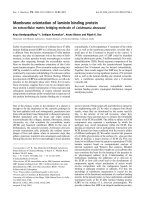
Fig. 2. Continuous sucrose gradient analysis of epidermal growth factor
receptor (EGFr) in CHO-K1 cell lines. (A) CHO-K1 cell clones tran-
siently expressing human EGFr, glycosylphosphatidylinositol-yellow
fluorescence protein (GPI-YFP), or vesicular stomatitis virus glyco-
protein-cyan fluorescence protein (VSVG-CFP) were lysed in lysis
buffer at 4 °C for 1 h and centrifuged (10 h, 150 000 g,4°C) on
continuous sucrose gradients (5–30%). Twelve fractions were collected
from the bottom of the sucrose density gradient using a fraction col-
lector. Proteins were resolved by electrophoresis through 8% (for
EGFr analysis) or 10% (for GPI-YFP and VSVG-CFP) SDS–PAGE
and analysed by Western blot. The antibodies used were anti-EGFr
andanti-GFPtorevealGPI-YFPandVSVG-CFP,respectively.The
positions (molecular masses) of recombinant proteins are indicated.
(B) Protein profile of the gradient, visualized by Ponceau S staining of
the nitrocellulose membrane.
Ó FEBS 2004 EGF receptors distribution in cell membranes (Eur. J. Biochem. 271) 2431
centrifuged (1 h, 100 000 g,4°C). Proteins from soluble
and insoluble fractions were detected by Western blot with
the appropriate antibody. EGFr was totally soluble to
Triton X-100 extraction (Fig. 3A). As expected, the GEM
marker (GPI-YFP) was 68% insoluble, whereas the non-
GEM marker (VSVG-CFP) was less than 20% insoluble
(Fig. 3A).
Finally, we explored the grade of association of EGFr on
the cell surface of wild-type CHO-K1 cells by using the
chemical cross-linking agent, BS
3
, which is membrane
impermeable and possesses a spacer arm of 1.14 nm.
Wild-type CHO-K1 cells, transiently expressing EGFr and
subjected to BS
3
, showed a low (less than 5%) cross-linking
efficiency [calculated as the percentage of cross-linked
molecules (dimer plus oligomer) with respect to total
protein], as expected for a homogenous distribution of a
non-GEM protein marker (compare with the VSVG-CFP
cross-linking efficiency of 10%; Fig. 3B). On the other
hand, GPI-CFP, a GEM protein marker, showed a cross-
linking efficiency of > 40%
7
, represented by dimer and
oligomer species [15]. As a control, and to demonstrate that
BS
3
acts on active EGFr, CHO-K1 cells expressing the
receptor were incubated with buffer, or with 100 n
M
EGF,
for 1 h at 4 °C, treated with the cross-linking agent BS
3
,
lysed and analysed by SDS/PAGE. It was found that EGF
was able to stimulate the appearance of EGFr homodimer
(Fig. 3C). Additionally, the EGFr homodimer was absent
when the receptor was not stimulated with EGF. That
EGFr has a homogeneous distribution (a scarce association
with microdomains) in membranes from CHO-K1 cells is
consistent with the behaviour of the EGFr in both sucrose
gradient centrifugation and solubilization by nonionic
detergent, indicated above. Although we show results from
solubility/insolubility and cross-linking experiments only for
wild type CHO-K1 cells, the behaviour of EGFr in all other
clones was essentially the same (results not shown).
Tyrosine-phosphorylated EGFr membrane distribution
after stimulation with EGF
Wild-type CHO-K1 cells, and cells from clone 2 (Fig. 1),
were transfected to transiently express EGFr and cultured in
the absence of fetal bovine serum for 12 h. Then, the cells
were incubated with EGF (100 n
M
)for5min,lysedwith
cold Triton X-100, centrifuged in a sucrose gradient and the
activated EGF receptor was analysed, in all fractions, by
Western blot using antibodies recognizing phosphorylated
EGFr on tyrosines located at positions 845, 992 and 1068.
The activated EGF receptor
8
was found to be distributed
similarly in sucrose gradients from both wild-type CHO-K1
cells (GM3
+
) and cells from clone 2 (GD3
+
) (Fig. 4). It
should be emphasized that in these experiments the total
amount of protein and EGFr expression levels in both cell
lines were not necessarily comparable. The membrane
distribution of the active EGFr fits well with that of the total
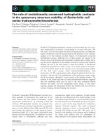
Fig. 3. Detergent solubility and membrane distribution of epidermal
growth factor receptor (EGFr), glycosphingolipid-enriched microdomain
(GEM) and non-GEM markers. (A) Homogenates from wild-type
CHO-K1 cells transiently expressing human EGFr, glycosylphospha-
tidylinositol-yellow fluorescence protein (GPI-YFP) (GEM marker)
or vesicular stomatitis virus glycoprotein-cyan fluorescence protein
(VSVG-CFP) (non-GEM marker) were extracted with Triton X-100 at
4 °C and then ultracentrifuged (1 h, 100 000 g,4°C). Proteins from
soluble (S) and insoluble (I) fractions were resolved by SDS/PAGE
and probed with the appropriate antibody. (B) Detection of protein
clusters by chemical cross-linking with bis(sulfosuccinimidil)suberato
(BS
3
) in membranes from CHO-K1 cells. Wild-type CHO-K1 cells,
transiently expressing EGFr, GPI-YFP and VSVG-CFP, were sub-
jected to cross-linking with 0.5 m
M
BS
3
. Protein extracts were resolved
in SDS/PAGE and detected by Western blot. (C) Chemical cross-
linking of EGFr after EGF stimulation. To demonstrate that BS
3
acts
on active EGFr, CHO-K1 cells expressing the receptor (lines 2–4) were
incubated with buffer (lanes 2 and 3) or with 100 n
M
EGF (lane 4).
Then, samples were cross-linked with BS
3
(lanes 3 and 4) or incubated
with buffer alone (line 2). As a control, an extract from mock-trans-
fected cells was run in lane 1. Protein extracts were resolved in SDS–
PAGE and detected by Western blot. Positions of monomers (m),
dimers (d) or oligomers (o) are indicated.
2432 A. R. Zurita et al. (Eur. J. Biochem. 271) Ó FEBS 2004
EGFr (Fig. 2A), suggesting that there were no changes
of membrane distribution associated with its activation
status.
EGFr and GD3 localization in CHO-K1 cell membranes
Having demonstrated a converse segregation of GD3 and
EGFr, by biochemical means, in membranes from clone 2
cells (this work) [15,16], we next investigated the spatial
localization of GD3 and EGFr in both its active and
inactive state. Studies were carried out by confocal micros-
copy immunofluorescence, using the monoclonal antibody,
R24, to detect GD3 and two anti-EGFr Igs that recognize
the extracellular or intracellular domains of the receptor.
GD3 was mainly detected in the plasma membrane,
showing a distribution in patches, while EGFr was observed
both in plasma membranes and intracellular membranes
(Fig. 5). A comparison between GD3 and EGFr membrane
distribution revealed a minor separation (Fig. 5A–D). To
better define the localization of GD3 and EGFr in the
plasma membrane of CHO-K1 cells in the absence of EGF,
confocal analysis was performed using formaldehyde-fixed
cells (nonpermeabilized cells) labelled with an anti-EGFr Ig
recognizing the extracellular domain. In addition, to ease
the separation between GEM and other parts of the plasma
membrane, the focal plane was mainly adjusted through the
top of the cell, allowing the visualization of larger plasma
membrane areas and the identification of small membrane
domains (Fig. 5E–H). These data clearly confirmed that
EGFr and GD3 are colocalized, to some extent, on the
plasma membrane of CHO-K1 cells.
Interestingly, when the cells were stimulated with EGF
for 10 min, a clear endocytosis of the EGFr was observed
but, under this condition, GD3 remains at the cell surface
(Fig. 5I–L). Altogether, these results showed that GD3 is
mainly expressed on the plasma membrane of cells from
clone 2, and that GD3 and EGFr co-localized, to some
extent, only in the absence of EGF stimulation, while, upon
addition of EGF, a clear separation of these two membrane
components was observed.
Endogenous ErbB2 membrane distribution in wild-type
CHO-K1 cells (GM3
+
) and cells from clone 2 (GD3
+
)
Next, we studied how other members of the ErbB family
would behave in terms of membrane distribution, as shown
previously for EGFr (ErbB1). To achieve this, we investi-
gated the endogenous expression, in CHO-K1 cells, of the
orphan receptor, ErbB2, the preferred heteroassociation
partner of all other ErbB proteins. ErbB2 expression was
analysed by Western blot with an antibody directed to its
intracellular domain, both in wild-type and human EGFr-
expressing CHO-K1 cells. First, we analysed the heterolo-
gous expression of EGFr in CHO-K1 cells. As expected,
EGFr is expressed in CHO-K1 cells as a functional protein
of 170 kDa [10]. Endogenous EGFr expression in CHO-K1
cells was below the limit of detection (Fig. 6A, upper panel).
Nonspecific lower bands were also detected with the anti-
EGFr Ig (see also Fig. 3C). Additionally, ErbB2 was
detected as a band of 185 kDa in both wild-type and EGFr-
expressing CHO-K1 cells (Fig. 6A, upper panel). As control
of protein loading, we analysed the constitutive expression
of p53. No substantial differences were observed in any of
the lanes analysed (Fig. 6A, lower panel). Next, we inves-
tigated the membrane distribution of endogenous ErbB2 by
sucrose gradient both in wild-type CHO-K1 cells (GM3
+
)
and in cells from clone 2 (GD3
+
). As also observed for
EGFr (Fig. 2A), there was no difference in the distribution
pattern of ErbB2 at high-density fractions (Triton-X-100-
soluble proteins) in wild-type CHO-K1 cells (GM3
+
)and
cells from clone 2 (GD3
+
) (Fig. 6B,C). However, it should
be noted that in both cell lines, a significant fraction of
ErbB2 (27%) was associated with low-density fractions
(fractions 7–12, see the distribution of GEM and non-GEM
markers in Fig. 2A), which probably represent a fraction
associated with GEM. On these fractions, a small shift in
Fig. 4. Tyrosine-phosphorylated epidermal growth factor receptor (EGFr) membrane distribution after EGF stimulation. Wild-type CHO-K1 cells
(CHO-K1 wt, GM3
+
) and cells from clone 2 (GD3
+
), transiently expressing human EGFr, were maintained in serum-free medium for 12 h. EGF
(100 n
M
) was added to the medium and, after 10 min, cells were lysed and centrifuged (10 h, 150 000 g,4°C) on continuous sucrose gradients
(5–30%). Twelve fractions were collected from the bottom of the sucrose density gradient. Proteins were resolved by electrophoresis through 8%
SDS–PAGE and probed with anti-(phospho-EGFr) (P-EGFr) Ig, which recognizes phosphorylated tyrosines (Tyr845, 992 and 1068), to detect the
active receptor.
Ó FEBS 2004 EGF receptors distribution in cell membranes (Eur. J. Biochem. 271) 2433
ErbB2 distribution was observed at the top of the gradient.
While the ErbB2 receptor from wild-type CHO-K1 cells
(CHO-K1 wt) was found mostly in fraction 12, the ErbB2
receptor from clone 2 cells was concentrated mainly in
fraction 11. In line with the results of ErbB2 distribution
in sucrose gradients, solubility/insolubility analysis of
ErbB2 to cold Triton X-100 extraction by velocity sedi-
mentation showed that 15% of the receptor was insoluble
(data not shown).
Discussion
The main goal of this work was to investigate the possibility
that changes in the expression of gangliosides could
modulate the membrane distribution of EGFr members
and thereby regulate their function. By studying the
solubility/insolubility of EGFr to extraction using nonionic
detergents, we demonstrated that changes in the composi-
tion of endogenous gangliosides did not significantly affect
the distribution of EGFr on the membranes of CHO-K1 cell
lines. In all clones analysed, this receptor behaved as a
soluble molecule to extraction with cold Triton X-100,
indicating that it is mainly excluded from GEM. Interest-
ingly, the behaviour of the receptor to extraction with cold
Triton X-100 was independent of its activation state because
binding of EGF to EGFr did not affect its membrane
distribution. The analysis of EGFr clusterization on the
plasma membrane of CHO-K1 cells, by using a chemical
cross-linking approach, is compatible with the notion of a
homogeneous membrane distribution of the growth factor
receptor.
Our initial evidence demonstrated the behaviour of
different gangliosides expressed in CHO-K1 cells to extrac-
tion with cold Triton X-100 in order to investigate their
association with GEM. The results revealed that the
majority of plasma membrane GD3 reside in GEM.
Confirming previous work, we found that after extraction
with nonionic detergent, GD3 was associated with low
Fig. 5. Epidermal growth factor receptor (EGFr) and GD3 localization in CHO-K1 cell membranes. Cells from clone 2 (GD3
+
)weretransiently
transfected to express human EGFr and maintained in serum-free medium for 12 h before incubation for 10 min at 37 °C in the absence (A–H) or
presence (I–L) of 100 n
M
EGF in the cell culture medium. EGFr expression was analysed by confocal microscopy immunofluorescence in acetone-
fixed (A–D, I–L) or formaldehyde-fixed (E–H) cells using a polyclonal anti-(intracellular domain) Ig (A and I) or a monoclonal (mouse IgG2) anti-
(extracellular domain) Ig (E) of EGFr, and rhodamine-conjugated donkey anti-rabbit IgG (A and I) or fluorescein-conjugated rat anti-mouse IgG2
(E) as secondary antibodies (pseudo-coloured red). GD3 was detected using the monoclonal antibody R24 (mouse IgG3) as primary antibody and
Alexa 488-conjugated goat anti-mouse IgG as secondary antibody (B and J, green). To reveal GD3 expression in cells shown in F, coverslips were
sequentially incubated with R24, goat anti-mouse IgG3 and finally with Alexa 546-conjugated donkey anti-goat Ig (pseudo-coloured green). C, G
andKaremergedimagesfromAandB,EandFandIandJ,respectively.AnenlargementoftheboxedareasinC,GandKareshowninD,Hand
L, respectively. Images shown in this figure are single xy confocal sections. The focal plane in E–H was adjusted through the top of the cell. Scale
bars: A, 20 lm (for A–C and I–K); E, 10 lm (for E–G, D and L); H, 3 lm.
2434 A. R. Zurita et al. (Eur. J. Biochem. 271) Ó FEBS 2004
buoyant density fractions in sucrose gradients, pelleted after
ultracentrifugation and expressed as detergent-resistant
patches on the plasma membrane of CHO-K1 cells
[15,16]. Additionally in this work, using confocal microsco-
py analysis we demonstrated that EGFr co-localizes only to
a minor extent with the disialoganglioside GD3, even after
stimulation with EGF. Taken together, these results make it
less probable that there is a direct effect of GD3 on EGFr
tyrosine phosphorylation [10] and suggests an indirect
effect, perhaps through its interaction with other mem-
brane-associated proteins. In this regard, it was recently
described that overexpression of GM3 in cells of the human
keratinocyte-derived cell line, SCC12F2, inhibited EGFr
tyrosine phosphorylation, while it did not affect EGFr
membrane distribution but shifted caveolin-1 to the deter-
gent-soluble, EGFr-containing region [29]. The authors
suggested that the GM3-induced shift of caveolin-1 mem-
brane distribution is critical for its EGFr-induced phos-
phorylation that is associated with the suppression of EGFr
activation.
The lack of EGFr in low buoyant density fractions in
sucrose gradients, and the complete solubilization of the
receptor to Triton X-100 extraction, strongly suggested that
EGFr expressed in CHO-K1 cells is mainly excluded from
GEM. However, our immunofluorescence microscopy
experiments showed that GD3 and EGFr co-localized, to
some extent, at the plasma membrane. A possible explan-
ation for the nondetectable EGFr in GEM fractions from
sucrose gradients is that the GEM-associated EGFr might
besensitivetoextractionwithTritonX-100,evenwhenused
at different concentrations (0.25, 0.5 and 1%), suggesting a
relatively weak interaction of EGFr with Triton X-100-
insoluble domains. These observations are in agreement
with a study in HeLa cells, where it was shown that most of
the EGFr is localized in lipid rafts containing the ganglio-
side GM1 and is sensitive to Triton-X-100 extraction but
insensitive to extraction with a less disrupting nonionic
detergent, Brij 58 [30].
The EGF receptor family comprises four members –
EGFr (ErbB1), ErbB2 (HER2 or Neu), ErbB3, and
ErbB4 [31]. The orphan receptor, ErbB2, is the preferred
heteroassociation partner of all other ErbB proteins,
enhancing signalling potency by its strong latent kinase
activity [32,33]. CHO cells express endogenous ErbB2, but
no other members of the ErbB family [34]. In SKBR-3
(a breast tumour cell line), ErbB2 was found to co-localize
with lipid rafts, identified by the GM1-binding B subunit
of cholera toxin [35]. Taking all these observations
together, we attempted to explore whether the effect of
GD3 on EGFr phosphorylation might be achieved
through the modulation of ErbB2 membrane distribution,
its potential heteroassociation partner. Clearly, our results
show that ErbB2 is expressed at a similar level in wild-
type CHO-K1 cells (GM3
+
) and in cells from clone 2
Fig. 6. Detergent solubility and continuous sucrose gradient analysis of
epidermal growth factor receptor 2 (ErbB2). (A) Homogenates from
wild-type CHO-K1 cells (CHO-K1) and CHO-K1 cells transiently
expressing human EGFr (CHO-K1 EGFr) were resolved in SDS–
PAGE (8%) and probed with anti-EGFr or anti-ErbB2 Ig. The
positions of EGFr (170 kDa) and ErbB2 (185 kDa) with molecular
masses are indicated. Then, antibodies were removed by treatment of
themembranewith1
M
NaOH for 5 min and p53 was determined by
Western blot. (B) Wild-type CHO-K1 cells (CHO-K1 wt) and cells
from clone 2 were lysed at 4 °C for 1 h and centrifuged (10 h,
150 000 g,4°C) on a continuous sucrose gradient (5–30%). Twelve
fractions were collected from the bottom of the sucrose density gra-
dient. Proteins were resolved by electrophoresis through 8% SDS/
PAGE, and ErbB2 was detected by Western blot with anti-ErbB2 Ig.
The position and molecular mass of ErbB2 (185 kDa) is indicated. (C)
A quantitative analysis of Western blots from Figs 6B and 2A (CHO-
K1 wt and clone 2 cells) was carried out to compare EGFr and ErbB2
gradient distribution. The receptor level in each fraction was normal-
ized to total receptor expression. White bars, EGFr; black bars, ErbB2.
Ó FEBS 2004 EGF receptors distribution in cell membranes (Eur. J. Biochem. 271) 2435
(GD3
+
). In addition, no significant changes in the
biochemical behaviour of ErbB2 to extraction using a
nonionic detergent were observed, suggesting that the
modulation of EGFr phosphorylation by endogenously
expressed GD3 does not occur because of a change in the
membrane distribution of ErbB2. It was also found that
an important fraction of the endogenous ErbB2 was
associated with low-density fractions after extraction with
cold Triton X-100 and sucrose gradient analysis. This
fraction of the ErbB2 receptor represents receptor mole-
cules, associated with GEM, in the membranes of CHO-
K1 cells. Interestingly, it was recently reported that
membrane clusters with a high concentration of ErbB2,
which is regulated by lipid rafts, strongly influence
homoassociation and the ligand-independent activation
of ErbB2 [35]. Considering that the orphan ErbB2 is the
only member of the ErbB family expressed in CHO-K1
cells, its activation by homodimerization is highly likely to
occur in plasma membrane clusters (GEMs) of this cell
line.
In conclusion, our studies demonstrate that most of the
EGFrs localize outside GEM in wild-type CHO-K1 cell
(GM3
+
) membranes. Contrary to results showing that
addition or depletion of cholesterol (another membrane
component that regulates GEM formation) can alter the
membrane distribution of EGFr [36,37], qualitative and
quantitative changes in ganglioside expression do not affect
the membrane distribution of EGFr and ErbB2. However,
we cannot entirely rule out the possibility that fine-tuning
mechanisms might be operating in the membrane distribu-
tion of EGFr. An interesting possibility to explain EGFr
regulation by gangliosides, particularly in GD3-expressing
CHO-K1 cells, is that gangliosides might regulate the
activity of ganglioside-stimulated receptor tyrosine phos-
phatases [38], or enhance the co-localization of EGFr with
its phosphatase, as recently suggested [7]. In this sense, our
work provides the basis for testing these possibilities and
gaining further insight into the regulation of the ErbB
family members.
Acknowledgements
This work was supported, in part, by Grants from the SECyT-
Universidad Nacional de Co
´
rdoba, ÔRamon Carrillo-Arturo On
˜
ativiaÕ
from Ministerio de Salud de la Nacio
´
n Argentina (2001 to J.L.D. and
2003 to N.P.K.), the International Society for Neurochemistry (Special
ISN One-Time Fund) and Fundacio
´
n Antorchas (Grant N°14116-112
toJ.L.D.andinpartbyGrantN°14022-10 to N.P.K.). We thank
F. Cerban and A. Gruppi for their donation of anti-mouse IgG subtype
antibodies and C. Alvarez for valuable reagents (Departamento de
Bioquı
´
mica Clı
´
nica, Facultad de Ciencias Quı
´
mica, UNC, Argentina).
The authors also thank G. Schachner and S. Deza for technical
assistance with the cell culture and C. Mas for excellent assistance with
confocal microscopy and image analysis. A.R.Z. and P.M.C. are
recipients of CONICET (Argentina) Fellowships. J.L.D. and N.P.K.
are Career Investigators of CONICET (Argentina).
References
1. Hakomori, S. & Igarashi, Y. (1995) Functional role of glyco-
sphingolipids in cell recognition and signaling. J. Biochem.
(Tokyo) 118, 1091–1103.
2. Fredman, P., Hedberg, K. & Brezicka, T. (2003) Gangliosides as
therapeutic targets for cancer. Biodrugs 17, 155–167.
3. Mutoh, T., Tokuda, A., Miyadai, T., Hamaguchi, M. & Fujiki, N.
(1995) Ganglioside GM1 binds to the Trk protein and regulates
receptor function. Proc. Natl Acad. Sci. USA 92, 5087–5091.
4. Fukumoto, S., Mutoh, T., Hasegawa, T., Miyazaki, H., Okada,
M., Goto, G., Furukawa, K. & Urano, T. (2000) GD3 synthase
gene expression in PC12 cells results in the continuous activation
of TrkA and ERK1/2 and enhanced proliferation. J. Biol. Chem.
275, 5832–5838.
5. Allende, M.L. & Proia, R.L. (2002) Lubricating cell signaling
pathways with gangliosides. Curr. Opin. Struct. Biol. 12, 587–592.
6. Yamashita, T., Hashiramoto, A., Haluzik, M., Mizukami, H.,
Beck, S., Norton, A., Kono, M., Tsuji, S., Daniotti, J.L., Werth,
N., Sandhoff, R., Sandhoff, K. & Proia, R.L. (2003) Enhanced
insulin sensitivity in mice lacking ganglioside GM3. Proc. Natl
Acad. Sci. USA 100, 3445–3449.
7. Miljan, E.A. & Bremer, E.G. (2002) Regulation of growth factor
receptors by gangliosides. Sci. STKE 160,RE15
9
.
8. Yates, A.J., Saqr, H.E. & Van Brocklyn, J. (1995) Ganglioside
modulation of the PDGF receptor. A model for ganglioside
functions. J. Neurooncol. 24, 65–73.
9. Mitsuda, T., Furukawa, K., Fukumoto, S., Miyazaki, H. &
Urano, T. (2002) Overexpression of ganglioside GM1 results in the
dispersion of platelet-derived growth factor receptor from glyco-
lipid-enriched microdomains and in the suppression of cell growth
signals. J. Biol. Chem. 277, 11239–11246.
10. Zurita, A.R., Maccioni, H.J. & Daniotti, J.L. (2001) Modulation
of epidermal growth factor receptor phosphorylation by
endogenously expressed gangliosides. Biochem. J. 355, 465–472.
11. Simons, K. & Ikonen, E. (1997) Functional rafts in cell mem-
branes. Nature 387, 569–572.
12. Prinetti, A., Iwabuchi, K. & Hakomori, S. (1999) Glyco-
sphingolipid-enriched signaling domain in mouse neuroblastoma
Neuro2a cells. Mechanism of ganglioside-dependent neurito-
genesis. J. Biol. Chem. 274, 20916–20924.
13. Simons, K. & Toomre, D. (2000) Lipid rafts and signal trans-
duction. Nat. Rev. Mol. Cell Biol. 1, 31–39.
14. Galbiati, F., Razani, B. & Lisanti, M.P. (2001) Emerging themes
in lipid rafts and caveolae. Cell 106, 403–411.
15. Crespo, P.M., Zurita, A.R. & Daniotti, J.L. (2002) Effect of
gangliosides on the distribution of a glycosylphosphatidylinositol-
anchored protein in plasma membrane from Chinese hamster
ovary-K1 cells. J. Biol. Chem. 277, 44731–44739.
16. Crespo, P.M., Zurita, A.R., Giraudo, C.G., Maccioni, H.J.F. &
Daniotti, J.L. (2004) Ganglioside glycosyltransferases and newly
synthesized gangliosides are excluded from detergent-insoluble
complexes of Golgi membranes. Biochem. J. 377, 561–568.
17.Daniotti,J.L.,Martina,J.A.,Giraudo,C.G.,Zurita,A.R.&
Maccioni, H.J. (2000) GM3 alpha2,8-sialyltransferase (GD3
synthase): protein characterization and sub-Golgi location in
CHO-K1 cells. J. Neurochem. 74, 1711–1720.
18. Giraudo, C.G., Rosales Fritz, V.M. & Maccioni, H.J. (1999)
GA2/GM2/GD2 synthase localizes to the trans-Golgi network of
CHO-K1 cells. Biochem. J. 342, 633–640.
19. Friedrichson, T. & Kurzchalia, T.V. (1998) Microdomains of
GPI-anchored proteins in living cells revealed by crosslinking.
Nature 394, 802–805.
20.Cochet,C.,Kashles,O.,Chambaz,E.M.,Borrello,I.,King,
C.R. & Schlessinger, J. (1988) Demonstration of epidermal
growth factor-induced receptor dimerization in living cells using a
chemical covalent cross-linking agent. J. Biol. Chem. 263, 3290–
3295.
21. Sorkina, T., Huang, F., Beguinot, L. & Sorkin, A. (2002) Effect of
tyrosine kinase inhibitors on clathrin-coated pit recruitment and
2436 A. R. Zurita et al. (Eur. J. Biochem. 271) Ó FEBS 2004
internalization of epidermal growth factor receptor. J. Biol. Chem.
277, 27433–27441.
22. Daniotti, J.L., Zurita, A.R., Trindade, V.M. & Maccioni, H.J.
(2002) GD3 expression in CHO-K1 cells increases growth rate,
induces morphological changes, and affects cell–substrate inter-
actions. Neurochem. Res. 27, 1421–1429.
23. Rosales Fritz, V.M., Daniotti, J.L. & Maccioni, H.J. (1997)
Chinese hamster ovary cells lacking GM1 and GD1a synthesize
gangliosidesupontransfectionwithhumanGM2synthase.
Biochim. Biophys. Acta 1354, 153–158.
24. Li, R., Manela, J., Kong, Y. & Ladisch, S. (2000) Cellular gang-
liosides promote growth factor-induced proliferation of fibro-
blasts. J. Biol. Chem. 275, 34213–34223.
25. Brown, D.A. & Rose, J.K. (1992) Sorting of GPI-anchored pro-
teins to glycolipid-enriched membrane subdomains during trans-
port to the apical cell surface. Cell 68, 533–544.
26. Kazui, A., Ono, M., Handa, K. & Hakomori, S. (2000) Glyco-
sylation affects translocation of integrin Src, and caveolin into or
out of GEM. Biochem. Biophys. Res. Commun. 273, 159–163.
27. Wang, F., Weaver, V.M., Petersen, O.W., Larabell, C.A., Dedhar,
S., Briand, P., Lupu, R. & Bissell, M.J. (1998) Reciprocal inter-
actions between beta1-integrin and epidermal growth factor
receptor in three-dimensional basement membrane breast cultures:
a different perspective in epithelial biology. Proc. Natl Acad. Sci.
USA 95, 14821–14826.
28. Yamada, K.M. & Even-Ram, S. (2002) Integrin regulation of
growth factor receptors. Nat. Cell Biol. 4, E75–E76.
29. Wang, X.Q., Sun, P. & Paller, A.S. (2002) Ganglioside induces
caveolin-1 redistribution and interaction with the epidermal
growth factor receptor. J. Biol. Chem. 277, 47028–47034.
30. Roepstorff, K., Thomsen, P., Sandvig, K. & van Deurs, B. (2002)
Sequestration of epidermal growth factor receptors in non-
caveolar lipid rafts inhibits ligand binding. J. Biol. Chem. 277,
18954–18960.
31. Yarden, Y. & Sliwkowski, M.X. (2001) Untangling the ErbB
signalling network. Nat. Rev. Mol. Cell Biol. 2, 127–137.
32.Karunagaran,D.,Tzahar,E.,Beerli,R.R.,Chen,X.,Graus-
Porta,D.,Ratzkin,B.J.,Seger,R.,Hynes,N.E.&Yarden,Y.
(1996) ErbB-2 is a common auxiliary subunit of NDF and EGF
receptors: implications for breast cancer. EMBO J. 15, 254–264.
33. Schlessinger, J. (2000) Cell signaling by receptor tyrosine kinases.
Cell 103, 211–225.
34. Chausovsky, A., Waterman, H., Elbaum, M., Yarden, Y., Geiger,
B. & Bershadsky, A.D. (2000) Molecular requirements for the
effect of neuregulin on cell spreading, motility and colony orga-
nization. Oncogene 19, 878–888.
35. Nagy,P.,Vereb,G.,Sebestyen,Z.,Horvath,G.,Lockett,S.J.,
Damjanovich, S., Park, J.W., Jovin, T.M. & Szollosi, J. (2002)
Lipid rafts and the local density of ErbB proteins influence the
biological role of homo- and heteroassociations of ErbB2. J. Cell
Sci. 115, 4251–4262.
36. Ringerike, T., Blystad, F.D., Levy, F.O., Madshus, I.H. & Stang,
E. (2002) Cholesterol is important in control of EGF receptor
kinase activity but EGF receptors are not concentrated in caveo-
lae. J. Cell Sci. 115, 1331–1340.
37. Pike, L.J. & Casey, L. (2002) Cholesterol levels modulate EGF
receptor-mediated signaling by altering receptor function and
trafficking. Biochemistry 41, 10315–10322.
38. SuarezPestana,E.,Tenev,T.,Gross,S.,Stoyanov,B.,Ogata,M.
& Bohmer, F.D. (1999) The transmembrane protein tyrosine
phosphatase RPTPsigma modulates signaling of the epidermal
growth factor receptor in A431 cells. Oncogene 18, 4069–4079.
Ó FEBS 2004 EGF receptors distribution in cell membranes (Eur. J. Biochem. 271) 2437