Báo cáo khoa học: Affinity of S100A1 protein for calcium increases dramatically upon glutathionylation docx
Bạn đang xem bản rút gọn của tài liệu. Xem và tải ngay bản đầy đủ của tài liệu tại đây (246.89 KB, 9 trang )
Affinity of S100A1 protein for calcium increases
dramatically upon glutathionylation
Graz_ yna Goch, Sergiusz Vdovenko, Hanna Kozłowska and Andrzej Bierzyn
˜
ski
Institute of Biochemistry and Biophysics, Polish Academy of Sciences, Poland
Calcium ions are one of the most important messen-
gers and regulate numerous vital biological processes.
A crucial role in calcium signal transduction is played
by EF-hand proteins, which upon incorporating cal-
cium change their conformation, exposing hydrophobic
patches to which target proteins bind.
S100 is a subfamily of EF-hand proteins regulating
an amazingly wide variety of biological processes in
either a calcium-dependent or calcium-independent
manner [1–3]. A typical S100 protein is composed of
two subunits, very strongly associated with each other,
and each containing two calcium-binding loops [4–9].
The glutamate residue at the C-terminal position in
both loops plays a crucial role in calcium binding.
To elucidate the calcium-dependent biological activit-
ies of S100 proteins it is of the utmost importance that
their microscopic calcium-binding constants be deter-
mined at physiological conditions. The results of this
study clearly illustrate this point. Only for calbindin D
9k
have such measurements been made [10–12]. These
results, although important, are of limited value in
understanding the calcium-binding mechanism typical
of S100 proteins because of the unique structural fea-
tures of calbindin D
9k
: it is a monomer, not a dimer,
Keywords
calcium binding; EF-hand proteins;
glutathionylation; S100A1
Correspondence
A. Bierzyn˜ ski, Institute of Biochemistry and
Biophysics, Polish Academy of Sciences, ul.
Pawin˜ skiego 5 A, 02–106 Warsaw, Poland
Fax: +48 22 823 71 94
Tel: +48 22 592 23 71
E-mail:
(Received 14 January 2005, revised 14
March 2005, accepted 22 March 2005)
doi:10.1111/j.1742-4658.2005.04680.x
S100A1 is a typical representative of a group of EF-hand calcium-binding
proteins known as the S100 family. The protein is composed of two a sub-
units, each containing two calcium-binding loops (N and C). At physiologi-
cal pH (7.2) and NaCl concentration (100 mm), we determined the
microscopic binding constants of calcium to S100A1 by analysing the
Ca
2+
-titration curves of Trp90 fluorescence for both the native protein
and its Glu32 fi Gln mutant with an inactive N-loop. Using a chelator
method, we also determined the calcium-binding constant for the S100A1
Glu73 fi Gln mutant with an inactive C-loop. The protein binds four
calcium ions in a noncooperative way with binding constants of K
1
¼
4±2· 10
3
m
)1
(C-loops) and K
2
10
2
m
)1
(N-loops). Only when both
loops are saturated with calcium does the protein change its global confor-
mation, exposing to the solvent hydrophobic patches, which can be detec-
ted by 2-p -toluidinylnaphthalene-6-sulfonic acid – a fluorescent probe of
protein-surface hydrophobicity. S-Glutathionylation of the single cysteine
residue (85) of the a subunits leads to a 10-fold increase in the affinity of
the protein C-loops for calcium and an enormous – four orders of magni-
tude – increase in the calcium-binding constants of its N-loops, owing to a
cooperativity effect corresponding to DDG ¼ )6 ± 1 kcalÆmol
)1
. A similar
effect is observed upon formation of the mixed disulfide with cysteine and
2-mercaptoethanol. The glutathionylated protein binds TRTK-12 peptide
in a calcium-dependent manner. S100A1 protein can act, therefore, as a
linker between the calcium and redox signalling pathways.
Abbreviations
Br
2
-BAPTA, 5,5¢-dibromo-1,2-bis(o-aminophenoxy)-ethane-N,N,N¢,N¢-tetraacetic acid; 5-NBAPTA, 5-nitro-1,2-bis(o-aminophenoxy)-ethane-
N,N,N¢,N¢-tetraacetic acid; TNS, 2-p-toluidinylnaphthalene-6-sulfonic acid.
FEBS Journal 272 (2005) 2557–2565 ª 2005 FEBS 2557
and it is a calcium buffer, not a regulatory protein, so its
structure does not change upon metal binding [13,14].
Therefore, we decided to measure, at physiological
pH (7.2) and NaCl concentration (0.1 m), the micro-
scopic calcium-binding constants of S100A1 protein,
which is in every respect a typical representative of the
family. This protein, and its close homologue S100B,
have been the subject of numerous intensive studies
since their discovery in 1965 [15], and they are by far
the best known members of the S100 family.
An accidental discovery that mixed disulfide forma-
tion between the S100a subunit and 2-mercaptoethanol
results in a dramatic increase in the affinity of the
S100A1 protein for calcium led us to the supposition
that glutathionylation or cysteinylation of the single
cysteine residue of a subunits (Cys85) – processes that
can occur in vivo – may also have a similar effect.
Therefore, the second goal of this study was to deter-
mine the binding constants and cooperativity of Ca
2+
binding to mixed disulfides of S100A1 with glutathi-
one, cysteine, and, for comparison, with 2-mercapto-
ethanol.
To separately study calcium binding to the C- and
N-terminal loops of the S100A1 molecule and deter-
mine their microscopic binding constants, we used
Glu32 fi Gln and Glu73 fi Gln mutants of the pro-
tein in which the calcium-binding activities of the
N- and C-loops, respectively, are switched off or, at
least, strongly reduced [16].
Results
Fluorescence properties of S100A1, its mutants
and derivatives
The fluorescence spectrum of S100A1 is dominated by
the fluorescence of the single tryptophan residues in its
subunits (Trp90) with the maximum at 346 nm. For all
oxidized forms of the protein a 4 nm batochromic shift
in fluorescence is observed. Fluorescence quantum
yields of all apo species are listed in Table 1. They are
affected in various ways by coordination with metals
(Fig. 1). Neither the E32 fi Q (Table 1) mutation nor
E73 fi Q (data not shown) has a measurable effect on
the fluorescence signal intensities of either apo S100A1
protein or its mixed disulfides with 2-mercaptoethanol
and glutathione.
Calcium binding to the reduced and oxidized
forms of the E32Q mutant
The fluorescence signal of E32Q (S100A1 mutant with
a nonactive N-binding loop) increases in the presence
of Ca
2+
ions. Its titration curve (Fig. 2) can be des-
cribed [17] using a simple model assuming that quite
independently, i.e. without any cooperativity effects,
each a subunit of the mutated protein binds only
one calcium ion with the binding constant K
1
¼
Table 1. Fluorescence efficiency U, relative changes in fluorescence signals after binding of the first (f
1
) and second (f
2
) calcium ion, and
macroscopic Ca
2+
-binding constants to S100A1, its E32Q mutants and their mixed disulfides.
Protein U apo K
1
[M
)1
] f
1
K
2
[M
)1
]f
2
E32Q reduced 0.053 4 ± 2 · 10
3
1.20 ± 0.04
E32Q)2-mercaptoethanol 0.033 7.6 ± 1.4 · 10
4
1.05 ± 0.02
E32Q–glutathione 0.066 1.1 ± 0.3 · 10
5
0.84 ± 0.08 2.2 ± 0.6 · 10
3
0.60 ± 0.03
S100A– reduced 0.053 4 ± 2 · 10
3
1.17 ± 0.03 60 ± 40 1.51 ± 0.04
S100A1–2-mercaptoethanol 0.032 7.6 ± 1.4 · 10
4
1.03 ± 0.03 3 ± 1 · 10
4
1.55 ± 0.02
S100A1–glutathione 0.065 1.1 ± 0.2 · 10
5
0.88 ± 0.03 7 ± 3 · 10
5
0.60 ± 0.03
S100A1–cysteine 0.052 7 ± 2 · 10
4
0.91 ± 0.02 1.2 ± 0.2 · 10
6
0.63 ± 0.02
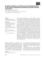
Fig. 1. Calcium titration curves for fluorescence signals of S100A1
(black) and its disulfides: S100A1–2-mercaptoethanol (red),
S100A1–glutathione (green) and S100A1–cysteine (yellow). In all
cases the protein concentration was 8 l
M. Interpolation curves
have been calculated as described in the text, using the parameters
listed in Table 1.
S100A1 affinity for calcium G. Goch et al.
2558 FEBS Journal 272 (2005) 2557–2565 ª 2005 FEBS
4±2· 10
3
m
)1
. (Details of the fluorescence titration
analysis are given in the supplementary material.)
In the case of E32Q)2-mercaptoethanol the relative
increase in the fluorescence signal upon calcium bind-
ing (f
1
) is much smaller but the titration curve has a
similar shape (not shown) and is also well described by
one binding constant K
1
¼ 7.6 ± 1.4 · 10
4
m
)1
.
The fluorescence signal of E32Q–glutathione does
not increase but rather decreases in the presence of cal-
cium. The shape of the titration curve (Fig. 2) clearly
indicates that subunits of E32Q–glutathione coordinate
not one, but two calcium ions, with very different
binding constants K
1
and K
2
(Table 1).
Remarkably, the relative change in fluorescence seen
upon the coordination of two calcium ions, as des-
cribed by the parameter f
2
, is the same for E32Q–
glutathione and glutathionylated native S100A1
(Table 1). Evidently, the second calcium ion is still
coordinated by the N-binding loop of the glutathionyl-
ated a subunit of E32Q, despite the Glu32 fi Gln
mutation, although the binding capacity is reduced by
a few orders of magnitude.
Calcium binding to the E73Q mutant and
its oxidized forms
The fluorescence signal of the E73Q mutant changes
only at very high CaCl
2
concentrations (data not
shown). Experiments with 5-nitro-1,2-bis(o-aminophen-
oxy)-ethane-N,N,N¢,N¢-tetraacetic acid (5-NBAPTA) as
a calcium chelator also show that the affinity of the
N-loop for calcium is very low. Both E73Q and
E73Q)2-mercaptoethanol bind calcium with binding
constants not exceeding 10
2
m
)1
, too low to be deter-
mined more precisely using 5-NBAPTA chelator with
aCa
2+
binding constant of the order of 10
4
m
)1
.
The results obtained for E73Q–glutathione are dif-
ferent. Its fluorescence signal increases in the presence
of calcium (data not shown) indicating that the metal
ion is coordinated in the vicinity of the tryptophan
residue with the binding constant determined either
by a chelator or by fluorescence measurements at
4.4 · 10
3
m
)1
. This observation can be rationalized in
the following way.
The C-terminal part of the S100A1 a subunit,
CNNFFWENS, contains numerous potential calcium
ligands provided by Glu91, Ser93 and three asparagine
residues: 86, 87 and 92. Glutathionylation of Cys85
introduces additional ligands – carboxylate groups of
the glutathione moiety – creating an efficient calcium-
binding site different from the N- and C-loops.
The results obtained for E32Q–glutathione and
S100A1–glutathione (see below) indicate that this addi-
tional metal binding site created by glutathionylation
is not active when the C-loop is saturated by calcium.
Similarly, as in the case of S100A1 and S100A1–
2-mercaptoethanol, only two, not three, calcium ions
are coordinated by subunits of these proteins.
Because of the appearance of an additional, non-
native calcium-biding site in E73Q–glutathione we
were not able to determine the microscopic calcium-
binding constants to N-loops of glutathionylated
subunits of the S100A1 protein. Nevertheless, because
formation of mixed disulfide between the subunits of
E73Q and 2-mercaptoethanol does not affect the affin-
ity of the protein N-loops for calcium, it can be safely
assumed that calcium binding to the N-loops of the
glutathionylated protein can be described by micro-
scopic constants of the order of 10
2
m
)1
as determined
for the reduced protein.
Calcium binding to native S100A1 protein
and its derivatives
The microscopic binding constants to the C-loop of
the S100A1 protein and its oxidized forms, as deter-
mined from studies of the E32Q mutant and its deri-
vatives (K
1
values listed in Table 1), are at least two
orders of magnitude greater than the values for the
N-loops (K
N
) of S100A1 and its derivatives, as evalu-
ated from studies of the E73Q mutant and its mixed
disulfides ( 10
2
m
)1
). This means that the subunits
of these proteins bind first to C-loops and then to
Fig. 2. Ca
2+
titration of relative fluorescence signals of 8 lM solu-
tions of E32Q (black) and its E32Q–glutathione derivative (green).
(Inset) The initial titration points for E32Q–glutathione are shown in
the linear scale.
G. Goch et al. S100A1 affinity for calcium
FEBS Journal 272 (2005) 2557–2565 ª 2005 FEBS 2559
N-loops, so that the population of subunits with free
C-loops and Ca
2+
-saturated N-loops is negligible.
Therefore, the titration curves for the fluorescence of
S100A1 and its derivatives can be analysed using a
simple consecutive model of metal binding with the
macroscopic binding constants K
1
and K
2
(supplement-
ary analysis of the fluorescence titration curves).
Two inflexions are seen in the titration curve of the
reduced protein (Fig. 1) indicating that the calcium-
binding constants K
1
and K
2
are quite different from
each other. For S100A1–2-mercaptoethanol, S100A1–
glutathione and S100A1–cysteine only one inflexion is
observed, although the data analysis clearly shows that
each of these molecules coordinates two calcium ions.
Evidently, the values of K
1
and K
2
are quite similar,
so that determination of all four parameters K
1
, f
1
, K
2
and f
2
using a curve-fitting procedure is not possible.
Therefore, we used K
1
and f
1
parameters found previ-
ously from the studies of E32Q mutant and its deriva-
tives and allowed them to change only within the error
limits of their determination (Table 1) during the fit-
ting procedure of K
2
and f
2
. The best-fit parameters
calculated in this way are listed in Table 1.
Only one inflexion was observed in the titration
curve of the cysteinylated protein. Nevertheless, in this
case, all calcium-binding parameters can be determined
directly using the fitting procedure because K
2
» K
1
(Table 1).
The stoichiometry and cooperativity of calcium
binding to S100A1–glutathione have been confirmed
by mass spectrometry. At a Ca
2+
concentration of
300 lm the protein spectrum is completely dominated
by signals corresponding, with 1 Da accuracy, to the
noncovalent dimer of a subunits of S100A1–glutathi-
one with four coordinated calcium ions, and various
numbers of water molecules (from two to 16). The
other, very weak, protein signals correspond to the
subunit dimer with two or no calcium ions, both
with various numbers of coordinated water mole-
cules. No signals coming from species containing one
or more than two Ca
2+
ions per a subunit have been
detected.
TNS binding to S100A1 and its S100A1–
glutathione derivative
It was shown that 2-p-toluidinylnaphtalene-6-sulfonic
acid (TNS) fluorescence increases dramatically upon
binding to hydrophobic patches exposed on protein
surfaces [18]. Therefore, TNS is frequently used to
monitor protein conformational transitions accompan-
ied by changes in the hydrophobic area exposed to
water.
Using a TNS probe such a conformational transition
induced by calcium was observed in the S100A1 pro-
tein by Leung et al. [19]. We obtained similar results,
although a strict, quantitative comparison is not pos-
sible because our experiments were carried out at
somewhat different pH and ionic strength.
In calcium-free solutions, TNS fluorescence in the
presence of S100A1 protein and its Cys85–glutathione
derivative is very weak, with the maximum at 420 nm
(Fig. 3). At 200 mm calcium concentration, when the
S100A1 protein is almost completely saturated with
metal ions (Fig. 3B) the maximum of TNS fluorescence
shifts to 440 nm and its intensity increases about four
times (Fig. 3A). Similar effects on TNS fluorescence
are observed when S100A1–glutathione is fully satur-
ated with calcium at a Ca
2+
concentration of 60 lm,
although the increase in the fluorescence signal is smal-
ler (Fig. 3C).
Because calcium binding to C- and N-loops of the
a subunits of S100A1 is not cooperative and the
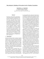
AB
CD
Fig. 3. TNS fluorescence spectra in the presence of S100A1 pro-
tein (A), and its mixed disulfide with glutathione (C), at the follow-
ing calcium concentrations: (A) 0 (s), 0.3 m
M (+), 3 mM (cyan),
21.5 m
M (dark cyan), 100 mM (grey) and 200 mM (black); (C) 0 (n),
17 l
M (green), 60 lM (dark green) and 250 lM (grey). The TNS and
protein concentrations were 21 and 6.0 l
M, respectively. The relat-
ive fluorescence signals of S100A1 protein and its disulfide with
glutathione, at the same calcium concentrations as in the TNS
experiments, are shown in Fig. 3B and D, respectively.
S100A1 affinity for calcium G. Goch et al.
2560 FEBS Journal 272 (2005) 2557–2565 ª 2005 FEBS
respective binding constants differ from each other by
at least two orders of magnitude, at calcium concentra-
tion of about 1 mm only the C-loop of the protein is
saturated with calcium. The fluorescence measurements
of TNS prove that calcium binding to the C-loop
alone does not result in protein conformational trans-
ition leading to exposure of a hydrophobic surface
(compare Fig. 3A,B). Such a transition is induced only
when both C- and N-loops are saturated with calcium.
TRTK-12 binding to glutathionylated S100A1
protein
TRTKIDWNKILS peptide, termed TRTK-12, was
derived from a consensus sequence (K ⁄ R)(L ⁄ I)XWX-
XIL identified in numerous S100A1 target proteins.
The peptide has been shown to compete with them for
calcium-dependent binding to S100A1 [1,20,21]. There-
fore, it is commonly used as a convenient probe for
calcium-induced biological activity in this protein.
In the absence of calcium, the fluorescence signal of
an equimolar mixture of 2 lm S100A1–glutathione and
TRTK-12 is equal (see Experimental procedures),
within the limits of error (2%), to the sum of the sig-
nals of each component. In the presence of 200 lm
Ca
2+
, the fluorescence of the protein–peptide mixture
is reduced by 20% relative to the sum of the fluores-
cence signals for TRTK-12 and the protein as meas-
ured separately in the presence of calcium. This proves
that the molecules interact with each other.
Discussion
Under physiological conditions (pH ¼ 7.2, 100 mm
NaCl) unmodified S100A1 protein coordinates calcium
via the C-loops of its subunits, with a binding constant
of K
1
¼ 4±2· 10
3
M
)1
. The N-loops of the protein
bind calcium very weakly, with K
2
values close to the
microscopic binding constant determined from studies
of the E73Q mutant ( 10
2
m
)1
). This proves that the
binding process is noncooperative.
These results confirm numerous previous reports of
the low affinity of S100A1 [22,23] and, in general, of
S100 proteins for calcium [24]; much lower than expec-
ted for calcium-signalling proteins. The intracellular
calcium concentration changes transiently from a basal
level of 0.1 lm to 1 lm [25]. Therefore, inside a
cell, the isolated S100A1 protein should always remain
in the apo state. Of course, the affinity of the protein
for calcium can increase when it binds to its target.
Indeed, Landar et al. [20] have shown that S100A1
binds TRTK-12 peptide at pH 7.4 in the presence of
milimolar concentrations of Ca
2+
ions, one order of
magnitude lower than the Ca
2+
dissociation constant
1 ⁄ K
2
0.01 m determined by us. Nevertheless, it has
also been shown [20] that the protein does not bind
the peptide when the calcium concentration decreases
to below 10 lm.
Therefore, it seems that some, as yet unknown,
cofactor(s) must be involved in the induction of cal-
cium-dependent intracellular activity of S100A1. Such
a cofactor would need to fulfil the following require-
ments: (a) It should increase the affinity of S100A1
for calcium. (b) Its interaction with S100A1 must not
lead to similar conformational changes as those
induced by calcium coordination. Otherwise, it would
replace, and therefore eliminate, calcium from the sig-
nal pathway because it would keep the protein in the
active conformation even in the absence of calcium.
(c) Calcium-saturated S100A1 protein modified by a
cofactor must preserve its ability to bind target
proteins.
Our results indicate that glutathionylation conforms
to all these requirements. The affinity of S100A1 pro-
tein for calcium is dramatically enhanced when the SH
groups of the cysteine residues of its subunits (Cys85)
are linked covalently to glutathione: the Ca
2+
-binding
constant for C-loops increases 10-fold and that for
N-loops increases by as much as four orders of magni-
tude. The glutathionylated protein binds TRTK-12
peptide in a calcium-dependent manner.
A regulatory role of S-glutathionylation has been
demonstrated for a number of proteins. It is postulated
[26,27] that this reversible protein modification, con-
trolled by the intracellular redox potential and enzy-
matic cleavage of S-S bonds, as well as by reactive
oxygen and nitrogen species, plays a crucial role in the
cell’s response to oxidative stress ’contributing to the
control of cell development, differentiation, growth,
death and adaptation’ [28]. Although S100A1 protein
is engaged in all of these processes it has not yet been
suspected that its biological activity may be regulated
by glutathionylation.
Our results indicate that under physiological condi-
tions the ability of S100A1 protein to act as a calcium
receptor can be turned on by glutathionylation (cyste-
inylation) of its Cys85 residue and off by reduction of
the mixed disulfide S100A1–glutathione (S100A1–cys-
teine) species. It is probable, therefore, that S100A1
acts as a linker between the two most important cell-
signalling pathways, i.e. between calcium and redox
signalling.
This hypothesis does not exclude the possibility that
some other cofactors may be involved in regulating the
calcium-induced biological activities of S100A1. Their
existence is substantiated by the observation that, in
G. Goch et al. S100A1 affinity for calcium
FEBS Journal 272 (2005) 2557–2565 ª 2005 FEBS 2561
the presence of 1 mm dithiothreitol, a protein with free
SH groups disassembles, in a calcium-dependent man-
ner, microtubules in triton-cytoskeletons from astro-
cyte and myoblast cell lines [29].
It is worth noting that in 10 of 20 sequences of S100
proteins homologous to S100A1, isolated from various
organisms, the cysteine residue is conserved at the 12th
position from the last ligand of Ca-binding loops (see
the SwissProt database). The affinity of some, if not
all, of these proteins for calcium may also be regulated
by post-translational modification of this residue.
The mechanism by which mixed disulfide formation
by Cys85 leads to an increase in the affinity of S100A1
for calcium does not seem to be related to the intro-
duction of some functional groups arranged in space
in any specific manner. Despite the different structure
and number of its carboxyl groups (one, instead of
two) cysteine appears to be an excellent substitute for
glutathione. Even the 2-mercaptoethanol molecule,
devoid of any charged groups, has a similar although
somewhat smaller effect, probably because of its small
size. A large increase in macroscopic Ca
2+
-binding
constants to S100A1 was observed by Baudier et al.as
a result of protein labelling with monobromo(trimethyl-
ammonio)bimane [30].
Remarkably, experiments with E73Q–glutathione
indicate that the microscopic binding constant K
N
does
not change, within the margins of error, and remains
low. Therefore, the tremendous increase in the affinity
of the N-loops for calcium upon protein glutathionyla-
tion is due to the appearance of a large cooperativity
effect, corresponding to Gibbs’ free energy determined
by the ratio of microscopic (K
N
) to macroscopic (K
2
)
binding constants for the N-loops of the protein:
DDG ¼ RT ln(K
N
⁄ K
2
). Because 10 < K
N
< 100 m
)1
and K
2
¼ 7±3· 10
5
m
)1
(Table 1, K
2
) DDG can be
estimated at )6 ± 1 kcalÆmol
)1
. Similar cooperativity
is observed for S100A1–cysteine and much smaller for
S100A1–mercaptoethanol (DDG )3.4 kcalÆmol
)1
).
The experimental results were analysed using a sim-
ple model assuming that the a subunits of S100A1
protein, although dimerized, bind calcium independ-
ently, in a noncooperative way. All our data conform
to this model. It seems, therefore, that the protein
subunits do not exchange any signals regarding their
conformational status. This observation is substan-
tiated by comparative NMR studies of the met and
apo forms of S100B protein [8]. The structure of the
interface between the protein subunits has been
shown to be unaffected by metal binding. Apparently,
it provides a barrier for propagation of calcium-
induced conformational changes from one subunit to
its neighbour.
Experimental procedures
Expression and purification of proteins and
TRTK-12 peptide
S100A1 protein and its mutants were expressed as described
previously [31]. The synthetic gene coding for the bovine
S100a subunit was constructed and cloned into a derivative
of pAED4 plasmid. Genes coding for Glu32 fi Gln and
Glu73 fi Gln mutants of S100a were obtained by site-
directed mutagenesis. The genes were expressed in Escheri-
chia coli utilizing the T7 expression system. The expression
products were isolated using a phenyl–Sepharose column,
purified by reverse-phase HPLC on a semi-preparative
Vydac C
18
column, and identified by the ESI-MS using a
Macromass Q-Tof spectrometer (supplementary Table S1).
Two forms of the proteins: (a) with sequences strictly
corresponding to the respective gene sequences, and (b)
containing the additional initiator methionine at the N-ter-
mini, come from HPLC as partly overlapping picks. NMR
measurements indicated that structural differences between
both forms are small and localized in the vicinity of the
N-terminal Met residue [4,32]. Our comparative fluorescence
experiments with both pure forms of S100A1 have shown
that they coordinate calcium and lanthanide ions with the
same, within the margins of error, binding constants and
that their fluorescence properties are similar. Therefore, the
mixtures of a and b species were used in experiments.
In an analogous way, TRTK-12 peptide with the
sequence TRTKIDWNKILS was produced in E. coli, puri-
fied using HPLC, and identified by ESI-MS using a Macro-
mass Q-Tof spectrometer.
Derivatives of S100A1 protein and its mutants
When a 1 mm concentration of 2-mercaptoethanol is main-
tained during E. coli cell sonification and isolation of the
recombinant proteins, the mixed disulfides: S100A1–2-merca-
ptoethanol, E32Q)2-mercaptoethanol and E73Q)2-merca-
ptoethanol, respectively, predominate in the preparation and
can be easily separated from their respective reduced forms
using reverse-phase HPLC.
Mixed disulfides of S100A1 and its mutants with cysteine
and glutathione were obtained by 15 min incubation of
2.5 mm protein solutions in 6 m guanidinium chloride at
pH 8 in the presence of a threefold excess of l-cystine or
oxidized glutathione, respectively. After 10-fold dilution,
the reaction products were purified by reverse-phase
HPLC.
The identity of all products was checked by MS using a
Q-Tof Micromass apparatus. The list of expected and
measured molecular masses is given (supplementary
Table S1). It was checked, using HPLC and MS, that each
derivative could be reduced to the respective original pro-
tein by short incubation with 1 mm dithiothreitol at pH 8.
S100A1 affinity for calcium G. Goch et al.
2562 FEBS Journal 272 (2005) 2557–2565 ª 2005 FEBS
Protein samples
Tris buffer (20 M M), pH 7.2, containing 100 mm NaCl in
MQ water filtered through a Chelex column was used as
the solvent in all experiments. All protein solutions used in
the fluorescence titration experiments were checked for
possible calcium contamination by comparing the fluores-
cence signals of samples measured in the presence and
absence of EDTA. If the difference exceeded 1% the solu-
tion was not used.
Protein stock solutions of 80 lm a subunits were centri-
fuged and stored for no longer than 2 weeks before experi-
ments. Concentrations of the native a subunit, its mutants
and their derivatives were determined from UV absorp-
tion at 280 nm using a molar extinction coefficient of
9300 m
)1
cm
)1
[33]. The absorption spectra were measured
on a Cary 3E spectrophotometer (Varian International AG,
Zug, Switzerland) in thermostated cells of 10 mm path
length. All measurements were made at 25 °C.
Fluorescence measurements
For fluorescence titration experiments we used an appar-
atus described previously [34]. The fluorescence was excited
at 280 nm using a xenon–mercury lamp L2482 (Hama-
matsu Photonics Deutschland, Herrsching, Germany) and a
double prism monochromator (M3, Cobrabid, Poland).
The emission signal was measured using UG1 glass filter
(Schott, Jena, Germany) with transmission of < 1% below
300 nm and R585 photomultiplier (Hamamatsu) working in
a single photon counting mode. The absolute values of pro-
tein fluorescence quantum yields U listed in Table 1 were
estimated by comparing the protein signals with that of N-
acetyl-l-tryptophanamide used as a standard with U ¼ 0.14
in water [35]. The measurements were repeated several
times. Their statistical error did not exceed ± 0.005.
The fluorescence test of TRTK-12 binding to S100A1–
gluthathione was performed as follows. Four solutions (A,
B, C and D) containing 4 lm of the protein (A and B) or
peptide (C and D) in standard buffer were prepared. EDTA
(10 lm) was added to solutions A and C and 200 lm of
CaCl
2
was added to solutions B and D. The fluorescence
signals of A, B, C, D and of 1 : 1 mixtures of A + C and
B + D were measured as described above at an excitation
wavelength of 298 nm.
The use of calcium chelators
Calcium binding to the E73Q mutant and its derivatives
was studied using 5-NBAPTA as a metal chelator. More-
over, calcium-binding constants to E32Q–glutathione and
S100A1–glutathione determined from fluorescence measure-
ments were confirmed by the chelator method using 5,5¢-di-
bromo-1,2-bis(o-aminophenoxy)-ethane-N,N,N¢,N¢-tetrraacetic
acid (Br
2
-BAPTA). Both chelators were purchased from
Molecular Probes (Leiden, the Netherlands).
The chelator concentrations were determined by the
absorbance in the presence of excess calcium using the
following molar extinction coefficients: e
340
¼ 6.0 ·
10
3
m
)1
Æcm
)1
and e
239.5
¼ 1.6 · 10
4
m
)1
Æcm
)1
for 5-NBAPTA
[36] and Br
2
-BAPTA [37], respectively. Two-millilitre sam-
ples of 80 lm a subunits and 20 lm of 5-NBAPTA or of
equimolar concentrations ( 25 l m) of protein subunits and
5,5¢-Br
2
-BAPTA in the standard buffer were titrated by addi-
tion of concentrated CaCl
2
in microlitre portions. The
absorbance at 430 nm for 5-NBAPTA solutions or at
263 nm for Br
2
-BAPTA solutions, corresponding to the
absorption maxima of the calcium-free chelators, was monit-
ored using Cary 3E spectrometer in thermostated 1 cm cells.
Titration curves were analysed according to the equation
given by Linse et al. [38].
MS measurements
MS experiments were carried out using an electrospray
Q-ToF1 (Micromass, Manchester, UK) instrument in the
positive ion mode. In noncovalent interaction studies a
33 lm solution of S100A1–glutathione in 10 mm ammo-
nium acetate (pH 7.5) and 300 lm calcium chloride was
analysed with a 10-fold increased pressure at the first
pumping stage of the instrument.
Acknowledgements
We are indebted to Dr Aleksandra Wysłouch-Cies-
zyn
˜
ska for interpretation of MS spectra, to Mrs Mari-
anna Neczypor for technical assistance in some of
fluorescence measurements, and to Professor Włodzi-
mierz Zago
´
rski for his contribution to the discussion
of our results. This study was supported by the Polish
Committee for Scientific Research Grant 6 P04 009 16.
References
1 Donato R (2001) S100: a multigenic family of calcium-
modulated proteins of the EF-hand type with intracellu-
lar and extracellular functional roles. Int J Biochem Cell
Biol 33, 637–668.
2 Heizmann CW, Fritz G & Scha
¨
fer BW (2004) S100 pro-
teins: structure, functions and pathology. Front Biosci 7,
d1356–d1368.
3 Scha
¨
fer BW & Heizmann CW (1996) The S100 family
of EF-hand calcium-binding proteins: functions and
pathology. Trends Biochem Sci 21, 134–140.
4 Rustandi RR, Baldisseri DM, Inman KG, Nizner P,
Hamilton SM, Landar A, Landar A, Zimmer DB &
Weber DJ (2002) Three-dimensional solution structure
G. Goch et al. S100A1 affinity for calcium
FEBS Journal 272 (2005) 2557–2565 ª 2005 FEBS 2563
of the calcium-signaling protein apo-S100A1 as deter-
mined by NMR. Biochemistry 41, 788–796.
5 Kilby PM, Van Eldik LJ & Roberts GCK (1996) The
solution structure of the bovine S100B protein dimer in
the calcium-free state. Structure 4, 1041–1052.
6 Drohat AC, Amburgey JC, Abildgaard F, Starich MR,
Baldisseri D & Weber DJ (1996) Solution structure of
rat apo-S100B (bb) as determined by NMR spectrosco-
py. Biochemistry 35, 11577–11588.
7 Potts BCM, Smith J, Akke M, Macke TJ, Okazaki
K, Hidaka H, Case DA & Chazin WJ (1995) The
structure of calcyclin reveals a novel homodimeric fold
for S100 Ca
2+
-binding proteins. Nat Struct Biol 2,
790–796.
8 Drohat AC, Baldisseri DM, Rustandi RR & Weber DJ
(1998) Solution structure of calcium-bound rat S100B
(bb) as determined by nuclear magnetic resonance
spectroscopy. Biochemistry 37, 2729–2740.
9 Smith SP & Shaw GS (1998) A novel calcium-sensitive
switch revealed by the structure of human S100B in the
calcium-bound form. Structure 6, 211–222.
10 Linse S, Brodin P, Drakenberg T, Thulin E, Sellers P,
Elmde
´
n K, Grundstro
¨
m T & Forse
´
n S (1987) Structure–
function relationships in EF-hand Ca
2+
-binding pro-
teins. Protein engineering and biophysical studies of cal-
bindin D
9k
. Biochemistry 26, 6723–6735.
11 Linse S, Johansson C, Brodin P, Grundstro
¨
mT,
Drakenberg T & Forse
´
n S (1991) Electrostatic contribu-
tions to the binding of Ca
2+
in calbindin D
9k
. Biochem-
istry 30, 154–162.
12 Linse S & Chazin WJ (1995) Quantitative measurements
of the cooperativity in an EF-hand protein with sequen-
tial calcium binding. Protein Sci 4, 1038–1044.
13 Ko
¨
rdel J, Skelton NJ, Akke M & Chazin WJ (1993)
High-resolution structure of calcium-loaded calbindin
D
9k
. J Mol Biol 231, 711–734.
14 Skelton NJ, Ko
¨
rdel J & Chazin WJ (1995) Determina-
tion of the solution structure of apo calbindin D
9k
by
NMR spectroscopy. J Mol Biol 249, 441–462.
15 Moore B (1965) A soluble protein characteristic of the
nervous system. Biochem Biophys Res Commun 19, 739–
744.
16 Maune JF, Klee CB & Beckingham K (1992) Ca
2+
binding and conformational change in two series of
point mutations to the individual Ca
2+
-binding sites of
calmodulin. J Biol Chem 267, 5286–5295.
17 Eftink MR (1997) Fluorescence methods for studying
equilibrium macromolecule–ligand interactions. Methods
Enzymol 278, 221–257.
18 McClure WO & Edelman GM (1966) Fluorescent
probes for conformational states of proteins. I. Mechan-
ism of fluorescence of 2-p-toluidinylnaphthalene-6-sulfo-
nate, a hydrophobic probe. Biochemistry 5, 1908–1918.
19 Leung IK, Mani RS & Kay CM (1987) Fluorescence
studies on the Ca
2+
and Zn
2+
binding properties of the
a-subunit of bovine brain S-100a protein. FEBS Lett
214, 35–40.
20 Landar A, Rustandi RR, Weber DJ & Zimmer DB
(1998) S100A1 utilizes different mechanisms for interact-
ing with calcium-dependent and calcium-independent
target proteins. Biochemistry 37, 17429–17438.
21 Osterloh D, Ivanenkov VV & Gerke V (1998) Hydro-
phobic residues in the C-terminal region of S100A1 are
essential for target protein binding but not for dimeriza-
tion. Cell Calcium 24 , 137–151.
22 Baudier J, Glasser N & Ge
´
rard D (1986) Ions binding
to S100 proteins. I. Calcium- and zinc-binding proper-
ties of bovine brain S100aa, S100a (ab), and S100b (bb)
protein: Zn
2+
regulates Ca
2+
binding on S100b protein.
J Biol Chem 261, 8192–8203.
23 Baudier J (1988) S100 proteins: structure and calcium
binding properties. In Calcium and Calcium Binding
Proteins (Gerday C, Gilles R, Boils L, eds), p. 102.
Springer-Verlag, Berlin.
24 Heizmann CW & Cox JA (1998) New perspectives on
S100 proteins: a multi-functional Ca
2+
-, Zn
2+
- and
Cu
2+
-binding protein family. Biometals 11, 383–397.
25 Berridge MJ, Lipp P & Bootman MD (2000) The vers-
ality and universality of calcium signaling. Nat Rev Mol
Cell Biol 1, 11–21.
26 Cotgreave IA & Gerdes RG (1998) Recent trends in
glutathione biochemistry – glutathione–protein interac-
tions: a molecular link between oxidative stress and
cell proliferation? Biochem Biophys Res Commun 242,
1–9.
27 Klatt P & Lamas S (2000) Regulation of protein func-
tion by S-glutathionylation in response to oxidative and
nitrosative stress. Eur J Biochem 267, 4928–4944.
28 Cooper CE, Patel RP, Brookes PS & Darley-Usmar
VM (2002) Nanotransducers in cellular redox signaling:
modification of thiols by reactive oxygen and nitrogen
species. Trends Biochem Sci 27, 489–492.
29 Sorci G, Agneletti AL & Donato R (2000) Effects of
S100A1 and S100B on microtubule stability. An in vitro
study using triton-cytoskeletons from astrocyte and
myoblast cell lines. Neuroscience 99, 773–783.
30 Baudier J, Glasser N & Duportail G (1986) Bimane-
and acrylodan-labeled S100 proteins. Role of cysteines-
85a and -84b in the conformation and calcium binding
properties of S100aa and S100b (bb) proteins. Biochem-
istry 25, 6934–6941.
31 Bolewska K, Kozłowska H, Goch G, Mikołajek B &
Bierzyn
˜
ski A (1997) Molecular cloning and expression
in Escherichia # of a gene coding for bovine S100A1
protein and its Glu32 ? Gln and Glu73 ? Gln mutants.
Acta Biochim Polon 44, 275–284.
32 Baldisseri DM, Rustandi RR, Zhang Z, Tang C, Bair
CL, Landar A, Zimmer DB & Weber DJ (1999)
1
H,
13
C
and
15
N NMR sequence-specific resonance assignments
for rat apo-S100A1 (aa). J Biomol NMR 14, 91–92).
S100A1 affinity for calcium G. Goch et al.
2564 FEBS Journal 272 (2005) 2557–2565 ª 2005 FEBS
33 Baudier J & Ge
´
rard D (1986) Ions binding to S100
proteins. II. Conformational studies and calcium-
induced conformational changes in S100aa protein: the
effect of acidic pH and calcium incubation on subunit
exchange in S100a (ab) protein. J Biol Chem 261,
8204–8212.
34 Dadlez M, Go
´
ral J & Bierzyn
˜
ski A (1991) Luminescence
of peptide-bound terbium ions. Determination of bind-
ing constants. FEBS Lett 282, 143–146.
35 Eftink MR & Hagaman KA (1985) Fluorescence
quenching of the buried tryptophan residue of cod par-
valbumin. Biophys Chem 22, 173–180.
36 Rand MD, Lindblom A, Carlson J, Villoutreix BO &
Stenflo J (1997) Calcium binding to tandem repeats of
EGF-like modules. Expression and characterization of
the EGF-like modules of human Notch-1 implicated
in receptor–ligand interactions. Protein Sci 6, 2059–
2071.
37 Tsien RY (1980) New calcium indicator and buffers
with high selectivity against magnesium and protons:
design, synthesis, and properties of prototype structures.
Biochemistry 19, 2396–2404.
38 Linse S, Johansson C, Brodin P, Grundstro
¨
mT,
Drakenberg T & Forse
´
n S (1991) Electrostatic contribu-
tions to the binding of Ca
2+
in calbindin D
9k
. Biochem-
istry 30, 154–162.
Supplementary material
The following material is available from http://www.
blackwellpublishing.com/products/journals/suppmat/
EJB/EJB4681/EJB4681sm.htm
Table S1. Analysis of the fluorescence titration curves.
Calculated and measured molecular masses of recom-
binant proteins and their disulfide derivatives.
G. Goch et al. S100A1 affinity for calcium
FEBS Journal 272 (2005) 2557–2565 ª 2005 FEBS 2565