Báo cáo khoa học: Differential expression pattern of the novel serine⁄threonine kinase, STK33, in mice and men doc
Bạn đang xem bản rút gọn của tài liệu. Xem và tải ngay bản đầy đủ của tài liệu tại đây (1.11 MB, 15 trang )
Differential expression pattern of the novel
serine⁄ threonine kinase, STK33, in mice and men
Alejandro O. Mujica
1
*, Bastienne Brauksiepe
1
*, Sigrid Saaler-Reinhardt
2
, Stefan Reuss
3
and
Erwin R. Schmidt
1
1 Institute of Molecular Genetics, Johannes Gutenberg-University, Mainz, Germany
2 Institute of Genetics, Johannes Gutenberg-University, Mainz, Germany
3 Department of Anatomy and Cell Biology, Johannes Gutenberg-University, Mainz, Germany
The serine ⁄ threonine kinase 33 gene (STK33 ⁄ Stk33)
was identified by comparative sequencing of human
chromosome region 11p15.3 and its syntenic region in
mouse chromosome 7 [1,2]. Chromosome 11p15.3 is a
gene-rich region of clinical importance because several
human diseases, including predisposition for some
types of cancer, have been mapped there [3,4]. It has
also been associated with several defects and malig-
nancies, such as the Beckwith–Wiedemann syndrome,
haemoglobinopathies, Long QT syndrome (Ward–
Romano Syndrome), insulin-dependent diabetes mellitus
I, Usher syndrome 1C, T-cell leukaemia, hypoparathy-
roidism and Nieman–Pick disease type A and B
(reviewed in [5]) as well as different types of cancer in
urinary bladder, ovary, testis, breast and lung [6,7].
By phylogenetic analysis, the STK33 ⁄ Stk33 protein
was classified as a member of the Ca
2+
⁄ calmodulin
dependent kinases family (CAMK) which was subse-
quently confirmed by the human and mouse kinome
catalogues [1,8–10]. Related members of the CAMK
family of serine ⁄ threonine kinases have been associated
with a variety of biological functions through regula-
tion of transcription by, for example, phosphorylating
cAMP response element-binding transcription factor
[11–13]. While CAMK1 and CAMK2 are expressed
ubiquitously [14], CAMK4 is differentially expressed in
certain neural tissues, T cells and testis [13]. In mice,
Camk2 and Camk4 expression has been associated
with functions as diverse as spermatogenesis, memory
formation and cardiac hypertrophy and heart failure
Keywords
serine ⁄ threonine kinase, spermatogenesis,
STK33 antibody, STK33 expression
Correspondence
E. R. Schmidt, Institute of Molecular
Genetics, Johannes Gutenberg-University,
J J. Becherweg 32, D-55099 Mainz,
Germany
Fax: +49 6131 3925346
Tel: +49 6131 3925748
E-mail:
*These authors contributed equally to the
paper
(Received 6 May 2005; revised 2 August
2005, accepted 3 August 2005)
doi:10.1111/j.1742-4658.2005.04900.x
Serine ⁄ threonine kinase 33 (STK33 ⁄ Stk33) is a recently discovered gene
whose inferred amino acid sequence translation displays characters typical
for a calcium ⁄ calmodulin dependent kinase (CAMK). In this study we ana-
lysed the STK33 ⁄ Stk33 RNA and protein distribution and the localization
of the protein. The STK33 ⁄ Stk33 expression pattern resembles those of
some related members of the CAMK group. STK33 ⁄ Stk33 displays a non-
ubiquitous and, in most tissues, low level of expression. It is highly
expressed in testis, particularly in cells from the spermatogenic epithelia.
Moreover, significant expression is detected in lung epithelia, alveolar
macrophages, horizontal cells in the retina and in embryonic organs such as
heart, brain and spinal cord. A possible role of STK33 ⁄ Stk33 in spermato-
genesis and organ ontogenesis is discussed.
Abbreviations
STK33 ⁄ Stk33, human and mouse serine ⁄ threonine kinase proteins; STK33 ⁄ Stk33: human and mouse serine ⁄ threonine kinase genes; CAMK:
Ca
2+
⁄ calmodulin dependent kinases group; CAMK1 ⁄ Camk1, CAMK2 ⁄ Camk2, CAMK4 ⁄ Camk4: genes for Ca
2+
⁄ calmodulin dependent
kinases I, II and IV from human (in capitals) and mouse; FCS, fetal calf serum; dpp, days postpartum; MTE, multiple tissue expression;
ISH, in situ hybridization; LSM, laser scanning microscopy; Hsa, Homo sapiens; Mmu, Mus musculus.
4884 FEBS Journal 272 (2005) 4884–4898 ª 2005 FEBS
[15–21]. Certain alternative splicing variants of
CAMK2 were found to be expressed preferentially in
tumour cells [22] and the Camk2d isoform is downreg-
ulated in both human and mouse tumour cells [23].
CAMK4 expression has also been associated with epi-
thelial ovarian cancer [24]. Alternative splicing may
also explain apparently contradicting results from
Camk4 knockout experiments: mice void of Camk4, as
a consequence of disrupted promoter structure and
exons I and II, exhibit impaired follicular development
and ovulation [25] and impaired spermiogenesis in
males [26]. On the other hand, mice defective in
Camk4 generated by disrupting exon III and hence still
able to produce the shorter alternative splice transcript
for calspermin, have shown neither spermatogenesis
dysfunction nor infertility [27]. The PhKgT gene (phos-
phorylase kinase, testis ⁄ liver, gamma-2) which displays
a close relationship with STK33 in phylogenetic analy-
ses, originally found to be expressed mainly in testis
[28], has been shown to be associated with hepatic
disorders [29,30]. DAP-kinases are serine ⁄ threonine
kinases involved in apoptosis that phosphorylate myo-
sin light chains in a calmodulin-dependent way and are
associated with the cytoskeleton [31].
The sequence of STK33 ⁄ Stk33 is divergent enough
from other kinase genes so that corresponding EST
entries are unequivocally identified [1] and examination
of the human and mouse genomes suggests that
STK33 and Stk33 are single-copy genes [1,8,10]. The
human chromosome 19 harbours a nonexpressed
STK33 pseudogene [8] but no Stk33 pseudogene is
detected in the mouse genome [10]. The NCBI’s Uni-
Gene [32] human build no. 184 contains 106 EST ent-
ries for the STK33 cluster, and the murine build no.
174 contains 50 for Stk33. This reflects significantly
lower total expression than, for example, housekeeping
genes such as GAPDH (16014 human EST ⁄ 1128
mouse EST) or b-actin (15776 ⁄ 4559). STK33 ⁄ Stk33
EST counts are similar to other CAMKs (CAMK1:
133 ⁄ 148, CAMK2A: 141 ⁄ 129, CAMK4:83⁄ 177) and
their distribution suggests a nonubiquitous expression
(Table 1 shows a human ⁄ mouse comparison together
Table 1. Comparison of human and mouse STK33 ⁄ Stk33 expression levels between our data and expression databases.
Organ ⁄ tissue
Hsa STK33 Mmu Stk33 Hsa STK33 Mmu Stk33 Hsa STK33 Mmu Stk33 Hsa STK33 Mmu Stk33
This paper
Positive experiments
UniGene
a
count [32]
Number ⁄ % of clones
EST profile viewer
a
[32]
Transcripts per million
SOURCE Gene
Report
b
[34]
% Normalized
Testis +++ +++ 17 ⁄ 16.0 34 ⁄ 68.0 124 329 10.89 71.57
Lung + + ++ 22 ⁄ 20.7 0 76 0 6.97 –
Fetal lung + – 0 0 – – – –
Heart – – 0 1 ⁄ 2 0 18 – 4.96
Fetal heart + + 0 0 – – – –
Heart’s interv septum + n.a. 0 0 – – – –
Brain – – 2 ⁄ 1.8 4 ⁄ 8.0 4 8 – 1.50
Fetal brain – + 0 0 – – – –
Pituitary gland + n.a. 0 2 ⁄ 4.0 45 11.29
Ovary – – 3 ⁄ 2.8 0 31 0 2.11 –
Kidney + – 2 ⁄ 1.8 0 14 0 – –
Stomach – n.a. 1 ⁄ 0.9 0 9 0 – –
Pancreas + n.a. 5 ⁄ 4.7 0 25 0 – –
Trachea + n.a. 0 0 – – – –
Thyroid gland + n.a. 1 ⁄ 0.9 0 – – – –
Prostate – n.a. 2 ⁄ 1.8 0 14 – 0.76 –
Uterus – n.a. 7 ⁄ 6.6 0 38 0 3.40 –
Cervix n.a. n.a. 1 ⁄ 0.9 0 24 – 2.33 –
Eye – + + 2 ⁄ 1.8 2 ⁄ 4.0 11 11 – 3.40
Lymph node – n.a. 2 ⁄ 1.8 0 15 0 1.33 –
Mammary gland – n.a. 0 1 ⁄ 2 0 2 – 0.73
Embryonic stem cells n.a. n.a. 2 ⁄ 1.8 0 – – – –
Neuroblastoma n.a. n.a. 1 ⁄ 0.9 0 – – 64.34 –
Bone, intestine, liver, skin,
spleen, muscle, amongst
others
–– 000–––
a
UniGene Hs.501833 STK33 and Mm.79075 Stk33. Consulted on 15.06.2005. ‘Other’ or ‘mixed’ entries not included.
b
H. sapiens UniGene
Build no. 184, and Mus musculus UniGene Build no. 147, released on 2005-06-09. ‘Other’ or ‘mixed’ entries not included.
A. O. Mujica et al. STK33 expression pattern
FEBS Journal 272 (2005) 4884–4898 ª 2005 FEBS 4885
with the expression pattern obtained in this paper).
ESTs from human lung and testis are most frequently
represented (20.7% and 16.0%, respectively). Embry-
onic and fetal tissues as well as diverse tumour tissues
and cancer cell lines are also represented with several
entries each. Some entries are present from tissues of
the nervous system as well as from auditory and ocular
systems. Prostate and uterus also show single STK33
entries. The EST coverage for Stk33 in the mouse
seems to be more limited. Only testis is very well repre-
sented with 68% of the entries, and interestingly there
are no entries from the lung. In addition to testis, there
are single ESTs from retina, pituitary gland and cir-
cumventricular organs of the brain such as subfornical
organ and area postrema.
In this first survey, we have addressed the expression
of STK33 ⁄ Stk33 focussing on RNA and protein distri-
bution with emphasis on the mouse as an animal
model. Manning and colleagues [8] explained the fail-
ure to detect some novel kinases, despite their similar-
ity to members of the superfamily, with the limited
expression of these proteins. The results presented here
suggest that this may be the case for STK33 and that
its expression pattern also resembles that of members
of the CAMK family of protein kinases. As a step
towards understanding their function, we have
analysed the distribution of STK33 ⁄ Stk33 RNA and
protein as well as the subcellular localization of the
protein.
Results
Presence of STK33 ⁄ Stk33 RNA in mouse and
human tissues
Northern blot experiments were performed with
4–20 lg immobilized poly(A)
+
RNA from various
murine organs. Simultaneously, hybridization with a
cDNA fragment of ribosomal protein L19 housekeep-
ing gene was performed as normalization probe. L19
gene shows a ubiquitous expression and, in particular,
high expression in testis, according to the Gene
Expression Atlas [33]. The L19 probe hybridizes to an
RNA with a significantly lower length (full length
mRNA 673 bp) and hence yields a signal in a clearly
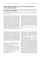
different position than Stk33. Results (Fig. 1A) show a
strong hybridization signal with Stk33 in RNA from
testis corresponding to an RNA of 3000 bp. In addi-
tion, there is a weak signal corresponding to a shorter
RNA which could be the result of a second Stk33-
RNA variant, possibly generated by alternative spli-
cing. In this analysis, no signals were detected in heart,
intestine, brain, kidney, ovary and lung.
A
B
C
Fig. 1. STK33 ⁄ Stk33 levels of expression in human and mouse
tissues. (A) Northern blot of immobilized poly(A)
+
RNA from adult
mouse organs with a Stk33-specific probe. The following
amounts of poly(A)
+
RNA were loaded (lg): heart, 4; intestine,
20; brain, 20; kidney, 6; ovary, 6; testis, 12; lung, 15. Normaliza-
tion was performed by simultaneous hybridization with a probe
from mouse ribosomal protein gene L19. The arrow shows a
possible shorter alternative transcript present in testis. (B) cDNA
dot-blot (MTE, Clontech) hybridization with an STK33 specific
probe, with samples from different regions of nervous system
(NS), heart (He), digestive system (DS), several organs (SO), can-
cer cell lines (Ca), fetal organs (FO) and diverse controls (Co).
The first column with practically no signal corresponds to several
regions of brain (see Fig. S1 for the identity of each dot). (C)
Quantification of the signal obtained in the cDNA dot-blot normal-
ized to the maximal signal in testis. Only results are shown from
tissues with signals higher than the highest value for the negat-
ive controls.
STK33 expression pattern A. O. Mujica et al.
4886 FEBS Journal 272 (2005) 4884–4898 ª 2005 FEBS
The expression pattern of STK33 in human was
investigated by cDNA dot-blot hybridization using the
human multiple tissue expression (MTE, Clontech,
Palo Alto, CA) and a STK33 specific probe. The
cDNA dot-blot contains 76 normalized dots of immo-
bilized cDNA, 61 from adult normal tissues, eight can-
cer cell lines and seven fetal tissues (see supplementary
Fig. S1). In two hybridization experiments weak but
reproducible STK33-specific signals were produced in a
small number of tissues. Significant hybridization sig-
nals were obtained in cDNA from testis, fetal lung,
fetal heart, pituitary gland and kidney. Weak but still
above background signals were found in interventricu-
lar septum of the heart, pancreas, trachea, and thyroid
gland (Fig. 1B,C). Hybridization signals were consid-
ered nonsignificant if they were below the highest pro-
duced by the negative controls (yeast total RNA, yeast
tRNA, Escherichia coli rRNA, E. coli DNA, poly
r(A), human Cot-1 DNA, human genomic DNA).
Such ‘nonsignificant’ signals were found in cDNAs
from a large number of tissues. The reason may be
low or very low amount of Stk33 RNA in those tissues
or unspecific binding of the hybridization probe.
To localize Stk33 transcripts at the cellular level,
mRNA in situ hybridization (ISH) on tissue sections of
various adult murine organs were performed. Controls
with sense RNA show negative results (Fig. S2) com-
pared with the antisense RNA probes. As expected from
the results from the northern analysis, strong hybridiza-
tion signals were found in mouse testis sections. Stk33-
specific signal appeared to be restricted to the cells of
the spermatid differentiation process, from the sperma-
togonia to the early spermatides with a remarkable
maximum of signal in the spermatocytes (Fig. 2A,B). In
all cases the signal was perinuclear, strongly supporting
its association to germinal cells. No signal was observed
in periluminar areas of the germinal epithelium or in
spermatozoa, in cells from the interstitial tissue, Leydig
cells, vascular cells or myoid cells in the lamina propria.
Stk33-specific ISH signal were also detected in lung
tissue sections, particularly in the epithelium of the
bronchi (Fig. 2D,E). Strong signals were also observed
in single alveolar cells (Fig. 2D,F), which, as revealed
by nuclear staining, probably are alveolar macro-
phages. In situ RNA hybridization in mouse retina
showed a signal in the outer plexiform layer (Fig. 2G).
Protein distribution
To investigate protein distribution, a polyclonal anti-
body against a Stk33-specific synthetic peptide was
generated. The specificity of the anti-Stk33 antibody
was determined by competition tests with synthetic
Stk33-specific peptide, anti-Stk33 signal disappeared in
testis sections, when antibody is preabsorbed with
12.5 ngÆlL
)1
synthetic peptide (Fig. 3, C3). Immuno-
staining on tissue sections with the Stk33 antibody
(Fig. 4) was observed in the same regions as the
mRNA in situ-hybridization (Fig. 2). In testis an
intensive staining was observed in only few cells per
tubulus, which may be classified as secondary sperma-
tocytes according to nucleus morphology and localiza-
tion (Figs 4 and 5). Round spermatides showed signals
of lower intensity compared to the spermatocytes.
Moreover, Stk33 positive and negative spermatides
were often seen in groups restricted to distinct tubular
profiles (Fig. 4A). Immunostaining signal was also vis-
ible in Sertoli cells, concentrated in the perinuclear
space. In all cases, the protein localization appeared to
be cytoplasmic.
In lung, immunostaining with anti-Stk33 antibody
produced strong signals in epithelial cells and suppo-
sedly alveolar macrophages. Laser scanning micro-
scopy (LSM) as well as comparison of immunostaining
with nuclear staining showed cytosplasmic localization
of Stk33 in the putative alveolar macrophages
(Fig. 4D, E, F and J). In the retina, a strong cytoplas-
matic immunostaining signal was found in horizontal
cells (Fig. 4K–M).
Immunostaining with Stk33 antibody was also per-
formed with 15-day mouse embryos (Fig. 6A). Stk33
specific signals were found in some areas of the ner-
vous system, in particular in the intermediate zone of
the cerebral hemisphere and between cerebellum prim-
ordium and medulla oblongata (Fig. 6, A1 and A2).
Also border regions between metencephalon and pons
with the third ventricle (arrows in Fig. 6, A1 and A2)
showed an augmentation of the signal. The spinal cord
showed a strong signal with a maximum in its lumbar
part, including neuronal processes (Fig. 6, A3). A clear
signal was observed in heart ventricles but not in the
atria (Fig. 6B). The signal is augmented in the endo-
cardium (Fig. 6, B1 to B3). Signals were also observed
in the trigeminal ganglion, rhinencephalon and tongue
(see Fig. S3B). All immunostaining negative controls
with no primary antibody reproducibly showed no
staining (see Figs S2 and S3).
Spermatogenesis specific signal
To confirm spermatogenesis-specific Stk33 signal,
Western blots with protein extracts from testis of mice
of different ages were performed. The Western blot
analysis showed signals in 20 and 30 days postpartum
(dpp) mice, whereas 10 dpp were Stk33 negative (see
Fig. 3D). Although the Western blot results were not
A. O. Mujica et al. STK33 expression pattern
FEBS Journal 272 (2005) 4884–4898 ª 2005 FEBS 4887
evaluated quantitatively, it seems obvious that the sig-
nal is stronger in older mice.
Discussion
Here we report the first survey of the distribution of the
recently discovered serine ⁄ threonine kinase 33 in mouse
and human tissues. Our results are well in accordance
with the STK33 ⁄ Stk33 expression pattern derived from
the expressed sequence databases (compiled in Table 1).
The predominant expression of STK33 ⁄ Stk33 in testis
is also observable in UniGene EST database [32]
( Gene Expres-
sion Atlas [33] ( and
Testis
A
B C
Lung
Retina
D
G
E
F
Fig. 2. Distribution of Stk33 mRNA in testis, lung and retina demonstrated by in situ hybridization (ISH). (A,B) ISH in testis. Strong Stk33 signal
is detected in spermatocytes (spc), spermatides (sd) and possibly in spermatogonia (sg). No signal is detected around Sertoli cell nucleus (sen)
or in spermatozoa (sz), neither in Leydig cells nor any kind of cell in the interstitial space (is). (C) Nuclear staining with DAPI was used for char-
acterization of the nuclei in all tissues and is shown here exemplary for testis. (D,E,F) ISH in lung. Stk33 signal was detected in epithelium (ep)
and alveolar macrophages (am). No signal was found in cartilage (ca), smooth muscle (sm), connective tissue (ct), bronchioli (bri), aleveolar
duct (avd), alveoli (av), artery (ar) and bronchus (bru). (G) ISH in mouse retina. Stk33 signal was visible in the outer plexiform layer (opl). No sig-
nal was detected in ganglio cells (gc), inner plexiform layer (ipl), inner nuclear layer (inl), outer nuclear layer (onl), choroid (ch) and sclera (sc).
Dark staining in the pigmented epithelium (pe) was also observable in negative controls (data not shown) and hence disregarded as signal.
STK33 expression pattern A. O. Mujica et al.
4888 FEBS Journal 272 (2005) 4884–4898 ª 2005 FEBS
Stanford’s SOURCE database [34] (http://source.
stanford.edu). The dataset of the GeneCards from the
Weizmann Institute of Science (http://bioinformatics.
weizmann.ac.il/cards), which is restricted to human
genes and does not provide results from testis, shows
expression of STK33 in lung and heart. However,
GeneCards expression data for STK33 are close to the
background level, which is fitting to the low level
expression in tissues other than testis. The only discrep-
ancy of the UniGene data, is the fact that we have
detected significant Stk33 expression in mouse lung in
two of three experiments. All expression data are sum-
marized in Table 1.
Clearly, the expression of Stk33 is only high enough
in testis to be detected by northern blot and cDNA
dot-blot experiments. More sensitive methods, such as
RNA in situ hybridization and immunostaining, reveal
expression in a broader spectrum of tissues but restric-
tion to particular cell types and populations. The
absence of signal in regions of the nervous system in
the human cDNA dot-blot analysis and mouse nor-
thern blot is remarkable, as several CAMK genes are
expressed there in high rates [35]. However, cells from
the nervous system in adult mice retina, probably hori-
zontal cells, display Stk33 expression as observed
by ISH and immunostaining. Additionally, Stk33
RNA ⁄ protein is expressed in some regions of the ner-
vous system in fetal mice (Fig. 6), and protein distribu-
tion in the adult brain shows a distinct distribution
that will be presented in a separate paper.
Human fetal lung yielded the second highest signal in
our cDNA dot-blot hybridization, whereas there was
remarkably weaker or no signal in human adult lung
samples. Northern blot analysis with mRNA also
showed no signal in adult mice lung, even though more
mRNA was immobilized from lung than from testis.
On the other hand, both mRNA in situ hybridization
and immunostaining experiments on tissue slices of
mouse adult lung showed a reproducible signal in bron-
chial epithelium and putative alveolar macrophages. It
seems evident that low expression of Stk33 is difficult
to examine by hybridization methods such as northern
blot and cDNA dot-blots using whole organs in all
tissues but testis. We were not able to detect Stk33
protein in mouse fetal lung. As we tested embryos
only at 15 days postcoitus, it is conceivable that expres-
sion in fetal lung occurs at different developmental
stages.
According to their predicted general biochemical
features, STK33 and Stk33 are probably soluble pro-
teins. psort analysis [36] found no notable known sig-
nal in STK33 ⁄ Stk33 primary structure except from
conserved C-terminal di-lysine motifs (511-Thr-Lys-
Lys-Lys-514 in human and 488-Gly-Lys-Lys-Arg-491
in mouse), which are recognized as putative endoplas-
matic reticulum membrane retention signals [37]. The
highest psort scores for subcellular localization based
on amino acid composition correspond to cytoplasm
and nucleus, weakly favouring nuclear localization.
The results of our immunostaining shown here
(Figs 4–6) demonstrate a predominantly cytoplasmic
localization of Stk33 protein.
STK33 could be involved in the normal development
of heart and other organs in embryonic and fetal stages.
The third highest signal in the cDNA dot-blot corres-
ponds to fetal heart and the sixth highest to the inter-
ventricular septum of the heart; this is also in line with
the strong immunostaining signal in mouse embryo
A
C
D
B
Fig. 3. Stk33 recombinant protein, antibody and spermatogenesis
specific signal in mice. Western blot of recombinant biotin–Stk33
fusion protein, affinity purified, detected with ExtrAvidinÒ-AP (A)
and with anti-Stk33 IgG (B). The 22.5-kDa band corresponds to the
E. coli native protein biotin carboxyl carrier protein (BCCP). Track 1
in (B) corresponds to the purge of the column and shows a trace
of antigen. Track 2 and 8 contain 572 ng and 0.425 ng of purified
protein, respectively. (C) Preabsorption of anti-Stk33 IgG in testis
slides with the following concentrations of synthetic peptide:
0ngÆlL
)1
(C1), 1.25 ngÆlL
)1
(C2) and 12.5 ngÆlL
)1
(C3). C2 and C3
were photographed with longer exposure times and accordingly
exhibit the light-coloured regions in the interstitial space also seen
in negative control (C4) with no primary antibody. (D) Stk33 sper-
matogenesis specific expression demonstrated by anti-Stk33 anti-
body staining. Immunoreactivity is observable in recombinant
protein (Rec), protein extracts from testis of mice at 20 and 30 dpp
but absent in 10 dpp. Coomassie blue staining of total protein
shows the homogeneous presence of 75 lg protein extract in each
track. Recombinant protein is not visible in the Coomassie blue
stained control track (Rec), as the amount loaded (80 ng) is below
the detection threshold [48].
A. O. Mujica et al. STK33 expression pattern
FEBS Journal 272 (2005) 4884–4898 ª 2005 FEBS 4889
heart (Fig. 6B) and the lack of signal in adult mouse
heart in our northern blot analysis (Fig. 1A). A study
on human heart malformations using DNA micro-
arrays [38] revealed STK33 among the most downregu-
lated genes in patients with tetralogy of Fallot, a
nonfatal congenital cardiovascular malformation in
which ventricles are not fully separated by the septum
and the pulmonary artery valve is narrowed, causing a
partial mixing of venal and arterial blood and a
decrease of blood flow to the lungs. On the other hand,
data from the source database [34] suggests a higher
expression of STK33 in neuroblastoma than in any
Testis
AB C
Lung
Retina
DG
EH
F
KL M
I
J
Fig. 4. Stk33 protein localization in testis, lung and retina. (A,B) Localization in testis. High signals in spermatocytes (spc) followed by round
spermatids (rsd) and around the Sertoli cell nucleous (sen). Stk33-positive and negative round spermatides (+ rsp, –rsp) exhibit a tubulus-spe-
cific distribution. (C) LSM image demonstrating cytoplasmic localization of the spermatocytes signal (anti-Stk33 in red and anti-a-tubulin in
green). (D–I) Immunostaining combined with nuclear staining in mouse lung preparations (anti-Stk33 in red and nuclear staining in blue).
(D,E,F) Alveolar macrophages showing cytoplasmic Stk33-signal, confirmed in (J) by LSM (anti-Stk33 in red and anti-a-tubulin in green).
(G,H,I) Lung epithelial cells with Stk33 specific signal. (K,L,M) Stk33 localization in mouse retina, in particular in horizontal cells. Immunostain-
ing with anti-Stk33 antibody showing strong signal in horizontal cells (hc) in the outer plexiform layer.
STK33 expression pattern A. O. Mujica et al.
4890 FEBS Journal 272 (2005) 4884–4898 ª 2005 FEBS
normal tissue (Table 1). Both neuroblastoma and
tetralogy of Fallot (more generally congenital cardio-
vascular malformations) are childhood diseases which
may be associated because of their common neural crest
origin [39,40]. The fact that STK33 may be downregu-
lated in tetralogy of Fallot but upregulated in neuro-
blastoma is intriguing and supports the idea of a role in
early organ development.
More cells in testis were labelled by ISH than by
immunostaining preparations, which may be due to a
high turnover of the protein. Moreover, the observa-
tion of a high immunostaining signal in spermatocytes
and grouped round spermatides with differential
expression pattern may reflect a cell division stage-spe-
cific synchrony of Stk33 expression during spermato-
genesis. This observation is strongly supported by the
Western blot results showing no signal in 10 dpp mice
and positive signal first by 20 dpp. The absence of sig-
nal at 10 dpp excludes involvement of Stk33 in Sertoli
cell or spermatogonia proliferation, which extends to
12 dpp [41]; but the signals at 20 and 30 dpp suggest a
meiosis and ⁄ or spermiogenesis specific function which
first occur at the periods of 8–18 dpp and 18–30 dpp
[41]. It has been hypothesized that novel cell division
regulatory checkpoints probably exist in the germ line
of higher eukaryotic organisms which are not necessar-
ily present in the basic ones already described for yeast
[42]. On the other hand, STK33 orthologs are present
in the genomes of several chordate organisms (Fig. 7),
but not found in yeast, fly or nematode genomes. This
is not too surprising as humans possess 74 members of
the CAMK group of protein kinases whereas only 21
are detectable in yeast, 32 in fly and 46 in worm [8].
We propose that STK33 ⁄ Stk33 expression occurs only
within a narrow time window during the spermatogen-
esis. It remains to be established whether this ‘pulse
like’ expression is directly associated with some germ
cell development checkpoint, which based on our data
seems suggestive, or rather reflects synchrony to other
events of the spermatogenesis. For instance, the mech-
anisms unleashing the pass of the haploid germ cells
through the tight junctions from the adluminal to the
luminal region are still unknown. It has been argued
that the germ cells themselves may signal the Sertoli
cells their entry to meiosis, triggering their transport
through the blood–testis barrier [43]. On the other
hand, apoptotic germ cells in wild-type rats, show a
similar frequency and distribution pattern as we show
here for Stk33 [44]. The molecular basis of these phe-
nomena is not yet fully established and a role for
Stk33 in some of these pathways is feasible. An
involvement of STK33 in human spermatogenesis may
also be postulated.
STK33 is clearly related to the canonical kinases
from the CAMK group and, as we demonstrate here,
shows similar expression pattern. Hence, similar func-
tions may be proposed. In particular, the maximum
expression signals of Stk33 in certain stages of sperma-
togenesis, in some embryonic fetal organs and its
involvement in child diseases, let us to postulate a role
in gametogenesis and organ ontogenesis that strongly
deserves to be further investigated.
Experimental procedures
Radioactive DNA labelling
Fifty to 500 ng of purified PCR-generated STK33 ⁄ Stk33-
DNA was used as template for radioactive probes. In vitro
random primed DNA labelling was carried out according
to manufacturer’s protocols (Roche Diagnostics, Mann-
heim, Germany). The labelling reaction was performed
overnight at room temperature, or for 2 h at 37 °C.
Typically, blot hybridizations were carried out overnight
at stringent temperatures depending on the G + C con-
tent of the probe (usually 64 °C for Southern blots
A
B
C
D
E
G
H
Fig. 5. Stk33 in mouse secondary spermatocytes. Double staining
with anti-Stk33 IgG (red) and nuclear staining with DAPI (green). (A)
Detail of a section of two seminiferous tubules showing a single
Stk33-positive cell. Note the dashed line marking the borders
between two different tubuli sections. The DAPI staining reveals
the different nuclear morphologies of: sertoli cell nucleous (sen),
spermatogonia (sg), primary spermatocytes (sc1), secondary sper-
matocytes (sc2), round spermatides (rsd) and spermatozoa (sz).
According to their chromatine condensation, Stk33-positive sperma-
tocytes were found in metaphase II (A), prophase II (B,C,D) and
anaphase II (E,F,G, chromosome segregation indicated by arrows).
A. O. Mujica et al. STK33 expression pattern
FEBS Journal 272 (2005) 4884–4898 ª 2005 FEBS 4891
and 42 °C +50% v ⁄ v formamide for northern blots).
After several washes with 2 · NaCl ⁄ Cit and 0.1 NaCl ⁄
Cit at room temperature membranes were exposed to
X-ray film with or without intensifying screens at
)70 °C. Exposure time varied from several hours to sev-
eral days.
Fig. 6. Localization of Stk33 in mouse embryo, day 15 postcoitius. (A) Overview of the mouse embryo immunostaining analysed with non-
confocal laser scanning. Regions with particular strong signal (a1–a3 and b) were photographed with the fluorescence microscope. Signal is
detected in some regions in the head, for instance between pons and medulla oblongata (A1), intermediate zone of mesencephalon (A2)
and medulla (A3). Strong signal is observed in heart ventricle (B) in particular in endocardium (B1–B3), here supported by nuclear staining in
green.
STK33 expression pattern A. O. Mujica et al.
4892 FEBS Journal 272 (2005) 4884–4898 ª 2005 FEBS
DNA probes
To avoid cross-hybridization, probes were designed exclu-
ding the region encoding the evolutionary conserved kinase
domain. The probes were checked for uniqueness by blast
searches against human and mouse DNA sequences in the
databases, in particular with the EST subset and whole gen-
ome assembly, and for repeat sequences by Repeatmasker
(Washington University, ). The
human DNA probe was amplified by PCR from uterus
cDNA as described [1] with the forward primer 5¢-AA
GCAATTTCCTGCAACC-3¢ and the reverse primer
5¢-CATGTGAATGACTGAAGC-3¢, yielding a 676-bp
DNA fragment. For the murine Stk33 a 570-bp probe was
amplified from lung cDNA spanning the last two exons
with the forward primer 5¢-AACCCAGAAAGTGATGAG-
3¢ and the reverse primer 5¢-TAGAACTAAGCGAG
CATG-3¢. For normalization purposes in hybridization
experiments with mouse tissues, a ribosomal protein gene
L19-specific probe was amplified by PCR from the
IMAGE:2648593 (kindly provided by GENterprise GmbH,
Mainz, Germany) using the standard primers T7 and SP6.
All PCR products were purified, checked by electro-
phoresis, quantified and sequenced before use as probes for
hybridization.
Northern blot
Total RNA isolation from mouse tissues was performed by
guanidinium thiocyanate ⁄ phenol ⁄ chloroform extraction
[45]. mRNA was isolated from total RNA with the Nucleo-
Trap
Ò
isolation kit (Macherey-Nagel, Du
¨
ren, Germany),
following the manufacturer’s instructions. mRNA was
quantified spectrophotometrically, and separated by electro-
phoresis on a 1.2% agarose gel under denaturing conditions
(5.5% formaldehyde), transferred to Nylon membranes
(Roche, Mannheim, Germany) and UV-cross linked. The
blot was hybridized with a DNA Stk33-specific probe and
a probe from the L19 housekeeping gene as normalization
control. BioMax MS autoradiography films were exposed
to the radioactive blot with intensifying screen at )70 °C.
Exposure time was determined empirically.
Fig. 7. Amino acid sequence alignment of
Stk33 kinase domain in some chordates.
Proteins sequences from human (Hsa) and
mouse (Mmu) as described in [1].
Sequences from Rattus norvegicus (Rno),
Fugu rubripes (Fru), Danio rerio (Dre) and
Ciona intestinales (Cin), were inferred from
their respective genome projects already
available [49–51]. Partial Stk33 EST
sequences are detected in Xenopus laevis
and X. tropicalis, but they still do not extend
the whole kinase domain and were in con-
sequence excluded from the analysis. The
putative ATP-binding subdomain, serine ⁄
threonine phosphorylation signature and the
Mg
2+
chelating Asp-Phe-Gly are shown, by
similarity with known kinase structures
[52–54]. Alignment accession number:
ALIGN_000866.
A. O. Mujica et al. STK33 expression pattern
FEBS Journal 272 (2005) 4884–4898 ª 2005 FEBS 4893
cDNA dot-blot hybridization
To evaluate expression of STK33 in human, the MTE array
(BD Biosciences Clontech, San Jose, CA, USA) was used.
This cDNA dot-blot contains samples of normalized total
RNA from 61 normal adult tissues, eight cancer cell lines,
seven fetal organs and eight diverse controls (see Fig. S1).
After hybridization, the array was exposed to Phospho-
Imager plates at room temperature from several hours to
overnight. Hybridization signals from the Phospho-Imager
plates were detected with a FUJIFILM BAS-1800 Phos-
phor-Imager and quantified using the software Aida V. 2.0
performing manual baseline subtraction.
Tissue sections
Anesthetized adult Balb ⁄ C mice were killed by an ether
overdose and immediately perfused transcardially with
NaCl ⁄ P
i
to which 15000 IU heparinÆ L
)1
were added, at
room temperature, followed by an ice-cold periodate-lysine-
paraformaldehyde solution [46]. The right atrium was
opened to enable venous outflow. Organs were prepared,
cryoprotected in a gradient of sucrose and stored in
NaCl ⁄ P
i
)30% sucrose. Cryosections (8–15 lm) were pre-
pared and mounted onto glass slides coated with silane.
Additionally, some tissues and 15-day embryos of sacrificed
Balb ⁄ C mice were taken, fresh frozen on dry ice and stored
at )80 °C. Cryosections of these tissues (8–15 lm) were
fixed in 4% paraformaldehyde–NaCl ⁄ P
i
for 10 min prior to
immunohistochemistry.
RNA in situ hybridization
Templates for the production of sense and antisense RNAs
were produced by adding T7 viral RNA polymerase pro-
moter adaptors to Stk33-PCR amplification products fol-
lowing standard procedures [47]. Accordingly, template for
the production of sense RNA was amplified with the
primers 5¢-CAGAGATGCATAATACGACTCACTATAGG
GAGAAACCCAGAAAGTGATGAG-3¢ and 5¢-TAGAA
CTAAGCGAGCATG-3¢, whereas the template for the
production of the antisense strand was amplified using the
primers 5¢-CAGAGATGCATAATACGACTCACTATAG
GGAGATAGAACTAAGCGAGCATG-3¢ and 5¢-AACC
CAGAAAGTGATGAG-3¢. The probes were used as tem-
plates for in vitro RNA synthesis and labelling with
digoxigenin (RNA in vitro transcription from Roche) fol-
lowing the manufacturer’s instructions. Briefly, RNA
probes were hybridized to mouse tissue sections, overnight
at 42 °C in the presence of 50% v ⁄ v formamide. Nonhy-
bridized RNA was digested with RNaseA and T1 (Roche).
RNA–RNA hybrids were detected with antidigoxigenin
Fab fragments conjugated to alkaline phosphatase (Roche).
Hybridized RNA was detected by staining with NBT ⁄ BCIP
solution (Roche). Nuclear staining was performed with
DAPI-Vectashield (Vector Laboratories, Inc., Burlingame,
CA, USA). Sections were mounted with 1 mgÆmL
)1
pheny-
lenediamine in 50% v ⁄ v phosphate-buffered glycerol.
Antibody production
Based on the Stk33 primary structure, peptide candidates
were screened for antigenicity. Regions outside the con-
served kinase domain were selected, to minimize cross-reac-
tivity with other members of the protein superfamily.
Furthermore, regions of high human–mouse conservation
were preferred in order to produce potential multispecies
antibody. The peptide selected is located C-terminal to the
kinase domain and has the sequence 387-PTNVLEMM-
KEWKNNPE-402, which is totally conserved between
human and mouse. The chosen region is hydrophilic and
harbours potential antigenic activity according to our
inspection of the sequence and analysis with the GCG pro-
gram from the HUSAR–DKFZ interface (http://genome.
dkfz-heidelberg.de/Heidelberg). The peptide contains nei-
ther targets of translational modifications according to pro-
site ( nor transmembranal
domains, according to tmhmm ( />services/TMHMM/) and tmpred (net.
org/software/TMPRED_form.html). It is likely to be exter-
nally oriented according to sspro2 (.
uci.edu/BRNN-PRED) and blastp searches with low strin-
gency parameters (expected value ¼ 1000, word size ¼ 2)
against human and mouse protein databases underline its
high Stk33 specificity ( />The peptide was synthesized adding a cysteine residue in
the N terminal for keyhole limpet haemocyanin conjuga-
tion. Two rabbits were immunized with the keyhole limpet
haemocyanin-conjugated peptide for 10 weeks. Peptide syn-
thesis and rabbit immunization were performed by Gene-
med Synthesis (San Francisco, CA, USA).
The anti-Stk33 polyclonal serum was purified by affinity
chromatography using immobilized sulfhydryl-containing
Stk33-peptide to SulfoLink coupling gel (Pierce Biotechno-
logy, Rockford, IL, USA). Coupling, blocking, prepar-
ation of the column and purification of the antiserum
were carried out following manufacturer’s instructions.
Antibody was eluted from the column with glycine buffer
(100 mm, pH 2.5). Fractions were neutralized by adding
1 m Tris pH 9. The affinity-purified antibody is in a final
concentration of 25 lgÆmL
)1
and preserved in 0.05%
sodium azide.
The activity of the antibody was tested by western blot-
ting with a recombinant biotin-Stk33 fusion protein
expressed in E. coli (Fig. 3B). In our hands, anti-Stk33
diluted 1 : 40 (0.625 lgÆ mL
)1
) detects as little as 0.425 ng
biotin-Stk33 fusion protein in a western blot (Fig. 3B,
track 8).
STK33 expression pattern A. O. Mujica et al.
4894 FEBS Journal 272 (2005) 4884–4898 ª 2005 FEBS
Bacterial expression of recombinant Stk33
A Stk33-cDNA was amplified with expand long template
PCR-system (Roche) using the forward 5¢-GAGCAAT
GTCACAGTAGG-3¢ and reverse 5¢ -TGCAGATGGAC
TGTAGAC-3¢ primers. The 1006-base-long Stk33 cDNA
fragment (database accession number AM056057) spans the
complete coding sequence for the kinase domain including
the peptide used for immunization and is in frame with the
start and stop codons of the PinPointä Xa)1T (Promega,
Madison, WI, USA) plasmid. This inducible expression vec-
tor contains a biotin coding region downstream from its
own start codon and upstream from the cloning site. Liga-
tion of the Stk33-PCR product with the vector was carried
out according to the manufacturer’s instructions. The
recombinant DNA was isolated using the E.Z.N.A. Plas-
mid-Miniprep-Kit (PeqLab, Erlangen, Germany). The
insert was checked for proper orientation by restriction
analysis and confirmed by sequencing with standard vector
primers. Transformation of E. coli JM109 and bacterial
expression of the biotinylated Stk33-fusion protein was
induced following the manufacturer’s protocol. Briefly, to
recover the fusion protein from culture, bacterial cells were
resuspended on ice in 10 vols of cell lysis buffer (50 mm
Tris ⁄ HCl pH 7.5, 50 mm NaCl, 5% glycerol) per gram cell
paste, followed by sonication on ice (Branson Sonifier Cell
Disruptor B15). The lysate was centrifugated at 10 000 g
for 15 min at 4 °C to remove cellular debris. The biotinyl-
ated Stk33-fusion protein was affinity-purified using Soft
Link
TM
Soft Release Avidin Resin (Promega) following the
manufacturer’s instructions.
Western blotting
Recombinant Stk33-Protein was separated on 12%
SDS ⁄ polyacrylamide gels and electrophoretically trans-
ferred to nitrocellulose membranes (Schleicher & Schuell,
Dassel, Germany) using 48 mm Tris, 39 mm glycine,
0.037% SDS pH 8.3 containing 20% methanol. Following
transfer, the nitrocellulose membrane was incubated in
blocking buffer (NaCl ⁄ P
i
containing 3% BSA fraction V,
Sigma, St Louis, MO, USA) for at least 1 h at 4 °C,
washed in TBS and then incubated with the anti-Stk33 anti-
body for at least 2 h at room temperature with gentle agita-
tion. The membrane was rinsed with TBS (five times for
5 min each), incubated with alkaline phosphatase-conju-
gated AffiniPure goat Anti-Rabbit IgG (H + L) (Dianova,
Hamburg, Germany) for 1 h at room temperature and
washed three times for 5 min with TBS and twice for 5 min
with buffer 100 mm Tris, 100 mm NaCl, 50 mm MgCl
2
pH 9.5. Bound anti-Stk33 antibodies were detected by
staining with NBT ⁄ BCIP solution (Roche).
To detect recombinant Stk33 via the biotin fusion tag,
ExtrAvidinÒ-Alkaline Phosphatase (Sigma) was used. After
blocking with TBST, the nitrocellulose membrane was
incubated with ExtraAvidin
Ò
-AP at 1 : 7500 in TBST for
30–45 min at room temperature. The membrane was
washed three times for 5 min with TBST and finally with
water to remove residual Tween. The signal was visualized
(Fig. 3A) by staining with NBT ⁄ BCIP solution (Roche).
Testis were prepared from 10, 20, 30 days postpartum
(dpp) C57 ⁄ BL ⁄ 6 mice. Proteins were extracted using RIPA
buffer [10 mm Tris, 1 mm CaCl
2
, 0.5% (v ⁄ v) deoxycholic
acid, 1% (w ⁄ v) SDS, 150 mm NaCl, 10 mm NaF, 20 mm
b-glycerophosphate, pH 7.4]. After mechanical dissociation
of the tissues, sonication on ice (10 s impulse, Branson
Sonifier Cell Disruptor B15) and centrifugation (3 min,
1000 g), the protein amounts in the supernatant were meas-
ured utilizing Roti-Quant (Carl Roth GmbH, Karlsruhe,
Germany). Aliquots from the protein extracts were dis-
solved in Laemmli sample buffer [125 mm Tris, 4% (w ⁄ v)
SDS, 20% (v ⁄ v) Glycerin, 10% (v ⁄ v) b-mercaptoethanol,
2mm EDTA, 0,04% (w ⁄ v) Bromophenol blue, pH 6.8] and
boiled for 5 min with occasional vortexing. Equal amounts
of protein extracts (75 lg) and 80 ng recombinant Stk33-
protein as control were loaded on a 12% polyacryla-
mide ⁄ SDS gel and electrophoresed (Mini-PROTEAN
Ò
3
cell, BIO RAD Laboratories). Separated proteins were
electrophoretically transferred (Mini Trans-Blot
Ò
Electro-
phoretic Transfer Cell, BIO RAD Laboratories) to nitro-
cellulose membranes (Schleicher & Schuell) using 48 mm
Tris, 39 mm glycine, 0.037% SDS, pH 8.3 containing 20%
methanol. Following transfer, the nitrocellulose membrane
was incubated in blocking buffer (NaCl/P
i
-T: phosphate-
buffered saline (NaCl/P
i
), 0.1% Tween-20 containing 5%
nonfat dry milk [Applichem, Darmstadt, Germany]) for at
least 1 h at room temperature, washed twice briefly in
NaCl ⁄ P
i
-T and then incubated with the anti-Stk33 antibody
(1 : 100 dilution in NaCl ⁄ P
i
-T) overnight at room tempera-
ture with gentle agitation. The membrane was rinsed with
NaCl ⁄ P
i
-T (twice briefly, once for 15 min, three times for
5 min), incubated with monoclonal anti-rabbit IgG Peroxi-
dase conjugate (Sigma) for 2 h at room temperature and
washed with NaCl ⁄ P
i
-T (twice briefly, once for 15 min,
three times for 5 min). A chemiluminescence substrate (Su-
perSignal
Ò
West Pico Trial Kit, Pierce) was applied to the
membrane according to the manufacturer’s instructions and
signals were detected by exposure to Fuji Medical X-ray
film Super RX, from 10 to 15 min at room temperature.
Duplicates of the SDS ⁄ PAGE described above were stained
by Commassie brilliant blue to confirm the presence of
equal amounts of protein. Control incubations were carried
out essentially as described in western blot analysis but
omitting primary antibodies.
Immunohistochemistry
After washing in 10 mm NaCl ⁄ P
i
pH 7.4, sections were
blocked for 20 min in 10% fetal calf serum (FCS) in
NaCl ⁄ P
i
and incubated overnight at 4 °C in a moist cham-
A. O. Mujica et al. STK33 expression pattern
FEBS Journal 272 (2005) 4884–4898 ª 2005 FEBS 4895
ber with the purified Stk33 antibody. Slides were washed
three times with NaCl ⁄ P
i
for 5 min before visualizing the
signal using Cy3-conjugated F(ab¢)
2
fragment donkey anti-
rabbit IgG (Dianova) at 1 : 100 dilution in 10%
FCS ⁄ NaCl ⁄ P
i
for 1 h at room temperature. Double-stain-
ing experiments with the monoclonal anti-a-tubulin anti-
body (Sigma) sections were additionally fixed with 20%
methanol at )20 °C for 10 min and incubated at 1 : 7000
dilution in 10% FCS ⁄ NaCl ⁄ P
i
overnight at 4 °C. Detection
of a-tubulin was performed using fluorescein goat anti-
mouse IgG (Oncogene Research Products, San Diego, CA,
USA). Nuclei were stained with DAPI prior to washing
with NaCl ⁄ P
i
. Sections were mounted with 1 gÆL
)1
pheny-
lenediamine in 50% v ⁄ v phosphate-buffered glycerol.
Control incubations were carried out essentially as the
immunostaining reactions but omitting primary antibodies.
The specificity of the immunostaining was demonstrated
performing preabsorption tests of the Stk33-antibody with
synthetic peptide. Before applying it to the testis cryostat
sections, the antibody was diluted 1 : 40 in NaCl ⁄ P
i
, 10%
FCS, incubated overnight with different concentrations of
antigen (1.25 ng ÆlL
)1
to 1.25 lgÆlL
)1
). Additionally, specif-
icity of the signal was demonstrated using rabbit preim-
munserum instead of the Stk33-antibody.
Microscopy and laser scanning imaging
Preparations of mRNA in situ hybridization and immuno-
staining were analysed using a Leitz Aristoplan microscope
equipped with a Photometrics SenSys digital camera and an
Olympus BX51 microscope equipped with an epifluores-
cence unit and a digital colour camera. Cellular localization
was investigated with the Leica TCS SP2 laser scanning
spectral confocal microscope using anti-a-tubulin as marker
in parallel to anti-Stk33.
DNA microarray scanning was used to get a full over-
view of the mouse embryo sections. Accordingly, the non-
confocal Scanner GeneTAC LSIV from Genomic Solutions
(Ann Arbor, MI) was used, which permits scanning inde-
pendently of thickness, flatness or placement of the object
due a 500-lm depth range. Pictures were taken with a per-
centage point of gain about 25–30% (250–300 V) for prepa-
rations with anti-Stk33 and about 50% for the negative
controls. The resolution varied between 1 and 5 lmÆpixel
)1
depending on the size of the section of the image. The
Adobe photoshop program was used to produce merged
images in the double-staining experiments.
Acknowledgements
The authors thank Tilmann Laufs for kindly providing
the mouse embryo slides, Christof Rickert for assist-
ance with the LSM and Oliver Bitz for assistance with
the Laser Scanner. Financial assistance was obtained
from the Rheinland-Pfalz Stiftung fu
¨
r Innovation and
through funding by the German Human Genome Pro-
ject (DHGP grant 01KW9624) which is gratefully
acknowledged.
References
1 Mujica AO, Hankeln T & Schmidt ER (2001) A novel
serine ⁄ threonine kinase gene, STK33, on human chro-
mosome 11p15.3. Gene 280, 175–181.
2 Amid C, Bahr A, Mujica A, Sampson N, Bikar SE,
Winterpacht A, Zabel B, Hankeln T & Schmidt ER
(2001) Comparative genomic sequencing reveals a strik-
ingly similar architecture of a conserved syntenic region
on human chromosome 11p15.3 (including gene ST5)
and mouse chromosome 7. Cytogenet Cell Genet 93,
284–290.
3 Bepler G & Koehler A (1995) Multiple chromosomal
aberrations and 11p allelotyping in lung cancer cell
lines. Cancer Genet Cytogenet 84, 39–45.
4 Redeker E, Alders M, Hoovers JM, Richard CW, 3rd
Westerveld A & Mannens M (1995) Physical mapping of
3 candidate tumor suppressor genes relative to Beckwith–
Wiedemann syndrome associated chromosomal break-
points at 11p15.3. Cytogenet Cell Genet 68, 222–225.
5 Nowak NJ & Shows TB (1995) Genetics of chromo-
some 11: loci for pediatric and adult malignancies,
developmental disorders, and other diseases. Cancer
Invest 13, 646–659.
6 Karnik P, Chen P, Paris M, Yeger H & Williams BR
(1998) Loss of heterozygosity at chromosome 11p15 in
Wilms tumors: identification of two independent
regions. Oncogene 17, 237–240.
7 Karnik P, Paris M, Williams BR, Casey G, Crowe J &
Chen P (1998) Two distinct tumor suppressor loci
within chromosome 11p15 implicated in breast cancer
progression and metastasis. Hum Mol Genet 7, 895–903.
8 Manning G, Whyte DB, Martinez R, Hunter T &
Sudarsanam S (2002) The protein kinase complement of
the human genome. Science 298, 1912–1934.
9 Kostich M, English J, Madison V, Gheyas F, Wang L,
Qiu P, Greene J & Laz TM (2002) Human members of
the eukaryotic protein kinase family. Genome Biol. 3,
RESEARCH0043.
10 Caenepeel S, Charydczak G, Sudarsanam S, Hunter T
& Manning G (2004) The mouse kinome: Discovery
and comparative genomics of all mouse protein kinases.
Proc Natl Acad Sci USA 101, 11707–11712.
11 Corcoran EE & Means AR (2001) Defining Ca
2+
⁄ cal-
modulin-dependent protein kinase cascades in transcrip-
tional regulation. J Biol Chem 276, 2975–2978.
12 Ikura M, Osawa M & Ames JB (2002) The role of cal-
cium-binding proteins in the control of transcription:
structure to function,. Bioessays 24, 625–636.
STK33 expression pattern A. O. Mujica et al.
4896 FEBS Journal 272 (2005) 4884–4898 ª 2005 FEBS
13 Soderling TR (1999) The Ca-calmodulin-dependent pro-
tein kinase cascade. Trends Biochem Sci 24, 232–236.
14 Hook SS & Means AR (2001) Ca
2+
⁄ CaM-dependent
kinases: from activation to function. Annu Rev Pharma-
col Toxicol 41, 471–505.
15 Bach ME, Hawkins RD, Osman M, Kandel ER &
Mayford M (1995) Impairment of spatial but not con-
textual memory in CaMKII mutant mice with a selec-
tive loss of hippocampal LTP in the range of the theta
frequency. Cell 81, 905–915.
16 Guo R, Yu Z, Guan J, Ge Y, Ma J, Li S, Wang S, Xue
S & Han D (2004) Stage-specific and tissue-specific
expression characteristics of differentially expressed
genes during mouse spermatogenesis. Mol Reprod Dev
67, 264–272.
17 Means AR, Cruzalegui F, LeMagueresse B, Needleman
DS, Slaughter GR & Ono T (1991) A novel Ca
2+
⁄ cal-
modulin-dependent protein kinase and a male germ cell-
specific calmodulin-binding protein are derived from the
same gene. Mol Cell Biol 11, 3960–3971.
18 Micheau J & Riedel G (1999) Protein kinases: which
one is the memory molecule? Cell Mol Life Sci 55, 534–
548.
19 Ohmstede CA, Bland MM, Merrill BM & Sahyoun N
(1991) Relationship of genes encoding Ca
2+
⁄ calmodu-
lin-dependent protein kinase Gr and calspermin: a gene
within a gene,. Proc Natl Acad Sci USA 88, 5784–5788.
20 Wei F, Qiu CS, Liauw J, Robinson DA, Ho N, Chatila
T & Zhuo M (2002) Calcium calmodulin-dependent
protein kinase IV is required for fear memory. Nat
Neurosci 5, 573–579.
21 Zhang T & Brown JH (2004) Role of Ca (2+) ⁄ calmod-
ulin-dependent protein kinase II in cardiac hypertrophy
and heart failure. Cardiovasc Res 63, 476–486.
22 Tombes RM & Krystal GW (1997) Identification of
novel human tumor cell-specific CaMK-II variants.
Biochim Biophys Acta 1355, 281–292.
23 Tombes RM, Mikkelsen RB, Jarvis WD & Grant S
(1999) Downregulation of delta CaM kinase II in
human tumor cells. Biochim Biophys Acta 1452,
1–11.
24 Takai N, Miyazaki T, Nishida M, Nasu K & Miyakawa
I (2002) Ca
2+
⁄ calmodulin-dependent protein kinase IV
expression in epithelial ovarian cancer. Cancer Lett 183,
185–193.
25 Wu JY, Gonzalez-Robayna IJ, Richards JS & Means
AR (2000) Female fertility is reduced in mice lacking
Ca
2+
⁄ calmodulin-dependent protein kinase IV. Endo-
crinology 141, 4777–4783.
26 Wu JY, Ribar TJ, Cummings DE, Burton KA,
McKnight GS & Means AR (2000) Spermiogenesis and
exchange of basic nuclear proteins are impaired in male
germ cells lacking Camk4. Nat Genet 25, 448–452.
27 Blaeser F, Toppari J, Heikinheimo M, Yan W, Wallace
M, Ho N & Chatila TA (2001) CaMKIV ⁄ Gr is dispen-
sable for spermatogenesis and CREM-regulated tran-
scription in male germ cells. Am J Physiol Endocrinol
Metab 281, E931–E937.
28 Hanks SK (1989) Messenger ribonucleic acid encoding
an apparent isoform of phosphorylase kinase catalytic
subunit is abundant in the adult testis. Mol Endocrinol
3, 110–116.
29 Burwinkel B, Shiomi S, Al Zaben A & Kilimann MW
(1998) Liver glycogenosis due to phosphorylase kinase
deficiency: PHKG2 gene structure and mutations asso-
ciated with cirrhosis. Hum Mol Genet 7, 149–154.
30 Maichele AJ, Burwinkel B, Maire I, Sovik O &
Kilimann MW (1996) Mutations in the testis ⁄ liver iso-
form of the phosphorylase kinase gamma subunit
(PHKG2) cause autosomal liver glycogenosis in the gsd
rat and in humans. Nat Genet 14, 337–340.
31 Krebs J (1998) The role of calcium in apoptosis. Bio-
metals 11, 375–382.
32 Wheeler DL, Church DM, Edgar R, Federhen S,
Helmberg W, Madden TL, Pontius JU, Schuler GD,
Schriml LM, Sequeira E et al. (2004) Database
resources of the national center for biotechnology infor-
mation: update. Nucleic Acids Res. 32, Database issue,
D35–40.
33 Su AI, Cooke MP, Ching KA, Hakak Y, Walker JR,
Wiltshire T, Orth AP, Vega RG, Sapinoso LM,
Moqrich A et al. (2002) Large-scale analysis of the
human and mouse transcriptomes. Proc Natl Acad Sci
USA 99, 4465–4470.
34 Diehn M, Sherlock G, Binkley G, Jin H, Matese JC,
Hernandez-Boussard T, Rees CA, Cherry JM, Botstein
D, Brown PO & Alizadeh AA (2003) SOURCE: a uni-
fied genomic resource of functional annotations, ontolo-
gies, and gene expression data. Nucleic Acids Res 31,
219–223.
35 Scott JD & Soderling TR (1992) Serine ⁄ threonine pro-
tein kinases. Curr Opin Neurobiol 2, 289–295.
36 Horton P & Nakai K (1997) Better prediction of
protein cellular localization sites with the k nearest
neighbors classifier. Proc Int Conf Intell Syst Mol Biol
5, 147–152.
37 Teasdale RD & Jackson MR (1996) Signal-mediated
sorting of membrane proteins between the endoplasmic
reticulum and the Golgi apparatus. Annu Rev Cell Dev
Biol 12, 27–54.
38 Kaynak B, von Heydebreck A, Mebus S, Seelow D,
Hennig S, Vogel J, Sperling HP, Pregla R, Alexi-
Meskishvili V, Hetzer R et al. (2003) Genome-wide
array analysis of normal and malformed human hearts.
Circulation 107, 2467–2474.
39 Holzer R & Franklin RC (2002) Congenital heart dis-
ease and neuroblastoma: just coincidence? Arch Dis
Child 87, 61–64.
40 George RE, Lipshultz SE, Lipsitz SR, Colan SD &
Diller L (2004) Association between congenital cardio-
A. O. Mujica et al. STK33 expression pattern
FEBS Journal 272 (2005) 4884–4898 ª 2005 FEBS 4897
vascular malformations and neuroblastoma. J Pediatr
144, 444–448.
41 Janca FC, Jost LK & Evenson DP (1986) Mouse testi-
cular and sperm cell development characterized from
birth to adulthood by dual parameter flow cytometry.
Biol Reprod 34, 613–623.
42 Wolgemuth DJ, Laurion E & Lele KM (2002) Regula-
tion of the mitotic and meiotic cell cycles in the male
germ line. Recent Prog Horm Res 57, 75–101.
43 Mruk DD & Cheng CY (2004) Sertoli-Sertoli and Ser-
toli–germ cell interactions and their significance in germ
cell movement in the seminiferous epithelium during
spermatogenesis. Endocrinol Rev 25, 747–806.
44 Jeyaraj DA, Grossman G & Petrusz P (2003) Dynamics
of testicular germ cell apoptosis in normal mice and
transgenic mice overexpressing rat androgen-binding
protein. Reprod Biol Endocrinol 1, 48.
45 Chomczynski P & Sacchi N (1987) Single-step method
of RNA isolation by acid guanidinium thiocyanate-
phenol-chloroform extraction. Anal Biochem 162,
156–159.
46 McLean IW & Nakane PK (1974) Periodate-lysine-par-
aformaldehyde fixative. A new fixation for immunoelec-
tron microscopy. J Histochem Cytochem 22 , 1077–1083.
47 Sambrook J & Russell DW (2001) Molecular Cloning: a
Laboratory Manual, 3rd edn. Cold Spring Harbor
Laboratory Press, Cold Spring Harbor, NY.
48 Rehm H (2002) Der Experimentator: Proteinbiochemie ⁄
Proteomics, 4th edn. Spektrum Akademischer Verlag,
Heidelberg.
49 Dehal P, Satou Y, Campbell RK, Chapman J, Degnan
B, De Tomaso A, Davidson B, Di Gregorio A, Gelpke
M, Goodstein DM et al. (2002) The draft genome of
Ciona intestinalis: insights into chordate and vertebrate
origins. Science 298, 2157–2167.
50 Aparicio S, Chapman J, Stupka E, Putnam N, Chia
JM, Dehal P, Christoffels A, Rash S, Hoon S, Smit A
et al. (2002) Whole-genome shotgun assembly and ana-
lysis of the genome of Fugu rubripes. Science 297,
1301–1310.
51 Gibbs RA, Weinstock GM, Metzker ML, Muzny DM,
Sodergren EJ, Scherer S, Scott G, Steffen D, Worley
KC, Burch PE et al. (2004) Genome sequence of the
Brown Norway rat yields insights into mammalian evo-
lution. Nature 428, 493–521.
52 Hanks SK & Hunter T (1995) Protein kinases 6. The
eukaryotic protein kinase superfamily: kinase (catalytic)
domain structure and classification. FASEB J 9, 576–
596.
53 Hanks SK, Quinn AM & Hunter T (1988) The protein
kinase family: conserved features and deduced phylo-
geny of the catalytic domains. Science 241, 42–52.
54 Taylor SS, Radzio-Andzelm E & Hunter T (1995) How
do protein kinases discriminate between serine ⁄ threo-
nine and tyrosine? Structural insights from insulin recep-
tor protein-tyrosine kinase. FASEB J 9, 1255–1266.
Supplementary material
The following material is available for this article
online:
Fig. S1. Distribution of cDNAs in Clontech’s MTE
array, catalog # 7775–1, Protocol # PT3307-1, Version
# PR89545.
Fig. S2. Exemplary negative controls of RNA Stk33 in
situ hybridization and immunostaining in testis.
Fig. S3. Immunohistochemistry with anti-Stk33 and
negative controls without primary antibody of mouse
embryos.
STK33 expression pattern A. O. Mujica et al.
4898 FEBS Journal 272 (2005) 4884–4898 ª 2005 FEBS