Báo cáo khoa học: Localization of fluorescence-labeled poly(malic acid) to the nuclei of the plasmodium of Physarum polycephalum pot
Bạn đang xem bản rút gọn của tài liệu. Xem và tải ngay bản đầy đủ của tài liệu tại đây (175.41 KB, 7 trang )
Localization of fluorescence-labeled poly(malic acid) to the nuclei
of the plasmodium of
Physarum polycephalum
Miachael Karl
1
, Bernd Gasselmaier
1
, Rene
´
C. Krieg
2
and Eggehard Holler
1
1
Institut fu
¨
r Biophysik und Physikalische Biochemie and
2
Institut fu
¨
r Pathologie der Universita
¨
t Regensburg, Germany
The nuclei in the plasmodium of Physarum polycephalum,as
of other myxomycetes, contain high amounts of polymalate,
which has been proposed to function as a scaffold for the
carriage and storage of several DNA-binding proteins
[Angerer, B. and Holler, E. (1995) Biochemistry 34, 14741–
14751]. By delivering fluorescence-labeled polymalate into a
growing plasmodium by injection, we observed microscopic
staining of nuclei in agreement with the proposed function.
The fluorescence intensity was highest during the recon-
struction phase of the nuclei. To examine whether the
delivery was under the control of polymalatase or related
proteins [Karl, M. & Holler, E. (1998) Eur. J. Biochem. 251,
405–412], the cellular distribution of these proteins was also
examined by staining with antibodies against polymalatase.
Double-stained plasmodia revealed a fluorescent halo
around each fluorescent nucleus during the reconsititution.
Fluorescent nuclei were not observed when the hydroxyl
terminus of polymalate, known to be essential for the
binding of polymalatase, was blocked by labeling with
fluorescein-5-isothiocyanate. By immune precipitation, it
was shown that polymalate and polymalatase or related
proteins were in the precipitate. It is concluded that
polymalate is delivered to the surface of nuclei in the com-
plex with polymalatase or related proteins. The complex
dissociates, and polymalate translocates into the nucleus,
while polymalatase or related proteins remain at the
surface.
Keywords: Physarum polycephalum; plasmodium; polymalic
acid; polymalatase; reconstituting nuclei.
Physarum polycephalum is a well characterized member of
the plasmodial slime molds (myxomycetes) that typically
have a life cycle involving haploid (spores, amoebae) and
diploid (plasmodia) cell forms [1]. Together with the cellular
slime mold Dictyostelium discoideum and other Mycetozoa,
P. polycephalum has been placed among the multicellular
eukaryotes on the basis of molecular phylogenetic criteria
[2]. The plasmodium undergoes mitosis without cytokinesis
and usually develops into a multinuclear giant cell (macro-
plasmodium). This can contain billions of nuclei (e.g. 10
9
nuclei for a cell having a diameter of 14 cm), which divide
synchronously within 3–4 min in an 8–9 h cycle. P. poly-
cephalum, especially the plasmodium, has been chosen as a
model to study biological and biochemical questions
concerning differentiation and cell cycle [3,4].
Considering the synchronous timing of cellular events and
the giant dimension of a plasmodium, an appropriate device
is necessary to achieve at any given moment an even
distribution of molecular constituents. A characteristic
oscillating protoplasma streaming [1] probably accounts
for the travelling of material along the veins over a certain
distance, although the mechanistic details are not known.
To achieve a coordinated delivery of equilocally functioning
proteins, molecular vehicles would have the advantage of
stockpiling and carrying a number of different molecules
jointly to defined loci, for example, nuclear proteins involved
in DNA replication to nuclei. Previously, we have isolated a
polyanion which could function in this regard ([5,6] and
references therein). Poly(b-
L
-malate) is synthesized only in
plasmodia and not in the other cell types of the life cycle. It is
found at a constant high level in the nuclei and at lower,
variable concentrations in the cytoplasm and the culture
medium. The polyanion consists of units of
L
-malate, which
are esterified between the hydroxyl group and the b-carboxyl
group to form a linear polyester of a number-averaged
molecular mass of 50 000. It binds replicative DNA poly-
merases, histones and other nuclear proteins reversibly [5,7–
10]. Polymalate synthetase has been studied in vivo [11], but
neither the locus nor the proteins involved in the synthesis
could be identified. It is assumed that polymalate transfers
through the cytoplasm, becomes loaded with cargo proteins,
and delivers them to the nucleoplasm. When exceeding a
certain cellular concentration threshold, polymalate is
exported to the culture medium and subsequently cleaved
to
L
-malate by polymalatase [12]. The cleavage activity is
maximal at pH 4.0. Although large amounts of the enzyme
and of other immunologically related proteins are found in
the cytoplasm, polymalate is not degraded due to the
unfavorable intracellular pH of 6.5 [13]. The high cytoplas-
mic abundance, the increased affinity to bind polymalate yet
at the same time a decreased hydrolytic activity at pH 6.5,
and the mode of binding to the HO-terminus of polymalate
Correspondence to E. Holler, Institut fu
¨
r Biophysik und Physikalische
Biochemie der Universita
¨
t Regensburg, Universita
¨
tstrasse 31,
Regensburg, Germany.
Fax: +49 941 943 2813, Tel.: +49 941 943 3030
E-mail:
Abbreviations: FITC, fluorescein-5-isothiocyanate; DAPI,
4,6-diamino-2-phenylindol.
Enzyme: malate dehydrogenase (EC 1.1.1.37).
(Received 21 October 2002, revised 7 February 2003,
accepted 12 February 2003)
Eur. J. Biochem. 270, 1536–1542 (2003) Ó FEBS 2003 doi:10.1046/j.1432-1033.2003.03521.x
suggested other additional activities of polymalatase, espe-
cially the function of an adapter binding to polymalate and
targeting it to nuclei [14,15].
Although polymalate has been proposed to function as a
mobile scaffold for the carriage of nuclear proteins, its
delivery to nuclei has not been demonstrated. We show here
that the polymer, which has been conjugated to fluorescent
dye, is quite rapidly transferred into nuclei after injection
into the cytoplasm. Moreover, the immunochemical detec-
tion of polymalatase or related proteins at the nuclear
surface is in agreement with the proposed adapter function.
Materials and methods
Materials
Physarum polycephalum, yellow strain M3CVII ATCC
204388 (high polymalate producer) (American Type Culture
Collection) or white strain LU 897 · LU 898 (low poly-
malate producer) (R. Anderson, Sheffield), was grown as
microplasmodia [16] or macroplasmodia (single cell) [17].
Polymalate was purified from the culture broth of micro-
plasmodia and had a number-averaged molecular mass of
50 000 (polydispersity of 2.0) [18].
The covalent coupling of Rhodamine-B-amine (Sigma)
via amide formation to polymalate present in a 25-fold
molar excess was achieved in water over 4 h at 27 °C
(10 m
M
sodium phosphate buffer, pH 7.5) applying an
equimolar amount (dye as reference) of the water-soluble
N-(3-dimethylaminopropyl)-N¢-ethylcarbodiimide [19].
The dye–polymalate conjugate was precipitated with
75% (v/v) ethanol in the presence of 0.27
M
KCl and
was further purified on Sephadex G25, eluting in the
break-through. In this conjugate, less than 1% of the malyl
moieties carried the fluorophor. The terminal hydroxyl
group was coupled to fluorescein-5-isothiocyanate (FITC)
in the presence of triethylamine following routine
techniques [20,21]. The terminal carboxyl group was
conjugated to 1-napthyl-ethylenediamine as described
previously [22]. The wavelength for excitation/emission
were (in nm wavelength): 522/555, 495/525, 320/432 for
the conjugate of Rhodamine-B-amine,FITC,N-(1-naph-
thyl)ethylenediamine. The number-averaged molecular
masses of the conjugates were 11 000, 7000, 17 000 by
size-exclusion HPLC with polystyrene sulfonates as stand-
ards. Protein A/G (UltraLink
TM
Immobilized) was pur-
chased from Pierce. Protease inhibitor tablets (Complete,
Roche Diagnostics, inhibiting serine, cysteine and metallo
proteases as well as calpains) and mitochondrial malate
dehydrogenase (EC 1.1.1.37) of pig heart were from Roche
Diagnostics. Chromatographically purified rabbit anti-
serum against polymalatase was the same as used previ-
ously [15]. The rabbit polyclonal antibody against BSA
was purchased from Sigma (B1520). All other reagents
were purchased from Merck (Germany), Pierce (USA) and
Sigma (USA) and were of the highest available grade.
Methods
Preparation of extracts from plasmodia. Microplasmodia,
grown for 2 days after inoculation, were collected on a
nylon sieve and washed briefly with distilled water. Excess
water was removed by spreading on a paper towel. An
amount of 1 g was suspended in 10 mL homogenisation
puffer containing 15 m
M
Tris/HCl (pH 7.5), 0.5 m
M
CaCl
2
,
15 m
M
MgCl
2
,5m
M
EGTA, 500 m
M
hexylenglycol, 10%
(v/v) glycerol, 14 m
M
2-mercaptoethanol, and complete
protease inhibitor cocktail. The cells were broken by 6–8
strokes in a Dounce homogenator, and pelleted at 4 °Cfor
5 min at 2000 g in a Heraeus Biofuge R17. The supernatant
was pelleted again for 15 min at 18 000 g, yielding the
cytoplasmic fraction.
Immunoprecipitation and hydrolysis of polymalate. One
ml of the extracted cytoplasm and 10 lL of rabbit
antiserum (20 lg) against p97 polymalatase or against
BSA (50 lg for control) were incubated with gentle
agitation at 4 °C for 2 h, before being coincubated for 2 h
at room temperature with 100 lL settled bed of Ultra-
Link
TM
Immobilized Protein-A/G (previously washed twice
with 20 m
M
sodium buffer, pH 7.5, containing 0.5
M
NaCl
and twice with 20 m
M
sodium phosphate buffer, pH 7.5).
After centrifugation at 2500 g for 3 min, the pellet was
washed three times, each with 500 lL of cell homogenation
buffer, and once with distilled water. Aliquots of the pellet
were either examined by SDS polyacrylamide gel electro-
phoresis or assayed for polymalate. In this case, 20 lLof
settled bed were washed three times with 40 lL distilled
water and eluted with 40 lL1
M
NaCl in water. The eluate
was incubated overnight at 37 °C with 120 lLof2
M
H
2
SO
4
to hydrolyze polymalic acid to
L
-malic acid. After
careful neutralization with 5
M
NaOH,
L
-malate was
assayed fluorimetrically. If slices of polyacrylamide gels
containing polymalate had to be assayed fluorimetrically,
extraction/hydrolysis was carried out in the presence of 2
M
NaOH followed by neutralization with concentrated HCl.
L
-Malic acid was assayed either photometrically or fluori-
metrically [34].
Polyacrylamide gel electrophoresis. Denaturing 7.5%
SDS/PAGE was carried out as described by Laemmli
[23]. Polyacrylamide gradient (3–10%) gel electrophoresis
under nondenaturing conditions was performed as des-
cribed by Holler et al. [24]. Silver staining of proteins was
according to Heukeshoven and Dernick [25] and Western
blotting as described by Towbin et al. [26], employing
anti-polymalatase Ig (diluted 5000-fold) as the primary
antibody, and peroxidase-coupled anti-(rabbit IgG) IgG
as the secondary antibody for staining with 3,3¢-diamino-
benzidinetetrahydrochloride as substrate according to
Adams [27].
Preparation of plasmodia for fluorescence microscopy.
Naturally grown plasmodia were too thick for image
processing. To obtain satisfactory optical resolution of
structures by fluorescence microscopy, ultra-thin macro-
plasmodia were grown for 14–17 h at 27 °C in the dark
before staining and fixation, according to the method of
Naib-Majani et al. [28]. In control experiments, the progress
of the mitotic cycle was followed by the phase contrast
technique of Mohberg [29]. For staining, the ultra-thin
plasmodia were overlayed with culture medium containing
10 mgÆmL
)1
of fluorescence-labeled polymalate or fluores-
cent dye alone (control) at the times 10, 30 and 40 min (in S
Ó FEBS 2003 Polymalate locates to nuclei of Physarum plasmodia (Eur. J. Biochem. 270) 1537
phase), and 200 min (in G
2
phase) after mitosis. Injection of
the fluorescent polymer into the ultra-thin plasmodia was
not possible. An efficient transfer from overlaid liquid into
plasmodia has been reported for histones [4,30]. Naturally
grown plasmodia, routinely of mass 300 mg, were injected
with 2 lL of solution containing 10–100 lg fluorescent
polymalate or dye alone, as described previously [11]. For
fixation, the plasmodia were washed three times with
phosphate-buffered saline (1.5 m
M
KH
2
PO
4
,8.1m
M
Na
2
HPO
4
, 140 m
M
NaCl, 2.7 m
M
KCl, pH 7.4), and
immersed for 1–2 min in ice-cold ethanol or for 10 min in
3% paraformaldehyde. After washing three times in
phosphate-buffered saline and once with 10% (w/v) BSA,
followed by phosphate-buffered saline for 1 h, the fixed
plasmodium was stained with anti-polymalatase Ig (diluted
250–500-fold) for 1 h at 24 °C. This was then washed once
with a 1% (w/v) solution of BSA, once with phosphate-
buffered saline, and then incubated with FITC-labeled anti-
rabbit IgG (diluted 80-fold) as the secondary antibody.
DNA was stained with 4,6-diamino-2-phenylindol (DAPI,
0.2 lgÆmL
)1
in a solution containing 17.5 gÆL
)1
NaCl and
8.82 gÆL
)1
sodium citrate). To control the cellular integrity
of treated plasmodia, representative samples were stained
with tetramethylrhodamin B-labeled phalloidin to demon-
strate F-actin bundles. For visualization under the fluores-
cence microscope, samples were washed five times with
phosphate-buffered saline and embedded in phosphate-
buffered saline/glycerol (1 : 1, v/v).
Fluorescence microscopy
For image processing, the preparations of ultra-thin
plasmodia were scanned as optical sections employing a
conventional microscope (Axiovert S100, Zeiss, Germany)
equipped with an epifluorescence adapter, a Plan-Apochro-
mat 63·, 1.40 NA oil immersion objective lens, no additional
magnification in front of the camera, a piezoelectric z-axis
focus device (resolution 10 nm) and computer-controlled
excitation light shutter. For optimal visualization, band pass
filters were used as follows: excitation at 480 ± 15 nm
(FITC-conjugate, Fig. 1A), 560 ± 15 nm (Rhodamine-
B-amine conjugate, Fig. 1B), and 360 ± 20 nm
(DAPI, Fig. 1D); emission at 535 ± 15 nm (FITC-conju-
gate, Fig. 1A), 630 ± 20 nm (Rhodamine-B–amine conju-
gate, Fig. 1B), and 460 ± 25 nm (DAPI, Fig. 1D). Images
were recorded with a high resolution (4096 levels of gray,
1317 · 1035 pixels) and peltier element cooled ()15 °C),
charge coupled (CCD) camera (Princeton Instruments)
employing
METAMORPH
software (Universal Imaging
Corp.). The light haze contributed by fluorescently labeled
structures located above and below the plane of optimal
focus was mathematically reassigned to its proper places of
origin (
EXHAUSTIVE PHOTON REASSIGMENT
software from
Scanalytics, Billerica, Massachusetts) after accurate charac-
terization of the blurring function of the optical system. The
blurring function of the optical system was characterized by
imaging a through-focus series of optical sections of a
0.22 lm-diameter fluorescent bead (Molecular Probes,
Leiden, the Netherlands) using the same optical conditions
as those used to obtain the specimen image. Each color was
separately recorded and processed, and at the end, images
were superimposed.
Results
All of the dye–polymalate conjugates could be delivered into
the plasmodia by injection or the overlay technique. Only
nuclei were stained, while other organelles remained dark.
The staining intensity depended on the amount of the
probes injected, the phase of the cell cycle, and the position
of the covalent linkage of the dye to the polymer. Results
were indistinguishable for the low (white) and the high
(yellow) polymalate-producing strains. Controls with the
free dyes replacing the polymalate–dye conjugates did not
show the staining.
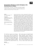
Figure 1B,C,F shows an ultra-thin plasmodium stained
by the overlay technique with Rhodamine-B-amine–
polymalate in early S phase. The red fluorescence indicates
polymalate–dye, the blue indicates chromatin and the green
shows polymalatase epitopes. The red fluorescence appears
together with the blue DAPI staining (Fig. 1F) indicating
the transfer of the probe to the nuclei. These were the only
organelles stained. By injection into normally growing
Fig. 1. Immunofluorescence microscopy of an ultra-thin macroplasmo-
dium (white strain LU 897 · LU 898) in early S phase. Very
similar results were obtained with the yellow strains. The picture at a
magnification of 630-fold was taken with a CCD-camera and is
computer-processed. The red fluorescence shows the position of
Rhodamine-B-amine–polymalate. The living plasmodium was at first
exposed to the reagent for 30 min. At 60 ± 20 min after metaphase,
the plasmodium was fixed and stained with anti-polymalatase Ig fol-
lowed by fluorescein-5-isothiocyanate labeled anti-(rabbit IgG) (green
fluorescence). In the last step, the nuclei were stained with DAPI
(blue fluorescence). For excitation and emission filters see the
Methods section. (A) Fluorescein-5-isothiocyanate-labeled antibody
staining polymalatase epitopes (green), (B) Rhodamine-amine-labeled
polymalate (red), (C) superposition of A and B. (D) DAPI staining of
DNA (blue), (E) superposition of A and D, (F) superposition of A, B
and D. The bar indicates 5 lm.
1538 M. Karl et al.(Eur. J. Biochem. 270) Ó FEBS 2003
macroplasmodia, the intensity of staining was probed as a
function of 10–100 lg of labeled polymalate. At low
concentration, the success of staining depended on the time
of injection in the cell cycle. The majority of nuclei (>90%)
were stained in the first half of S phase. Only a few appeared
in G
2
phase and were referred to as unscheduled nuclei. At
the higher concentration, all nuclei were simultaneously
stained, and the intensity was independent of the time of
injection.
Staining by the overlay method as in Fig. 1 or by
microinjection of 10 lg or less of the conjugate resulted in
the same type of fluorescent nuclei that was typical for early
S phase. The red fluorescence was distributed unevenly in
patches (Fig. 1F). The fluorescence was arranged around
the center of the nucleus, and matched only the peripheral
area of the blue DAPI fluorescence of chromatin. The
patches were reminiscent of the patchwork-like structure of
reconstituting nuclei in early S phase seen by phase contrast
illumination (e.g. [29]). In the case of staining with high
amounts of the dye–polymalate conjugate, the nuclei were
evenly stained.
A very similar result was obtained when the plasmodium
was stained with polymalate conjugated to N-(1-naph-
thyl)ethylenediamine at the carboxyl terminus (results not
shown). However, the staining of nuclei could not be
observed for polymalate conjugated to FITC at its hydroxyl
terminus. These plasmodia showed an enhanced back-
ground fluorescence only. It may be concluded from these
results that for successful delivery into the nucleus the
terminal hydroxyl group has to be unsubstituted, whereas
pending and terminal carboxyl groups could be derivatized.
The kinetics of staining were followed after the injection
of 100 lg Rhodamine-B-amine–polymalate in G
2
phase by
observing nuclei at a distance of 5 cm from the injection
point. Fluorescence was detected after 2 min and displayed
at maximum intensity after 8 min. While the fluorescence
gradually decreased in the nuclei over the next few hours
and finally disappeared after 24 h, it concomitantly
appeared in the agar medium. The dye extracted from the
agar showed the fluorescence of Rhodamine-B-conjugated–
polymalate and the number-averaged molecular mass of
2000. The results suggested that the fluorescent polymalate
probe was delivered rapidly to nuclei, thereby traversing
large cellular distances. Over a prolonged period of time, the
conjugate was secreted into the culture medium, and
exocytosis was followed by a trimming of the polymer
chain as has been observed previously [12].
Polymalate and polymalatase-like protein(s) migrate
jointly to nuclei
It has been suggested that polymalate may not be free in the
plasmodium but bound to an adapter [15]. Polymalatase has
been proposed, because it specifically binds to the hydroxyl-
terminus of polymalate in vitro [14] and is devoid of enzyme
activity at pH 6.5 in the cytoplasm. We investigated whether
the putative adapter was delivered together with the injected
Rhodamine-B-amine–polymalate to the nuclei. After the
injection of low amounts of polymalate–dye conjugate and
fixation, the plasmodium was incubated with antiserum
against polymalatase and stained with FITC-labeled secon-
dary antibody. Green fluorescent material was found
deposited around nuclei, which harbored the red fluorescent
Rhodamine-B-amine–polymalate (compare Figs 1A,C,F).
The green and the red fluorescence were completely
separated from each other (Fig. 1C). The fluorescent
material scattered elsewhere in Fig. 1 is thought to belong
to pieces of halos not focussed to the plane. The occurrence
together of the green halo and the red fluorescence was
verified in more than 90% of the fluorescent nuclei. Thus, in
early S phase almost all of the nuclei could be stained at low
concentrations of the red polymalate–dye conjugate, and
these nuclei exhibited the halo of green FITC-labeled
polymalatase. Plasmodia which had not received the
polymalate–dye conjugate nevertheless showed fluorescent
halos, indicating that the conjugated dye was not a stimulus
for halo formation. These halo intensities compared with
those for polymalate-injected plasmodia, suggesting that the
amount of the intrinsic polymalate was not limiting. Halos
were absent in G
2
phase except around those very few red
fluorescent nuclei that were thought to represent unsched-
uled S phase nuclei. The nuclei, which became fluorescent
only after injection of high amounts (100 lg) of polyma-
late–dye conjugate, did not display the halo. An explanation
was that these nuclei incorporated small amounts of
polymalate and that the level of deposited halo was in these
cases below detection limit. The interior of the nuclei did not
show green fluorescence (Fig. 1A), indicating that poly-
malatase did not enter nuclei. The results are in support of
the simultanous transfer of polymalate and polymalatase to
the surface of the nuclei, the dissociation of this complex,
and the entrance of polymalate only into the nucleus.
Polymalate and polymalatase are constituents
of an
in vivo
complex
While a direct in vivo demonstration of the polymalate–
polymalatase complex was technically not possible, the
following results show support for it. In a first approach, a
complex was demonstrated by nondenaturing electropho-
resis of the cytoplasmic fraction of the yellow strain on
gradient polyacrylamide gels. Immunoblotting revealed
polymalatase as a broad band centered at 450 kDa
(Fig. 2A). SDS gel electrophoresis of the cut-out band
and immunoblotting indicated bands at 220 kDa, 125 kDa,
97 kDa, 68 kDa and 45 kDa as in previous investigations,
which were supportive of a precurser, two forms of
polymalatase and proteolytic fragments [31]. Analysis of
the polymalate content in the cut and eluted nondenaturing
gel indicated a peak in the 200–600 kDa range (Fig. 2B). In
contrast, a sample of purified polymalate migrated at a
position of 100 kDa and below. The addition of spermine
hydrochloride to the sample from the cytoplasm caused a
shift in the polymalate content of the peak towards positions
of lower molecular masses, as was expected on the basis of
the dissociative effect of sperminium ions [5,9]. The amount
of polymalate in the sample of the cytoplasmic fraction was
1 lg. When 0.5 lg of purified polymalate was added to this
sample, the polymer content in the 100–600 kDa position
increased marginally, indicating that the free capacity of the
protein(s) in the cytoplasm was limited in binding further
polymalate. The results are in agreement with the assump-
tion of a polymalatase–polymalate complex, but the binding
to other proteins cannot be excluded.
Ó FEBS 2003 Polymalate locates to nuclei of Physarum plasmodia (Eur. J. Biochem. 270) 1539
In a second approach, polymalate and polymalatase in
the cytoplasmic fraction of the yellow strain were copreci-
pitated by specific anti-polymalatase Igs immobilized on
protein-A/G-Sepharose. Aliquots of 20 lLoftheloaded
Sepharose beads were shown by SDS gel electrophoresis
and Western blotting to contain polymalatase (data not
presented). Another sample (20 lL) was eluted in the
presence of 1
M
NaCl. Polymalate in the eluate was hydro-
lysed and quantitated by the fluorimetric malate dehydro-
genase assay. An average amount of 0.069 ± 0.003 lg
polymalate (SD, three measurements) was detected com-
pared to 0.003 ± 0.003 lg in the control (anti-polymala-
tase serum substituted by rabbit antiserum against BSA).
Variation of the salt concentration in the precipitation
buffer indicated that 0.15
M
NaCl sufficed to dissociate
polymalate from the Sepharose-bound immune complex.
Regarding the balance, polymalate in 1 mL of the cyto-
plasmic fraction was 6 lg (100%), the corresponding
amount in the precipitate was 0.35 lg (6%). This low
amount of coprecipitated polymalate is in agreement with
the large excess of polymalate over polymalatase in the
cytoplasmic fraction.
Discussion
Polymalate has been proposed to function as a molecular
scaffold that binds nuclear proteins and delivers them to the
nuclei of the plasmodium [9]. The details of the in vivo
delivery are largely unknown. To investigate the delivery,
the polymer was labeled with Rhodamine-B-amine or other
fluorescent dyes and microinjected or administered by the
overlay technique. The amounts of polymalate probes
introduced into a typical macroplasmodium (300 mg) were
of the order of 10–100 lg, corresponding to 5–50% of the
total polymalate content (200–250 lg [13]). The immediate
delivery to the nuclei, indicated by their fluorescence
staining, was followed by microscopy, while the amount
of the probe and the time of administration relative to the
cell cycle were varied. After the administration of small
amounts of the probe, high numbers (>90%) of nuclei were
stained only in early S phase. The few nuclei in G
2
phase
were referred to as lacking synchrony. At large amounts of
the probe, nuclei were fluorescent independently of the
phase of the cell cycle and were evenly stained. At low probe
concentrations, however, the fluorescence intensity was
distributed in patches, and these did not coincide with the
intensity of the DAPI-stained DNA. In the plasmodium,
the early S phase immediately follows mitosis and coincides
with the phase of nuclear reconstitution [29]. The fluorescent
patchwork could correspond to the existence of the reported
higher order complexes of polymalate and nuclear proteins
[9,10]. The bright spots are indicative of the reconstituting
nucleolus [29], reflecting complexes of polymalate with
nucleolus-specific proteins, such as the basic fibrillarin-like
protein B-36 [32]. The high degree of staining in the early
S phase correlates with the reported massive incorporation
of
14
C-polymalate into nuclei during feeding of plasmodia
with
D
-[
14
C]glucose [13]. Because the request for nuclear
Fig. 2. Comigration of polymalatase and polymalate in nondenaturing polyacrylamide gradient (3–10%) gel electrophoresis. (A) Western blot with
anti-polymalatase serum. (B) The gel was cut at the positions of molecular masses indicated in the figure; the pieces were extracted and assayed for
polymalate. Molecular mass standards were BSA, ferritin and their oligomers. The samples were: 0.5 lg of purified polymalate; a mixture of
purified polymalate and 50 nmol spermine hydrochloride; 100 lL cytoplasmic fraction (1 lg polymalate); a mixture of 100 lL cytoplasmic fraction
and purified polymalate; a mixture of 100 lL cytoplasmic fraction, purified polymalate, and 50 nmol spermine hydrochloride. The mixtures were
incubated for 10 min on ice before electrophoresis.
1540 M. Karl et al.(Eur. J. Biochem. 270) Ó FEBS 2003
proteins is high in early S phase, the observed specific
and rapid delivery of polymalate to nuclei in the reconsti-
tution phase is in agreement with the proposed carriage
function. The necessity for high concentrations of the
probe to achieve staining of nuclei in G
2
phase is explained
by a decreased rate of polymalate delivery and could
reflect the transport of different proteins in G
2
phase
compared to early S phase. Polymalate recycles to the
cytoplasm for new rounds of delivery. As new molecules of
the polymer are continuously synthesized, a fraction of the
recycled molecules will be secreted into the culture medium
to maintain a constant nuclear level [13]. This is observed as
a depletion of the fluorescence from the nuclei and an
accumulation of fluorescent polymalate in the culture
medium.
Polymalatase has been proposed to function as an
adapter for polymalate [15] and the binding site has been
mapped by inhibitor studies [14]. The hydroxyl-terminus of
the polymer is recognized by a structurally rigid subsite on
the protein. The chemical substitution of the hydroxyl
group by a bulky residue would abolishes the binding for
sterical reasons. Indeed, after substitution with fluorescein-
5-isothiocyanate the staining of the nuclei was abolished.
This result supported the assumption that the delivery of
polymalate to the nuclei required the binding to poly-
malatase. Evidence for a polymalate–polymalatase complex
was provided by the results of electrophoretic and copre-
cipitation experiments. Simultaneous staining observed by
polymalate–dye and anti-polymalatase Igs suggested that
polymalate and polymalatase comigrated to the nuclei. The
assumption of a comigration is corroborated by the fact that
polymalate and polymalatase are soluble in the cytoplasm
and form a complex with each other. The deposition of
polymalatase on the surface of the nuclear envelope is
thought to involve the docking of polymalatase to certain
envelope proteins that induced the dissociation of the
adapter–polymalate complex. While these docking proteins
are not known, it is interesting to refer to the observation
that the dissociation of the adenovirus Ad2 capsid from
virus DNA is mediated by its docking to the nuclear pore
complex of the receptor CAN/Nup214 and histone H1 in
rat [33]. Alternatively, the dissociation of the polymalate–
polymalatase complex could be due to a local ionic strength
effect, disrupting the electrostatic interactions between
polymalate and the adapter protein [14]. A third possibility
could be myristoylation and/or palmitoylation of poly-
malatase by a corresponding transacylase located in the
outer nuclear membrane, resulting in the binding of the
adapter to this membrane and the concomitant release
of polymalate. The primary sequence structure of poly-
malatase suggests such fatty acid acylations (unpublished
data). Whether specific docking proteins, local ionic effects
or fatty acid acylation and membrane binding are involved
has yet to be clarified.
After the dissociation, the released polymalate translo-
cates into the nuclei. Translocation through the nuclear pore
could involve nuclear location signals of proteins carried by
the polymer as cargo (proteins such as histone H1 or DNA
polymerases [9]). It also seems possible that naked poly-
malate could permeate through the nuclear pore because of
its high degree of structural flexibility [34]. Future research
has to unravel the details.
Plasmodia of myxomycetes may have solved their
transport problems in the particularly giant plasmodium
by choosing polymalate. It is of interest how other syncytia
solved this problem and whether the polymalate theme is
used elsewhere in phylogeny or whether it has been replaced
by functionally similar but structurally different devices.
References
1. Wick, R.J. & Sauer, H.W. (1982) Developmental biology of slime
molds: An overview. In Cell Biology of Physarum and Didymium
(Aldrich, H.C. & Daniel, J.W., eds), pp. 3–20. Academic Press,
New York, USA.
2. Baldauf, S.L. & Doolittle, W.F. (1997) Origin and evolution of the
slime molds (Mycetozoa). Proc. Natl. Acad. Sci. 94, 12007–12012.
3. Burland, T.G., Bailey, J., Adam, L., Mukhopadhyay, M.J., Dove,
W.F. & Pallotta, D. (1992) Transient expression in Physarum of a
chloramphenicol acetyltransferase gene under the control of actin
gene promoters. Curr. Genet. 21, 393–398.
4. Thiriet, C. & Hayes, J.J. (1999) Histone proteins in vivo: Cell-cycle-
dependent physiological effects of exogeneous linker histones
incorporated into Physarum polycephalum. Methods: a Companion
to Methods in Enzymology 17, 140–150.
5. Fischer,H.,Erdmann,S.&Holler,E.(1989)Anunusualpoly-
anion from Physarum polycephalum that inhibits homologous
DNA polymerase a in vitro. Biochemistry 28, 5219–5226.
6. Doehoefer, S., Windisch, C., Angerer, B., Lavrik, O.I., Lee, B S.
& Holler, E. (2002) The DNA-polymerase inhibiting activity of
poly (b-
L
-malic acid) in nuclear extract during the cell cycle of
Physarum polycephalum. Eur. J. Biochem. 269, 1253–1258.
7. Achhammer, G., Angerer, B., Windisch, C., Uhl, A. & Holler, E.
(1992) DNA Polymerase a-primase complexes of Physarum
polycephalum. Cell. Biol. Int. Reports 16, 1047–1053.
8. Achhammer, G., Winkler, A., Angerer, B. & Holler, E. (1995)
DNA polymerase d of Physarum polycephalum. Curr. Genet. 28,
534–545.
9. Angerer, B. & Holler, E. (1995) Large complexes of b-poly
(
L
-malate) with DNA polymerase a, histones, and other proteins
in nuclei of growing plasmodia of Physarum polycephalum.
Biochemistry 34, 14741–14751.
10. Doerhoefer, S., Khodyreva, S., Safronov, I.V., Wlassoff, W.A.,
Anarbaev, R., Lavrik, O.I. & Holler, E. (1998) Molecular con-
situents of the replication apparatus in the plasmodium of Phy-
sarum polycephalum: identification by photoaffinity labelling.
Microbiology 144, 3181–3193.
11. Willibald,B.,Bildl,W.,Lee,B S.&Holler,E.(1999)Isb-Poly
(
L
-malate) synthesis catalysed by a combination of b-
L
-malyl-
AMP-ligase and b-poly(
L
-malate) polymerase? Eur. J. Biochem.
265, 1085–1090.
12. Korherr, C., Roth, M. & Holler, E. (1965) Poly(b-
L
-malate)
hydrolase from plasmodia of Physarum polycephalum. Can. J.
Microbiol. 41 (Suppl. 1), 192–199.
13. Schmidt, A., Windisch, C. & Holler, E. (1996) Nuclear accumu-
lation and homeostasis of the unusual polymer b-poly(
L
-malate) in
plasmodia of Physarum polycephalum. Eur. J. Cell. Biol. 70,373–
380.
14.Gasslmaier,B.,Krell,C.M.,Seebach,D.&Holler,E.(2000)
Synthetic substrates and inhibitors of b-poly(
L
-malate)-hydrolase
(polymalatase). Eur. J. Biochem. 267, 5101–5105.
15. Karl, M. & Holler, E. (1998) Multiple polypeptides immuno-
logically related to b-poly(
L
-malate) hydrolase (polymalatase) in
the plasmodium of the slime mold Physarum polycephalum. Eur. J.
Biochem. 251, 405–412.
16. Daniel, J.W. & Baldwin, H.H. (1964) Methods of culture for
plasmodial myxomycetes. Methods Cell Physiol. 1, 9–14.
Ó FEBS 2003 Polymalate locates to nuclei of Physarum plasmodia (Eur. J. Biochem. 270) 1541
17. Nygaard, O.P. & Guttes, S.R.H.P. (1960) Nucleic acid metabolism
in a slime mold with synchronous mitosis. Biochim. Biophys. Acta
38, 298–306.
18. Holler, E. (1997) Poly(malic acid) from natural sources. Handbook
of Engineering Polymeric Materials (Cheremisinoff, N.P., ed.), pp.
93–103. Marcel Dekker, Inc, New York, USA.
19. Means, G.E. & Feeney, R.E. (1971) Chemical Modification of
Proteins. Holden-Day, San Franciso, USA.
20. Geiger, B. & Volberg, T. (1998) Cell Biology. Academic Press,
New York, USA.
21. Walter, W. & Bode, K D. (1967) Synthesen von Thiourethanen.
Angewandt Chemie 79, 285–328.
22. Gasslmaier, B. & Holler, E. (1997) Specificity and direction of
depolymerization of b-poly(
L
-malate) catalysed by polymalatase
from Physarum polycephalum. Fluorescence labeling at the
carboxy-terminus of b-poly(
L
-malate). Eur. J. Biochem. 250,
308–314.
23. Laemmli. U.K. (1970) Cleavage of structural proteins during
the assembly of the head of bacteriophage T4. Nature 227,
680–685.
24. Holler, E., Fischer, H. & Simek, H. (1985) Non-disruptive detec-
tion of DNA polymerases in nondenaturing polyacrylamide gels.
Eur. J. Biochem. 151, 311–317.
25. Heukeshoven, J.V. & Dernick, R. (1988) Improved silver staining
for fast staining in Phast System Development Units. Staining of
SDS-Gels. Electrophoresis 2, 28–32.
26. Towbin, H., Staehlin, T. & Gordon, J. (1979) Electrophoretic
transfer of proteins from polyacrylamide gels to nitrocellulose
sheets: Procedure and some applications. Proc. Natl. Acad. Sci.
USA 76, 4350–4354.
27. Adams, J.C. (1981) Heavy metal intensification of DAB-based
HRP reaction product. J. Histochem. Cytochem. 29,775.
28. Naib-Majani, W., Osborn, M., Weber, K., Wohfarth-Bottermann,
K E., Hinssen, H. & Stockem, W. (1983) Immunocytochemistry
of the acellular slime mold Physarum polycephalum. J.CellSci.60,
13–28.
29. Mohberg, J. (1982) Recognition of mitosis. Cell Biology of
Physarum and Didymium (Aldrich, H.C. & Daniel, J.W., eds), pp.
273–276. Academic Press, New York, NY.
30. Prior, C.P., Cantor, C.R., Johnson, E.M. & Allfrey, V.G. (1980)
Incorporation of exogenous pyrene-labeled histone into Physarum
chromatin: a system for studying changes in nucleosome
assembledinvivo.Cell 20, 597–608.
31. Holler,E.,Achhammer,G.,Angerer,B.,Gantz,B.,Hambach,C.,
Reisner, H., Seidel, B., Weber, C., Windisch, C., Braud, C.,
Guerin, P. & Vert, M. (1992) Specific inhibition of Physarum
polycephalum DNA-polymerase-a-primase by poly(
L
-malate) and
related polyanions. Eur. J. Biochem. 206,1–6.
32. Christensen, M.E. & Fuxa, K.P. (1988) The nucleolar protein B-36
contains a glycine and dimethylarginine-rich sequence conserved
in several other nuclear RNA-binding proteins. Biochem. Biophys.
Res. Commun. 155, 1278–1283.
33. Trotman, L.C., Mosberger, N., Fornerod, M., Stidwill, R.P. &
Greber, U.F. (2001) Import of adenovirus DNA involves the
nuclear pore complex receptor CAN/Nup214 and histone H1.
Nature Cell Biol. 3, 1092–1100.
34. Lee, B S., Vert, M. & Holler, E. (2002) Water-soluble aliphatic
polyesters: poly(malic acid)s. In Biopolymers 3a (Doi,Y.&
Steinbu
¨
chel, A., eds), pp. 75–103. Wiley-VCH, Weinheim
(Bergstrasse), Germany.
1542 M. Karl et al.(Eur. J. Biochem. 270) Ó FEBS 2003