Báo cáo Y học: O-GalNAc incorporation into a cluster acceptor site of three consecutive threonines Distinct specificity of GalNAc-transferase isoforms pot
Bạn đang xem bản rút gọn của tài liệu. Xem và tải ngay bản đầy đủ của tài liệu tại đây (447.89 KB, 11 trang )
O-GalNAc incorporation into a cluster acceptor site of three
consecutive threonines
Distinct specificity of GalNAc-transferase isoforms
Hideyuki Takeuchi
1
, Kentaro Kato
1
, Helle Hassan
2
, Henrik Clausen
2
and Tatsuro Irimura
1
1
Laboratory of Cancer Biology and Molecular Immunology, Graduate School of Pharmaceutical Sciences, The University of Tokyo,
Japan;
2
Department of Oral Diagnostics, Faculty of Health Sciences, School of Dentistry, University of Copenhagen, Denmark
O-Glycosylation of three consecutive Thr residues in a
fluorescein-conjugated peptide PTTTPLK ) which mimics
a portion of mucin 2 ) by four isozymes of UDP-N-ace-
tylgalactosaminyltransferases (pp-GalNAc-T1, T2, T3, or
T4) was investigated. Partially glycosylated versions of this
peptide, PT*TTPLK, PTTT*PLK, PT*TT*PLK,
PTT*T*PLK, PT*°TTPLK, and PTTT*°PLK (*, N-acetyl-
galactosamine; °, galactose), were also tested. The products
were separated by RP-HPLC and characterized by MALDI-
TOF MS and peptide sequencing. The first and the third Thr
residues act as the peptide’s initial glycosylation sites for
pp-GalNAc-T4, which were different from the sites for
pp-GalNAc-T1 and T2 (the first Thr residue) or T3 (the third
Thr residue) shown in our previous report. All pp-GalNAc-
T isozymes tested exhibited distinct specificities toward
glycopeptides. The most notable findings were: (a) prior
incorporation of an N-acetylgalactosamine residue at the
third Thr greatly enhanced N-acetylgalactosamine incor-
poration into the other Thr residues when pp-GalNAc-T2,
T3, or T4 were used; (b) the enhancing effect of the N-ace-
tylgalactosamine residue on the third Thr was completely
abrogated by galactosylation of this N-acetylgalactosamine;
(c) prior incorporation of an N-acetylgalactosamine at the
first Thr did not have any enhancing effect; (d) pp-GalNAc-
T2 was unique as it transferred N-acetylgalactosamine into
the second Thr residue only when N-acetylgalactosamine
was attached to the third one.
Keywords: O-glycosylation; mucin; polypeptide N-acetylga-
lactosaminyltransferase; Tn antigen; UDP-GalNAc.
Biosynthesis of O-glycans is mediated by the step-wise
addition of monosaccharides by a variety of glycosyl-
transferases, where topology and kinetic properties of
Golgi-resident glycosyltransferases are believed to generate
additional diversity of carbohydrate structures [1]. The
initial O-glycosylation is thought to be a highly selective
process where the sequence context determines where
O-glycans are attached to proteins, although the rules
governing this selection are still poorly understood [2–11].
Mucins form a large family of membrane-associated or
secretory glycoproteins rich in O-glycans. They are pro-
duced by epithelial cells and function as a physical and
biological barrier protecting mucous epithelia. There are
also leukocyte and erythrocyte markers with mucin-like
structures. The core polypeptides of mucins are not only
rich in serines and threonines but they also contain Ser and
Thr repeats, and tandem repeats of Ser/Thr-rich stretches
[12,13]. Sequences with consecutive Thr and Ser residues
seem to play important roles in recognition events. Trun-
cated O-glycans displayed on consecutive Thr residues serve
as ligands for endogenous C-type lectins on macrophages
and carcinoma-specific anti-Tn antibodies [14]. Many
mucin-like leukocyte markers such as CD34, CD45 and
CD68 bear sequences containing consecutive Ser and Thr
residues at their outermost segments [15–17]. Therefore, it is
tempting to speculate that these consecutive Ser/Thr
sequences with various arrangements of O-glycans are
structural motifs having specific biological relevance [18].
The first step of mucin O-glycosylation is initiated by
a family of UDP-N-acetyl-
D
-galactosamine : polypeptide
UDP-N-acetylgalactosaminyltransferases (pp-GalNAc-Ts,
EC 2.4.1.41) that transfer N-acetylgalactosamine(GalNAc)
residues to Ser and Thr residues in a polypeptide. To date,
nine members of the mammalian pp-GalNAc-T family have
been cloned and characterized [19–31]. Although the kinetic
properties and substrate specificities of some of these
recombinant isozymes have been investigated by in vitro
studies using several synthetic peptides as substrates, we are
still far from understanding the regulation of O-glycosyla-
tion [32–35]. When the peptide PTTTPITTTTK [that
represents a portion of the mucin 2 (MUC2) tandem
repeat] was used as a substrate with detergent-soluble
microsome fractions from the human colon carcinoma cell
line LS174T (which expresses several members of the
GalNAc-Ts family), GalNAc was transferred to these Thr
Correspondence to T. Irimura, Laboratory of Cancer Biology and
Molecular Immunology, Graduate School of Pharmaceutical
Sciences, The University of Tokyo, 7-3-1 Hongo, Bunkyo-ku,
Tokyo 113-0033, Japan.
Fax: + 81 3 5841 4879, Tel.: + 81 3 5841 4870,
E-mail:
Abbreviations: pp-GalNAc-T, UDP-N-acetyl-
D
-galactosaminide,
polypeptide N-acetylgalactosaminyltransferase.
Enzymes:UDP-N-acetyl-
D
-galactosamine : polypeptide UDP-
N-acetylgalactosaminyltransferases (EC 2.4.1.41).
(Received 19 May 2002, revised 22 August 2002,
accepted 28 October 2002)
Eur. J. Biochem. 269, 6173–6183 (2002) Ó FEBS 2002 doi:10.1046/j.1432-1033.2002.03334.x
residues in a specific and distinct order [36,37]. Also, we
reported that pp-GalNAc-T isoforms (GalNAc-T1, -T2,
and -T3) exhibited different orders of incorporation of
GalNAc residues into consecutive Thr residues of the
PTTTPLK acceptor peptide [38].
These results suggest that some pp-GalNAc-T isoforms
work in a cooperative fashion transferring to different
acceptor sites in clusters. Evidence demonstrating negative
effects of GalNAc attachments for subsequent activities of
pp-GalNAc-Ts [39], suggests that the order by which
GalNAc-T isoforms initiate glycosylation may lead to
different pathways of biosynthesis resulting in different
patterns of O-glycan occupancy. Furthermore, it has been
proposed recently that some GalNAc-T isoforms function
as follow-up enzymes in that they are directed by the initial
action of other isoforms [21,23,28,35,40]. This latter mech-
anism is not fully understood, but recent data by Hassan
and coworkers indicate that the putative lectin domains of
these isoforms are responsible for the unique GalNAc-
glycopeptide specificities [41]. We therefore hypothesized
that different subsets of pp-GalNAc-T isoforms are desig-
nated to generate different arrangement of O-glycans on
mucins having consecutive Thr residues. Using a simple
model substrate with three consecutive Thr acceptor
residues, we examined whether vicinal effects, positive as
well as negative, of GalNAc and Galb1–3GalNAc residues
on the efficacy and pathway of incorporation of the second
and the third GalNAc residues with four pp-GalNAc-Ts
were observed.
EXPERIMENTAL PROCEDURES
Synthesis of acceptor substrates
A synthetic oligopeptide PTTTPLK, was used as the
acceptor substrate for the pp-GalNAc-T isozymes. Its
sequence was derived from the tandem repeat domain of
the MUC2 core polypeptide (PTTTPITTTTTVTPTPTPT
GTQT) [42]. It was synthesized on a Model 9020 peptide
synthesizer (Milligen, Burlington, MA, USA) with a lysine
as the C-terminal residue. The peptide was labelled at
pH 7.5 (adjusted with 100 m
M
Hepes buffer) with fluores-
cein isothiocyanate (FITC) at its N-terminal amino acid
under conditions in which the e-amino groups of lysine
residues were not modified. The lysine was added to allow
further modifications to study the interaction of resultant
glycopeptides with carbohydrate recognition molecules [14]
but such experiments are not described in the present report.
Using FITC–PTTTPLK as a substrate, glycopeptides
containing GalNAc residues were prepared enzymatically.
Two glycopeptides, designated FITC–PT*TTPLK or
FITC–PT*TT*PLK (where * stands for a GalNAc residue),
were generated with recombinant pp-GalNAc-T1. The
remaining two glycopeptides, denoted FITC–PTTT*PLK
and FITC–PTT*T*PLK, were prepared with recombinant
pp-GalNAc-T3. Glycopeptides with Galb1–3GalNAc resi-
dues were prepared enzymatically using FITC–PT*TTPLK
or FITC–PTTT*PLK as acceptor substrates, UDP-Gal
(final 1 m
M
) as donor substrates, and detergent-soluble
microsome fractions of human laryngeal carcinoma H.Ep.2
cells as the source of UDP-Gal:N-acetylgalactosaminide
b1–3 galactosyltransferase(s). The incubation conditions
and the preparations of microsome fractions are described
in the following sections. All glycopeptides were purified by
RP-HPLC on a C
18
column. Sites of GalNAc attachment
were confirmed by protein sequencing using the PE
Biosystems 490 Procise protein sequencing system [38]. To
test the effect of the FITC residue on the acceptor specificity
of pp-GalNAc-Ts, the same peptide without an FITC
residue was synthesized, used as an acceptor substrate, and
conjugated with FITC for the HPLC separation. In another
experiment, the same peptide with additional six alanine
residues at the N terminus was synthesized, conjugated with
FITC, and used as an acceptor.
Preparation of recombinant pp-GalNAc-Ts
Soluble recombinant pp-GalNAc-T1, T2, and T3 were
prepared as described previously [43]. Briefly, each of the
plasmids pAcGP67-GalNAc-T1-sol, pAcGP67-GalNAc-
T2-sol, and pAcGP67-GalNAc-T3-sol were cotransfected
with Baculo-Gold DNA (Pharmingen) to Sf9 cells. The
recombinant pp-GalNAc-T1, T2, and T3 were purified
from the spent media. pp-GalNAc-T4 was prepared from
the secretions of a stably transfected Chinese hamster ovary
(CHO) cell line (CHO/GalNAc-T4/21 A) as described
previously [22]. One unit of recombinant enzyme was
defined as the amount of enzyme that transferred 1 nmol of
GalNAc residues in 30 min onto FITC–PTTTPITTTTK at
a final concentration of 5 l
M
in 50 lL-incubation mixtures.
Preparation of detergent-soluble microsome fractions
of H.Ep.2 cells
Human laryngeal carcinoma H.Ep.2 cells were cultured in
modified Eagle’s medium supplemented with 10% fetal
bovine serum. Cells were homogenized in 50 m
M
Tris/HCl
buffer pH 7.5 containing 250 m
M
sucrose, 1 lgÆmL
)1
aprotinin (Sigma), 1 lgÆmL
)1
leupeptin (Peptide Institute
Inc.,Osaka,Japan),and0.5lgÆmL
)1
pepstatin A (Sigma).
After centrifugation at 3000 g at 4 °C for 10 min, the
decanted supernatant was centrifuged at 100 000 g for 1 h.
The pellet was re-suspended in the buffer used during the
homogenization containing an additional 0.1% Triton
X-100 (Sigma). Protein concentrations were determined
using Protein Assay Kit (Bio-Rad) with BSA as a standard.
The solutions were stored in aliquots at )80 °C until use.
Enzymatic GalNAc incorporation into peptide and
glycopeptide acceptors
The standard enzyme reaction mixture consisted of 50 m
M
Hepes buffer pH 7.5, 5 m
M
MnCl
2
,5m
M
2-mercapto-
ethanol, 0.1% Triton X-100, 1 m
M
UDP-N-acetyl-
D
-gal-
actosamine (Sigma), 5 l
M
acceptor peptides or
glycopeptides, and 0.2 U recombinant enzyme pp-GalNAc
T1, T2, T3, or T4 (0.2268 lg, 1.098 lg, 1.365 lg, and
2.768 lg, respectively), in a final volume of 100 lL.
Reactions were performed at 37 °C for 16 h and were
terminated by adding 20 lL of 500 m
M
EDTA.
Monitoring of in vitro O-glycosylation by RP-HPLC
The glycosylated peptides were separated by RP-HPLC
(JASCO, Tokyo, Japan). A Cosmosil column (C
18
,
10 · 250 mm; Nacalai tesque, Japan) was used. The
6174 H. Takeuchi et al. (Eur. J. Biochem. 269) Ó FEBS 2002
column was eluted with a linear gradient ranging from 0 to
50% solvent B (0.05% trifluoroacetic acid in 70% 2-pro-
panol in acetonitrile) in solvent A (0.05% trifluoroacetic
acid in water) at a flow rate of 2 mLÆmin
)1
for 30 min.
Eluates were monitored by fluorescence intensity at 520 nm.
MALDI-TOF MS of glycosylated peptides
Glycosylated peptides were applied on a tip and mixed with
a10mgÆmL
)1
solution of a-cyano-4-hydroxycinnamic acid
dissolved in 0.1% trifluoroacetic acid/50% ethanol in water.
All mass spectra were obtained on a Voyager Elite
instrument (Nippon PerSeptive Biosystems, Tokyo, Japan)
operating at an accelerating voltage of 20 kV (grid voltage
93.5%, guide wire voltage 0.05%) in the linear mode with
the delayed extraction setting. Recorded data were pro-
cessed by using GRAMS/386 software.
Amino acid sequencing
Pulsed liquid Edman degradation amino acid sequencing
of glycopeptides was performed with the Applied Biosys-
tems 490 Procise protein sequencing system (Perkin
Elmer). With this system, a phenylthiohydantoin-deri-
vative of GalNAc-attached Thr was identified as a pair of
peaks eluted near the positions of phenylthiohydantoin-
Ser and phenylthiohydantoin-Thr [44]. Amino acid
sequencing of fully glycosylated peptide (FITC–
PT*T*T*PLK) confirmed the eluting positions. The
peptides used in the present study were modified at the
N terminals and the amino acid (Pro) was not detected.
The second amino acid (Thr2) was detected at the first
cycle of Edman degradation.
RESULTS
Fractionation of products resulting from glycosylation
of FITC–PTTTPLK peptide by pp-GalNAc-T4
An FITC-labelled oligopeptide PTTTPLK that mimicked
the tandem repeat portion of MUC2 was chemically
synthesized and labelled with FITC at its N-terminal
amino acid residue. Theoretically, seven different products
could be generated from this peptide upon incubation
with a pp-GalNAc-T isozyme in the presence of UDP-
N-acetyl-
D
-galactosamine. When FITC–PTTTPLK was
incubated with recombinant pp-GalNAc-T4 for various
periods ranging up to 24 h and then subjected to
RP-HPLC, six peaks, including the unaltered peptide,
were observed depending on the incubation period
(Fig. 1). These fractions (a–e) were collected separately
and analysed by MALDI-TOF MS, which showed that
the fractions corresponded to FITC–PTTTPLK bearing
either one, two, or three glycosylated residues (Fig. 2).
Thus, peaks (b) and (c) apparently contained two
GalNAc residues and peaks (d) and (e) apparently
contained a single GalNAc residue. Peak (a) appears to
be the fully glycosylated peptide. The associated peaks on
the MALDI-TOF MS profiles are not likely to be due to
contaminating glycopeptides with smaller numbers of
attached GalNAc residues, judging from the clear separ-
ation of glycopeptides with given numbers of GalNAc
residues on the RP-HPLC. These peaks in MALDI-TOF
MS profiles should be the result of degradation during
the matrix-assisted ionization of these glycopeptides. The
degree of glycosylation depended on the duration of
incubation. Up to 6 h, the six peaks could be detected,
with the major fraction being unglycosylated peptide. At
24 h, peptides bearing one or three GalNAc residues were
prominent. After the addition of fresh enzyme and UDP-
GalNAc, the proportion of the peptide bearing three
GalNAc residues increased (data not shown).
Characterization of the pp-GalNAc-T4 glycosylation
products
The sites of GalNAc attachment to the peptide were
analysed by amino acid sequencing. As shown in Fig. 3,
peak (a) isolated by RP-HPLC, indicated that all three Thr
residues are glycosylated, while peak (b) contained two
GalNAc residues at Thr-3 and Thr-4. Peak (d), which
constituted the major peak in the HPLC, consisted of the
peptide glycosylated at Thr-2. Thus, it is clear that peak (d)
is not the precursor of peak (b). Amino acid sequencing of
the minor peaks corresponding to peptides with one or two
GalNAc residues, namely, peaks (c) and (e), was unsuc-
cessful because of their minute quantity. According to their
retention times, peaks (e) and (c) are likely to contain FITC–
PTTT*PLK and FITC–PT*TT*PLK, respectively,
although the possibility that they are FITC–PTT*TPLK
and FITC–PT*T*TPLK, cannot be excluded. The presence
of the three major products in this incubation mixture can
be explained by the unique acceptor specificity of
pp-GalNAc-T4, which is different from the activities of
pp-GalNAc-T1, T2, or T3 on FITC–PTTTPLK, as repor-
ted previously [38].
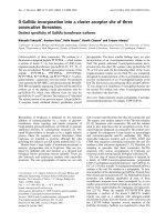
Fig. 1. Elution profiles of products separated by RP-HPLC after incu-
bation of FITC–PTTTPLK peptide with recombinant pp-GalNAc-T4
for the indicated periods.
Ó FEBS 2002 Regulation of peptide O-glycosylation (Eur. J. Biochem. 269) 6175
Ability of partially glycosylated FITC–PTTTPLK with
GalNAc to act as acceptor substrate for all
four isozymes
To understand further the regulation of GalNAc transfer to
consecutive Thr residues in a mucin, acceptor specificities
should be investigated with a glycopeptide whose Thr
residues have already been partly occupied. Thus, the effects
of prior attachment of GalNAc residues to this peptide on
the activities of pp-GalNAc-T1, T2, T3 or T4 were
examined. We enzymatically synthesized four GalNAc
peptides with one or two GalNAc residues. Using these
four glycopeptides and FITC–PTTTPLK as acceptors (all
at a final concentration of 5 l
M
), GalNAc-T assays in a 100-
lL reaction mixture with 0.2 U pp-GalNAc-T1, T2, T3, or
T4 were performed for 16 h. Incubation products were
subjected to RP-HPLC (Fig. 4). The separated fractions
were concentrated and analysed by MALDI-TOF MS and
theresultsaresummarizedin
1
Table 1.
As we had reported previously, a maximum of two, one
or three GalNAc residues was transferred onto the ungly-
cosylated FITC–PTTTPLK by pp-GalNAc-T1, T2 or T3,
respectively [38]. When FITC–PT*TTPLK was used as an
acceptor with pp-GalNAc-T1, T2, T3, or T4, 5.1%, 3.2%,
23.8%, and 10.8% of the products bore an additional
GalNAc residue, respectively, while 0%, 3.4%, 3.7%, and
3.4% were fully glycosylated, respectively.
When FITC–PT*TT*PLK was used as an acceptor
substrate, incorporation of an additional GalNAc residue
did not significantly occur with any of the pp-GalNAc-T
isozymes. When FITC–PTTT*PLK was used as an accep-
tor, glycopeptide products with an additional GalNAc
constituted 65.1%, 19.0%, 16.3%, and 10.3% of the total
products for pp-GalNAc-T1, T2, T3 or T4, respectively.
The product resulting from the action of pp-GalNAc-T1
was FITC–PT*TT*PLK and the proportion of this product
was relatively high partly because it was not converted
further. pp-GalNAc-T2, T3, or T4 efficiently converted
FITC–PTTT*PLK into the fully glycosylated form.
Fig. 2. Representative profiles of MALDI-TOF MS of FITC–
PTTTPLK peptide glycosylated by recombinant pp-GalNAc-T4 and
separated by RP-HPLC. Mass indicates the (M + H) + form. The
profiles a–f represent the materials retrieved from peaks a–f indicated
in Fig. 1. (a) The predicted mass (1755.9) corresponds to FITC–
PTTTPLK peptide with three attached GalNAc residues. (b and c)
The predicted mass (1552.7) corresponds to FITC–PTTTPLK peptide
with two attached GalNAc residues. (d and e) The predicted mass
(1349.5) corresponds to FITC–PTTTPLK peptide with a single
attached GalNAc residue. (f) The predicted mass (1146.3) corresponds
to FITC–PTTTPLK peptide with no GalNAc residue.
Fig. 3. Profiles of amino acid sequencing chromatograms of FITC–
PTTTPLK peptide and its major derivatives glycosylated by recombin-
ant pp-GalNAc-T4. (A) The profile of FITC–PTTTPLK with three
GalNAc residues attached [peak (a) in Fig. 1]. (B) FITC–PTTTPLK
peptide with two GalNAc residues attached [peak (b) in Fig. 1]. (C)
FITC–PTTTPLK peptide with a single GalNAc residue attached
[peak (d) in Fig. 1]. (D) Untreated FITC–PTTTPLK peptide [peak (f)
in Fig. 1]. Asterisks indicate phenylthiohydantoin (PTH)-derivatized
a-GalNAc-Thr, which was detected as a pair of peaks.
Fig. 4. Elution profiles of products separated by RP-HPLC after incu-
bation of FITC–PTTTPLK peptide or its glycosylated derivatives with
recombinant pp-GalNAc-T1, T2, T3, or T4 for 16 h. Acceptor sub-
strates were as follows: (A) FITC–PTTTPLK; (B) FITC–PT*TTPLK;
(C) FITC–PT*TT*PLK; (D) FITC–PTTT*PLK; (E) FITC–
PTT*T*PLK (GalNAc-Thr was indicated by T*). Broken lines indi-
cate the retention time of each substrate.
6176 H. Takeuchi et al. (Eur. J. Biochem. 269) Ó FEBS 2002
Two products containing two GalNAc residues, namely,
FITC–PT*TT*PLK and FITC–PTT*T*PLK, were both
generated by the action of pp-GalNAc-T2, T3, or T4.
FITC–PTT*T*PLK was the apparent intermediate product
to be converted into the fully glycosylated form because
FITC–PTT*T*PLK was efficiently converted to the fully
glycosylated form by pp-GalNAc-T2, T3, or T4, as shown
in Table 1.
Effects of galactosylation of GalNAc residues at Thr-2
or the Thr-4
Vicinal effects of attachment of a Gal residue to GalNAc at
the first Thr residue (Thr-2) or the third Thr residue (Thr-4)
were investigated. Using FITC–PT*TTPLK and FITC–
PTTT*PLK as acceptors, two glycopeptides with Galb1–
3GalNAc at Thr-2 or Thr-4 were prepared. The structures
of these glycopeptides, FITC–PT*°TTPLK and FITC–
PTTT*°PLK (T*° indicates Galb1–3GalNAca-Thr), were
confirmed by MALDI-TOF MS, by their binding to peanut
agglutinin specific for Galb1–3GalNAc, and sensitivity to
b-galactosidase from Bacillus circulans specific for 1–3
linked b-galactoside [45]. Using four glycopeptides (FITC–
PT*TTPLK, FITC–PT*°TTPLK, FITC–PTTT*PLK and
FITC–PTTT*°PLK) as acceptors, GalNAc-T assays were
Table 1. Relative quantity of glycopeptides formed after incubation of
FITC-PTTTPLK with pp-GalNAc-T1, T2, T3 or T4 and UDP-GalNAc
for 16 h.
Acceptor Enzyme
Retention
time
(min)
Number of
GalNAc
attached
Percent of
total
products
PTTTPLK T1 23.1 2 22.4
23.9 1 59.8
24.3 1 15.0
25.0 0 2.8
T2 21.8 3 3.1
23.1 2 2.1
23.8 1 71.5
24.2 1 16.5
24.9 0 6.7
T3 21.9 3 68.6
22.6 2 10.4
23.1 2 2.8
23.8 1 4.5
24.2 1 13.6
T4 21.9 3 30.2
22.7 2 5.4
23.1 2 1.9
23.8 1 29.1
24.2 1 14.6
24.9 0 18.8
PT*TTPLK T1 23.2 1 5.1
24.0 0 94.9
T2 21.8 2 3.3
23.0 1 3.2
23.7 0 93.4
T3 21.8 2 3.7
23.0 1 23.8
23.7 0 72.4
T4 21.9 2 3.3
22.8 1 7.3
23.2 1 3.4
23.9 0 85.8
PT*TT*PLK T1 23.7 0 100
T2 22.0 1 1.6
23.2 0 98.4
T3 21.9 1 2.1
23.0 0 97.9
T4 22.0 1 4.7
23.1 0 95.3
PTTT*PLK T1 23.3 1 65.1
24.1 0 34.9
T2 22.0 2 72.8
22.8 1 10.4
23.2 1 8.5
23.9 0 8.3
T3 22.0 2. 72.7
22.7 1 12.6
23.1 1 3.7
23.9 0 11.0
T4 21.8 2 82.2
22.6 1 6.3
23.0 1 3.9
23.7 0 7.5
Table 1. (Continued).
Acceptor Enzyme
Retention
time
(min)
Number of
GalNAc
attached
Percent of
total
products
PTT*T*PLK T1 21.8 1 8.2
22.5 0 91.8
T2 21.9 1 92.8
22.6 0 7.2
T3 22.0 1 87.4
22.7 0 12.5
T4 22.0 1 93.4
22.7 0 6.6
Fig. 5. Elution profiles of products separated by RP-HPLC after incu-
bation of glycosylated derivatives of FITC–PTTTPLK with recombinant
pp-GalNAc-T1, T2, T3, or T4 for 16 h. Acceptor substrates were as
follows: (A) FITC–PT*TTPLK; (B) FITC–PT*°TTPLK; (C) FITC–
PTTT*PLK; (D) FITC–PTTT*°PLK (GalNAc-Thr and Galb1–
3GalNAc-Thr were indicated by T* and T*°, respectively). Broken
lines indicate the retention time of each substrate.
Ó FEBS 2002 Regulation of peptide O-glycosylation (Eur. J. Biochem. 269) 6177
performed in a 100-lL reaction mixture with 0.2 U
pp-GalNAc-T1, T2, T3, or T4. After a 16-h incubation
products were subjected to RP-HPLC (Fig. 5). The peak
fractions were pooled, concentrated by evaporation, and
analysed by MALDI-TOF MS. The results indicated that
the influence of prior Gal transfer on the vicinal GalNAc
transfer depended on the site and the isozyme type of
pp-GalNAc-T
1
as summarized in Table 2. Gal transfer to
GalNAc on Thr-2 did not increase the efficiency of
GalNAc-incorporation by pp-GalNAc-T1. pp-GalNAc
T2, 3, or 4, transferred GalNAc to a very low extent when
Thr-2 was occupied by GalNAc or Galb1–3GalNAc. The
effect of Gal transfer to the GalNAc attached to the Thr-4
was very slight as far as pp-GalNAc-T1 is concerned.
As stated in the previous sections, greater proportions of
Thr-2 and Thr-3 residues in FITC–PTTT*PLK received
transfer of GalNAc residues with pp-GalNAc-T2, T3, or T4
than FITC–PTTTPLK. The first site of the incorporation
was apparently Thr-3 then to Thr-2. This vicinal enhancing
effect was abrogated by the addition of a Gal residue to the
GalNAc residue in FITC–PTTT*PLK. The position of the
GalNAc incorporation was Thr-2 resulting in the formation
of FITC-PT*TT*°PLK in the case of the action of
pp-GalNAc-T1 and T2 according to the protein sequencing
analysis (Fig. 6).
Effects of modification of N terminals of PTTTPLK
Differences in the incorporation of GalNAc into underiva-
tized and the fluorescein-labeled PTTTPLK by pp-
GalNAc-T2 or T3 were compared. The products from the
underivatized peptide were reacted with FITC and applied
to RP-HPLC. The number of GalNAc incorporated
residues was estimated by MALDI-TOF MS. A glycopep-
tide with one GalNAc residue was the predominant product
after 16 h incubation with pp-GalNAc-T2. Peptide sequen-
cing analysis indicated that the GalNAc residue was
attached to Thr-2. Four peaks of glycopeptides with three,
two, two, or one GalNAc residues were identified in the
reaction mixture with pp-GalNAc-T3. By peptide sequen-
cing analysis, FITC–PTT*T*PLK and FITC–PT*TT*PLK
were identified as indicated in Fig. 7A. These results
indicated that modification of the N terminus of acceptor
peptides with FITC had no significant effect on the order or
maximum number of attachment of GalNAc residues.
When FITC–PTTTPLK was used as an acceptor and
incubated with pp-GalNAc-T3 for 16 h, a glycopeptide
with three GalNAc residues was the major product, whereas
PTTT*PLK was the major product from underivatized
peptide incubated under the same conditions.
Peptide AAAAAAPTTTPLK was synthesized and
labelled with FITC. FITC–AAAAAAPTTTPLK was
Table 2. Relative quantity of glycopeptides formed after incubation of
FITC-PTTTPLK containing a Galb1-3GalNAca residue with pp-Gal-
NAc-T1, T2, T3 or T4 and UDP-GalNAc for 16 h.
Acceptor Enzyme
Retention
time
(min)
Number of
GalNAc
attached
Percent of
total
products
PT*TTPLK T1 23.2 1 1.5
24.0 0 98.5
T2 21.9 2 2.2
23.1 1 0.8
23.8 0 97.0
T3 21.8 2 2.5
23.0 1 19.7
23.7 0 77.8
T4 21.8 2 2.6
22.6 1 8.6
23.0 1 2.0
23.7 0 86.7
PTTT*PLK T1 23.3 1 68.6
24.0 0 31.4
T2 21.8 2 74.1
22.6 1 6.8
23.0 1 4.8
23.8 0 14.2
T3 21.9 2 71.9
22.6 1 12.1
23.0 1 3.7
23.8 0 12.3
T4 21.8 2 80.7
22.6 1 6.6
23.8 0 12.7
PT*°TTPLK T1 22.9 0 100
T2 23.0 0 100
T3 22.4 1 6.0
23.1 0 94.0
T4 21.5 1 10.6
23.1 0 89.4
PTTT*°PLK T1 22.4 1 81.0
23.4 0 19.0
T2 22.4 1 33.2
23.4 0 66.8
T3 22.5 1 8.5
23.5 0 91.4
T4 22.6 1 9.6
23.5 0 90.4
Fig. 6. Profiles of amino acid sequencing chromatograms of products
after incubation of FITC–PTTT*°PLK peptides with (A) recombinant
pp-GalNAc-T1 or (B) pp-GalNAc-T2 and those of untreated substrates
(C) FITC–PTTT*°PLK and (D) FITC–PT*°TTPLK. Asterisks indi-
cate PTH-derivatized a-GalNAc-Thr, which is detected as a pair of
peaks.
6178 H. Takeuchi et al. (Eur. J. Biochem. 269) Ó FEBS 2002
incubated with pp-GalNAc-T2 or T3 for 16 h. Elution
profiles of the products on the RP-HPLC were shown in
Fig. 7B. Peptide sequencing analysis indicated that
pp-GalNAc-T2 transferred one GalNAc residue to Thr-8,
the first Thr in the Thr triad. The first GalNAc residue
transferredbypp-GalNAc-T3seemedtoattachtoThr-8
and Thr-10 and a product FITC–AAAAAAPT*TTPLK
did not seem to be further modified. The ratio of FITC–
T*TTPLK to FITC–PT*T*T*PLK was smaller than the
ratio of FITC–AAAAAAPT*TTPLK to FITC–AAAAA
APT*T*T*PLK.
DISCUSSION
We hypothesize that the arrangement of O-glycans on
consecutive Ser/Thr residues in mucins and mucin-like cell
surface receptors generate structural motifs. If this really is
the case, then the biosynthetic pathway of O-glycans on
consecutive Ser/Thr should strictly be regulated regarding
where and what order the O-glycosylation occurs. In the
study presented here, the initial sites of O-glycosylation and
the subsequent order of attachment of GalNAc to a
sequence containing three consecutive Thr residues by four
glycosyltransferase isoforms were investigated. The prefer-
ential site of glycosylation in FITC–PTTTPLK and parti-
ally modified peptides by the action of each pp-GalNAc-T
are summarized in Fig. 8. The initial site of GalNAc
attachment to FITC–PTTTPLK with pp-GalNAc-T1, T2,
and T3 was predominantly Thr-2, Thr-2, and Thr-4,
respectively, as described previously [38]. pp-GalNAc-T4
appears to have two preferential initial glycosylation sites,
which results in the formation of FITC–PT*TTPLK [peak
(d) in Fig. 2] and FITC–PTTT*PLK [putative sequence
of peak (e) in Fig. 2]. Other investigators using various
synthetic peptide acceptors have already reported that each
pp-GalNAc-T has preference for different flanking amino
acid sequences surrounding the Thr residue. It has not been
demonstrated that the order of GalNAc incorporation into
three Thr and/or Ser residues in the vicinity is strictly
determined. Most of the previous studies focused on
probability that one site was more likely to be glycosylated
Fig. 7. Elution profiles of (A) PTTTPLK and (B) FITC–AAA
AAAPTTTPLK peptides. (A) Elution profiles of PTTTPLK peptides
incubated with pp-GalNAc-T2 (a), pp-GalNAc-T3 (b) or buffer alone
(c), for 16 h prior to labelling with FITC on the RP-HPLC. The
estimated structures of the glycopeptides corresponding to the peaks
are depicted schematically. (B) Elution profiles of FITC–AAA
AAAPTTTPLK peptides incubated with pp-GalNAc-T2 (a),
pp-GalNAc-T3 (b) or buffer alone (c), for 16 h on the RP-HPLC. The
estimated structures of the glycopeptides corresponding to the peaks
are depicted schematically.
Fig. 8. Summary of actions of four recombinant pp-GalNAc-Ts toward
TTT stretch in FITC–PTTTPLK peptide and its partially glycosylated
derivatives. (A) pp-GalNAc-T1 (B) pp-GalNAc-T2 (C) pp-GalNAc-
T3, and (D) pp-GalNAc-T4 are shown. The products are indicated by
shaded squares according to the proportion among whole products. d,
Gal residues in acceptor substrates; h, GalNAc residues in acceptor
substrates. The percentage of GalNAc incorporated was calculated
based on the total amount of acceptor substrates.
Ó FEBS 2002 Regulation of peptide O-glycosylation (Eur. J. Biochem. 269) 6179
over another site. Our present results show that the order,
i.e. which Thr is glycosylated first and which Thr is second,
is determined almost exclusively when a peptide sequence
and a pp-GalNAc-T are fixed. The preferential pathways of
O-glycosylation of a peptide containing three consecutive
Thr residues (FITC–PTTTPLK) are indicated in Fig. 9.
Very interestingly and importantly, the preferential order
did not change when a 10-fold concentration of the acceptor
substrates were used and additional components were
not generated when the incubation period was extended
up to 48 h with an addition of the same amounts of
pp-GalNAc-Ts.
Previous reports indicated that Pro residues positively
influenced GalNAc incorporation into a particular Thr
residue [33]. Statistical studies on various peptides contain-
ing O-glycans suggested that Pro residues located at )1and
+3 positions relative to the glycosylation site had positive
effects, although the pp-GalNAc-T having this preference
was not clear [2–5,8]. The FITC–PTTTPLK used in the
present study have two Pro residues, which potentially
provide positive effects on Thr-2 according to the previous
reports [32]. These Pro residues may contribute to the initial
glycosylation site by pp-GalNAc-T1 and T2 but obviously
not by pp-GalNAc-T3. The specificity of each pp-GalNAc-
T seems to be unique toward consecutive Thr residues and
their partially glycosylated derivatives. For example, when
partially glycosylated FITC–PTTTPLK were used as
acceptor substrates, the effect of the attached GalNAc
residues on the activity of pp-GalNAc-T1 was obvious.
Although the initial glycosylation site for pp-GalNAc-T1 is
Thr-2, this isozyme could not glycosylate Thr-2 of FITC–
PTT*T*PLK. Thus, the ability of pp-GalNAc-T1 to
transfer GalNAc onto a Thr immediately upstream ()1)
of an existing GalNAc-Thr residue is likely to be suppressed.
Neither a GalNAc residue nor a Galb1–3GalNAc residue at
Thr-4 of FITC–PTTTPLK significantly influenced the
activity of pp-GalNAc-T1 which could transfer one
GalNAc residue to Thr-2, resulting in the formation of
FITC–PT*TT*PLK or FITC–PT*TT*°PLK.
pp-GalNAc-T2, T3, and T4 behaved differently from
pp-GalNAc-T1 in that the presence of GalNAc-Thr at the
penultimate position (+1) promoted their efficacy. Thus,
FITC–PTTT*PLK could be rapidly converted to the fully
glycosylated form by all of these isozymes via the interme-
diate FITC–PTT*T*PLK. The preferential glycosylation of
the peptide with one GalNAc residue was inhibited by the
addition of a Gal residue to this GalNAc residue in FITC–
PTTT*PLK.
Several issues regarding the use of a relatively short
peptide with fluoresceine at the N terminus as a substrate
should be carefully evaluated. The kinetic parameters
reported for three FITC–conjugated peptides in our previ-
ous publication were not distinct from those for unmodified
MUC2 peptide (PTTTPISTTTMVTPTPTPTC) reported
by Wandall and coworkers [43]. We also examined the
specificity of detergent-soluble microsome fraction of
human colon carcinoma LS174T cells towards larger
GalNAc-glycosylated peptides than FITC–PTTTPLK used
in the present study [37]. RT/PCR and immunocytological
analysis indicated that LS174T cells expressed at least
pp-GalNAc-T1, T2, T3, and T4. In vitro GalNAc-T assays
were performed using FITC–PTTT*PITTTTK, FITC–
PT*TTPITTTTK, FITC–PTT*T*PITT*T*TK, and
FITC–PT*TTPIT*T*T*TK as substrates. Similar results
on the specificity to that of our present results were also
observed in these assays, although the microsome fraction
contained more than two pp-GalNAc-Ts. FITC–
PTTT*PITTTTK were efficiently glycosylated and conver-
ted to FITC–PT*T*T*PIT*T*T*T*K. When FITC–
PTTT*PITTTTK was used as a substrate, the order of
incorporation of GalNAc residues was restricted in the
formation of PT*T*T*P. Within this motif, PTTT*P, a
GalNAc residue was incorporated at Thr-3 at first, and after
that, one more GalNAc residue was incorporated at Thr-2.
Similarly, FITC–PTT*T*PITT*T*TK were converted to
fully glycosylated FITC–PT*T*T*PIT*T*T*T*K. Thus,
the presence of extra C-terminal sequence did not seem to
influence the order of GalNAc incorporation. We did not
examine the effect of two Pro residues on specificity of
pp-GalNAc-Ts by mutation analysis, although Pro residues
in a flanking sequence may influence the initial GalNAc-
attachment site in a polypeptide as mentioned above.
There are few previous reports regarding the acceptor
specificity of GalNAc transfer by pp-GalNAc-T isozymes
on unglycosylated and partially glycosylated sequences.
Hanisch and coworkers reported that the addition of a
GalNAc residue by pp-GalNAc-T isozymes, in particular
pp-GalNAc-T2, to Ser-16 in the tandem repeat of the
MUC1 mucin was accelerated when the adjacent Thr-17
Fig. 9. Putative pathways of GalNAc incorporation into FITC–
PTTTPLK by the action of pp-GalNAc-T1 (A), pp-GalNAc-T2 (B), pp-
GalNAc-T3 (C), and pp-GalNAc-T4 (D). *, GalNAc residues; s,Gal
residues; bold arrows, reactions in which > 50% GalNAc was incor-
porated; broken arrows, the reactions in which < 50% GalNAc was
incorporated; shaded letters, hypothetical glycosylation products
which were not detected in the present investigations.
6180 H. Takeuchi et al. (Eur. J. Biochem. 269) Ó FEBS 2002
residue was glycosylated [39,40]. Bennett and coworkers
also reported that the catalytic activity of pp-GalNAc-T4
with a peptide corresponding to a MUC2 sequence was
enhanced fivefold by prior incorporation of 1–2 mole of
GalNAc by pp-GalNAc-T2 [23]. However, the structural
characteristics responsible for this effect were not elucidated.
In the study by Bennett and coworkers with a MUC1
peptide, pp-GalNAc-T4 preferentially transferred GalNAc
onto a Ser residue adjacent to a glycosylated Thr [21]. Thus,
our findings are consistent with prior reports. In addition,
we are also able now to delineate the structural basis that
regulates GalNAc incorporation into three consecutive Thr
residues.
The present work indicates that GalNAc attachment to
one of three consecutive Thr residues is an important factor
that negatively or positively affects subsequent transfers of
GalNAc residues. The mechanisms behind this remain to be
explored in detail, but factors should include sequence
context, influence of GalNAc residues to conformation and
recognition of acceptor and modulation of kinetic proper-
ties potentially through the lectin domain. GalNAc residues
attached to the peptides via the lectin motifs contained
within their sequences, as has been postulated previously
[46]. Hagen and coworkers showed that mutations in the
C-terminal ricin-like lectin motif of murine pp-GalNAc-T1
did not alter its catalytic properties [27].
Attachment of Gal to GalNAc at Thr-4 of FITC–
PTTTPLK inhibited the transfer of GalNAc to Thr-3 by
pp-GalNAc-T2, T3, and T4. This suggests that pp-Gal
NAc-T isozymes may recognize directly GalNAc residues in
the vicinity. It is an interesting possibility that GalNAc-Ts
compete with glycosyltransferases responsible for the
extension of O-glycans. Brockhausen and coworkers
showed that galactose incorporation by UDP-Gal:glyco-
protein-GalNAc 3-b-
D
-galactosyltransferase (core 1
b3-Gal-T) purified from rat liver became less efficient when
acceptor peptides were heavily converted with GalNAc
[47,48]. From results to determine the glycosylation pattern
of porcine submaxillary mucin tandem repeats, Gerken and
coworkers suggested that local glycopeptide structures, such
as GalNAc density, regulate the in vivo elongation of the
O-glycan by the porcine core 1 b3-Gal-T [44,49,50].
Although many factors potentially modulate attachment
and elongation of O-glycans remain unknown, coordinated
actions of pp-GalNAc-Ts and Gal-Ts should play a major
role in generating a variety of structural motifs on consecu-
tive Thr residues. The present study suggests that a decrease
in galactosylation of GalNAc residues in consecutive Thr
residues in mucins does not only expose GalNAc residues
but also promotes the formation of GalNAc clusters. This
should result in an efficient binding to parasitic protozoa
such as Entamoeba histolytica through their lectins specific
for clusters of O-linked GalNAc residues [51]. The O-glycan
structures of a mucin-like molecule, CD43, were shown to
be modulated upon the exposure of epithelial cells to bac-
terial lipopolysaccharides [52], which appeared to be similar
to the change observed in T cells [53]. However, the present
report is one of very few to show that glycan extension
directly affects the glycosylation of backbone peptides.
In conclusion, we show that a peptide mimicking a
portion of MUC2 containing three consecutive Thr residues
(FITC–PTTTPLK) can be glycosylated by pp-GalNAc-T1,
T2, T3, T4, or combinations of these isozymes, into a variety
of differently glycosylated peptides through their unique
acceptor specificities. Each isozyme was unique in the
specificity not only to this peptide but also to the peptides
with one or two GalNAc residues or Galb1–3GalNAc
residues at different positions.
ACKNOWLEDGEMENT
This work was supported by grants-in-aid from the Ministry of
Education, Science, Sports and Culture of Japan (07407063, 09254101,
11557180, and 11672162), the Research Association for Biotechnology,
the Program for the Promotion of Basic Research Activities for
Innovative Biosciences, and the Danish Cancer Society. We thank C.
Hiraiwa for her assistance in preparing this manuscript.
REFERENCES
1. Brockhausen, I. (1999) Pathways of O-glycan biosynthesis in
cancer cells. Biochim. Biophy. Acta 1473, 67–95.
2. Wilson, I.B., Gavel, Y. & von Heijne, G. (1991) Amino acid dis-
tributions around O-linked glycosylation sites. Biochem. J. 275,
529–534.
3. O’Connell,B.C.,Hagen,F.K.&Tabak,L.A.(1992)Theinfluence
of flanking sequence on the O-glycosylation of threonine in vitro.
J.Biol.Chem.267, 25010–25018.
4. O’Connell,B.,Tabak,L.A.&Ramasubbu,N.(1991)Theinflu-
ence of flanking sequences on O-glycosylation. Biochem. Biophys.
Res. Commun. 180, 1024–1030.
5. Hansen, J.E., Lund, O., Tolstrup, N., Gooley, A.A., Williams,
K.L. & Brunak, S. (1998) NetOglyc: prediction of mucin type
O-glycosylation sites based on sequence context and surface
accessibility. Glycoconjugate J. 15, 115–130.
6. Chou, K.C., Zhang, C.T., Kezdy, F.J. & Poorman, R.A. (1995)
A vector projection method for predicting the specificity of Gal-
NAc-transferase. Proteins 21, 118–126.
7.Elhammer,A.P.,Poorman,R.A.,Brown,E.,Maggiora,L.L.,
Hoogerheide, J.G. & Kezdy, F.J. (1993) The specificity of UDP-
GalNAc: polypeptide N-acetylgalactosaminyltransferase as in-
ferred from a database of in vivo substrates and from the in vitro
glycosylation of proteins and peptides. J. Biol. Chem. 268, 10029–
10038.
8. Hansen, J.E., Lund, O., Engelbrecht, J., Bohr, H. & Nielsen, J.O.
(1995) Prediction of O-glycosylation of mammalian proteins:
specificity patterns of UDP-GalNAc: polypeptide N-acet-
ylgalactosaminyltransferase. Biochem. J. 308, 801–813.
9. Stadie, T.R., Chai, W., Lawson, A.M., Byfield, P.G. & Hanisch,
F.G. (1995) Studies on the order and site specificity of GalNAc
transfer to MUC1 tandem repeats by UDP-GalNAc: polypeptide
N-acetylgalactosaminyltransferase from milk or mammary carci-
noma cells. Eur. J. Biochem. 229, 140–147.
10. Nishimori, I., Johnson, N.R., Sanderson, S.D., Perini, F.,
Mountjoy, K., Cerny, R.L., Gross, M.L. & Hollingsworth, M.A.
(1994) Influence of acceptor substrate primary amino acid
sequence on the activity of human UDP-N-acetylgalactosamine:
polypeptide N-acetylgalactosaminyltransferase. Studies with the
MUC1 tandem repeat. J. Biol. Chem. 269, 16123–16130.
11. Nishimori, I., Perini, F., Mountjoy, K.P., Sanderson, S.D.,
Johnson, N., Cerny, R.L., Gross, M.L., Fontenot, J.D. &
Hollingsworth, M.A. (1994) N-acetylgalactosamine glycosylation
of MUC1 tandem repeat peptides by pancreatic tumor cell
extracts. Cancer Res. 54, 3738–3744.
12. Gendler, S.J. & Spicer, A.P. (1995) Epithelial mucin genes. Annu.
Rev. Physiol. 57, 607–634.
13. Kim, Y.S., J.G. Jr & Brockhausen, I. (1996) Mucin glycoproteins
in neoplasia. Glycoconjugate J. 13, 693–707.
14. Iida, S., Yamamoto, K. & Irimura, T. (1999) Interaction of human
macrophage C-type lectin with O-linked N-acetylgalactosamine
Ó FEBS 2002 Regulation of peptide O-glycosylation (Eur. J. Biochem. 269) 6181
residues on mucin glycopeptides. J.Biol.Chem.274, 10697–
10705.
15. Simmons, D.L., Satterthwaite, A.B., Tenen, D.G. & Seed, B.
(1992) Molecular cloning of a cDNA encoding CD34, a sialo-
mucin of human hematopoietic stem cells. J. Immunol. 148,267–
271.
16. Streuli, M., Hall, L.R., Saga, Y., Schlossman, S.F. & Saito, H.
(1987) Differential usage of three exons generates at least five
different mRNAs encoding human leukocyte common antigens.
J.Exp.Med.166, 1548–1566.
17. Holness, C.L. & Simmons, D.L. (1993) Molecular cloning of
CD68, a human macrophage marker related to lysosomal glyco-
proteins. Blood 81, 1607–1613.
18.Irimura,T.,Denda,K.,Iida,S.,Takeuchi,H.&Kato,K.
(1999) Diverse glycosylation of MUC1 and MUC2: potential
significance in tumor immunity. J. Biochem. (Tokyo) 126,975–
985.
19. White, T., Bennett, E.P., Takio, K., Sorensen, T., Bonding, N. &
Clausen, H. (1995) Purification and cDNA cloning of a human
UDP-N-acetyl-a-D-galactosamine: polypeptide N-acetylgalactos-
aminyltransferase. J.Biol.Chem.270, 24156–24165.
20. Zara, J., Hagen, F.K., Ten Hagen, K.G., Van Wuyckhuyse, B.C.
& Tabak, L.A. (1996) Cloning and expression of mouse UDP-
GalNAc: polypeptide N-acetylgalactosaminyltransferase-T3.
Biochem. Biophys. Res. Commun. 228, 38–44.
21. Bennett, E.P., Hassan, H., Mandel, U., Mirgorodskaya, E.,
Roepstorff, P., Burchell, J., Taylor-Papadimitriou, J., Hollings-
worth,M.A.,Merkx,G.,vanKessel,A.G.,Eiberg,H.,Steffensen,
R. & Clausen, H. (1998) Cloning of a human UDP-N-acetyl-a-D-
Galactosamine: polypeptide N-acetylgalactosaminyltransferase
that complements other GalNAc-transferases in complete O-gly-
cosylation of the MUC1 tandem repeat. J. Biol. Chem. 273,
30472–30481.
22. Bennett, E.P., Hassan, H., Mandel, U., Hollingsworth, M.A.,
Akisawa, N., Ikematsu, Y., Merkx, G., van Kessel, A.G.,
Olofsson, S. & Clausen, H. (1999) Cloning and characterization of
a close homologue of human UDP-N-acetyl-a-
D
-galactosamine:
Polypeptide N-acetylgalactosaminyltransferase-T3, designated
GalNAc-T6. Evidence for genetic but not functional redundancy.
J. Biol. Chem. 274, 25362–25370.
23. Bennett, E.P., Hassan, H., Hollingsworth, M.A. & Clausen, H.
(1999) A novel human UDP-N-acetyl-
D
-galactosamine: polypep-
tide N-acetylgalactosaminyltransferase, GalNAc-T7, with specifi-
city for partial GalNAc-glycosylated acceptor substrates. FEBS
Lett. 460, 226–230.
24. Bennett, E.P., Hassan, H. & Clausen, H. (1996) cDNA cloning
and expression of a novel human UDP-N-acetyl-a-D-galactos-
amine. Polypeptide N-acetylgalactosaminyltransferase, GalNAc-
T3. J.Biol.Chem.271, 17006–17012.
25. Hagen, F.K., Ten Hagen, K.G., Beres, T.M., Balys, M.M.,
VanWuyckhuyse, B.C. & Tabak, L.A. (1997) cDNA cloning and
expression of a novel UDP-N-acetyl-D-galactosamine: polypep-
tide N-acetylgalactosaminyltransferase. J.Biol.Chem.272, 13843–
13848.
26. Ten Hagen, K.G., Hagen, F.K., Balys, M.M., Beres, T.M., Van
Wuyckhuyse, B. & Tabak, L.A. (1998) Cloning and expression of
a novel, tissue specifically expressed member of the UDP-GalNAc:
polypeptide N-acetylgalactosaminyltransferase family. J. Biol.
Chem. 273, 27749–27754.
27. Hagen, F.K., Hazes, B., de Raffo, R., Sa, D. & Tabak, L.A. (1999)
Structure-function analysis of the UDP-N-acetyl-D-galacto-
samine: polypeptide N-acetylgalactosaminyltransferase. Essential
residues lie in a predicted active site cleft resembling a lactose
repressor fold. J. Biol. Chem. 274, 6797–6803.
28. Ten Hagen, K.G., Tetaert, D., Hagen, F.K., Richet, C.,
Beres, T.M., Gagnon, J., Balys, M.M., Van Wuyckhuyse, B.,
Bedi, G.S., Degand, P. & Tabak, L.A. (1999) Characterization of a
UDP-GalNAc: polypeptide N-acetylgalactosaminyltransferase
that displays glycopeptide N-acetylgalactosaminyltransferase
activity. J.Biol.Chem.274, 27867–27874.
29. White, K.E., Lorenz, B., Evans, W.E., Meitinger, T., Strom, T.M.
& Econs, M.J. (2000) Molecular cloning of a novel human
UDP-GalNAc: polypeptide N-acetylgalactosaminyltransferase,
GalNAc-T8, and analysis as a candidate autosomal dominant
hypophosphatemic rickets (ADHR) gene. Gene 246, 347–356.
30. Simmons, A.D., Musy, M.M., Lopes, C.S., Hwang, L.Y., Yang,
Y.P. & Lovett, M. (1999) A direct interaction between EXT
proteins and glycosyltransferases is defective in hereditary multiple
exostoses. Hum. Mol. Genet. 8, 2155–2164.
31.Toba,S.,Tenno,M.,Konishi,M.,Mikami,T.,Itoh,N.&
Kurosaka, A. (2000) Brain-specific expression of a novel human
UDP-GalNAc: polypeptide N-acetylgalactosaminyltransferase
(GalNAc-T9). Biochim. Biophys. Acta 1493, 264–268.
32. Yoshida, A., Suzuki, M., Ikenaga, H. & Takeuchi, M. (1997)
Discovery of the shortest sequence motif for high level mucin-type
O-glycosylation. J. Biol. Chem. 272, 16884–16888.
33. Hennebicq, S., Tetaert, D., Soudan, B., Boersma, A., Briand, G.,
Richet, C., Gagnon, J. & Degand, P. (1998) Influence of the amino
acid sequence on the MUC5AC motif peptide O-glycosylation
by human gastric UDP-GalNAc: polypeptide N-acetylgalacto-
saminyltransferase(s). Glycoconjugate J. 15, 275–282.
34. Tetaert, D., Richet, C., Gagnon, J., Boersma, A. & Degand, P.
(2001) Studies of acceptor site specificities for three members of
UDP-GalNAc: N-acetylgalactosaminyltransferases by using a
synthetic peptide mimicking the tandem repeat of MUC5AC.
Carbohydr. Res. 333, 165–171.
35. Tetaert, D., Ten Hagen, K.G., Richet, C., Boersma, A., Gagnon,
J. & Degand, P. (2001) Glycopeptide N-acetylgalactosaminyl-
transferase specificities for O-glycosylated sites on MUC5AC
mucin motif peptides. Biochem. J. 357, 313–320.
36. Iida,S.,Takeuchi,H.,Kato,K.,Yamamoto,K.&Irimura,T.
(2000) Order and maximum incorporation of N-acet-
ylgalactosamine into threonine residues of MUC2 core peptide
with microsome fraction of human colon carcinoma LS174T cells.
Biochem. J. 347, 535–542.
37. Kato, K., Takeuchi, H., Miyahara, N., Kanoh, A., Hassan, H.,
Clausen, H. & Irimura, T. (2001) Distinct orders of GalNAc
incorporation into a peptide with consecutive threonines.
Biochem. Biophys. Res., Commun. 287, 110–115.
38. Iida, S., Takeuchi, H., Hassan, H., Clausen, H. & Irimura, T.
(1999) Incorporation of N-acetylgalactosamine into consecutive
threonine residues in MUC2 tandem repeat by recombinant
human N-acetyl-D-galactosamine transferase-T1, T2 and T3.
FEBS Lett. 449, 230–234.
39. Hanisch, F.G., Muller, S., Hassan, H., Clausen, H., Zachara, N.,
Gooley, A.A., Paulsen, H., Alving, K. & Peter-Katalinic, J. (1999)
Dynamic epigenetic regulation of initial O-glycosylation by
UDP-N-acetylgalactosamine: peptide N-acetylgalactosaminyl-
transferases. Site-specific glycosylation of MUC1 repeat peptide
influences the substrate qualities at adjacent or distant Ser/Thr
positions. J.Biol.Chem.274, 9946–9954.
40.Hanisch,F.G.,Reis,C.A.,Clausen,H.&Paulsen,H.(2001)
Evidence for glycosylation-dependent activities of polypeptide
N-acetylgalactosaminyltransferases rGalNAc-T2 and -T4 on
mucin glycopeptides. Glycobiology 11, 731–740.
41. Hassan, H., Reis, C.A., Bennett, E.P., Mirgorodskaya, E.,
Roepstorff, P., Hollingsworth, M.A., Burchell, J., Taylor-Papad-
imitriou, J. & Clausen, H. (2000) The lectin domain of UDP-N-
acetyl-
D
-galactosamine: polypeptide N-acetylgalactosaminyl-
transferase-T4 directs its glycopeptide specificities. J. Biol. Chem.
275, 38197–38205.
42. Gum,J.R.Jr,Hicks,J.W.,Toribara,N.W.,Siddiki,B.&Kim,
Y.S. (1994) Molecular cloning of human intestinal mucin (MUC2)
cDNA. Identification of the amino terminus and overall sequence
6182 H. Takeuchi et al. (Eur. J. Biochem. 269) Ó FEBS 2002
similarity to prepro-von Willebrand factor. J.Biol.Chem.269,
2440–2446.
43. Wandall,H.H.,Hassan,H.,Mirgorodskaya,E.,Kristensen,A.K.,
Roepstorff, P., Bennett, E.P., Nielsen, P.A., Hollingsworth, M.A.,
Burchell, J., Taylor-Papadimitriou, J. & Clausen, H. (1997)
Substrate specificities of three members of the human UDP-
N-acetyl-a-
D
-galactosamine: Polypeptide N-acetylgalactosami-
nyltransferase family, GalNAc-T1-T2, and -T3. J.Biol.Chem.
272, 23503–23514.
44. Gerken, T.A., Owens, C.L. & Pasumarthy, M. (1997) Determi-
nation of the site-specific O-glycosylation pattern of the porcine
submaxillary mucin tandem repeat glycopeptide. Model proposed
for the polypeptide: galnac transferase peptide binding site. J.Biol.
Chem. 272, 9709–9719.
45. Fujimoto, H., Miyasato, M., Ito, Y., Sasaki, T. & Ajisaka, K.
(1998) Purification and properties of recombinant b-galactosidase
from Bacillus circulans. Glycoconjugate J. 15, 155–160.
46. Imberty, A., Piller, V., Piller, F. & Breton, C. (1997) Fold
recognition and molecular modeling of a lectin-like domain in
UDP-GalNac: polypeptide N-acetylgalactosaminyltransferases.
Protein Eng. 10, 1353–1356.
47. Brockhausen, I., Moller, G., Merz, G., Adermann, K. & Paulsen,
H. (1990) Control of mucin synthesis: the peptide portion of
synthetic O-glycopeptide substrates influences the activity of
O-glycan core 1 UDPgalactose: N-acetyl-alpha-galactosaminyl-
R-b 3-galactosyltransferase. Biochemistry 29, 10206–10212.
48. Granovsky, M., Bielfeldt, T., Peters, S., Paulsen, H., Meldal, M.,
Brockhausen, J. & Brockhausen, I. (1994) UDPgalactose: glyco-
protein-N-acetyl-
D
-galactosamine 3-beta-
D
-galactosyltransferase
activity synthesizing O-glycan core 1 is controlled by the amino
acid sequence and glycosylation of glycopeptide substrates. Eur. J.
Biochem. 221, 1039–1046.
49. Gerken, T.A., Owens, C.L. & Pasumarthy, M. (1998) Site-specific
core 1 O-glycosylation pattern of the porcine submaxillary gland
mucin tandem repeat. Evidence for the modulation of glycan
length by peptide sequence. J. Biol. Chem. 273, 26580–26588.
50. Gerken, T.A., Gilmore, M. & Zhang, J. (2002) Determination of
the site-specific oligosaccharide distribution of the O-glycans at-
tached to the porcine submaxillary mucin tandem repeat. Further
evidence for the modulation of O-glycans side chain structures by
peptide sequence. J.Biol.Chem.277, 7736–7751.
51. Yi, D., Lee, R.T., Longo, P., Boger, E.T., Lee, Y.C., Petri,W.A. Jr
& Schnaar, R.L. (1998) Substructural specificity and polyvalent
carbohydrate recognition by the Entamoeba histolytica and rat
hepatic N-acetylgalactosamine/galactose lectins. Glycobiology 8,
1037–1043.
52. Amano, J., Morimoto, C. & Irimura, T. (2001) Intestinal epithelial
cells express and secrete the CD43 glycoform that contains core 2
O-glycans. Microbe. Infect. 3, 723–728.
53. Fukuda, M. (1991) Leukosialin, a major O-glycan-containing
sialoglycoproetin defining leukocyte differentialtion and malig-
nancy. Glycobiology 1, 347–356.
Ó FEBS 2002 Regulation of peptide O-glycosylation (Eur. J. Biochem. 269) 6183