Báo cáo khoa học: Maturation of Pichia pastoris-derived recombinant pro-Der p 1 induced by deglycosylation and by the natural cysteine protease Der p 1 from house dust mite doc
Bạn đang xem bản rút gọn của tài liệu. Xem và tải ngay bản đầy đủ của tài liệu tại đây (300.95 KB, 9 trang )
Maturation of
Pichia pastoris
-derived recombinant pro-Der p 1
induced by deglycosylation and by the natural cysteine protease
Der p 1 from house dust mite
Erica van Oort, Pleuni G. de Heer, W. Astrid van Leeuwen, Ninotska I. L. Derksen, Marcel MuÈ ller,
Stephan Huveneers, Rob C. Aalberse and Ronald van Ree
CLB Department of Immunopathology and Laboratory for Experimental and Clinical Immunology, Academic Medical Center,
University of Amsterdam, the Netherlands
The mature cysteine protease from Dermatophgoides
pteronyssinus, Der p 1, is a ma jor house dust mite a llergen.
Its enzymatic activity has been shown to have pro-in¯am-
matory eects that could also negatively in¯uence ecacy of
allergen-speci®c immunotherapy. The aim of this study was
to express recombinant pro-Der p 1 (rpro-Der p 1) in the
yeast Pichia pastoris and to study its maturation. Expression
was achieved at a concentration ranging from 45 mgáL
)1
(methanol-induced expression) to 168 mgáL
)1
(constitutive
expression). No signi®cant spontaneous maturation of the
secreted proenzyme was observed. rpro-Der p 1 with a
sequence-based molecular mass of 34 kDa was hypergly-
cosylated by the yeast, migrating at 50±60 kDa on SDS/
PAGE. Compared w ith i ts natural counterpart (nDer p 1),
the recombinant proenzyme demonstrated decreased IgE
reactivity, resulting in a 30-fold lower capacity to induce
histamine release from human basophils. D ecreased immu-
noreactivity was also shown by competitive RIA and
sandwich ELISA with Der p 1-speci®c antibody reagents.
CD spectra of r pro-Der p 1 and nDer p 1 revealed signi®-
cant structural di erences. Deglycosylation of rpro-Der p 1
with endoglycosidase H resulted in a decr ease in apparent
molecular mass f rom 5 0 k Da to 34 kDa, but did not aect
nDer p 1. On removal of N-glycans from rpro-Der p 1,
which h arbours t wo putative N-glycosylation sites in both
propeptide and mature sequence, the mature rDer p 1
appeared. This suggests that hyperglycosylation hampers
spontaneous maturation. Maturation of the recombinant
pro-enzyme was also achieved by addition of the active
natural cysteine protease, nDer p 1. In conclusion,
high-level expression of rpro-Der p 1 in P. pastoris results in
a stable h ypoallergenic proenzyme with potential for u se in
allergen-speci®c immunotherapy.
Keywords: allergy; Der p 1; house dust mite; pro-allergen;
yeast.
Group 1 allergen produced by the house dust mite
Dermatophagoides pteronyssinus (Der p 1) has be en
described as an aeroallergen with a molecular mass of
27 kDa, carried (mostly) on mite faeces [1±3]. It is a
glycoprotein with cysteine protease activity and is able to
cleave human CD25 and CD23 [4±7]. This activity enhances
total and speci®c IgE production in mice immunized with
proteolytically active Der p 1 [8±10]. Protease activity of
Der p 1 has also been reported to increase the perm eability
of the human respiratory e pithelium [ 11]. The structure of
Der p 1 w as determined by comparative modeling with
papain, actinidin and papaya proteinase W of the cysteine
proteinase family [12], and epitopes responsible for binding
to IgE and IgG could be identi®ed [13±15].
To produce a fully reactive recombinant version of
Der p 1, several expression systems have been tested. An
Escherichia coli-derived recombinant (as fusion protein) [16]
showed < 50% of the IgE-binding activity of that of the
natural allergen. Expression of Der p 1 in the yeast
Saccharomyces cerevisiae revealed high IgE reactivity,
although clear differences from the natural allergen were
demonstrated [17]. Recently, the precursor form of D er p 1
produced in Drosophila and mammalian cells has been
characterized [18,19]. Although enzymatically inactive, it
was claimed to have similar IgE reactivity to that of the
natural allergen, even though the prosequence was still
attached. This contrasts with results obtained for pro-
Der f 1 expressed in the baculovirus s ystem, where cleavage
of the prosequence was necessary to obtain a fully IgE-
reactive recombinant [20]. The autocatalytic processing of
pro-Der f 1 was achieved by incubation in acidic pH as
described for other cysteine proteases [21]. Jacquet et al. [18]
described rpro-Der p 1 autocatalytic processing by incuba-
tion at 60 °C, acidic pH and addition of up to 20 m
M
cysteine. M aturation of up to 80% was reported, which d id
not impr ove over time [ 18]. E xpression of Der f 1 a nd
Der p 1 in Pichia pastoris was recently r eported by Yasuhara
et al.[23]andBestet al. [24,25], respectively. Maturation of
rpro-Der f 1 was shown to be induced by dialysis against
pH 4.0, resulting in complete IgE-binding capacity and
biological activity. Best et al. [ 24,25] r eported spontaneous
maturation of both rpro-Der f 1 and rpro-Der p 1 during
induced and constitutive expression in P. pastoris.
In our study, secretory e xpression and immunochemical
characterization of the precursor form of Der p 1 in
Correspondence to R. van Ree, Plesmanlaan 125, 1066 CX Amster-
dam, the Netherlands. Fax: + 31 205123170, Tel.: + 3 1 205123242,
E-mail:
Abbreviations: Endo H, endoglycosidase H; RAST, radioallergosor-
bent assay; YPD, yeast extract peptone.
(Received 18 September 2001, revised 23 November 2001, accepted 26
November 200 1)
Eur. J. Biochem. 269, 671±679 (2002) Ó FEBS 2002
P. p as tori s is reported. Methanol-induced expression in two
different strains (SMD1168H and GS115) a nd constitutive
expression in strain X-33 were obtained. Partial cleavage o f
the prosequence was achieved spontaneously after degly-
cosylation or by incubation with nDer p 1 .
MATERIALS AND METHODS
Cloning and sequencing of mature and pro-Der p 1
From a house dust mite kgt11 library (kindly provided by
W. R. Thomas, Princess Margaret Children's Medical
Research Foundation, Perth, Australia) cDNAs of m ature
and pro-Der p 1 were obtained by PCR (Table 1). Subse-
quently, they were cloned into pPICZaA(andpGAPZaA)
in-frame with the s ecretion peptide (Table 1). DNA
sequences were d etermined by automated sequencing
(Applied Biosystems) using t he DYEnamic
TM
ET termina-
tor cycle sequencing premix kit (Amersham Pharmacia
Biotech Inc) according to the manufacturer's instructions.
Sequence primers were used as described in Table 1.
Expression in
P. pastoris
Pro-Der p 1 w as expressed in P. pas toris strain SMD116 8h
(PEP4 mutant, de®cient in protease A, his4+), GS115
(his4+) or X-33 (wild-type), and mature Der p 1 only in
SMD1168h. Transformation was performed as described by
the manufacturer (Invitrogen, San Diego, CA, USA).
Positive clones were selected from yeast extract/peptone/
dextrose medium (YPD plates) containing zeocine
(100 lgámL
)1
) as a selection marker. Selected clones were
inoculated in YPD with zeocine and grown overnight at
29 °C. Cells transformed with pPICZaA were then trans-
ferred to buffered glycerol-complex medium for 24 h, after
which they were centrifuged (glycerol inhibits expression)
andtransferredtoexpressionmedium(bufferedmethanol-
complex medium, pH 5.0) at D
600
10 (SMD1168h) or
D
600
1 (GS115) for methanol-induced expression. After
96 h, the supernatant was harvested.
For constitutive expression, cells containing pro-Der p 1
in pGAPZaA remained in YPD medium after inoculation
of a single colony from a YPD/zeocine plate. From an
overnight culture, 0.225 mL was transferred to inoculate
125 m L YPD medium as described by the manufacturer
(Invitrogen). After 96 h the supernatant was harvested.
Puri®cation of (recombinant) allergens
Recombinant p ro-Der p 1 was puri®ed from culture super-
natant by af®nity chromatography with Sepharose-coupled
monoclonal antibody against nDer p 1 [26]. After the
Der p 1 had been allowed to b ind, the column was washed
with NaCl/P
i
and subsequently eluted with 50% ethylene
glycol/5 m
M
lysine, pH 11. Purity was assessed by SDS/
PAGE/silver staining (Novex, San Diego, CA, USA).
nDer p 1 was af®nity-puri®ed from spent medium extract
[2% (w/v) in NaCl/P
i
/0.01% poly(ethylene glycol) 6000/
0.01% sodium azide (CSL, Melbourne, Australia)]. Pro-
tein concentrations were determined using the BCA method
as described by the manufacturer (Pierce, Rockford, IL,
USA).
SDS/PAGE and immunoblotting
Proteins were separated by SDS/PAGE (4±12%) (Novex)
as described b y the manufacturer, and silver-stained
according to the ExcelGel pr otocol (Amersham Pharmacia
Biotech, Uppsala, Sweden). Western blotting was per-
formed by transferring the proteins on to nitrocellulose
membrane as described by the manufacturer (Novex).
Subsequently, the blots were blocked with NaCl/P
i
/1%
BSA and incubated o vernight with polyclonal rabbit anti-
(Der p 1) Ig. After being washed, t he blots were incubated
overnight with
125
I-labeled sheep anti-(rabbit IgG) Ig (CLB)
and exposed to an autoradiographic ®lm (Eastman Kodak
Company, Rochester, NY, USA).
Radio Allergo Sorbent test (RAST)
RAST was p erformed as described previously [27]. Brie¯y,
both natural and recombinant proteins were coupled to
CNBr-activated Sepharose 4B (250 lg o f allergen per
100 mg of Sepharose; Amersham Pharmacia Biotech).
The Sepharose was resuspended to 2 mgámL
)1
in NaCl/P
i
/
0.3% BSA/0.1% Tween-20, 250 lL of which was incubat-
ed with 50 lL human serum. After incubation overnight,
unbound material was washed away, and 50 lL
125
I-labeled sheep anti-(human IgE) Ig (CLB) was added.
After incubation overnight and a wash, bound radioactiv-
ity w as measured in a c counter. The results were e xpressed
as IUámL
)1
, which were calculated from a standard curve
of serial dilutions of a human/mouse chimeric I gE
antibody directed to Der p 2 a nd Seph arose-coupled
rDer p 2 [28]. A result greater than 0.30 IUámL
)1
was
regarded as positive.
Radiolabeling
Radiolabeling of puri®ed Der p 1 samples ( 25 lg) with
125
I (37 MBq) was performed by the chloramine-T m ethod.
Radiolabeled allergen and free iodine were separated by
size-exclusion chromatography (ACA 54) (Life Technolo-
gies, BioSepra SA Cergy-Saint-Christophe, France).
Table 1. Primers used for PCR, cloning and s equencing o f mature and pro-Der p 1.
Primer name Primer sequence
pPICZaA5¢ cloning primer pro-Der p 1 5¢)GGGCTCGAGAAAA-
GACGTCCATCATCGATCAAAACTTTTG-3¢
pPICZaA3¢cloning primer mature and
pro-Der p 1
5¢)GGGGAGCTCTTAGAGAATGACAACATATGG-3¢
pPICZaA5¢cloning primer mature Der p 1 5¢)GGGCTCGAGAAAAGAACTAACGCCTGCAGTATCAAT-3¢
Sequence primers used for vector pPICZaA5¢AOX, 3¢AOX and a-factor primer
672 E. van Oort et al.(Eur. J. Biochem. 269) Ó FEBS 2002
Competitive RIA
In a competitive RIA [29,30], 50 lL rabbit anti-(Der p 1)
(1 : 2500) [26] was preincubated for 2 h at room tem-
perature with 50 lL of serial dilutions of the inhibitor
(rpro-Der p 1, nDer p 1, mite extract, or Pichia culture
supernatants), before addition of 250 lL P rotein A±Sepha-
rose (2 mgámL
)1
), and 50 lL
125
I-labeled nDer p 1. After
overnight incubation (end-over-end rotation at room tem-
perature), samples were washed, and bound radioactivity
was counted. For the uninhibited value, polyclonal anti-
body was preincubated with NaCl/P
i
/0.3% BSA/0.1%
Tween-20 instead of allergen. All tests were performed in
duplicate.
Der p 1 ELISA
A Der p 1 ELISA was obtained from Indoor Biotechnol-
ogies (Cardiff, UK) and carried out according to the
manufacturer's instructions, except for the substrate system,
which was modi®ed for 3,3¢,5,5¢-tetramethylbenzidine
usage. Consequently, color development was initiated by
adding 100 lL3,3¢,5,5¢-tetramethylbenzidine (10 mgámL
)1
)
in sodium acetate, pH 5.5, and 10 lL3%H
2
O
2
.The
reaction was stopped b y adding 2
M
H
2
SO
4
,afterwhichthe
absorbance was measured at 450/540 nm. All tests were
performed in duplicate.
In vitro
histamine-release assays
White b lood cells were isolated from blood of a nonallergic
donor by Percoll c entrifugation and stripped from IgE by
lactic acid treatment a s d escribed elsewhere [31,32]. Subse-
quently, cells were resensitized with patie nts' sera (n 6)
that tested positive (RAST) on Der p 1. Histamine release
was performed with puri®ed natural and recombinant
Der p 1 (0.1 ngámL
)1
to 10 lgámL
)1
). Liberated h istamine
was measured b y the ¯uorim etric method essentially as
described by Siraganian [33]. The protocol was approved by
the medical ethical committee (MEC) of the Amsterdam
Medical Center under project number: MEC97/030.
Endoglycosidase H (Endo H) cleavage of recombinant
pro-Der p 1
Onevolumeofprotein( 5 lg) was combined w ith 1 vol.
100 m
M
ammonium acetate, pH 5.5, and a ®nal concen-
tration of 0.2% SDS, which was incubated for 10 min at
80 °C. Subsequently, 1 .5 mU Endo H ( Boehringer, Mann-
heim, Germany) was added and incubated at 37 °C
overnight. Endo H is active on N-linked oligosaccharides
of glycopeptides/proteins and cleaves only high-mannose
structures and hybrid structures (AcNeu-Gal-GlcNAc). The
results were analyzed by SDS/PAGE (silver staining),
immunoblot with rabbit anti-(Der p 1) Ig, and concanav-
alin A binding.
Glycan analysis
Natural Der p 1, recombinant pro-Der p 1 (SMD1168h
and X-33) and Endo H-treated rpro-Der p 1 ( SMD1168 h)
were electroblotted o n to nitrocellulose membrane and then
incubated overnight in NaCl/Tris/0.1% Tween 20. Subse-
quently the blot was incubated with concanavalin A
(25 lgámL
)1
;Sigma,StLouis,MO,USA)inNaCl/Tris/
0.1% Tween 20, containing 1 m
M
MgCl
2and
1m
M
CaCl
2
for 90 min. After a wash with N aCl/Tris/0.1% Tween 20,
containing 1 m
M
MgCl
2
and 1 m
M
CaCl
2
, the membrane
was incubated with horseradish peroxidase for 60 min
(50 lgámL
)1
; Sigma) [34]. The bands were visualized with
one tablet of diaminobenzidine in a qua dest (10 mg
diaminobenzidine tetrahydrochloride; Kem-En-Tec,
Copenhagen, Denmark). T he reaction was started with
40 lL30%H
2
O
2
.
Further glycan analysis was carried out with the DIG
Glycan Differentiation Kit (Roche Diagnostics GmbH,
Mannheim, Germany) using the following lectins: Galanthus
nivalis agglutinin, S ambuc us nigra agglutinin, Maackia
amurensis agglutinin, peanut agglutinin, and Datura stra-
monium agglutinin. The Der p 1 samples were dot-blotted
or electroblotted on nitrocellulose after separation by SDS/
PAGE.
Circular dichroism
In CD experiments, ellipticity measurements were per-
formed with nDer p 1 (740 lgámL
)1
and 370 lgámL
)1
)and
rpro-Der p 1 (300 lgámL
)1
) dissolved in 10 m
M
Tris/
EDTA buffer, pH 7.5. The proteins were measured in a
0.05-mm cuvette and subjected to 20 cycles with a resolution
of 0.2 nm and a speed of 20 nmámin
)1
.Thespectrawere
calculated after s ubtraction of the blank (spectra obtained
with 10 m
M
Tris/ 1 m
M
EDTA, pH 7.5). Both spectra were
also corrected with respect to concentration and number o f
amino acids. The percentages of a helices, b sheets and
random structures were interpreted from known reference
spectra.
Autoprocessing of rpro-Der p 1
Puri®ed r pro-Der p 1 ( 37 lgámL
)1
; P ierce), was dialyz ed
for 2 days against 0.2
M
sodium acetate, pH 4.0, which was
reported to induce autocatalyzed cleavage of the prose-
quence in case of Der f 1 [20]. Alternatively, puri®ed
recombinant pro-Der p 1 ( 100 lgámL
)1
) w as applied to
a PD-10 column (Sephadex G-25, bed vol. 9.1 mL; Amer-
sham Pharmacia Biotech AB) equilibrated in 50 m
M
sodium acetate, pH 4.0, to exchange buffer. Cysteine was
added to a concentration of 20 m
M
, and the sample was
incubated at 60 °C for 1.5 h [18]. The effect of SDS (0.05±
0.2%) under these conditions was also studied. Samples
were analyzed by SDS/PAGE.
Proteolytic processing with nDer p 1
125
I-Labeled rpro-Der p 1 (2 lL) was incubated with
nDer p 1 ( 1 lg) at room temperature or 37 °Cin
NaCl/P
i
, pH 7.4, or sodium acetate, pH 5.5, for 4 h in a
®nal volume of 20 lL. Incubation was ended by the
addition of reducing s ample buffer. Samples were analyzed
by autoradiography after separation by S DS/PAGE on
Excel gel (8±18%) (Amersham Pharmacia Biotech).
nDer p 1 was coupled to Sepharose (400 lgnDer p1per
100 mg
)1
Sepharose) and taken up in NaCl/P
i
at
32 mgámL
)1
.rPro-Derp1(6lg, volume 34 lL) was
incubated with 100 lL of this solid phase at room
Ó FEBS 2002 Expression and maturation of recombinant pro-Der p 1 (Eur. J. Biochem. 269) 673
temperature for times ranging from 2 t o 72 h. Supernatant
was harvested after centrifugation and analyzed by SDS/
PAGE/silver staining (Novex).
N-Terminal sequencing
rpro-Der p 1 was separated by SDS/PAGE (4±12% gel;
Novex) and electroblotted on poly(vinylidene di¯uoride)
membrane. The blot was stained with Coomassie R-250
(Bio-Rad, Hercules, CA, USA) in 50% methanol. The band
corresponding to rpro-Der p 1 was excised and sequenced
on a PerkinElmer/Applied Biosystems 476A gas-phase
sequencer (Edman degradation).
Sera
Sera (n 198) with speci®c IgE antibodies against house
dust mite a llergens ( > 0 .3 IU ámL
)1
) were used for RAST
analysis.
Statistical analysis
RAST results for natural and recombinant proDer p 1 were
compared by Spearmann rank correlation and Student's
t-test after log t ransformation. Responses in Der p 1
ELISA and competitive RIA were compared by parallel-
line analyses.
RESULTS
Sequence analyses of mature and pro-Der p 1
cDNAs of mature and pro-Der p 1 were picked up by PCR
from a kgt11 D. pteronyssinus cDNA library. All clones had
identical s equences (81E, 124A, 136S, 149A and 215E) with
those published by Chua et al. [15]. Of the six reported
polymorphisms, only one was observed, being either a
tyrosine or a histidine at postition 50. The clone containing
polymorphism 50Y was selected for expression, because
T-cell responses to peptides containing 50H were decreased
compared with peptides containing 50Y [35].
Expression of mature and pro-Der p 1 in
P. pastoris
strain SMD1168h
Both cDNAs were cloned i nto pPICZaA and transformed
to Pichia strain SMD1168 h. Mature Der p 1 w as not
expressed at a detectable level (< 1 ngámL
)1
) as judged by
competitive R IA. Pro-Der p 1 expression resulted in a ®nal
yield of 55 mgáL
)1
(competitive RIA) [29]. Af®nity puri®-
cation of rpro-Der p 1 gave a ®nal puri®cation yield of 15%.
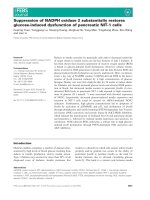
nDer p 1, rpro-Der p 1 and Endo H-treated rpro-
Der p 1 were separated by SDS/PAGE (4±12% gel) and
silver stained (Fig. 1A). rpro-Der p 1 with a theoretical
molecular mass of 34 kDa migrated as a broad band of
50 kDa without any detectable mature Der p 1 at the
level of n Der p 1 (25 kDa). Endo H treatment resulted in a
shift from 50 kDa to 34 kDa, being similar to the
theoretical molecular mass of rpro-Der p 1. This implies
that the high molecular mass of rpro-Der p 1 was caused by
glycosylation. In addition, at least two weaker bands of
lower molecular mass a ppeared on Endo H treatment, one
with molecular mass identical with that of nDer p 1. The
other band of 20 kDa was also present in nDer p 1.
Immunoblot analysis with rabbit antibodies against Der p 1
con®rmed t he Der p 1 nature o f all three bands (Fig. 1B).
Endo H treatment did not affect nDer p 1, suggesting the
absence of N-linked glycosylation (at least the absence of
N-linked glycans for which Endo H has speci®city).
Blot analysis with concanavalin A con®rmed the hyper-
glycosylation of rpro-Der p 1 (Fig. 1C). Concanavalin A
staining almost completely disappeared on Endo H treat-
ment. Concanavalin A staining o f nDer p 1 was weak but
signi®cant. Of t he different l ectins tested with rpro-Der p 1
and nDer p 1 on dot blot, only peanut agglutinin gave a
positive reaction with n Der p 1 (not shown). This s uggests
the presence of O-glycans on nDer p 1, which were not
present on the recombinants. These glycans have been
Fig. 1. (A) SDS/polyacrylamide gel (silver stained), (B) immunoblot
with rabbit anti-(Der p 1) Ig, and (C) concanavalin A blot. (A) L ane 1,
Mark 12 protein ladder (Novex); lane 2, r pro-Der p 1 ( X-33); lane 3,
Endo H-treated rpro-Der p 1 (X-33); lane 4, rpro-Der p 1
(SMD1168h); lane 5, rpro-Der p 1 Endo H-tre ated (SMD1168h); lane
6, rpro-Der p 1 (GS115); lane 7, rpro-Der p 1 Endo H-treated
(GS115); lane 8, Endo H (control); lane 9, nDer p 1 ; lane 10, nDer p 1
(Endo H treated). (B) Lane 1, rpro-Der p 1 (X-33); lane 2, rpro-
Der p 1 (SMD1168 h); lane 3, Endo H-treated rpro-Der p 1
(SMD1168h); lane 4, nDer p 1 . (C) Lane 1, rpro-Der p 1 (X-33); lane
2, rpro-Der p 1 (SMD1168h); lane 3, Endo H-treated rpro-Der p 1;
lane 4, nDer p 1 ; lane 5, prestained, broad-range precision ladder
(Bio-Rad).
674 E. van Oort et al.(Eur. J. Biochem. 269) Ó FEBS 2002
described as having a core disaccharide galactose b(1±3)
N-acetylgalactosamine w hich forms the core unit of
O-glycans (except in yeast glycoproteins).
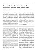
N-Terminal sequencing and CD spectra
N-Terminal sequencing was performed on rpro-Der p 1 to
investigate whether inef®cient cleavage of the yeast secretion
peptide could also be involved in the higher apparent
molecular mass observed on SDS/PAGE. Sequencing
revealed that the recombinant proenzyme starts with the
correct sequence (RPSSIKTFEE) and that no signal peptide
was left attached [15]. Analysis of the CD spectra resulted in
the following predictions for the secondary structures of
nDer p 1 and rpro-Der p 1: 50% a helical and 50%
b pleated sheets compared with an a/b combination with
30% random coil, respectively (Fig. 2).
IgE reactivity (RAST and histamine-release assays)
Patients allergic to house dust mites were tested in a RAST
(n 198) for IgE-speci®c antibodies against nDer p 1 and
rpro-Der p 1 (not shown). IgE b inding to rpro-Der p 1
showed signi®cant correlation with that to nDer p 1
[R
s
0.9077 (+0.8774 to +0.9308), p
s
< 0.01]. How-
ever, binding to nDer p 1 was twice as potent than to the
recombinant protein (2.2 mean ratio; 95% con®dence
interval 2.0 to 2.4). Endo H treatment did not alter the
results signi®cantly (n 14; not shown), although it
cannot be excluded that SDS treatment and low pH
(pH 5 .5) during deglycosylation masked a possible favor-
able effect on the IgE binding of rpro-Der p 1.
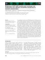
In histamine-release assays, six mite allergic sera were
used to test the ability of the pro-allergen compared with
nDer p 1 to induce histamine re lease ( 0.1 n g mL
)1
to 10 lgámL
)1
). The reco mbinant pro-allergen showed a
greatly decreased biological activity. A 25% histamine
release was achieved with 2 ngámL
)1
nDer p 1, w hereas the
recombinant required a concentration of 60 ng ámL
)1
.In
addition, the mean maximum release was 31% for rpro-
Der p 1 compared with 41% for nDer p 1 (Fig. 3). No
signi®cant release (< 3%) from stripped cells was detected
(data not shown).
Major allergen tests (competitive RIA, sandwich ELISA)
Af®nity-puri®ed nDer p 1 and r pro-Der p 1 were also
compared in a competitive RIA with
125
I-labeled n Der p 1.
nDer p 1 was 9.2-fold more ef®cient as an inhibitor than
rpro-Der p 1(Fig. 4).
Comparison of nDer p 1 and rpro-Der p 1 in a sandwich
ELISA with two Der p 1-speci®c monoclonal antibodies
resulted in much smaller differen ces. Here, the recombinant
was only 2.5-fold less potent (Fig. 5).
Expression of pro-Der p 1 in
Pichia
strain GS115
and X-33
As no mature Der p 1 spontaneously appeared in the
protease-de®cient strain SMD1168h, expression was per-
formed in a nonprotease-de®cient strain, GS115
(45 mgáL
)1
). Again no mature protein was detected (Fig. 1).
The molecular mass of G S115-produced rpro-Der p 1 was
even slightly higher than of the allergen produced in
SMD1168h. On Endo H treatment no signi®cant difference
between recombinant products from either strain was
observed. Degly cosylated GS115-derived rpro-Der p 1 also
migrated at 34 kDa and mature rDer p 1 appeared.
Finally, constitutive expression in strain X-33
(168 mgáL
)1
) was performed t o i nvestigate whether this
Fig. 2. CD spectrum of nDer p 1 vs. rpro-Der p 1. Spe ctra obtained
with 740 lgámL
)1
and 370 lgámL
)1
nDer p 1 are r epresented by bl ue
and red lines, re spectively. r pro-Der p 1 (300 lgámL
)1
)isrepresented
by the dashed and dotted line.
Fig. 3. Histamine-release assays with six Der p 1 allergic patients.
(A±F) represent patients 1 to 6. (j) Re lease induc ed with nD er p 1;
(h) relea se ind uced b y r pro- Der p 1. Concentratio n o f t he a llergen
ranged from 0.1 ngámL
)1
to 10 lgámL
)1
. Histamine release induced by
rpro-Der p 1 was signi®cantly lower th an th at indu ced b y n Der p 1,
varying from a factor o f 10 ( A) to a factor o f 100 (E).
Ó FEBS 2002 Expression and maturation of recombinant pro-Der p 1 (Eur. J. Biochem. 269) 675
wild-type strain facilitates maturation of Der p 1. Results
were, however, essen tially identical with t hose observed for
GS115-produced rpro-Der p 1 (Fig. 1). No spontaneous
maturation o ccurred. Only after deglycosylation was some
mature Der p 1 detected.
Autocatalytic processing of rpro-Der p 1
Methods described for autocleavage of cysteine proteases
[21,22] which were performed for rDer f 1 [20] (buffer
exchange to pH 4.0) and rpro-Der p 1 [18] [buffer exchange
to pH 4.0, addition of cysteine, and heating to 60 °C(with/
without SDS)] did not result in maturation of the recom-
binant pro-allergen (data not shown).
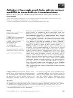
Proteolytic cleavage of recombinant pro-Der p 1
As autocatalytic cleavage was not achieved, enzymatically
active natural Der p 1 was evaluated as a tool to induce
maturation of rpro-Der p 1. Incubation of
125
I-labeled
rpro-Der p 1with crude mite extract and af®nity-puri®ed
nDer p 1 for 4 h at room temperature did result in dose-
dependent cleavage (Fig. 6A). A band with s imilar molec-
ular mass to that of the prosequence appeared with
increasing intensity on addition of increasing doses of
nDer p 1. Surprisingly, no clear band of mature Der p 1
was detected, although a smear became visible slightly
below the molecular mass of rpro-Der p 1. The approach
was repeated with nonradiolabeled rpro-Der p 1. To sep-
arate natural and recombinant mature Der p 1, enzymat-
ically active nDer p 1 was immobilized on Sepharose. Then,
the Sepharose was incubated with rpro-Der p 1. Time-
dependent maturation was observed, with weak but signif-
icant a ppearance of both mature D er p 1 (25 kDa) and the
cleaved propeptide (Fig. 6B). The 25-kDa mature band w as
recognized by rabbit antibodies against nDer p 1, con®rm-
ing the identity of the 25-kDa band as Der p 1 (not shown).
The 10-kDa fragment referred t o as the propeptide was also
recognized by these polyclonal rabbit antibodies. The total
cleavage product was subsequently radiolabeled and sepa-
rated by size-exclusion chromatography. Four peaks were
detected, two of which were again identi®ed a s mature
Der p 1 and the prosequence, respectively (Fig. 6C).
DISCUSSION
In this study, successful high-level expression of recombi-
nant pro-Der p 1 is repo rted. The recombinant protein
proves to be hypoallergenic as it has less than 5% of the
biological activity of its natural counterpart, although IgE
binding in RAST decreases only twofold. Immunoreactivity
as studied by competitive RIA and sandwich ELISA was
also effected. The limited decrease in reactivity observed i n
the sandwich ELISA suggests that both monoclonal
antibodies used are relatively insensitive to the structural
differences between rpro-Der p 1 and nDer p 1. These
discrepancies stress t he need to analyze allergenicity of
candidate hypoallergenic recombinants not only i n IgE-
binding tests such as RAST, ELISA, and immunoblot,
where allergen saturation is usually reached, but also in
biological assays such as histamine-release assays and the
skin prick test. Discrepancies between serological and
biological activity were also reported in studies on Bet v 1,
in which i t was shown that some mAbs e nhanced IgE
binding up to ®vefold, without in¯uencing h istamine-
releasing capacity [36,37]. In the sandwich ELISA, puri®ed
nDer p 1 was also compared with a crude D. p teronyssinus
extract that was calibrated on the WHO standard in
international units (not shown). This analysis showed that
the conversion factor that is generally used, of 1 IU Der p 1
being equivalent to 0.125 ng, is too high. Our calculations
gave similar results as those found by Yasueda et al.[38]:
1IU 0.05 ng Der p 1.
None of the expression systems used in this study
resulted in spontaneous maturation of rpro-Der p 1. To
Fig. 4. Compe tit iv e R IA . rpro-Der p 1 was 9.2 times less eective as an
inhibitor than nDer p 1 in a competitive RIA with rabb it anti-
(Der p 1 ) Ig and radiolabeled puri®ed n Der p 1. Error b ars show t he
range between du plicates.
Fig. 5. Der p 1 ELISA. rpro-Der p 1 was 2 times less potent in
binding to the monoclonal antibodies used in this ELISA than
nDer p 1. Error b ars show the range b etween duplicates.
676 E. van Oort et al.(Eur. J. Biochem. 269) Ó FEBS 2002
the best of our knowledge, we have copied the conditions
for expression that were claimed to result in spontaneous
maturation by Best et al. [24]. The only difference is that
they optimized codon usage for expression in Pichia.It
seems unlikely that codon usage can be at the basis of
differences in post-translational processing. The lack of
induction of maturation of rpro-Der p 1 after dialysis to
pH 4.0 observed in our study contrasts with observations
reported by Yasuhara et al. [23] for rpro-Der f 1. The
main difference between their approach and ours is that in
the present study maturation was attempted with af®nity-
puri®ed rpro-Der p 1 whereas Yasuhara et al. directly
used Pichia culture medium containing the proenzyme.
Possibly yeast-derived proteases facilitated the maturation
process.
Both the propeptide a nd the m ature sequence o f Der p 1
contain a putative N-glycosylation site, although Jacquet
et al. h ave reported that only the asparagine in the
propeptide is glycosylated [18]. In accordance with this,
lack of detectable N-linked glycans on the m ature natural
allergen was implicated by the observation that Endo H
treatment (cleaving off high-mannose and hybrid
N-glycans) did not affect nDer p 1. In contrast, Endo H
treatment of our rpro-Der p 1 resulted in a shift of
20 kDa i n apparent molecular mass on SDS/PAGE.
From these results, it cannot, however, be concluded
whether this i s a result of cleavage of N-glycans from o ne
or both glycosylation sites present in the sequence of pro-
Der p 1. The i nsensitivity of nDer p 1 to End o H does not
mean that the original claim that nDer p 1 is a g lycoprotein
is incorrect [1]. Analysis with several lectins revealed that
nDer p 1 most likely carries O-linked glycans with a core
disaccharide galactose b(1±3) N-acetylgalactosamine that
forms the core unit of O-glycans (except in yeast glycopro-
teins). Endo H treatment did have a strong effect on
rpro-Der p 1. On removal of N-glycans, spontaneous
maturation was observed. These data suggest that hyper-
glycosylation of rpro-Der p 1 in P. pastoris might be an
important factor in preventing maturation. The r esults with
Endo H support the hypothesis that a large high-mannose
structure on the pro-allergen could block cleavage of the
propeptide. Maturation was also observed when t he
recombinant proenzyme was incubated with its enzymati-
cally active natural counterpart. T his process was, however,
still far from ef®cient. Cleavage of radiolabeled rpro-
Der p 1 did not result in any detectable m ature rDer p 1.
Cleavage was, however, occurring because the propeptide
was clearly detected. When the enzymat ic cleavage was
repeated with nonradiolabeled rpro-Der p 1 and nDer p 1
immobilized on Sepharose, mature rDer p 1 was detected.
Most likely, the mature part of r pro-Der p 1 is not
ef®ciently substituted with
125
I in the presence of the
propeptide, in contrast with the recombinant mature
Der p 1 after removal of the p ropeptide.
In summary, enzymatically inactive rpro-Der p 1 with
signi®cantly decreased IgE-binding capacities was produced
Fig. 6. Cleavage of rpro-Der p 1 with nDer p 1. (A) SDS-PAGE/autoradiography. Cleavage of
125
I-labelled recombinant pro-Der p 1 facilitated by
puri®ed nDer p 1. Lane 1, 0 h rpro-Der p 1; lane 2, +0.37 lg nDer p 1; lane 3, +0.74 lg nDer p 1; lane 4, +1.48 lg nDer p 1; lane 5, +2.96 lg
nDer p 1; lane 6, +4.44 lg nDer p 1; and lane 7, +5.92 lg nDer p 1. All incubated for 5 h at room temperature. M
r
compared to SeeBlue Plus 2
pre-stained standards (N ovex). (B) SDS -PAGE /silverst aining. r pro-Der p 1 incubated with nDer p 1 coupled to Sepharose. Lane 1, 10 kDa ladder
(Life techno logies); lane 2, contro l NaCl/P
i
; l ane 3 , 2 h i ncubation; lane 4, 1 night; lane 5 , 2 nights; lane 6, 3 ni ghts. ( C) SDS-PAGE /autor adi-
ography. rpro-Der p 1 was incubated for 2 nights with Sepharose coupled nDer p 1, subsequently radiolabele d (
125
I) and separated by ACA 54 size
exclusion chromatography. Five dierent fractions were analyzed by SDS-PAGE/autoradiography, revealing: lane 1, dimerized rpro-Der p 1; lane
2, monomeric non-cleaved rpro-Der p 1; lane 3, mature rDer p 1 ; lane 4, containing bo th mature rDer p 1 and pro-peptide; lane 5, pro-peptide. M
r
compared to SeeBlue Plus 2 pre-stained standards (Novex).
Ó FEBS 2002 Expression and maturation of recombinant pro-Der p 1 (Eur. J. Biochem. 269) 677
at high expression levels in Pichia. Both the lack of
enzymatic activity and the hypoallergenic character make
this recombinant a potential safe candidate for a pplication
in allergen-speci®c immunotherapy. To further evaluate the
potential of this app roach, future investigations must
examine whether naturally occurring human cysteine pro-
teases could transform hypoallergenic rpro-Der p 1 into
biologically active mature Der p 1.
ACKNOWLEDGEMENTS
We thank W. R . Thomas for k indly providing the ho use dust mite
kgt11 library, Fridolin van der Lecq and others for t heir quick and
excellent work on the protein s equences (Sequentie centrum, Utrecht,
the Netherlands), and Dr Maurits de Planque for his explanations,
time, and help, which made it possible to measure the CD spectra (UU
Biochemie, Utrecht, the Netherlands). This study was ®nancially
supported by Stallerge
Á
nes S.A., Alta dis, ANVAR a nd CNRS.
REFERENCES
1. Chapman, M.D. & Platts-Mills, T.A. (1980) Puri®cation and
characterization of the major allergen from Dermatophagoides
pteronyssinus-antigen P1. J. Immunol. 125, 587±592.
2. Tovey, E.R., Chapman, M.D. & Platts-Mills, T.A. (1981) Mite
faeces are a major source of house dust allergens. Nature (London)
289, 592±593.
3. De Luc ca, S., Sporik, R., O'Meara, T.J. & Tovey, E.R. ( 1999)
Mite allergen (Der p 1) is not only carried on mite feces. J. Allergy
Clin. I mmunol. 103, 174±175.
4. Schulz, O., Se well, H.F. & Shakib, F. (1998) Proteolytic cleavage
of CD25, the alpha subun it of the human T cell interleukin 2
receptor, by Der p 1, a major mite allergen with cysteine protease
activity. J. Exp. Med. 187 , 271±275.
5. Shakib, F., Schulz, O. & Sewell, H. (1998) A mite subversive:
cleavage of CD23 and CD25 by Der p 1 enhances allergenicity.
Immunol. Today 19, 3 13±316.
6. Schulz, O., Laing, P., S ewell, H.F. & Shakib, F. (1995) Der p I, a
major allergen of the house dust mite, proteolytically cleaves the
low-anity receptor for human IgE (CD23). Eur. J. Immunol. 25,
3191±3194.
7. Schulz, O., Sutton, B.J., Beavil, R.L., Shi, J., Sewell, H.F., Gould,
H.J., Laing, P. & Shakib, F . (1997) Cleavage of the low-anity
receptor for human IgE (CD23) by a mite cysteine protease: nature
of the cleaved fragment in relation to the structure and function of
CD23. Eu r. J. Immunol. 27, 584±588.
8. Goug h, L., Schulz, O., Sewell, H.F. & Shakib, F. (1999) The
cysteine protease a ctivity of the major d ust mite allergen De r p 1
selectively enh ances the immunoglobulin E antibody response.
J. Exp. Med. 190, 1897±1902.
9. Schulz , O., Sewell, H.F. & Shakib, F. (1999) The interaction
between the dust mite antigen Der p 1 and cell-signalling molecules
in amplifying allergic disease. Clin. Exp. Allergy 29, 439±444.
10.Chapman,M.D.,Smith,A.M.,Vailes,L.D.&Arruda,L.K.
(1997) Recombinant mite allergens. New technologies for the
management of patien ts with asthma. Allergy 52, 374±379.
11. Wan, H., Winton, H.L., Soeller, C., Tovey, E.R., Gruenert, D.C.,
Thompson, P.J., Stewart, G.A., Taylor, G.W., Garrod, D.R.,
Cannell, M.B. & Robinson, C. (1999) Der p 1 facilitates trans-
epithelial allergen delivery by disruption of tight junctions. J. Clin.
Invest . 104, 123±133.
12. Topham, C.M., Srinivasan, N., Thorpe, C.J., Overington, J.P. &
Kalsheker, N.A. (1994) Comparative modelling of major house
dust mite allergen Der p 1: structure validation using an extended
environmental a mino acid propensity table. Protein Eng. 7,869±
894.
13. Greene, W.K., Cyster, J.G., Chua, K.Y., O'Brien, R.M. &
Thomas, W.R. (1991) IgE and IgG b inding of peptides expressed
from fragments of cDNA encoding the major house dust mite
allergen Der p 1. J. Immunol. 147 , 3768±3773.
14. Greene, W.K. & Thomas, W.R. (1992) Ig E binding structures of
the major house dust mite allergen Der p 1. Mol. Immunol. 29,
257±262.
15. Chua, K.Y., Kehal, P.K. & Thomas, W.R. (1993) Sequence
polymorphisms of cDNA clones encoding the mite allergen Der p
1. Int. Arch. Allergy Immunol. 101, 364±368.
16. Thomas, W .R., Stewart, G .A., Simpson, R.J., C hua, K.Y., Ploz za,
T.M., Dilworth, R.J., Nisbet, A. & Turner, K.J. (1988) Cloning
and expression of DNA co ding for th e major ho use dust mite
allergen Der p 1 in Escherichia coli. Int. Arch. Allergy Appl.
Immunol. 85, 127±129.
17. Chua, K.Y., Kehal, P.K., Thomas, W.R., Vaughan, P.R. &
Macreadie, I.G. ( 1992) High-fre quency b inding of IgE to the Der p
allergen expressed i n yeast. J. Allergy C lin. Immunol. 89 , 95±102.
18. Jacquet, A., Haumont, M., Massaer, M., Daminet, V., Garcia, L.,
Mazzu, P., Jacobs, P. & Bollen , A. (2000) Bioc hemical and
immunological c haracterization of a recombinant precursor form
of the house dust mite allergen Der p 1 produced by Drosophila
cells. Clin.Exp.Allergy30, 677±684.
19. Massaer,M.,Mazzu,P.,Haumont,M.,Magi,M.,Daminet,V.,
Bollen, A. & Jacquet, A. (2001) High-level expression in mam-
malian cells of recombin ant house dust mite allergen ProDer p 1
with optimized codon usage. Int. Arch. Allergy Immunol. 125,
32±43.
20. Shoji, H., Hanawa, M., Shibuya, I., Hirai, M., Yasuhara, T.,
Okumura, Y. & Yamakawa, H. (1996) Production of recombinant
mite allergen Der fI in insect cells and characterization of products:
removal of pro-sequence is e ssential to IgE-b inding activity. Biosci.
Biotechnol. B iochem. 60 , 621±625.
21. Mach,L.,Mort,J.S.&Glossl,J.(1994)Maturationofhuman
procathepsin B. Proenzym e activation a n d proteolytic processing
of the precursor to th e m ature proteinase, in vitro, a re primarily
unimolecular processes. J. Biol. C he m 269, 13030±13035.
22. Vernet,T.,Khouri,H.E.,La¯amme,P.,Tessier,D.C.,Musil,R.,
Gour-Salin, B.J., Storer, A.C. & Thomas, D.Y. (1991) Processing
of the papain precursor. P uri®cation of the zymogen and c har-
acterization of its mechanism of processing. J. Biol. Chem. 266,
21451±21457.
23. Yasuhara,T.,Takai,T.,Yuuki,T.,Okudaira,H.&Okumura,Y.
(2001) Biologically active recombinant forms of a major house
dust mite group 1 allergen Der f 1 with full activities of both
cysteine protease and IgE binding. Clin. Exp. Allergy 31, 116±124.
24. Best, E.A., Morales, T., Kane, S., Stedman, K.E., Hunter, S.W.,
McCall, C.A. & M cDermott, M.J. (2001 ) R ecombinant Der p 1
expressed in Pichia pastoris is fully processed and binds serum IgE
with activity com parable to the n at ural allergen. J. Al lergy Clin.
Immunol. 107, S18±S19 (Abstract).
25. Best, E.A., Stedman, K.E., Bozic, C.M., Hunter, S.W., Vailes, L.,
Chapman, M.D., McCall, C.A. & McDermott, M.J. (2000) A
recombinant group 1 house dust mite allergen, rDer f 1, with
biological activities similar to those of t he native a llergen. Protein
Expr. Purif. 20, 462±471.
26. van der Zee, J.S., van Swieten, P., Jansen, H.M. & Aalberse, R.C.
(1988) Skin tests and histamine release with P1-depleted Derma-
tophagoides pteronyssinus body extracts and puri®ed P1. J. Allergy
Clin. I mmunol. 81, 8 84±896.
27.Aalberse,R.C.,Koshte,V.&Clemens,J.G.(1981)Immuno-
globulin E antibodies that crossreact with vegetable foods, pollen,
and Hymenoptera venom. J. Allergy Clin. I mmunol. 68, 356±364.
28. Schuu rman, J., Perdok, G.J., Lourens, T.E., Parren, P.W.,
Chapman, M.D. & Aalberse, R.C. (1997) Production of a
mouse/human chimeric IgE monoclonal antibody to the house
dust mite allergen Der p 2 and its use for the absolute quanti-
678 E. van Oort et al.(Eur. J. Biochem. 269) Ó FEBS 2002
®cation of allergen-speci®c IgE. J. Allergy Clin. Immunol. 99,
545±550.
29. van Ree, R., van Leeuwen, W.A. & Aalberse, R.C. (1998) How far
can we simplify in vitro diagnostics for grass pollen allergy? A
study with 17 whole pollen extracts and puri®ed natural and
recombinant major allergens. J. A llergy Clin. Immunol. 102,
184±190.
30. van Ree, R., van Leeuwen, W.A., van den Berg, M., Weller, H.H.
& Aalberse, R.C. (1994) IgE and IgG cross-reactivity among Lol p
I and Lol p II/III. Identi®cation of the C-termini of Lol p I, II, and
III as cross-reactive structures. Allergy 49 , 254±261.
31. Knol, E.F., Kuijpers, T.W., Mul, F.P. & Roos, D . (1993) Stim-
ulation of hu man basophils results in homotypic aggregation. A
response in dependent of degranulation. J. Immunol. 151, 4926±
4933.
32. Klein Budde, I ., Aalbers, M., A alberse, R.C., v an der Zee, J .S. &
Knol, E.F. (2000) Reactivity to IgE-dependent histamine-releasing
factor is due to monomeric IgE. Allergy 55 , 653±657.
33. Siraganian, R.P. (1975) Re®nements in the automated ¯uoro-
metric histamine analysis system. J. Immunol. Methods 7,
283±290.
34. Faye, L. & Chrispeels, M.J. (1985) Characterization of N-linke d
oligosaccharides by anoblotting with concanavalin A-peroxi-
dase and t reatment of the b lots with glycosidases. Anal. B iochem.
149, 218 ±224.
35. Thomas, W.R., Smith, W., Hales, B.J. & Carter, M.D. (1997)
Functional eects of polymorphisms of house dust mite allergens.
Int. Arch. Allergy Immunol. 113, 96±98.
36. Lebecque, S., Dolecek, C., Laer, S., Visco, V., Denepoux, S., Pin,
J.J.,Guret,C.,Boltz-Nitulescu,G.,Weyer,A.&Valenta,R.
(1997) Immunologic characterization of monoclonal antibodies
that modulate human IgE binding to the major birch pollen
allergen Bet v 1. J. Allergy C lin. Immunol. 99, 374±384.
37. Visco, V., Dolecek, C., Denepoux, S., Le Mao, J., Guret, C.,
Rousset,F.,Guinnepain,M.T.,Kraft,D.,Valenta,R.,Weyer,A.,
Banchereau, J. & Labecque, S. (1996) Human IgG m onoclonal
antibodies that modulate the binding of speci®c IgE to birch pollen
Bet v 1. J. Immunol. 157, 956±962.
38. Yasued a, H., Saito, A., Akiyama, K., Maeda, Y., Shida, T.,
Sakaguchi, M. & Inouye, S. (1994) Estimation of Der p & Der f I
quantities in the reference preparations of Dermatophagoides mite
extracts. Clin. Exp. A llergy 24, 1030±1035.
Ó FEBS 2002 Expression and maturation of recombinant pro-Der p 1 (Eur. J. Biochem. 269) 679