Báo cáo Y học: Ruk is ubiquitinated but not degraded by the proteasome ppt
Bạn đang xem bản rút gọn của tài liệu. Xem và tải ngay bản đầy đủ của tài liệu tại đây (191.77 KB, 7 trang )
Ruk is ubiquitinated but not degraded by the proteasome
Fre
´
de
´
rique Verdier
1
, Taras Valovka
1
, Alexander Zhyvoloup
1,4
, Ludmila B. Drobot
5
, Vladimir Buchman
2,3
,
Mike Waterfield
1
and Ivan Gout
1,4
1
Ludwig Institute for Cancer Research, University College of London Medical School, London, UK;
2
Department of Preclinical
Veterinary Sciences, the University of Edinburgh, UK;
3
Institute of Gene Biology, Russian Academy of Sciences, Moscow, Russia;
4
Institute of Molecular Biology and Genetics, Kyiv, Ukraine;
5
Institute of Cell Biology, Lviv, Ukraine
The regulator of ubiquitous kinase (Ruk) protein, also
known as CIN85 or SETA, is an adaptor-type protein
belonging to the CD2AP/CMS family. It was found in
complexes with many signaling proteins, including phos-
phoinositol (PtdIns) 3-kinase (EC 2.7.1.137)
1
,Cbl,GRB2,
p130Cas and Crk. Functional analysis of these interactions,
implicated Ruk in the regulation of apoptosis, receptor
endocytosis and cytoskeletal rearrangements. We have
recently demonstrated that overexpression of Ruk induces
apoptotic death in neurons, which could be reversed by
activated forms of PtdIns 3-kinase and PKB/Akt. Further-
more, Ruk was shown to be a negative regulator of PtdIns
3-kinase activity through binding to its P85 regulatory sub-
unit [Gout, I., Middleton, G., Adu, J., Ninkina, N. N.,
Drobot, L. B., Filonenko, V., Matsuka, G., Davies, A. M.,
Waterfield, M. & Buchman, V. L. (2000) Embo J. 19,
4015–4025]. Here, we report for the first time, that all three
isoforms of Ruk (L, M and S) are ubiquitinated. Specific
interaction between the E3 ubiquitin ligase Cbl and all three
Ruk isoforms was demonstrated by coexpression studies in
Hek293 cells. The interaction of Ruk M and S isoforms with
Cbl was found to be mediated via heterodimerization with
Ruk L. The use of proteosomal and lysosomal inhibitors
clearly indicated that ubiquitination of Ruk L does not lead
to its degradation. Based on this study, we propose a possible
mechanism for the regulation of Ruk function by ubiquiti-
nation.
Keywords: signal transduction; adaptor protein; ubiquitina-
tion; proteasomal degradation.
Ubiquitination of proteins plays a major role in the
regulation of various cellular processes. The best studied
function of ubiquitination is its role in protein degradation,
where polyubiquitinated proteins are recognized by the 26S
proteasome or, in certain cases, by the lysosomes/vacuole
and rapidly degraded. Ubiquitination of target proteins
involves a cascade of reactions catalysed by the E1, E2 and
E3 enzymes. Ubiquitin (Ub) is first activated by an
activating enzyme (E1) to form a high energy thioester
bond between Ub and E1 and is then transferred to a
conjugating enzyme (E2). The Ub protein ligases or E3s are
responsible for specific substrate recognition and for
promoting covalent Ub ligation to the target protein. Thus,
the E3s provide specificity to the Ub system [1,2].
The Cbl proteins form a family of related proteins
harboring several highly conserved domains, such as an
N-terminal variant SH2 domain, a RING finger and a
C-terminal proline-rich domain containing potential tyro-
sine phosphorylation sites. Previous studies have shown that
c-Cbl and Cbl-b
2
function as adaptor proteins by interacting
with other signaling molecules through their various pro-
tein–protein interacting motifs [3].
Biochemical and genetic studies have shown that Cbl
family proteins, including those from Drosophila and
Caenorhabditis elegans, attenuate intracellular signaling
induced by the engagement of cell surface receptors. The
mechanism underlying the negative regulation of activated
receptors by Cbl proteins has been recently described. Cbl
functions as an E3 Ub protein ligase, which mediates the
ubiquitination of activated receptor tyrosine kinases [4–8] or
nonreceptor tyrosine kinases (e.g. Fyn, Syk [9,10]) and
targets them for degradation.
We have recently identified a novel adaptor-type protein
named Ruk [11]. It has been cloned by other groups and
named Cin85 or SETA [12,13]. Based on sequence homology
and domain organization, Ruk L was integrated into a new
subfamily of adaptor molecules that includes the protein
CD2AP, also named CMS. All members of this family
contain three SH3 domains at the N-terminus, followed by a
proline-rich region, a PEST
3
region, and a coiled-coil domain
towards the C-terminus. A variety of signaling molecules,
including Grb2, Crk, Sos, Cbl were shown to interact with
Cin85 via these protein–protein interaction domains [12,14].
We previously described the existence of three Ruk isoforms
named Ruk L, Ruk M and Ruk S, which are products of
alternative splicing and differential promoter usage
(Fig. 1A). These isoforms share a common C-terminal
region but are truncated at their N-termini. Ruk M possesses
only one SH3 domain but all downstream domains are the
same as in the Ruk L protein. In contrast, Ruk S retains only
the C-terminal coiled-coil region.
Although the precise role of this Ruk/CD2AP family is
still unknown, they are involved in the control of apoptosis
and the regulation of cytoskeletal architecture [11,13,15–18].
Correspondence to F. Verdier, De
´
partement d’He
´
matologie, Institut
Cochin, 27 Rue du Faubourg Saint Jacques, 75014 Paris, France.
Fax:+33140516510,Tel.:+33140516501,
E-mail:
Abbreviations: Ub, ubiquitin; Ruk, regulator of ubiquitous kinase;
PtdIns 3-kinase, phosphoinositol 3-kinase; HA, hemaglutinin
(Received 13 March 2002, revised 14 May 2002,
accepted 30 May 2002)
Eur. J. Biochem. 269, 3402–3408 (2002) Ó FEBS 2002 doi:10.1046/j.1432-1033.2002.03031.x
We found that Ruk L binds to PtdIns-3 kinase via the P85
regulatory subunit of the enzyme and inhibits its catalytic
activity. Furthermore, Ruk L overexpression in primary
neurons induces apoptosis, which could be rescued by
coexpression of constitutively activated forms of PtdIns-3
Kinase or its downstream effector PKB/Akt [11]. In
agreement with these findings, overexpression of SETA
was shown to trigger apoptosis in astrocytes [13]. Specific
associations between SETA and proteins involved in
apoptotic processes, such as AIP1, Alg2, Sb-1 further
confirm its importance in maintaining cellular homeostasis
[13,18]. Cin85 has been also implicated in the regulation of
cytoskeletal architecture since it interacts with p130 Cas and
colocalizes with actin cytoskeleton in epithelial cells [18] and
more recently Ruk L has been involved in the regulation of
receptor-mediated endocytosis [19].
Because Ruk L contains PEST sequences that are usually
found in proteins with short half lives [20], and because it
associates with the E3 ubiquitin ligase Cbl, we wondered
whether Ruk proteins are modified by ubiquitination.
In this report, we demonstrate that all three Ruk isoforms
are ubiquitinated. Co-expression studies in Hek293 cells
allowed us to detect specific association between the E3 Ub
ligase Cbl and all three Ruk isoforms. Moreover, binding of
c-Cbl to Ruk M and Ruk S was found to be dependent on
heterodimerization with Ruk L, via its coiled-coil domain.
Detailed analysis of the stability of Ruk indicated that
ubiquitination does not trigger its degradation by proteo-
somes.
MATERIALS AND METHODS
Reagents and antibodies
Protease inhibitors N-Ac-Leu-Leu-norLeucinal (ALLN)
and lactacystin were purchased from Sigma and Calbio-
chem, respectively. Rabbit polyclonal anti-hemagglutinin
(HA)
4
Ig and anti-Cbl Ig were purchased from Santa Cruz.
Mouse monoclonal anti-EE
5
Ig were kindly provided by
L. Stephens (AFRC Babraham Institute, Cambridge).
Mouse monoclonal anti-(b-actin) Ig were obtained from
Sigma, and mouse anti-(b catenine) Ig from Transduction
Laboratories. Rabbit polyclonal anti-Ruk antibodies,
directed against the C-terminal peptide were produced as
described previously [11].
Expression constructs
The full-length coding sequences corresponding to all three
splicing forms of Ruk (Ruk L, Ruk M and Ruk S) were
amplified by PCR using rat cDNAs as templates. Amplified
cDNA fragments were then cloned into pRc/CMV2 vector
(Invitrogen, Life Technologies) in-frame with the N-ter-
minal EE-tag epitope (MEFMPME). Generated constructs
were verified by restriction enzyme digestion and DNA
sequencing. pcDNA3/Cbl plasmid was a generous gift from
Y. Yarden (The Weizmann Institute of Science, Rehovot,
Israel). Mammalian expression vector encoding Ub–HA
was a gift from D. Bohmann (EMBL, Heidelberg,
Germany).
Cell culturing and transient transfection
Human embryonic kidney cells (HEK293) were cultured at
37 °Cand5%CO
2
in Dulbecco’s modified Eagle’s medium
(DMEM) supplemented with 10% fetal bovine serum (Life
Technologies, Inc.), 2 m
ML
-glutamine, 50 UÆmL
)1
penicil-
lin and 50 lgÆmL
)1
streptomycin. Transient transfections
were carried out by using LipofectAMINE according to the
manufacturer’s recommendations (Life Technologies, Inc.).
Twenty-four hours post-transfection cells were treated with
500 l
M
cycloheximide, 50 l
M
ALLN, 50 l
M
lactacystin,
20 m
M
NH
4
Cl, 200 l
M
chloroquine or vehicle alone for
indicated time.
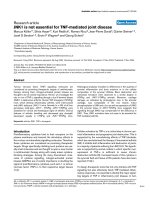
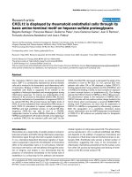
Fig. 1. Ruk L, Ruk M and Ruk S ubiquitination. (A) Schematic representations of the domain organization of the three Ruk proteins. SH3, Src
homology 3 domain; Pro, proline rich region; PEST, sequences enriched in proline, glutamic acid, serine and threonine; CC, Coiled-Coil domain.
(B) Hek293 cells were transfected with 1.5 lg of plasmid containing EE-tagged Ruk L cDNA, EE-tagged Ruk M cDNA or EE-tagged Ruk S in the
absence or presence of HA-tagged Ub plasmid (1.5 lg). As a control, Hek293 cells were transfected with an empty vector. Cell lysates were
immunoprecipitated with anti-EE Ig. The immunoprecipitates were subjected to immunoblotting with anti-HA antibody (top panel). The positions
of polyubiquitinated Ruk species [(Ub)n-Ruk] are indicated. The arrowheads mark the locations of the unmodified forms of Ruk L, M, S. The
membrane was reprobed with anti-EE antibody (bottom panel).
Ó FEBS 2002 Ruk is ubiquitinated but not degraded by the proteasome (Eur. J. Biochem. 269) 3403
Immunoprecipitation and Western blot analysis
Hek293 cells were washed with ice-cold NaCl/P
i
and lysed
with buffer containing 1% Brij 98, 10 m
M
Tris, 150 m
M
NaCl, 5 m
M
EDTA, 5 m
M
EGTA, 20 m
M
NaF, 20 l
M
2-glycero-phosphate, 1 m
M
sodium pyrophosphate, 1 m
M
vanadate, 10% glycerol, 1 m
M
phenylmethanesulfonyl
fluoride and a cocktail of protease inhibitors (Roche) at
4 °C for 20 min. Lysates were cleared by centrifugation for
30 min at 27 000 g and supernatants used for further
experiments. Immunoprecipitating antibodies were incuba-
ted with solubilized cell extracts for 1 h at 4 °C before the
addition of protein G–Sepharose beads, prewashed in lysis
buffer. After a 2-h incubation on the wheel, the beads were
washed three times with lysis buffer and twice with buffer
containing 0.1% Brij 98
6
. Immune complexes were removed
from beads by boiling in Laemmli sample buffer and
separated by SDS/PAGE. Resolved proteins were trans-
ferred onto a poly(vinylidene difluoride) membrane, which
was incubated for 1 h with blocking solution (5% milk in
Tris/NaCl/0.1%Tween) followed by specific antibody over-
night at 4 °C. After extensive washing with NaCl/Tris/0.1%
Tween, the membrane was incubated for 1 h with horse-
radish peroxidase-conjugated secondary antibody. The
antigen–antibody complexes were detected using enhanced
chemiluminescence (ECL) system (Amersham Pharmacia
Biotech). When immunoblots had to be reprobed, the
membranes were initially stripped and reblocked prior to
incubation with another type of primary antibody.
Detection of ubiquitinated proteins
Ub is highly conserved among eukaryotes, as only three of
76 amino acids differ between the human and yeast proteins.
Therefore, Ub is not an optimal antigen and anti-Ub Ig
rarely possess good affinity. Taking this into account and
that anti-Ruk Ig are not very efficient in immunoprecipi-
tation experiments, we decided to cotransfect Hek293 cells
with plasmids encoding HA-tagged ubiquitin and EE-
tagged versions of Ruk isoforms. (EE–Ruk L, EE–Ruk M
and EE–Ruk S). Transiently expressed Ruk isoforms were
immunoprecipitated with anti-EE Ig and resolved by SDS/
PAGE. Modification of Ruk by Ub was determined by
immunoblotting with anti-HA Ig.
RESULTS
All three Ruk isoforms are ubiquitinated
in vivo
Sequence analysis of Ruk L indicated the presence of
multiple PEST motives located between a stretch of proline-
rich sequences and the C-terminal coiled-coil domain. The
appearance of PEST motifs in protein sequences is often
associated with reduced protein stability and a short half-life
[20]. Taking this into account and the fact that Cin85/SETA
binds the E3 ligase c-Cbl [12,18], we decided to investigate
ubiquitination of Ruk isoforms in vivo. In this experiment,
EE-tagged versions of all three isoforms of Ruk were
cotransfected into Hek293 cells together with HA-tagged
Ub. Two days after transfection, Ruk isoforms were
immunoprecipitated using anti-EE antibodies, separated
by electrophoresis and analysed by immunoblotting using
anti-HA Ig. The results presented in Fig. 1B clearly
demonstrate the appearance of multiple high molecular
mass bands detected by anti-HA antibody. No signal was
observed when Hek293 cells were transfected only with
EE-tagged isoforms of Ruk, indicating the specificity of
anti-HA Ig. Reprobing of the membrane with anti-Ruk Ig,
which is specific for all three isoforms, confirmed that equal
amounts of EE-tag fusions were coimmunoprecipitated
from transfected cells (Fig. 1B, lower panel). The appear-
ance of multiple bands on the anti-HA Western blot, clearly
demonstrate that Ruk isoforms are polyubiquitinated.
Specific interaction between Ruk isoforms and the E3
ubiquitin ligase Cbl
Specific interaction between c-Cbl and Cin85/SETA, which
corresponds to the Ruk L isoform, has been recently
reported [12,18]. These findings and the results presented
above prompted us to investigate whether all Ruk isoforms
can interact with Cbl. To assess these interactions,
EE-tagged versions of Ruk L, M or S were cotransfected
into Hek293 cells with c-Cbl or vector alone. Immune
complexes were precipitated with anti-EE Ig and analysed
by Western blotting using anti-Cbl Ig. Figure 2A clearly
demonstrates that all three isoforms of Ruk interact with
exogenously expressed Cbl. Moreover, we also observed
coimmunoprecipitation of endogenous Cbl with anti-EE Ig
from cells, which were transfected with EE-tagged isoforms
of Ruk. Observed interactions were also detected in
reciprocal experiments, when anti-Cbl immunoprecipitates
were probed in Western blotting with anti-EE Ig. Ruk S is
detected only on a much longer exposure. (Fig. 2B). We
have also noted that the longest isoform of Ruk (Ruk L)
exhibits the strongest association with endogenous and
exogenously expressed Cbl. The association of Ruk M and
Ruk S with c-Cbl was quite unexpected, as these isoforms
do not possess the two N-terminal SH3 domains, found
previously to mediate the interaction with c-Cbl [12,18].
Heterodimerization of Ruk L with Ruk M and Ruk S
All Ruk isoforms share a common C-terminal region which
possesses a coiled-coil domain. Numerous studies have
implicated coiled-coil domains in mediating protein–protein
interaction via homodimerizations and heterodimerizations
[21]. Taking this into account, we speculated that associ-
ation of Ruk M and Ruk S with Cbl is not direct, but could
be mediated via heterodimerization with the longest
isoform, Ruk L. In order to test this hypothesis, we
cotransfected nontagged Ruk L together with each
EE-tagged Ruk isoforms. Following anti-EE Ig immuno-
precipitations, immune complexes were subjected to West-
ern blot analysis with the C-terminal anti-Ruk Ig, which
recognizes all three isoforms. As shown in Fig. 3A, untagged
Ruk L specifically coimmunoprecipitates with all three
EE-tagged isoforms. The expression of EE-tagged Ruk
isoforms in transfected cells was confirmed by immuno-
blotting of total cell lysates with anti-EE antibodies
(Fig. 3B). These data suggest that Ruk isoforms have the
potential to form homodimers and heterodimers and that
the coiled-coil domain mediates the formation of these
complexes. Moreover, specific interaction between Cbl and
two shorter isoforms of Ruk (Ruk M and Ruk S) was
found to be mediated by heterodimerization with Ruk L.
3404 F. Verdier et al. (Eur. J. Biochem. 269) Ó FEBS 2002
Ruk L is ubiquitinated but not degraded
by proteasomes
Ubiquitination, in most cases, targets modified proteins for
degradation by 26S proteasomes. In order to determine
whether ubiquitination of Ruk isoforms would induce their
degradation, a panel of proteosome- and lysosome-specific
inhibitors has been used. It is well established that lactacys-
tine and the peptide aldehyde ALLN inhibit proteasome-
mediated proteolysis, causing an accumulation of proteins
that are usually degraded by this pathway [22]. In contrast
to lactacystine, which is a highly specific proteasomal
inhibitor [23], ALLN inhibits also nonproteasomal
proteases, such as calpains and cathepsins.
Initially, we tested the stability of EE-tagged Ruk L
transiently overexpressed in Hek293 cells. Two days after
transfection, cells were treated with various inhibitors or
with the vehicle alone. Equal amounts of total cell lysates
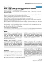
Fig. 3. Heterodimerization of Ruk L with Ruk M and Ruk S. Hek293 cells were transfected with 1.5 lg of plasmid containing nontagged version of
Ruk L cDNA, and cotransfected with the same quantity of either EE-tagged Ruk L, EE-tagged Ruk M, or EE-tagged Ruk S cDNA. As a control,
Hek293 cells were transfected with an empty vector (NT). Cell lysates were immunoprecipitated with anti-EE antibody. The immunoprecipitates
were analysed by Western Blot using anti-Ruk antibody raised against the last 17 C-terminal amino acids and thereby able to recognize all Ruk
isoforms (Ruk L, EE-tagged Ruk L, EE-tagged Ruk M and EE-tagged Ruk S). Ruk L and EE-tagged Ruk L can be separated by SDS/PAGE,
since their molecular masses differ from approximatly one kDa (A). An aliquot of each cell lysate was immunoblotted with anti-EE Ig to check the
expression of EE-Ruk isoforms (B). The blot in the lower panel of (B) was exposed 10 times longer than the upper panel.
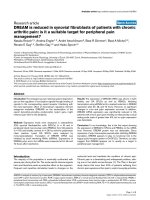
Fig. 2. Ruk L, Ruk M and Ruk S coprecipitate
with Cbl. Hek293 cells were transfected with
1.5 lg of plasmid containing EE-tagged RukL
cDNA, EE-tagged Ruk M cDNA or EE-tag-
ged Ruk S cDNA in the absence or presence of
Cbl plasmid (1.5 lg). As a control, Hek293
cells were transfected with an empty vector
(NT).Celllysatesweresplitintwoandwere
immunoprecipitated either with anti-EE Ig (A)
or with anti-Cbl Ig (B). The immunoprecipi-
tates were subjected to immunoblotting with
anti-Cbl Ig (A) or with anti-EE Ig (B).
An aliquot of each cell lysate was immuno-
blotted with anti-EE antibody to check
expression of Ruk isoforms (C).
Ó FEBS 2002 Ruk is ubiquitinated but not degraded by the proteasome (Eur. J. Biochem. 269) 3405
were separated by SDS/PAGE and immunoblotted with
anti-EE Ig. No changes in the level of exogenously expressed
Ruk L was detected, when the activities of proteosomal and
lysosomal proteases were blocked by specific inhibitors
(Fig. 4A). We reprobed the membrane with anti-(b-actin) Ig
to confirm equal loading of proteins in each lane (Fig. 4A,
lower panel). One can argue that only ubiquitinated form of
Ruk L would be targeted for degradation and if this fraction
is small, it might be difficult to detect the changes in the level
of total Ruk L protein. To overcome this problem, we
coexpressed EE-tagged Ruk L with HA-Ub in Hek293
cells. Then, cells were treated with proteosomal inhibitor
ALLN or vehicle alone. As shown in Fig. 4B, no accumu-
lation of ubiquitinated EE-Ruk L is detected in the presence
of ALLN.
We then measured the half-life of endogenous Ruk L,
which is expressed at high level in Hek293 cells. In most
cases, the half-life of ubiquitinated proteins is short due to
their rapid degradation by the proteasome. Time-course
treatment of cells with a protein synthesis inhibitor cyclo-
heximide showed no variations in the level of endogenous
Ruk L, suggesting a long half-life for the protein (Fig. 4C).
Furthermore, we examined the effect of selected inhibitors
of protein degradation on the level of endogenous Ruk L.
As can be seen in Fig. 4D, neither ALLN nor lactacystine
induce any accumulation of endogenous Ruk L in Hek293
cells, even 6 h after treatment. These results were also
confirmed in human monocytic cell line U937 (data not
shown). To verify that proteolytic activities of proteasomes
were effectively inhibited by the use of indicated inhibitors,
we checked the expression level of b-catenin, which is
degraded via the Ub-proteasome pathway [24,25].
Re-probing of the membrane with anti-(b-catenin) Ig clearly
demonstrated the accumulation of ubiquitinated forms of
b-catenin upon ALLN and lactacystine treatment (Fig. 4D,
lower panel). No effect on the stability of Ruk L protein was
also observed when cells were treated with lysosomal
inhibitors: NH
4
Cl or chloroquine
7
(Fig. 4D). Taken together,
these experiments clearly demonstrate that neither exogen-
ously expressed nor native Ruk L isoform is degraded via
proteasome or lysosome pathways.
DISCUSSION
Ubiquitination is now recognized as a regulatory protein
modification whose functional significance is comparable to
that of phosphorylation. Degradation of cellular proteins by
the ubiquitin system encompasses two successive steps: (a)
covalent attachment of ubiquitin molecules to selected
proteins; and (b) degradation of ubiquitin-conjugated
Fig. 4. Exogenously expressed EE-Ruk L (A,B) or Endogenous Ruk L (C,D) are not degraded by the proteasome. (A) Hek293 cells were transfected
with 2 lg of plasmid containing EE-tagged Ruk L cDNA. After 48 h, cells were incubated for the time indicated with various inhibitors or with
vehicle alone [Final concentrations: 50 l
M
for ALLN, 50 l
M
for Lactacystin (Lacta), 20 m
M
for NH
4
Cl and 200 l
M
for chloroquine (Chloro)].
Cells were lysed and the quantity of protein measured by Bradford assay. 10 lg of total protein from each cell lysate was separated by SDS/PAGE.
The level of exogenous EE-Ruk L was determined by Western Blot analysis using anti-EE Ig (upper panel). The membrane was reprobed with anti-
(b-actin) Ig to confirm equal loading of protein in each lane (bottom panel). (B) Hek293 cells were cotransfected with 1.5 lg of plasmid containing
EE-tagged Ruk L cDNA and 1.5 lg of Ub-HA plasmid. After 48 h, cells were incubated for 3 h with (+) or without (–) ALLN. Cell lysates were
immunoprecipitated with anti-EE Ig. The immunoprecipitates were subjected to immunoblotting with anti-HA Ig. The position of polyubiqui-
tinated Ruk species [(Ub)n-Ruk] are indicated. The arrowheads mark the locations of unmodified EE-Ruk L. (C) Hek293 cells were incubated with
500 l
M
Cycloheximide (CHX) for the times indicated. As described in (A), 30 lg of proteins from the total cell lysates was separated by SDS/PAGE
electrophoresis, and the expression level of endogenous Ruk L determined by Western blot (WB) using anti-Ruk Ig (upper panel). The membrane
wasreprobedwithanti-(b-actin) Ig to confirm equal loading of protein in each lane (lower panel). (D) Hek293 cells were incubated for the time
indicated with various inhibitors or with vehicle alone as described for panel A. Cells were lysed and the quantity of proteins measured by Bradford
assay. 30 lg of proteins from the total cell lysates was separated by SDS/PAGE electrophoresis. The expression level of endogenous Ruk L was
determined by Western blot analysis using anti-Ruk Ig (upper panel). The membrane was reprobed with anti-(b-catenin) Ig (bottom panel). The
position of polyubiquitinated b-catenin species [(Ub)n-b-catenin] are indicated. The arrowheads mark the locations of unmodified b-catenin.
3406 F. Verdier et al. (Eur. J. Biochem. 269) Ó FEBS 2002
proteins. Many ubiquitinated proteins are targeted for
degradation by 26S proteasomes, but some undergo
endocytosis, leading to proteolysis in the lysosome. New
findings show that ubiquitination is not always associated
with the degradation of modified proteins, but could be also
involved in regulation of enzymatic activities and intranu-
clear trafficking of tagged proteins [26].
The data presented in this study clearly demonstrate that
a recently identified adaptor-type protein Ruk L, also
known as Cin85 or SETA, is ubiquitinated in vivo.
Furthermore, we showed that shorter splicing variants of
Ruk, termed Ruk M and Ruk S, are also modified by
covalent attachment of ubiquitin. Ubiquitination of all three
isoforms of Ruk indicates that ubiquitin conjugation occurs
at the C-terminal region, which is common between them. It
is well established that ubiquitin is conjugated to target
proteins through lysine residues. Sequence analysis of Ruk
isoforms showed that their common C-terminal region
contains numerous lysine residues, which could be potential
sites for ubiquitination. The identification and characteri-
zation of Ruk ubiquitination sites is currently in progress.
Recently, specific association between Cbl and Cin85/
SETA was demonstrated [12,18]. This interaction was found
to be mediated by the first two SH3 domains of Cin85/
SETA and the proline-rich region of Cbl. In addition to
that, constitutive binding between both proteins was further
induced by stimulation of cells with EGF and found to be
dependent on tyrosine phosphorylation of the C-terminal
region of Cbl ([12] and our data, not shown). The same
mode of interaction has been recently reported between Cbl
and CMS/CD2AP, which requires the same domains and is
also regulated by tyrosine phosphorylation of Cbl [27]. It is
believed that tyrosine phosphorylation at the C-terminus of
Cbl induces a conformational change of the protein from a
closed to an open conformation, thereby unmasking
putative SH3-domain motifs.
In agreement with these findings, we show, in vivo,an
interaction between Cbl and the longest isoform of Ruk,
Ruk L. Furthermore, specific association of Cbl with
Ruk M and Ruk S was also demonstrated. As neither
Ruk M nor Ruk S possess the first two SH3 domains,
which are involved in complex formation with Cbl, the
mechanism of these interactions has been investigated. We
found that the C-terminal region, which contains a coiled-
coil domain common to all Ruk isoforms, is responsible for
their heterodimerization. These results suggest that binding
between Cbl/Ruk M and Cbl/Ruk S are indirect and
require heterodimerization with Ruk L.
As all isoforms are in complex with Cbl and are
ubiquitinated, Cbl is a potential E3 ligase responsible for
ubiquitination of Ruk isoforms. This hypothesis is currently
under investigation.
The discovery of Ruk isoform ubiquitination in cells
prompted us to investigate the importance of this modifi-
cation, especially in regulating its stability. Using proteo-
somal and lysosomal inhibitors, we found that
ubiquitination of exogenously expressed and native Ruk L
does not induce its degradation via these proteolytic
pathways. If ubiquitination of Ruk L is not a signal for
its degradation, what is the role of this post-translational
modification?
Proteolysis-independent regulation by ubiquitination has
recently been reported in several systems. Ubiquitination of
a number of cell surface receptors in response to ligand
binding serves as an internalization signal [28]. Moreover,
ubiquitination-dependent processing of precursor proteins
[29] and the regulation of multienzyme complex formation
have also been described [30]. An interesting paper by Fang
et al. demonstrates that like Ruk L, the P85 regulatory
subunit of PtdIns-3 kinase is ubiquitinated, but not
degraded by the proteasome pathway [31,32]. In addition,
Cbl was shown to be the E3 ligase responsible for P85
ubiquitination and to regulate the recruitment of PtdIns-3
kinase to CD28 and T cell antigen receptor complexes,
thereby inhibiting PtdIns-3 kinase activation.
Two contradictory mechanisms have been proposed for
Cbl binding to the P85 subunit: one involves the proline-rich
domain of Cbl and the SH3 domain of P85 and while the
other implicates a phosphorylated C-terminal tyrosine
(Y731) of Cbl and an undefined domain of P85 [32,33].
We previously reported specific association between Ruk L
and the P85 regulatory subunit of PtdIns-3 kinase, which is
mediated via proline-rich sequences and the SH3 domain,
respectively [11]. This interaction is not inducible by growth
factor stimulation and has an inhibitory effect on the
activity of PtdIns-3 kinase. The mechanism by which
PtdIns-3 kinase could be released from the inhibitory
complex with Ruk L is still unknown. Ruk L polyubiquiti-
nation could induce conformational changes in the molecule
which may modify its binding specificity towards the p85
subunit of the PtdIns-3 Kinase. We are currently investi-
gating whether ubiquitination of Ruk L and P85 could
affect the association between them and the activity of
PtdIns-3 kinase. The use of in vitro binding and ubiquiti-
nation assays will allow us to better understand the
mechanism of the interaction between Ruk L, E3 ligase
Cbl and PtdIns-3 kinase.
ACKNOWLEDGEMENTS
Fre
´
de
´
rique Verdier is supported by an EMBO (European Molecular
Biology Organization) Fellowship. We are grateful to Mark Griffin for
expert technical assistance and to H. Rebholz and T. Fenton for critical
reading of the manuscript.
REFERENCES
1. Ciechanover, A. (1998) The ubiquitin-proteasome pathway: on
protein death and cell life. EMBO J. 17, 7151–7160.
2. Hershko, A. & Ciechanover, A. (1998) The ubiquitin system.
Annu.Rev.Biochem.67, 425–479.
3. Tsygankov, A.Y., Teckchandani, A.M., Feshchenko, E.A. &
Swaminathan, G. (2001) Beyond the RING: CBL proteins as
multivalent adapters. Oncogene 20, 6382–6402.
4. Joazeiro, C.A., Wing, S.S., Huang, H., Leverson, J.D., Hunter, T.
& Liu, Y.C. (1999) The tyrosine kinase negative regulator c-Cbl as
a RING-type, E2-dependent ubiquitin-protein ligase. Science 286,
309–412.
5. Miyake, S., Lupher, M.L. Jr, Druker, B. & Band, H. (1998) The
tyrosine kinase regulator Cbl enhances the ubiquitination and
degradation of the platelet-derived growth factor receptor alpha.
Proc. Natl Acad. Sci. USA 95, 7927–7932.
6. Levkowitz, G., Waterman, H., Ettenberg, S.A., Katz, M.,
Tsygankov,A.Y.,Alroy,I.,Lavi,S.,Iwai,K.,Reiss,Y.,Ciec-
hanover, A., Lipkowitz, S. & Yarden, Y. (1999) Ubiquitin ligase
activity and tyrosine phosphorylation underlie suppression of
growth factor signaling by c-Cbl/Sli-1. Mol. Cell 4, 1029–1040.
Ó FEBS 2002 Ruk is ubiquitinated but not degraded by the proteasome (Eur. J. Biochem. 269) 3407
7. Levkowitz, G., Waterman, H., Zamir, E., Kam, Z., Oved, S.,
Langdon, W.Y., Beguinot, L., Geiger, B. & Yarden, Y. (1998)
c-Cbl/Sli-1 regulates endocytic sorting and ubiquitination of the
epidermal growth factor receptor. Genes Dev. 12, 3663–3674.
8. Yokouchi, M., Kondo, T., Houghton, A., Bartkiewicz, M.,
Horne, W.C., Zhang, H., Yoshimura, A. & Baron, R. (1999)
Ligand-induced ubiquitination of the epidermal growth factor
receptor involves the interaction of the c-Cbl RING finger and
UbcH7. J. Biol. Chem. 274, 31707–31712.
9. Andoniou, C.E., Lill, N.L., Thien, C.B., Lupher, M.L. Jr, Ota, S.,
Bowtell, D.D., Scaife, R.M., Langdon, W.Y. & Band, H. (2000)
The Cbl proto-oncogene product negatively regulates the Src-
family tyrosine kinase Fyn by enhancing its degradation. Mol. Cell
Biol. 20, 851–867.
10. Rao,N.,Ghosh,A.K.,Ota,S.,Zhou,P.,Reddi,A.L.,Hakezi,K.,
Druker, B.K., Wu, J. & Band, H. (2001) The non-receptor tyrosine
kinase Syk is a target of Cbl-mediated ubiquitylation upon B-cell
receptor stimulation. EMBO J. 20, 7085–7095.
11. Gout,I.,Middleton,G.,Adu,J.,Ninkina,N.N.,Drobot,L.B.,
Filonenko, V., Matsuka, G., Davies, A.M., Waterfield, M. &
Buchman, V.L. (2000) Negative regulation of PI 3-kinase by Ruk,
a novel adaptor protein. EMBO J. 19, 4015–4025.
12. Take, H., Watanabe, S. & Takeda, K., YuZ.X., Iwata, N. &
Kajigaya, S. (2000) Cloning and characterization of a novel
adaptor protein, CIN85, that interacts with c-Cbl. Biochem. Bio-
phys. Res. Commun. 268, 321–328.
13. Chen, B., Borinstein, S.C., Gillis, J., Sykes, V.W. & Bogler, O.
(2000) The glioma-associated protein SETA interacts with AIP1/
Alix and ALG-2 and modulates apoptosis in astrocytes. J. Biol.
Chem. 275, 19275–19281.
14. Watanabe,S.,Take,H.&Takeda,K.,Yu,Z.X.,Iwata,N.&
Kajigaya, S. (2000) Characterization of the CIN85 adaptor pro-
tein and identification of components involved in CIN85 com-
plexes. Biochem. Biophys. Res. Commun. 278, 167–174.
15. Dustin, M.L., Olszowy, M.W., Holdorf, A.D., Li, J., Bromley, S.,
Desai, N., Widder, P., Rosenberger, F., van der Merwe, P.A.,
Allen, P.M. & Shaw, A.S. (1998) A novel adaptor protein
orchestrates receptor patterning and cytoskeletal polarity in T-cell
contacts. Cell 94, 667–677.
16. Kirsch, K.H., Georgescu, M.M., Ishimaru, S. & Hanafusa, H.
(1999) CMS: an adapter molecule involved in cytoskeletal
rearrangements, Proc. Natl Acad. Sci. USA 96, 6211–6216.
17. Shih, N.Y., Li, J., Karpitskii, V., Nguyen, A., Dustin, M.L.,
Kanagawa, O., Miner, J.H. & Shaw, A.S. (1999) Congenital
nephrotic syndrome in mice lacking CD2-associated protein.
Science. 286, 312–315.
18. Borinstein, S.C., Hyatt, M.A., Sykes, V.W., Straub, R.E.,
Lipkowitz,S.,Boulter,J.&Bogler,O.(2000)SETAisamulti-
functional adapter protein with three SH3 domains that binds
Grb2, Cbl, and the novel SB1 proteins. Cell Signal. 12, 769–779.
19. Soubeyran, P., Kowanetz, K., Szymkiewicz, I., Langdon, W. &
Dikic, I. (2002) Cbl/Cin85/Endophilin complex mediates
ligand-induced downregulation of EGF receptors. Nature 416,
183–187.
20. Rechsteiner, M. & Rogers, S.W. (1996) PEST sequences and
regulation by proteolysis. Trends Biochem. Sci. 21, 267–271.
21. Burkhard, P., Stetefeld, J. & Strelkov, S.V. (2001) Coiled coils: a
highly versatile protein folding motif. Trends Cell. Biol. 11, 82–88.
22. Coux, O., Tanaka, K. & Goldberg, A.L. (1996) Structure and
functions of the 20S and 26S proteasomes. Annu. Rev. Biochem.
65, 801–847.
23. Fenteany, G. & Schreiber, S.L. (1998) Lactacystin, proteasome
function, and cell fate. J. Biol. Chem. 273, 8545–8548.
24. Orford, K., Crockett, C., Jensen, J.P., Weissman, A.M. & Byers,
S.W. (1997) Serine phosphorylation-regulated ubiquitination and
degradation of beta-catenin. J. Biol. Chem. 272, 24735–24738.
25. Aberle, H., Bauer, A., Stappert, J., Kispert, A. & Kemler, R.
(1997) beta-catenin is a target for the ubiquitin-proteasome
pathway. EMBO J. 16, 3797–3804.
26. Pickart, C.M. (2001) Ubiquitin enters the new millennium. Mol.
Cell. 8, 499–504.
27. Kirsch, K.H., Georgescu, M.M., Shishido, T., Langdon, W.Y.,
Birge, R.B. & Hanafusa, H. (2000) The adapter type protein
CMS/CD2AP binds to the proto-oncogenic protein c-Cbl through
a tyrosine phosphorylation-regulated Src homology 3 domain
interaction. J. Biol. Chem. 276, 4957–4963.
28. Hicke, L. (1997) Ubiquitin-dependent internalization and
down-regulation of plasma membrane proteins. Faseb J. 11, 1215–
1226.
29. Hoppe, T., Matuschewski, K., Rape, M., Schlenker, S., Ulrich,
H.D. & Jentsch, S. (2000) Activation of a membrane-bound
transcription factor by regulated ubiquitin/proteasome-dependent
processing. Cell 102, 577–586.
30. Kaiser, P., Flick, K., Wittenberg, C. & Reed, S.I. (2000) Regula-
tion of transcription by ubiquitination without proteolysis: Cdc34/
SCF (Met30)-mediated inactivation of the transcription factor
Met4. Cell 102, 303–314.
31. Fang, D. & Liu, Y.C. (2001) Proteolysis-independent regulation of
PI3K by Cbl-b-mediated ubiquitination in T cells. Nat. Immunol.
2, 870–875.
32. Fang, D., Wang, H.Y., Fang, N., Altman, Y., Elly, C. & Liu, Y.C.
(2001) Cbl-b, a RING-type E3 ubiquitin ligase, targets phospha-
tidylinositol 3-kinase for ubiquitination in T cells. J. Biol. Chem.
276, 4872–4878.
33. Elly, C., Witte, S., Zhang, Z., Rosnet, O., Lipkowitz, S., Altman,
A. & Liu, Y.C. (1999) Tyrosine phosphorylation and complex
formation of Cbl-b upon T cell receptor stimulation. Oncogene 18,
1147–1156.
3408 F. Verdier et al. (Eur. J. Biochem. 269) Ó FEBS 2002