- Trang chủ >>
- Khoa Học Tự Nhiên >>
- Vật lý
polymer - embedded stannic oxide nanoparticles as humidity sensors
Bạn đang xem bản rút gọn của tài liệu. Xem và tải ngay bản đầy đủ của tài liệu tại đây (602.31 KB, 4 trang )
Polymer-embedded stannic oxide nanoparticles as humidity sensors
Shadie Hatamie
a
, Vivek Dhas
b
, B.B. Kale
c
, I.S. Mulla
b
, S.N. Kale
a,
⁎
a
Department of Electronic-Science, Fergusson College, Pune 411 004, India
b
Physical and Materials Chemistry Division, National Chemical Laboratory, Pune 411 008, India
c
Center for Materials for Electronics Technology (C-MET), Panchawati, Pashan Road, Pune 411 008, India
abstractarticle info
Article history:
Received 22 April 2008
Received in revised form 21 June 2008
Accepted 29 July 2008
Available online 8 August 2008
Keywords:
Stannic oxide
Nanoparticles
Polymer
Humidity sensor
Stannic oxide (SnO
2
) nanoparticles have been suspended in polyvinyl alcohol (PVA) matrix in different PVA:
SnO
2
molar ratios ranging from 1:1 to 1:5 using simple chemical route. This suspension was deposited on
ceramic substrate and upon drying was carefully detached from the substrate. SnO
2
-embedded self-standing,
transparent and flexible thin films were hence synthesized. Transmission electron microscopy (TEM) and X-
ray diffraction (XRD) techniques show the rutile tetragonal structure of SnO
2
with particle size ~5 nm. UV–
Visible spectroscopy demonstrates the band gap of 3.9 eV, which does not alter when embedded in polymer.
Fourier transform infrared spectroscopy (FTIR) reveals that the properties of SnO
2
do not modify due to
incorporation in the PVA matrix. The structures work as excellent humidity sensors at room temperature. For
a critical PVA:SnO
2
molar ratio of 1:3, the resistance changes to fi ve times of magnitude in 92% humidity
within fraction of second when compared with resistance at 11% humidity. The sample regains its original
resistance almost instantaneously after being removed from humid chamber. Nanodimensions of SnO
2
particles and percolation mechanism related to transport through polymer matrix and water molecule as a
carrier has been used to understand the mechanism.
© 2008 Elsevier B.V. All rights reserved.
1. Introduction
Relationship between nanostructures and their implications on the
electrical, optical and thermal properties of materials is an extremely
interesting area of material science. Metal-oxides form attractive
domain therein due to their wide range of properties like ferroelec-
tricity, superconductivity and piezoelectricity. Synthesizing porous
nanoparticulate thin films using soft chemistry approach or incor-
poration of metal-oxide nanoparticles in polymer matrix are two
different fascinating approaches, which have been recently adopted as
routes to explore interesting physics of self-assembly and study the
range of properties exhibited by these oxide structures [1–8].In
polymer-embedded metal-oxide thin films, polymer controls viscosity
and binds the metal-oxide ions, resulting in their homogeneous
distribution in the film. These uniform, flexible and crack-free metal-
oxides — polymer films can be synthesized on much larger scale, in
bigger dimensions and for variety of applications. Some interesting
attempts have been made in recent past by Q.X. Jia et al. and N.V.
Kolytcheva et al. [1,5] on metal-oxide nanoparticles embedded in
polymer matrix. According to Jia et al. titanium dioxide thin films can
be synthesized in epitaxial manner using simple polymer-assisted
deposition technique, and the route promises good sensing devices. In
an attempt to synthesize and explore nanoparticulate thin films in
porous configurations, Brousse et al. [6] and Horillo et al. [9] have
explored nanomaterials of tin oxide and compared them with bulk
systems, for their gas sensing properties. It has been argued that since
nanoparticles have higher surface-to-volume ratio, surface states are
more, which increase the gas molecules adsorbed on nanoparticles, as
compared to bulk systems; thereby improving the sensing ability in
their nanoforms. Further, as indicated by Mizsei [10] the faceted or
non-faceted grains, and hence the surface morphology has its impact
on surface characteristics, which further controls sensing properties.
For such reasons, porous SnO
2
films are projected to be superior by M.
Honore et al. [11] and their transduction to conductivity changes have
been studied. Y. Shimizu et al. [12] have studied porous ZnO films as
varistors and using similar arguments have studied the non-linear
response of these materials as a function of particle size. It is hence
important to address the issue of polymer-embedded sensors, under-
stand the interesting science therein and explore its technological
importance. Finding out the role of polymer and exact ratio of
polymer-to-sens ing material in a given thin fil m, which gives
maximum response to the incident gas/humid ambience, is the key
to apply these materials to technology and finally establish a base to
yield extremely good, selective room temperature sensors.
In this communication, we report synthesis of stannic oxide (SnO
2
)
nanoparticles embedded in polyvinyl alcohol (PVA) matrix. Nearly
mono-dispersed nanoparticles of SnO
2
having size of ~5 nm and band
gap of 3.9 eV have been formed and when embedded in PVA, yielded
self-supporting thin films which were highly flexible, transparent and
non-degradable in ambient atmosphere. These films when subjected
Materials Science and Engineering C 29 (2009) 847–850
⁎ Corresponding author. Department of Electronic-Science, Fergusson College, F.C.
Road, Pune 411004, India. Tel.: +91 20 2565 5119.
E-mail address: (S.N. Kale).
0928-4931/$ – see front matter © 2008 Elsevier B.V. All rights reserved.
doi:10.1016/j.msec.2008.07.039
Contents lists available at ScienceDirect
Materials Science and Engineering C
journal homepage: www.elsevier.com/locate/msec
to humid environment showed change in resistance. The jump in
resistance in given humidity is a function of PVA:SnO
2
ratio, which has
been varied from molar ratios of 1:1 to 1:5. It has been found that
maximum change in resistance occurs for film with 1:3 molar ratio
and that the sensitivity response decreases on either side. The possible
reasons have been related to the adsorption sites offered by SnO
2
,
their interconnectivity and active polymer medium as a tunneling
percolation track.
2. Experimental
2.1. SnO
2
nanoparticle synthesis
SnO
2
nanoparticles were prepared by a simple co-precipitation
method [13]. Stannic chloride (SnCl
4
.5H
2
O (A.R), 0.01 M) was
dissolved in deionized water and stirred for 30 min at room
temperature. 8 ml ammonia solution was added drop wise to the
above solution to attain pH ~10. The resultant gel was filtered and
dried for 24 h in ambient temperature and then later for 2 h at 100 °C
to ensure that the powder was totally devoid of water. The powder
was ground for 10 min, and heated in oven at 400 °C for 3 h and cooled
to room temperature.
2.2. PVA/SnO
2
nanocomposite film
PVA was dissolved in deionized water (1 M) and the solution was
heated in a water bath at 90 °C for 1 h. Then the SnO
2
nanoparticles
suspended in water (with molarity varying from 1 to 5 M) were added
to the PVA solution and well dispersed using ultrasonication for
20 min. The homogeneous solution was then spin-coated on ceramic
substrate and dried in air at ambient temperature. After drying, this
composite film was easily removed from the ceramic substrate. The
similar procedure was obtained and multiple films were synthesized
with different PVA:SnO
2
molar ratios viz. varying from 1:1 to 1:5.
2.3. Humidity measurements
Different films were subjected to supersaturated solutions for
humidity measurements. Two different solutions, namely, 50 ml of
lithium chloride (LiCl) and potassium nitrate (KNO
3
) were put in the
air tight 250 ml plastic chambers, which provide different constant
relative humidity (% RH) at 30 °C of 11%, 92% after 24 h, respectively.
Two electrical contacts were made on the composite films using silver
paste and copper wires for contacts to meters. The probes were long
wires, which were connected to voltage source, and current meter,
which were placed outside the humidity chamber. The chamber cover
had sealed microholes that allowed the wires to come out of the
chamber without offering any leak during measurements. The films
were first subjected to 11% humidity chamber and current (and hence,
resistance (R
11
)) was noted down. This was done to ensure that all
samples had uniform reference for comparison. Then the sample was
shifted to 92% humidity chamber and current (and hence, resistance
(R
92
)) was noted down. For all samples, the voltage was kept constant
at 15 V. The change in current, which was converted to resistance, was
studied as a function of PVA:SnO
2
ratio. All samples had dimensions of
1 cm×1 cm× 0.03 cm. The time analysis was also done to find out the
amount of time the film takes to regain its original value of resistance
(basically R
11
) after the sample has been removed from 92% humidity
chamber and re-subjecting it to 11% humidity chamber. This gave us
information of recovery time and reusability.
The synthesized nanoparticles and nanocomposite films were
characterized for structural, compositional properties using Fourier
transform infrared spectroscopy (FTIR, Shimadzu 8400S Spectro-
meter), X-ray Diffraction Technique (XRD, Philips PW 1830 40 kV,
30 mA, CuK
α
λ =0.154178 nm), High-resolution Transmission
Electron Microscopy (HRTEM, JEOL model 1200EX) and UV–Vis
Spectroscopy (Jasco V570UV–VIS–NIR). Keithley meters were used
for transport measurements.
3. Results and discussion
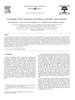
Fig. 1(a) shows the XR D patt ern o f the sample synthe sized using co-
precipitation method described abo ve. Th e spectrum has been compar ed
with standard commercial bulk powder (Fig. 1(b)). Typical tetragonal
rutile structure can be clearly seen in the nanopowder and peak
broadening confirms t he smaller particle s ize. Th e Miller in dices gave
lattice constants asa=b=4.738 andc= 3. 187, which matched w ell with
bulk SnO
2
(JCPDS File No. 41-1445). No impurity peaks were observed,
Fig.1. XRD patter n of SnO
2
nanoparticles (a) and compare d w ith SnO
2
bulk (b). As c an be seen
rutile structur e was for med with the broad sp ectr um indicating for mation of nanoparti cles.
The in set shows the graph o f (αE)
1/2
versus (E)showingthebandgapof3.9eV.
Fig. 2. FTIR spectrum of SnO
2
nanoparticles (a), PVA (b) and PVA: SnO
2
composite (c).
All signatures in PVA and SnO
2
are seen in the composite with no modifications in the
positions, indicating the formation of a composite. The inset shows photographs of the
films detached from ceramic substrates, which are highly flexible, self-supporting and
transparent.
848 S. Hatamie et al. / Materials Science and Engineering C 29 (2009) 847–850
indicating the high purity of the final p roducts. The average crystal
size of SnO
2
calculated from the Scherrer's formula (D =Kλ / β cosθ,
where D is the average diameter of the crystalline particles (nm), λ is
the wavelength of the X-ray beam, β is the full width and half
maximum intensity (in radians) for a certain powder peak and θ is the
corresponding angle) was found to be 5.32 nm. The UV–Vis spectro-
scopy data was used and from the absorption coefficient (α)and
energy values, band gap was determined by extrapolating the linear
portion of the plot of (αE)
1/2
versus (E), which indicated band gap to
be 3.9 eV, as is shown in the inset of Fig. 1, which was blue-shifted
from the bulk value (3.6 eV) confirming the nanoparticle formation.
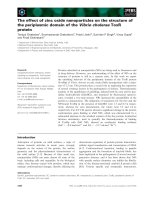
Fig. 2 shows FTIR sp ectroscopy data for SnO
2
nanoparticles
(Fig. 2(a)), Polyvinyl alcohol (Fig. 2(b)) and the PVA: SnO
2
composite (Fig. 2(c)). The results have been analyz ed using
standard FTIR reference book [14]. The typical signatures in PVA
which were due to O–H stretching (3333 cm
− 1
), C–Hstretching
(2912–2945 cm
− 1
), C–H bending (1416–1331 cm
− 1
), O–Hbending
(1416–1333 and 650 cm
− 1
)andC–O(1090cm
− 1
)wereseeninPVA
spectrum as well as the PVA:SnO
2
composi te. Additionally the
signature of Sn–Oat610cm
− 1
in the composite were also observed,
which were also seen in pure SnO
2
. As can be seen from this figure,
there was no modification of bonds of PVA after the composite had
been formed; nor there was any shift or intensity modification after
formation of the PVA:SnO
2
sample. Owing to the procedure that was
being used and using the well-established fact that SnO
2
is a highly
stable oxide, the spectrum was well anticipated. Thus we can conclude
here that the film formed was merely by embedding the SnO
2
nanoparticles in the PVA matrix, homogeneously. The insets of Fig. 2
show the photographs of nanocomposite films, exhibiting their
transparency and flexibility. However, as the percentage of SnO
2
was
changed in the composite, though FTIR did not show any changed
signature, one could expect that sample becomes denser and that can
affect the transport property of the sample.
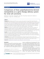
Fig. 3 confirmed the results of FTIR using TEM pictures. As seen in
Fig. 3, we observed nearly mono-dispersed SnO
2
nanoparticles with
particle size of ~5 nm (this was also confirmed using particle size
analysis). Lower inset shows a detailed TEM image at 10 nm length-
scale and upper left inset shows selected area electron diffraction
(SAED) pattern, which exhibited characteristics of polycrystalline
particles and the rings could be easily indexed with reference to the
rutile tetragonal SnO
2
structure, which was highly consistent with the
XRD results.
The most telling results are shown in Fig. 4. Humidity measure-
ments were done as described in the experimental section. Since we
were not very sure of the dimensional differences of different samples,
the comparison was done of the resistance ratios (R
11
/R
92
). The inset
of Fig. 4(a) shows the change in absolute values of resistance R
11
and
R
92
with the change in the molar ratios. This experiment was
performed for films with different molar ratios, namely 1:1, 1:2,
1:2.5, 1:3, 1:3.5, 1:4 and 1:5. To get fair comparison, results were also
checked with bulk SnO
2
powder and only PVA polymer. Very
interestingly it was found that the change in current with the change
in SnO
2
proportion in the sample was not monotonic, as is shown in
Fig. 4(a). It was seen that the ratio R
11
/R
92
increased initially with
SnO
2
, reached maximum (at 1:3 ratio) and decreased again. The
maximum change in resistance (current) was almost 5 times in the
1:3 sample. When compared to bulk SnO
2
, it was seen that the change
Fig. 3. TEM viewgraph of PVA: SnO
2
film, showing particle size ~5 nm. The inset below
shows the viewgraph on the scale of 10 nm. The inset on the top shows the SAED pattern
of the sample.
Fig. 4. (a). Plot of sensitivity (R
11
/R
92
) as a function of PVA:SnO
2
ratio. The inset shows
the change in resistance in 11% humidity and 92% humidity versus PVA:SnO
2
ratio.
(b). Plot of change in resistance of the 1:3 molar ratio film as a function of humidity. The
inset shows schematic with lower PVA: SnO
2
ratio (i) and critical threshold ratio.
(b). Schematic exhibits a percolation threshold in (ii) which shows a conductivity
between SnO
2
nanoparticles (orange balls) via tunneling through the PVA matrix and
water molecule (blue ball). (For interpretation of the references to colour in this figure
legend, the reader is referred to the web version of this article.)
849S. Hatamie et al. / Materials Science and Engineering C 29 (2009) 847–850
in resistance with the change in humidity from 11% to 92% was only by
few ohms, which suggested that SnO
2
in nanoparticles form and PVA
matrix was important in the sensing phenomenon. However, when
the similar experiment was done using only PVA polymer (1 M), we
did observe some change in current (R
11
/R
92
~1.89) indicating that
PVA was itself contributing to the humidity sensing. However, the
change was of the order of few ohms, which was quite insignificant as
compared to the SnO
2
embedded film. It is important to state here that
the response time was of the order of few msec. Also, similar rate of
recovery was observed as we removed the sample from 92% humidity
chamber and put it back in chamber at 11% humidity. The readings of
all films were taken at least two times to confirm these observations,
and same experiments were done on different batches of synthesized
samples. These findings gave us two different hints: i) role of PVA was
quite important in the sensing phenomenon; probably it offered ap-
preciable conducting tracks in between SnO
2
nanoparticles, and
ii) nanoparticles improvised the ability to sense humidity. After
getting the optimized molar ratio for maximum sensitivity, studies
were done using the 1:3 sample for practical applicability. The
samples were subjected to different humidity values ranging from 11%
to 92%. Fig. 4(b) shows the corresponding behavior, which depicts that
as the humidity increases, the resistance of the sample decreases.
With proper fitting of this data the sample can be calibrated for
outdoor applications. For further checking the reusability of the
synthesized samples, every sample was measured twice, using dif-
ferent contact positions. The samples were preserved in natural
ambience and after about three months, the samples exhibited the
readings within an error of b 5%.
Looking carefully into the literature, it can be envisaged that any
sensing device needs more adsorbing surface area, to adsorb moisture
(in our case) and yield some property changes. Since nanoparticles are
well known to have more surface-to-volume ratio, the increased jump
with nanoparticles as compared to their bulk value can be anticipated
[6,9]. Further, the polymer PVA helps this activity in two ways: firstly,
it works as a weak sensor and secondly, it is a hydrophilic polymer,
which hold the adsorbed water in the matrix. This helps the system-
as-a-whole to connect via the water molecules, to yield large current
values. Similar results have been observed by Andreev et al. [15],on
their system of PVA-calcium chloride. Their system offers relevant
conductivity variation by 4.5–5 times the magnitude with relative
humidity change from 0–86%. The humidity sensing was based on
hydrophilic polymers doped with metal salts, which increase the
sensor sensitivity due to the appearance of ionic conductivity during
water sorption. Similarly, Ogura et al. [16] have studied a humidity-
sensitive composite film that consists of conducting polyaniline and
water-loving PVA. Polyaniline gave a percolation threshold at a
particular volume fraction. The results have been interpreted on the
basis of doping level, which was affected by the concentration of water
molecules surrounding the conducting polymer.
In our case, on similar lines, we propose an explanation on
observed significantly higher humidity sensitivity by making a
nanocomposite of two materials (PVA and SnO
2
) which have far
weaker humidity sensitivity of their own. The high s ensitivity
occurred in a percolation regime where SnO
2
grains would almost
begin to touch each other building chain like configurations and
eventually leading to full percolation. Such tunneling phenomenon
has been reported in various systems, in recent past [17,18]. From our
standpoint the high humidity sensitivity at intermediate ratio of
nanocomponents can be explained based on the nature of the two
components of the system at their electronic proximity. SnO
2
is an n-
type conductor with high electron concentration at room tempera-
ture, hence although humidity may affect this concentration by
adsorption, the corresponding percentage change is very small. On the
other hand PVA has hopping c onduction, which leads to low
sensitivity because of limited mobility. At the optimum intermediate
molar concentration (1:3) the layer of PVA polymer would just be in
the tunneling regime with the humidity adsorption (as shown in the
schematic in the inset of Fig. 4(b) by introducing electronic states
which aid tunneling, effectively weakening the barrier. Hence we
envisage here that this is a novel system, which can be explored to
yield extremely sensitive devices. The film is highly flexible and
reusable. The synthesis mechanism promises useful applications of
this system as micro-sensors.
4. Conclusion
In conclusion, we have synthesized polycrystalline rutile structures
of SnO
2
nanoparticles of ~5 nm size using co-precipitation method.
Using simple chemical route SnO
2
has been embedded in PVA matrix.
The molarity ratio of PVA:SnO
2
was varied from 1:1 to 1:5. It was seen
that all films worked as humidity sensors. At a characteristic ratio of
1:3, the response of film was maximum and it decreased on either
sides of the optimum ratio. The results have been understood by
considering the critical tunneling regime, increased surface area of the
nanocomposites and the active role of PVA in the system.
Acknowledgements
Authors sincerely thank Dr. S.B. Ogale from National Chemical
Laboratory, Pune for his valuable guidance and suggestions. S.N. Kale
acknowledges International Centre for Theoretical Physics (ICTP), Italy
for her Associate affiliation and for the rich library access used for this
work.
References
[1] Q.X. Jia, T.M. Mccleskey, A.K. Burrell, Y. Lin, G.E. Collis, H. Wang, A.D.Q. Li, S.R.
Foltyn, Nat. Mater 3 (2004) 529.
[2] H. Kozuka, M. Kajimura, T. Hirano, K. Katayama, J. Sol-Gel Sci. Technol. 19 (2000)
205.
[3] S. Takenaka, H. Kozuka, Appl. Phys. Lett. 79 (2001) 3485.
[4] T.M. Racheva, G.W. Critchlow, Thin Solid Films 292 (1997) 299.
[5] N.V. Kolytcheva, H. Muller, J. Marstalerz, Sens. Actuators, B, Chem. 58 (1999) 456.
[6] T. Brousse, D.M. Schleich, Sens. Actuators, B, Chem. 31 (1996) 77.
[7] R. Luoh, H.T. Hahn, Compos. Sci. Technol. 66 (2006) 2436.
[8] S. Shukla, S. Seal, R. Vij, S. Bandopadhyay, Rev. Adv. Mater. Sci. 4 (2003) 1.
[9] M.C. Horrillo, A. Serventi, D. Rickerby, J. Gutierrez, Sens. Actuators, B, Chem. 58
(1999) 474.
[10] J. Mizsei, Sens. Actuators, B, Chem. 23 (1995) 173.
[11] M. Honore, S. Lenaerts, J. Desmet, G. Huyberechts, J. Roggen, Sens. Actuators, B,
Chem. 19 (1994) 621.
[12] Y. Shimizu, E.D. Bartolomeo, E. Traversa, G. Gusmano, T. Hyodo, K. Wada, M.
Egashira, Sens. Actuators, B, Chem. 60 (1999) 118.
[13] Ming-you MA, Ze-Qiang HE, Zhuo-bing Xiao, Ke-long Huang, Li-zhi Xiong, Xian-
ming Wu, Trans. Nonferr Met. Soc. China 16 (2006) 791.
[14] R.M. Silverstein, F.X. Webster, Spectrometric Identification of Organic Compounds,
sixth ed.John Wiley & Sons Inc Publications, 20 07.
[15] D.V. Andreev, L.L. Makarshin, V.N. Parmon, React. Kinet. Catal. Lett. 80 (2003) 181.
[16] K. Ogura, T. Saino, M. Nakayama, H. Shiigi, J. Mater. Chem. 7 (1997) 2363.
[17] S. Ju, K.W. Yu, Z.Y. Li, Phys. Rev. B 71 (2005) 014416.
[18] S.N. Kale, J. Mona, S.E. Lofland, S.D. Kulkarni, S.B. Ogale, Appl. Phys. Lett. 92 (2008)
012512.
850 S. Hatamie et al. / Materials Science and Engineering C 29 (2009) 847–850