- Trang chủ >>
- Khoa Học Tự Nhiên >>
- Vật lý
preparation and properties of magnetic iron oxide nanotubes
Bạn đang xem bản rút gọn của tài liệu. Xem và tải ngay bản đầy đủ của tài liệu tại đây (810.64 KB, 6 trang )
A
vailable online at www.sciencedirect.com
Particuology 6 (2008) 334–339
Preparation and properties of magnetic iron oxide nanotubes
Baoliang Lv
a,b
, Yao Xu
a,∗
, Dong Wu
a
, Yuhan Sun
a
a
State Key Laboratory of Coal Conversion, Institute of Coal Chemistry, Chinese Academy of Sciences, Taiyuan 030001, China
b
Graduate University of the Chinese Academy of Sciences, Beijing 100039, China
Received 7 March 2008; accepted 4 April 2008
Abstract
Magnetite (Fe
3
O
4
) nanotubes were prepared by reducing synthesized hematite (␣-Fe
2
O
3
) nanotubes in 5% H
2
+95% Ar atmosphere, and then
maghemite (␥-Fe
2
O
3
) nanotubes were obtained by re-oxidizing the Fe
3
O
4
nanotubes. The nanotube structure was kept from collapsing or sintering
throughout the high temperature reducing and re-oxidizing processes. The coercivities of the Fe
3
O
4
and ␥-Fe
2
O
3
nanotubes synthesized were
found to be 340.22 Oe and 342.23 Oe, respectively, both higher than other nanostructures with the same phase and of similar size. Both adsorbed
phosphate and the nanotube structure are considered responsible for this high coercivity.
© 2008 Chinese Society of Particuology and Institute of Process Engineering, Chinese Academy of Sciences. Published by Elsevier B.V.
All rights reserved.
Keywords: Nanostructures; Iron oxides; Nanotubes; Magnetic properties
1. Introduction
Magnetic materials with special nanostructures are scientif-
ically interesting and technologically important in research for
future applications (Sui, Skomski, Sorge, & Sellmyer, 2004a).
Iron oxides as an important class of magnetic materials have
been widely used in catalysis (Zhang et al., 2005), magnetic
devices (Zeng, Li, Liu, Wang, & Sun, 2002), environment pro-
tection (Wu, Qu, & Chen, 2005), sensors (Sun, Yuan, Liu, Han,
& Zhang, 2005), drug delivery (Wu et al., 2007) and water split-
ting (Cesar, Kay, Gonzalez Martinez, & Grätzel, 2006). Up to
now, iron oxides with nanostructures have attracted a great deal
of attention because of their promising properties and appli-
cations. Many iron oxide particles with zero-, one-, two- and
three-dimensional (0D, 1D, 2D and 3D) nanostructures have
been synthesized. Ferromagnetic nanotubes were considered
as candidates for recording head, biomagnetic sensors, cata-
lysts, etc., because of their expected vortex magnetization state
and floatability in liquid as a result of their hollow structure
(Goldstein, Gelb, & Yager, 2001; Haberzettl, 2002; Khizroev,
Kryder, Litvinov, & Thomson, 2002). Iron oxide nanotubes
have been synthesized mostly via the so-called template-directed
growthmethod.For example, Sui, Skomski, Sorge, and Sellmyer
∗
Corresponding author. Tel.: +86 351 4049859; fax: +86 351 4041153.
E-mail address: (Y. Xu).
(2004b), Wang, Wang, Li, Xu, and Zhou (2006), and Shen et
al. (2004) used porous anodic aluminium oxide (AAO) as tem-
plate to prepare Fe
3
O
4
and ␣-Fe
2
O
3
nanotube arrays; Sun et
al. (2005) used carbon nanotubes as templates to fabricate ␣-
Fe
2
O
3
nanotubes; Liu et al. (2005) used MgO nanowires as
templates to fabricate single-crystal Fe
3
O
4
nanotubes. How-
ever, templates not only introduced extraneous impurities but
also increased production cost, not to say the many prob-
lems to prepare these materials at large scale. Therefore, it
is of practical significance to develop a template-free and
somewhat easier method to synthesize magnetic iron oxides
nanotubes.
Recently, Jia et al. (2005) synthesized ␣-Fe
2
O
3
nanotubes by
a hydrothermal method without using template. In this work, we
improved their work by first synthesizing ␣-Fe
2
O
3
nanotubes,
followed by reducing ␣-Fe
2
O
3
and re-oxidizing the Fe
3
O
4
to
␥-Fe
2
O
3
nanotubes. The magnetic properties of the Fe
3
O
4
and
␥-Fe
2
O
3
nanotubes were investigated by using vibrating sample
magnetometry (VSM).
2. Experimental
All the reagents were A.R. grade and were used in prepara-
tion without further purification: ferric chloride (FeCl
3
·6H
2
O,
China Medicament Co.), sodium dihydrogen phosphate
(NaH
2
PO
4
·2H
2
O, Tianjin Chemical Reagent Co.), double-
1674-2001/$ – see inside back cover © 2008 Chinese Society of Particuology and Institute of Process Engineering, Chinese Academy of Sciences. Published by Elsevier B.V.All rights reserved.
doi:10.1016/j.partic.2008.04.006
B. Lv et al. / Particuology 6 (2008) 334–339 335
distilled water. Reduction gas was composed of 5 v% H
2
(high
purity) and 95 v% Ar (high purity).
The preparation of ␣-Fe
2
O
3
nanotubes was an improved
approach based onliterature (Jia et al., 2005).In a typical synthe-
sis, 40 mL of FeCl
3
aqueous solution (46.2 mmol/L) and 40 mL
of NaH
2
PO
4
aqueous solution (1.9 mmol/L) were first mixed
and then dispersed uniformly by ultrasonic irradiation. The solu-
tion was then sealed in a 100-mL Teflon-lined stainless steel
autoclave and hydrothermally treated for 36 h at 240
◦
C. At last,
a red precipitate was obtained at the bottom of the autoclave and
was separated by centrifugation. The precipitate was washed
three times with distilled water, and then dried at 60
◦
C in air.
The resulting powder was ␣-Fe
2
O
3
nanotubes, named as S1.
Fe
3
O
4
nanotubes were obtained by reducing S1 in a tubular
oven at 500
◦
Cfor2.5hina5%H
2
+95% Ar atmosphere, and
the resulting black powder was named as S2. In this process,
the temperature and reduction time were very important, or else
␣-Fe
2
O
3
or FeO would be present in the reduction product. To
prepare ␥-Fe
2
O
3
nanotubes, the as-prepared Fe
3
O
4
nanotubes
were oxidized by air at 300
◦
C for 2 h, to produce a red powder,
named as S3.
X-ray diffraction (XRD) measurement was performed on a
D8 Advance Bruker AXS diffractometer using Cu K␣ radia-
tion (λ=1.5406 Å). Raman spectra were recorded using a Horiba
Labram HR800 spectrometer equipped with a Spectra Physics
514 nm argon ion laser. The morphologies of the samples were
observedbyscanningelectronmicrograph(SEM,LEO 1530VP)
and transmission electron micrograph (TEM, Hitachi H-600).
X-ray photoelectron spectroscopy (XPS) measurements were
performed on a PHI 5300× multi-technique system with Mg K␣
X-ray source (PerkinElmer Physical Electronics).Magnetichys-
teresis loops were measured by vibrating sample magnetometry
(VSM, Lakeshore 7407).
3. Results and discussion
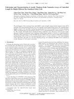
Fig. 1 shows the XRD patterns of samples S1 (a), S2 (b) and
S3 (c). In Fig. 1(a), the initial synthesized product (sample S1)
Fig. 1. XRD patterns of samples S1 (a), S2 (b) and S3 (c).
Fig. 2. Raman spectra of samples S2 (a) and S3 (b).
can be exclusively indexed to ␣-Fe
2
O
3
, according to standard
data (JCPDS 33-0664). In Fig. 1(b) and (c), the reflection peaks
of XRD patterns of S2 and S3, can be well assigned to a spinel
structure with the characteristic reflections of ␥-Fe
2
O
3
(JCPDS
39-1346) or Fe
3
O
4
(JCPDS 19-0629). However, it is well-known
that clear identification of ␥-Fe
2
O
3
and Fe
3
O
4
based on ordi-
nary XRD pattern is an arduous task due to their same spinel
structure and their similar lattice parameters (Xiong, Ye, Gu, &
Chen, 2007). Although the color of S2 was black and S3, red,
corresponding to Fe
3
O
4
and ␥-Fe
2
O
3
, respectively, the purity
of the samples cannot be simply identified by their appearance.
To differentiate samples S2 and S3 clearly, further characteriza-
tion is needed for more convincing evidence, for which Raman
spectrum was resorted to (Daou et al., 2006; Pinna et al., 2005;
Xiong et al., 2007). A representative Raman spectrum of sam-
ple S2, shown in Fig. 2(a), exhibits two clear peaks at 665 and
540 cm
−1
, which can be indexed to the A1g and T2g transitions
of the Fe
3
O
4
phase (Shebanova & Lazor, 2003). In Fig. 2(b),
the Raman spectrum of sample S3, the different characteristic
bands of ␥-Fe
2
O
3
(700, 500 and 350 cm
−1
) can be observed
(Varadwaj, Panigrahi, & Ghose, 2004). Consequently, it should
be reasonable to think that sample S2 is Fe
3
O
4
and sample S3
is ␥-Fe
2
O
3
.
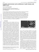
Fig. 3 presents the SEM and TEM images of samples S1,
S2 and S3. Fig. 3(a) and (b) show the morphologies of ini-
tial synthesized ␣-Fe
2
O
3
nanotubes (sample S1), in which the
nanotubes can be seen clearly, with length of 160–300 nm,
336 B. Lv et al. / Particuology 6 (2008) 334–339
Fig. 3. SEM and TEM images of ␣-Fe
2
O
3
nanotubes in sample S1 (a and b), Fe
3
O
4
nanotubes in sample S2 (c and d) and ␥-Fe
2
O
3
nanotubes in sample S3 (e and f).
B. Lv et al. / Particuology 6 (2008) 334–339 337
and outer and inner diameters of 70–120 nm and 45–80 nm,
respectively. Fig. 3(c) and (d) show the morphologies of Fe
3
O
4
nanotubes (sample S2), with their well retained nanotube struc-
ture. Fig. 3(e) and (f) show the nearly same morphologies of
␥-Fe
2
O
3
(sample S3) together with their well retainedtubestruc-
ture. Fig. 3(d) and (e) show that there was no obvious change
in the length and diameter of the nanotubes. Comparison of the
TEM images of the three samples indicates that the Fe
3
O
4
and
␥-Fe
2
O
3
nanotubes are conglomerated with each other, while
␣-Fe
2
O
3
nanotubes are well dispersed, apparently due to the
mutual attraction of the magnetic Fe
3
O
4
and ␥-Fe
2
O
3
particles,
though no obvious difference can be found from the morpholo-
gies of the three samples.
Generally, nanostructures areoften destroyed due to sintering
or collapsing during treatment at high temperature. But Fig. 3
showsthatthenanotubestructurewas well preserved after reduc-
tion and re-oxidization at high temperature. There might be two
reasons for this. First, according to Jiao et al. (2006), conversion
of ␣-Fe
2
O
3
to Fe
3
O
4
involves a change from a hexagonal close-
packed oxide ion array (␣-Fe
2
O
3
) to a cubic close-packed array
(Fe
3
O
4
). This conversion is not merely topotactic, but involves
a sheave of oxide ion planes from AB to ABC stacking, and this
significant structural change can occur without much destroy-
ing the tube structure. The thin walls of the nanotubes endowed
the solids with a structural flexibility that made such solid/solid
transformation smooth while preserving the tube structure. Sec-
ond, according to Jia et al. (2005), phosphate could be adsorbed
on ␣-Fe
2
O
3
by reacting with the singly coordinated surface
hydroxy groups to form a monodentate orbidentate inner-sphere
complex. Here, the amount of adsorbed phosphate was so small
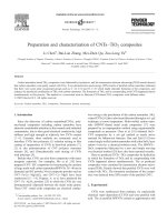
that it could not be detected by XRD. To confirm the existence
of phosphate on the surface of the nanotubes, XPS analysis was
carried out on the surface element composition of the initial ␣-
Fe
2
O
3
nanotubes, with the result shown in Fig. 4(a). The binding
energies obtained in the XPS analysis were corrected by refer-
encing the C1s line to 284.5 eV. Seen from Fig. 4(a), the binding
energy of P2p was found at 133.6 eV in the spectrum, which
agreed with the reported value of PO
4
3−
(Wang et al., 2003).
To further identify the existence of the phosphate layer, a high-
magnification image of sample S1 was obtained on TEM, as
shown in Fig. 4(b), indicating the presence of a 2.5-nm adsorp-
tion layer, thus confirming the presence of a phosphate layer on
the surface of synthesized ␣-Fe
2
O
3
nanotubes. The adsorbed
phosphate would be very stable in the reduction process, and act
as a framework or a protection shell for the nanotubes. When
the reduction of ␣-Fe
2
O
3
went on, the phosphate on the surface
could not be reduced, and only the inner ␣-Fe
2
O
3
wasreducedby
hydrogen. Therefore, the nanotubes could be kept from sintering
or collapsing. To confirm the stabilization of phosphate on nan-
otubes, pure iron phosphate (FePO
4
) sample was treated under
the same reduction condition as that for ␣-Fe
2
O
3
reduction. The
XRD pattern (not given here) of the reduction product showed
that FePO
4
was reduced to Fe
2
PO
5
. Oxidation of Fe
3
O
4
nan-
otubes to ␥-Fe
2
O
3
nanotubes involved a decrease in the number
of Fe atoms per unit cell of 32 oxygen ions, from 24 in Fe
3
O
4
to 21(1/3) in ␥-Fe
2
O
3
. This reaction proceeded with outward
migration of the Fe
2+
cations towards the surface of the crystal
Fig. 4. (a) XPS spectrum of ␣-Fe
2
O
3
nanotubes and (b) high magnification
TEM image of a nanotube in sample S1.
together with the creation of cation vacancies and the addition
of oxygen atoms. At the surface the Fe
2+
cations were oxidized
through interacting with adsorbed oxygen to form of ␥-Fe
2
O
3
,
too. The whole process involved a topotactic reaction in which
the original crystal morphology was preserved throughout the
process (Cornell & Schwertmann, 2003).
Magnetic nanoparticles, especially those with special struc-
tures, often exhibit unusual magnetic behaviors different from
that of bulk solids, owing to finite size effects and microstructure
(Bødker, Hansen, Bender Koch, Lefmann, & Mørup, 2000). To
investigate the magnetic properties of the as-synthesized nan-
otubes, magnetic hysteresis (M–H) loop measurements were
carried out in an applied magnetic field at room temperature,
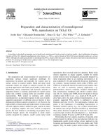
with the field sweeping from −18 to 18 kOe. Fig. 5 shows
the M–H loops of Fe
3
O
4
(a) and ␥-Fe
2
O
3
(b) nanotubes at
room temperature. From Fig. 5(a), the M–H loop of Fe
3
O
4
nanotubes shows ferromagnetic behavior with a saturation mag-
netization (Ms) of 60.92 emu/g, a remanent magnetization (Mr)
of 18.56 emu/g and a coercivity of 340.22 Oe at room tem-
perature. Compared to bulk Fe
3
O
4
(Ms = 92 emu/g, coercivity
115–150 Oe) (Liu, Fu, & Xiao, 2006), the Ms was obviously
lower and the coercivity was obviously higher. Fe
3
O
4
nanotubes
also possess higher coercivity than other Fe
3
O
4
nanostruc-
tures of similar size, such as octahedral nanoparticles (141 Oe),
nanocubes (62 Oe) and hollow spheres (40 Oe) (Daou et al.,
2006; Huang & Tang, 2005; Xiong et al., 2007; Yu et al., 2006).
From Fig. 5(b), the M–H loop of ␥-Fe
2
O
3
nanotubes shows
ferromagnetic behavior with a Ms of 42.71 emu/g, a Mr of
338 B. Lv et al. / Particuology 6 (2008) 334–339
Fig. 5. M–H loops of Fe
3
O
4
nanotubes (a) and ␥-Fe
2
O
3
nanotubes (b). The
inset diagrams are their corresponding expanded low-field curves.
13.56 emu/g and a coercivity of 342.23 Oe at room temperature.
Compared to bulk ␥-Fe
2
O
3
(Ms = 76 emu/g, coercivity 300 Oe)
(Zhang, Tang, and Hu, 2008), the Ms is obviously lower and
the coercivity is somewhat higher. Similar to Fe
3
O
4
nanotubes,
␥-Fe
2
O
3
nanotubes also have a higher coercivity than other
reported ␥-Fe
2
O
3
nanostructures, such as nanofibres (78.11 Oe),
nanoparticles (106 Oe), and some reported superparamagnetic
␥-Fe
2
O
3
particles (0 Oe) (Han et al., 2007; Jing, 2006; Zhang et
al., 2008). It is noted that boththesetwo magneticironoxidenan-
otubes have a higher coercivity than other nanostructures with
the same phase and of similar size. Furthermore, the M–H loops
of Fe
3
O
4
nanotubes and ␥-Fe
2
O
3
nanotubes indicate the similar
magnetic domain type. On the basis of the criteria given by Dun-
lop (Cornell & Schwertmann, 2003), the Mr/Ms value should
be larger than 0.5 for single domain (SD) particles, between 0.1
and 0.5 for pseudosingle-domain (PSD) particles and lower than
0.1 for multidomain (MD) particles. From Fig. 5, both the two
samples possess PSD-type magnetic domains, and their Mr/Ms
values are 0.30 and 0.32, respectively.
There might be two reasons for the high coercivity. First is
the influence of adsorbed phosphate at the surface of these nan-
otubes, which has been confirmed previously by XPS, and the
phosphate is not a magnetic material. From Fig. 5, both the two
samples have a Mr/Ms value between 0.1 and 0.5, indicating that
they may possess the magnetic properties of SD and MD struc-
tures simultaneously. If the synthesized products possess more
properties of MD structures, the magnetic domain walls would
exist inside the particles. For MD materials, the movement of
magnetic domain walls is the main reason for coercivity. It is
well known that there exist surface domain walls for MD par-
ticles. Here, the surface domain walls should be present at the
interface between iron oxide and the adsorbed phosphate. The
phosphate as an uninterrupted adsorbed layer can easily block
the movement of the surface domain walls and result in domain
wall pinning, which contributesto the high coercivity. Even if the
synthesized products possess more properties of SD particles,
the coercivity would also increase. For SD magnetic material,
magnetic domain wall does not exist, and spin flip conversion
is mainly responsible for the coercivity. In this case, the coor-
dination bonds between adsorbed phosphate ions and iron ions
would form spin pinning and block spin flip conversion, directly
resulting in the increase of coercivity of the samples. Second, the
nanotube structure may be another reason for the high coerciv-
ity. Torres-Heredia, López-Urías, and Mu
˜
noz-Sandoval (2005)
simulated the micromagnetic property of iron nanorings, and
they found large coercive fields for d
in
/d
out
> 0.5 (d
in
and d
out
are the inner and outer diameters of the rings, respectively) and
t = 160–200 nm (t is the thickness of the rings or length of the
tubes) nanorings due to the absence of the vortex states and the
presence of out-plane and in-plane spin configurations. In our
samples, the average d
in
/d
out
valueof nanotubes isabout 0.7, and
the length of many nanotubes is about 200 nm, which snugly fall
into the thickness range of nanorings mentioned in the literature
(Torres-Heredia et al., 2005). So the nanotubes can be thought
as nanorings with immensely large thickness, and this structure
can contribute to the high coercivity.
4. Conclusions
Fe
3
O
4
nanotubes were prepared by reducing synthesized ␣-
Fe
2
O
3
nanotubes with a gas mixture of 5% H
2
+95% Ar at 500
◦
C
for 2.5 h, and then ␥-Fe
2
O
3
nanotubes were obtained by re-
oxidizing the Fe
3
O
4
nanotubes with air at 300
◦
C for 2 h. The
nanotube structure was well retained without collapsing or sin-
tering, for which, adsorbed phosphate and the type of crystal
structure conversion should be the two most important reasons.
Investigation of the magnetic properties of Fe
3
O
4
and ␥-Fe
2
O
3
nanotubes revealed that both the two magnetic iron oxide nan-
otubes possess higher coercivity than other nanostructures with
same phase and of similar size. The adsorbed phosphate and
the tube structure should be responsible for the high coercivity.
Research on applications of these two magnetic nanotubes is in
progress.
References
Bødker, F., Hansen, M. F., Bender Koch, C., Lefmann, K., & Mørup, S. (2000).
Magnetic properties of hematite nanoparticles. Physical Review B, 61(10),
6826–6838.
Cesar, I., Kay, A., Gonzalez Martinez, J. A., & Grätzel, M. (2006). Translu-
cent thin film Fe
2
O
3
photoanodes for efficient water splitting by sunlight:
Nanostructure-directing effect of Si-doping. Journal of the American Chem-
ical Society, 128(14), 4582–4583.
B. Lv et al. / Particuology 6 (2008) 334–339 339
Cornell, R. M., & Schwertmann, U. (2003). The iron oxides: Structure, proper-
ties, reactions, occurrences and uses. Weinheim: Wiley-VCH.
Daou, T. J., Pourroy, G., Bégin-Colin, S., Grenèche, J. M., Ulhaq-Bouillet, C.,
Legaré, P., et al. (2006). Hydrothermal synthesis of monodisperse magnetite
nanoparticles. Chemistry of Materials, 18(18), 4399–4404.
Goldstein, A.S.,Gelb, M.H., & Yager, P. (2001). Continuousand highly variable
rate controlled release of model drugs from sphingolipid-based complex
high axial ratio microstructures. Journal of Controlled Release, 70(1), 125–
138.
Haberzettl, C. A. (2002). Nanomedicine: Destination or journey. Nanotechnol-
ogy, 13(4), R9–R13.
Han, Q., Liu, Z., Xu, Y., Chen, Z., Wang, T., & Zhang, H. (2007). Growth and
properties of single-crystalline ␥-Fe
2
O
3
nanowires. The Journal of Physical
Chemistry C, 111(13), 5034–5038.
Huang, Z. B., & Tang, F. J. (2005). Preparation, structure, and magnetic prop-
erties of mesoporous magnetite hollow spheres. Journal of Colloid and
Interface Science, 281(2), 432–436.
Jia, C. J., Sun, L. D., Yan, Z. G., You, L. P., Luo, F., Han, X. D., et al. (2005).
Single-crystalline iron oxide nanotubes. Angewandte Chemie International
Edition, 44(28), 4328–4333.
Jiao, F., Jumas, J C., Womes, M., Chadwick, A. V., Harrison, A., & Bruce,
P. G. (2006). Synthesis of ordered mesoporous Fe
3
O
4
and ␥-Fe
2
O
3
with
crystalline walls using post-template reduction/oxidation. Journal of the
American Chemical Society, 128(39), 12905–12909.
Jing, Z. (2006). Preparation and magnetic properties of fibrous gamma iron
oxide nanoparticlesvia anonaqueous medium.Materials Letters, 60(17–18),
2217–2221.
Khizroev, S., Kryder, M. H., Litvinov, D., & Thomson, D. A. (2002). Direct
observation of magnetization switching in focused-ion-beam-fabricated
magnetic nanotubes. Applied Physics Letters, 81(12), 2256–2257.
Liu, X. M., Fu, S. Y., & Xiao, H. M. (2006). Fabrication of octahedral magnetite
microcrystals. Materials Letters, 60(24), 2979–2983.
Liu, Z., Zhang,D., Han, S., Li, C.,Lei, B., Lu, W., et al. (2005).Single crystalline
magnetite nanotubes. Journal of the American Chemical Society, 127(1),
6–7.
Pinna, N.,Grancharov, S., Beato,P., Bonville, P., Antonietti, M.,& Niederberger,
M. (2005). Magnetite nanocrystals: Nonaqueous synthesis, characterization,
and solubility. Chemistry of Materials, 17(11), 3044–3049.
Shebanova, O. N.,&Lazor,P.(2003).Raman study ofmagnetite (Fe
3
O
4
): Laser-
induced thermal effects and oxidation. Journal of Raman Spectroscopy,
34(11), 845–852.
Shen, X. P., Liu, H. J., Pan, L., Chen, K. M., Hong, J. M., & Xu, Z. (2004).
An efficient template pathway to synthesis of ordered metal oxide nanotube
arrays using metal acetylacetonates as single-source molecular precursors.
Chemistry Letters, 33(9), 1128–1129.
Sui, Y. C., Skomski, R., Sorge, K. D., & Sellmyer, D. J. (2004a). Nanotube
magnetism. Applied Physics Letters, 84(9), 1525–1527.
Sui, Y. C., Skomski, R., Sorge, K. D., & Sellmyer, D. J. (2004b). Magnetic
nanotubes produced by hydrogen reduction. Journal of Applied Physics,
95(11), 7151–7153.
Sun, Z. Y., Yuan, H. Q., Liu, Z. M., Han, B. X., & Zhang, X. R. (2005). A highly
efficient chemical sensor material for H
2
S: ␣-Fe
2
O
3
nanotubes fabricated
using carbon nanotube templates. Advanced Materials, 17, 2993–2997.
Torres-Heredia, J. J., López-Urías, F., & Mu
˜
noz-Sandoval, E. (2005). Micro-
magnetic simulation of iron nanorings. Journal of Magnetism and Magnetic
Materials, 294(2), e1–e5.
Varadwaj, K. S. K., Panigrahi, M. K., & Ghose, J. (2004). Effect of capping and
particle size on Raman laser-induced degradation of ␥-Fe
2
O
3
nanoparticles.
Journal of Solid State Chemistry, 177(11), 4286–4292.
Wang, T., Wang, Y., Li, F., Xu, C., & Zhou, D.(2006). Morphology and magnetic
behaviorofan Fe
3
O
4
nanotube array.Journal of Physics: Condensed Matter,
18(47), 10545–10551.
Wang, X., Wang, Y., Tang, Q., Guo, Q., Zhang, Q., & Wan, H. (2003). MCM-
41-supported iron phosphate catalyst for partial oxidation of methane to
oxygenates with oxygen and nitrous oxide. Journal of Catalysis, 217(2),
457–467.
Wu, P. C., Wang, W. S., Huang, Y. T., Sheu, H. S., Lo, Y. W., Tsai, T. L., et al.
(2007). Porous iron oxide based nanorods developed as delivery nanocap-
sules. Chemistry A: European Journal, 13(14), 3878–3885.
Wu, R. C., Qu, J. H., & Chen, Y. S. (2005). Magnetic powder MnO-Fe
2
O
3
composite—A novel material for the removal of azo-dye from water. Water
Research, 39(4), 630–638.
Xiong, Y., Ye, J., Gu, X. Y., & Chen, Q. W. (2007). Synthesis and assembly
of magnetite nanocubes into flux-closure rings. The Journal of Physical
Chemistry C, 111(19), 6998–7003.
Yu, W., Zhang, T., Zhang, J., Qiao, X., Yang, L., & Liu, Y. (2006). The syn-
thesis of octahedral nanoparticles of magnetite. Materials Letters, 60(24),
2998–3001.
Zeng, H., Li, J., Liu, J. P., Wang, Z. L., & Sun, S. H. (2002). Exchange-
coupled nanocompositemagnets by nanoparticleself-assembly. Nature, 420,
395–398.
Zhang, J. L.,Wang, Y., Ji, H.,Wei, Y. G., Wu, N.Z., Zuo, B. J.,et al. (2005). Mag-
netic nanocomposite catalysts with high activity and selectivity for selective
hydrogenation of ortho-chloronitrobenzene. Journal of Catalysis, 229(1),
114–118.
Zhang, Y.C., Tang,J. Y.,& Hu,X. Y. (2008).Controllable synthesisand magnetic
properties of pure hematite and maghemite nanocrystals from a molecular
precursor. Journal of Alloys and Compounds, 462(1–2), 24–28.