- Trang chủ >>
- Khoa Học Tự Nhiên >>
- Vật lý
chronopotentiometric stripping analysis of gelatinase b, collagen and their interaction
Bạn đang xem bản rút gọn của tài liệu. Xem và tải ngay bản đầy đủ của tài liệu tại đây (426.39 KB, 6 trang )
Full Paper
Chronopotentiometric Stripping Analysis of Gelatinase B, Collagen
and Their Interaction
Dalibor Huska,
a
Vojtech Adam,
a, b
Ondrej Zitka,
a, c
Jiri Kukacka,
d
Richard Prusa,
d
Rene Kizek
a
*
a
Department of Chemistry and Biochemistry, Faculty of Agronomy, Mendel University of Agriculture and Forestry, Zemedelska 1,
CZ-613 00 Brno, Czech Republic
*e-mail:
b
Department of Animal Nutrition and Forage Production, Faculty of Agronomy, Mendel University of Agriculture and Forestry,
Zemedelska 1, CZ-613 00 Brno, Czech Republic
c
Department of Biochemistry, Faculty of Science, Masaryk University, Kotlarska 2, CZ-611 37 Brno, Czech Republic
d
Department of Clinical Biochemistry and Pathobiochemistry, 2
nd
Faculty of Medicine, Charles University, V Uvalu 84, CZ-150 06
Prague 5, Czech Republic
Received: July 23, 2008
Accepted: October 12, 2008
Abstract
Matrix metalloproteinases (MMP) belong to a group of zinc-dependent proteins that play a central role in the
breakdown of extracellular matrices. Collagen, elastin, gelatin and casein are the main components of extracellular
matrix cleaved by MMP. This paper aims to analyze the interaction between gelatinase B (MMP-9) and collagen using
chronopotentiometric stripping analysis with adsorptive transfer stripping technique (AdTS CPSA). Under optimized
experimental conditions (time accumulation of 90 s, supporting electrolyte 0.2 M acetate buffer pH 5, stripping
current 1 mA), the detection limit (3 signal/noise) for MMP-9 was estimated as being 100 pM. The interaction between
MMP-9 and collagen was studied according to the following scheme: i) HMDE surface was renewed. ii) Renewed
surface of HMDE collagen (1 mg/mL) was accumulated for 90 s under open circuit. iii) The electrode was rinsed in
ACS grade water and immersed in 5 mL drop of MMP-9. iv) The interaction between MMP-9 with collagen took place
at open circuit. v) The electrode was then rinsed in ACS grade water. vi) The rinsed electrode was transferred into an
electrochemical cell and measured in acetate buffer (pH 5). The CPSA signal of collagen after its interaction with
MMP-9 increased more than 30% compared to that of only collagen. This increase in signal is likely due to the
cleavage of collagen by MMP-9, hence its easy access to the electrodes surface.
Keywords: Matrix metalloproteinases, Chronopotentiometric stripping analysis, Collagen, Protein– protein
interaction, Cancer
DOI: 10.1002/elan.200804440
Dedicated to Professor Joseph Wang, on the Occasion of His 60
th
Birthday
1. Introduction
The matrix metalloproteinases (MMP), also known as
matrixins, belong to a group of zinc-dependent proteins,
which are thought to play a central role in the breakdown of
extracellular matrix. Collagen, elastin, gelatin, and casein
are the main components cleaved by MMP. The breakdown
of these components is essential for many physiological
processes such as embryonic development, morphogenesis,
reproduction, and tissue resorption and remodeling [1].
MMP also participate in pathological processes such as
arthritis, cancer, cardiovascular and neurological diseases
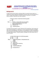
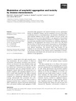
[2 – 6]. The primary structure of MMP, for twenty different
vertebrates, is comprised of several domain motifs, as
illustrated in Figure 1. The domains have been divided
according to their structure and function: collagenases,
stromelysins, matrilysins, gelatinases, membrane type MMP
and others MMP [7, 8].
Chronopotentiometric stripping analysis (CPSA) meas-
ures the evolution of hydrogen from the supporting electro-
lyte catalyzed by the presence of a protein. This method is a
highly sensitive techniquecommonlyused for the analysisof
proteins with detection limits at subnanomolar and lower
levels. Disadvantages include high standard deviations and
time of analysis at high stripping currents [9]. CPSA has
been used for thedetectionofseveralbiologically important
peptides [10, 11] and proteins such as metallothionein [12 –
18], a-synuclein protein [19], MutS protein [20], gluta-
thione-S-transferase [21],thrombin[22]. Moreover, Ostatna
et al. showed this electrochemical method can be employed
to studystructuralchanges of bovineserumalbumin [23, 24].
Serrano et al. studied metal – protein interactions using
CPSA [25, 26]. Redox states of peptides and proteins can
also be determined using CPSA [27]. However, CPSA has
not been utilized for the detection of MMP, yet. The main
aim of this paper is to characterize MMP-9, collagen and
536
Electroanalysis 2009, 21, No. 3-5, 536 – 541 2009 Wiley-VCH Verlag GmbH &Co. KGaA, Weinheim
their interaction by using chronopotentiometric stripping
analysis with adsorptive transfer stripping technique.
2. Experimental
2.1. Chemicals and pH Measurements
Human MMP-9 was purchased from Chemicon Interna-
tional (Temecula, USA). Collagen was supplied from
Vyzkumny ustav pletarsky (Brno, Czech Republic). ACS
grade Co(NH
3
)
6
Cl
3
and otherchemicals(chemicals meetthe
specifications of the American Chemical Society) used were
purchased from Sigma Aldrich (Sigma-Aldrich, USA)
unless noted otherwise. The stock standard solutions (10
mg/mL) were prepared with ACS water (Sigma-Aldrich,
USA) and stored in the dark at À208C. Working standard
solutions were prepared daily by the dilution of the stock
solutions with ACS certifiedwater.The pHandconductivity
were measured using inoLab Level 3 (Wissenschaftlich-
Technische Werkst¾tten GmbH; Weilheim, Germany).
2.2. Electrochemical Measurements
Electrochemical measurements were performed with AU-
TOLABAnalyzer (EcoChemie, Netherlands) connected to
VA-Stand 663 (Metrohm, Switzerland), using a standard
cell with three electrodes. A hanging mercury drop elec-
trode (HMDE)witha drop areaof0.4 mm
2
was employedas
the working electrode. An Ag/AgCl/3 M KCl electrode
served as the reference electrode. Glassy carbon electrode
was used as the auxiliary electrode. For smoothing and
baseline corrections, the software GPES 4.9 supplied by
EcoChemie was employed. The analyzed samples were
deoxygenated prior to measurements by purging with argon
(99.999%) and saturated with water for 120 s. All experi-
ments were carried out at room temperature. The temper-
ature of supporting electrolyte was maintained by the flow
electrochemical cell coupled with thermostat JULABO
F12/ED (Labortechnik GmbH, Germany).
Adsorptive transfer stripping technique (AdTS) with
chronopotentiometric stripping analysis (CPSA) was used
to determine the presence of MMP-9 and/or collagen by
recording the inverted time derivationofpotential(d E/dt)
À1
as a function of potential E [12]. Peptides and proteins
produce a well-developed peak at highly negative potentials
[10]. The behavior of this peak suggests the presence of
catalytic evolutionofhydrogen [16]. CPSAparameters were
optimized as seen in Section 3.
2.4. Descriptive Statistics and Estimation of Detection
Limit
Data were analyzed using MICROSOFT EXCEL (USA).
Results are expressed as mean ÆSD unless noted otherwise.
The detection limits (3 signal/noise, S/N) were calculated
Fig. 1. Characterization of single MMPs according to their structural differences.
537CPSA of Gelatinase B, Collagen and Their Interaction
2009 Wiley-VCH Verlag GmbH &Co. KGaA, Weinheim www.electroanalysis.wiley-vch.de Electroanalysis 2009, 21, No. 3-5, 536 – 541
according to Long and Winefordner [28], whereas N was
expressed as standard deviation of noise determined in the
signal domain unless stated otherwise.
3. Results and Discussion
The analysis of MMP raises methodical questions and
concerns such as the type of sample (serum or plasma or
blood), target molecule (total content of MMP or specific
MPP) or form of MMP detected (MMP molecules or
proMMP). Most commonly usedmethodsfor MMP analysis
are immunochemistry and enzymatic-based ones [29].
3.1. Chronopotentiometric Stripping Analysis of MMP-9
Coupling adsorptive transfer stripping technique with
chronopotentiometric stripping analysis has several advan-
tages which include lowdetectionlimitsfor target molecules
[9, 12, 13, 17, 23]. This coupled technique was employed to
detect MMP-9 (Fig. 2A). Even though the amino acid
cysteine forms only 3% (total count 19) of the total amino
acid content in MMP-9 (total count 707), they are known to
be responsible for most of the CPSA measured electro-
activity of MMP-9. Experimental conditions were opti-
mized to detect MMP-9 by AdTS CPSA. Time accumula-
tion of MMP-9 onto HMDE was the first parameter
optimized. Working MMP-9 concentrations used (1 ng/
mL) were low, which showed high CPSA sensitivity to
proteins. Measurements were carried out in borate buffer
(pH 7.6) according to previously published results [12, 15,
16, 30]. MMP-9 was adsorbed onto HMDE at various times
of accumulation: 30, 60, 90, 120, 150 and 180 s. Dependence
of peak height on accumulation time is shown in Figure 2B.
Peak height enhanced up to 90 s, and then decreased more
than 50% possibly due to increase formation of complex
structures on the working electrodes surface. Similar
behavior of protein adsorption on HMDE surface was
observed by Petrlova et al. [13, 31] and Adam et al. [32].
The pH of the supporting electrolyte plays an essential
role in CPSA analysis. Hence, the electrochemical behavior
of MMP-9 was studied in the following buffers: acetate
buffer (pH 4.6), Britton – Robinson buffer (pH 6.5), phos-
phate buffer (pH 6.95) and borate buffer (7.6). MMP-9
signals measured in the buffers are shown in Figure 2C.
MMP-9 gave signals at different potentials according to the
pH and type of buffer: acetate buffer at À1.47 V; Britton –
Robinson buffer at À1.62 V, phosphate buffer at À1.71 V
and borate buffer at À1.74 V. The highest MMP-9 signal
response was detected in acetatebuffer(Fig. 2D). The lower
pH values were determined to be more suitable for metal-
loproteinase electrochemical analysis than higher pH val-
ues. The value of MMP-9 isoelectric point is roughly 5.7
( and is overall
positively charged making MMP-9 detection advantageous.
Fasciglione et al. showed experiments were unsuitable
below pH 6.0 for accurate evaluation of MMP-9 enzymatic
activity. Nevertheless, based on their results, MMP-9 is a
relatively stable protein at pH values above 4.
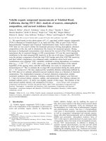
Fig. 2. A) Scheme of adsorptive transfer stripping technique used for the detection of collagen and/or MMP-9 or for the study of
interactions between these molecules; 1) renewing of the hanging mercury drop electrode (HMDE) surface; 2) adsorption of MMP-9 or
collagen in a drop solution onto the HMDE surface at open circuit; 3) rinsing electrode in water of ACS purity; 4) measuring by
chronopotentiometric stripping analysis. B) Dependences of MMP-9 (1 ng/mL) peak height on accumulation time and C), D) type of
supporting electrolyte.
538 D. Huska et al.
Electroanalysis 2009, 21, No. 3-5, 536 – 541 www.electroanalysis.wiley-vch.de 2009 Wiley-VCH Verlag GmbH& Co. KGaA, Weinheim
The dependence of MMP-9 (1 ng/mL) peak height on
various pH acetate buffers were investigated. This depend-
ence is shown in Figure 3A. The highest CPSA response was
observed at pH 5 0.2 M acetate buffer. Additionally, its
potential shifted to more negative values with increasing
pH. Lower pH values possibly facilitate hydrogen evolution
from the supporting electrolyte during the catalytic reac-
tion. Stripping current (1, 2, 4, 6, 8, 10 and 12 mA) was
another experimental condition that influenced MMP-9
peaks (Fig. 3B). The lower stripping current resulted in a
higher signal response. However, lower stripping currents
(below 1 mA) produced lower reoccurring signals and
increased relative standard deviations up to 10%. Based
on results obtained a stripping current of 1 mA was selected
for the following experiments.
The shape of the dependence of the CPSA peak height on
MMP-9 concentration was obtained (Fig. 3C). Concentra-
tion ranging from 1 to 10 nM MMP-9 assigned a linear
dependence (y ¼2299.6x þ220.36, R
2
¼0.9975). The detec-
tion limit (3 S/N) was estimated to be 100 pM.
3.2. Collagen Modified HMDE
Preparation of standard collagen solutions is a difficult task.
Dissolving collagen in water is limited due to its low
solubility and its compact structure. With agitation and
stirring the solubility of collagen can be enhanced, but the
natural folding structure could be lost. Estimating collagen
concentration by using spectrometry is also difficult. Elec-
trochemical methods including chronopotentiometr y are
convenient alternative methods for estimating collagen
concentration. The effect of two solvents, deionized water
and HCl (9% m/m), tested solubility properties of collagen.
Collagen suspensions were agitated using Vortex 2
(Eppendorf, Germany) at 400 rpm for 15 min. Collagen
decomposition improved in HCl compared to water
(Fig. 4A).
The effect of hydrochloric acid on collagen solubility was
studied in greater detail. HCl solutions with concentrations
ranging from 0.1 to 20%(m/m) were used to dissolve 100 mg
of collagen. This solution (1 mL) was placed onto a shaker
and agitated for 30 min. at 400 rpm. Collagen disintegration
increased with increasing hydrochloric acid concentration.
However acidic conditions (pH 0.5 – 1.5), can negatively
influence the native structure of a protein. Usha and
Ramasami found charge repulsion disrupts the stability of
rat tail tendon collagen fiber at low pH values [33]. At pH
lower then 6, there is a significant decrease in shrinkage
temperature. This may partly be due to osmotic forces that
lead to acid swelling. Extensive hydration could lead to
significant volume changes and the rupture of the matrix
structure. Furthermore, protonation of the ionizable group
may dominate at pH values lower than the isoelectric point
which could decrease intermolecular ion pair formation.
Lower pH does not digest collagen fibers although MMP
does. Collagen was dissolved with 9% HCl (m/m) and used
in the following experiments.
3.3. Interaction of MMP-9 with Collagen
This work studied the interaction of MMP-9 with collagen
using AdTS CPSA. The dependence of collagen peak height
(1 mg/mL) on time accumulation is shown in Figure 4B. The
highest response was measured at 90 s. In order to maintain
the optimum conditions for the enzymatic collagen cleavage
by MMP-9, MMP-9 dissolving solution, which contained
0.05 M Tris-HCl pH 7.6 þ0.2 M NaCl þ0.01 M CaCl
2
,was
used. At this pH, MMP-9 is activated and cleaves collagen
[34]. MMP-9 concentration of 1 ng/mL gave a signal at
À1.65 V (Fig. 4C). The signal of collagen appeared at
slightly more positive potentials (À1.64 V). The interaction
itself was studied according to the following scheme: i)
HMDE surface was renewed. ii) Collagen (1 mg/mL)
accumulated (90 s) on renewed HMDE surface at open
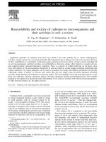
Fig. 3. Dependences of MMP-9 (1 ng/mL) peak height on A) pH of acetate buffer, B) stripping current and C) and inset, MMP-9
concentration.
539CPSA of Gelatinase B, Collagen and Their Interaction
2009 Wiley-VCH Verlag GmbH &Co. KGaA, Weinheim www.electroanalysis.wiley-vch.de Electroanalysis 2009, 21, No. 3-5, 536 – 541
circuit. iii) The electrode was rinsed in ACS grade water and
immersed in 5 mL MMP-9 solution (1 ng/mL). iv) The
interaction between MMP-9 and collagen was studied from
30 to 300 s under open circuit. v) The electrode was then
rinsed in ACS water. vi) The electrode was transferred into
an electrochemical cell and measured in acetate buffer
(pH 5). The change in CPSA peak is shown in Figure 4C.
CPSA signal of collagen after interaction with MMP-9
increased more than 30% compared to CPSA signal of
collagen only. The potential of the signal was shifted 20 mV
toward positive values. With increased MMP-9 interaction,
the signal of collagen adsorption onto HMDE enhanced.
The experiment was repeated with lower collagen and
MMP-9 concentrations. The concentration of both compo-
nents were halved: 0.5 mg/mL collagen itself (Fig. 4D-
column 3) and collagen after interaction with MMP-9
(0.5 ng/mL) (Fig. 4D-column 4). The signals measured were
cut in half compared to previous results. Based on the
obtained results, it is possible collagen is cleaved into
smaller fragments by MMP-9. These fragments are conven-
iently accessible to the HMDEs surface, resulting in a
higher signal (Fig. 4D). Similar phenomenon were observed
during the analysis of denatured protein p53 [35, 36], urease
[37] and lactoferrin [38–40].
4. Conclusions
Study of protein-protein interactions in the past have
required expensive, time consuming and labor intensive
methods, techniques and approaches. Adsorptive transfer
stripping technique coupled with chronopotentiometric
stripping analysis is an easy and low cost approach to detect
MMP-9 interaction with collagen. This technique deter-
mines the cleavage of collagen catalyzed by MMP-9 using
enhanced CPSA signals. The well observed signal is
probably due to the collagen moieties open access to the
electrodes surface.
5. Acknowledgements
Financial support from the Grants IGA MZLU MP 12/AF
and 2A-1591/122-MPO is highly acknowledged. The au-
Fig. 4. A) Height of peaks of collagen dissolved in ACS water or 9% HCl. B) Dependence of collagen peak height on accumulation
time. C) Signals of collagen, MMP-9 and collagen after interaction with MMP-9 measured by AdTS CPSA (interaction time: 30 s). D)
Height of CPSA peaks of collagen (0.5 or 1 mg/mL) after interaction with MMP-9 (0.5 or 1 ng/mL).
540 D. Huska et al.
Electroanalysis 2009, 21, No. 3-5, 536 – 541 www.electroanalysis.wiley-vch.de 2009 Wiley-VCH Verlag GmbH& Co. KGaA, Weinheim
thors wish to express their thanks to Dr. Grace Chavis for
English correction and discussion.
6. References
[1] H. Nagase, J. F. Woessner, J. Biol. Chem. 1999, 274, 21491.
[2] H. J. Ra, W. C. Parks, Matrix Biol. 2007, 26, 587.
[3] P. L. Jones, F. S. Jones, Matrix Biol. 2000, 19, 581.
[4] S. Ye, Matrix Biol. 2000, 19, 623.
[5] G. Murphy, V. Knauper, Matrix Biol. 1997, 15, 511.
[6] P. Basset, A. Okada, M. P. Chenard, R. Kannan, I. Stoll, P.
Anglard, J. P. Bellocq, M. C. Rio, Matrix Biol. 1997, 15, 535.
[7] H. E. Vanwart, H. Birkedalhansen, Proc. Natl. Acad. Sci.
USA 1990, 87, 5578.
[8] J. W. Becker, A. I. Marcy, L. L. Rokosz, M. G. Axel, J. J.
Burbaum, P. M. D. Fitzgerald, P. M. Cameron, C. K. Esser,
W. K. Hagmann, J. D. Hermes, J. P. Springer, Protein Sci.
1995, 4, 1966.
[9] E. Palecek, V. Ostatna, Electroanalysis 2007, 19, 2383.
[10] M. Tomschik, L. Havran, M. Fojta, E. Palecek, Electro-
analysis 1998, 10, 403.
[11] R. Selesovska-Fadrna, M. Fojta, T. Navratil, J. Chylkova,
Anal. Chim. Acta 2007, 582 , 344.
[12] R. Kizek, L. Trnkova, E. Palecek, Anal. Chem. 2001, 73,
4801.
[13] J. Petrlova, S. Krizkova, O. Zitka, J. Hubalek, R. Prusa, V.
Adam, J. Wang, M. Beklova, B. Sures, R. Kizek, Sens.
Actuators B, Chem. 2007, 127, 112.
[14] I. Sestakova, M. Kopanica, L. Havran, E. Palecek, Electro-
analysis 2000, 12, 100.
[15] M. Strouhal, R. Kizek, J. Vecek, L. Trnkova, M. Nemec,
Bioelectrochemistry 2003, 60, 29.
[16] L. Trnkova, R. Kizek, J. Vacek, Bioelectrochemistry 2002
, 56,
57.
[17] S. Krizkova, O. Zitka, V. Adam, M. Beklova, A. Horna, Z.
Svobodova, B. Sures, L. Trnkova, L. Zeman, R. Kizek, Czech
J. Anim. Sci. 2007, 52, 143.
[18] I. Fabrik, S. Krizkova, D. Huska, V. Adam, J. Hubalek, L.
Trnkova, T. Eckschlager, J. Kukacka, R. Prusa, R. Kizek,
Electroanalysis 2008, 20, 1521.
[19] M. Masarik, A. Stobiecka, R. Kizek, F. Jelen, Z. Pechan, W.
Hoyer, T. M. Jovin, V. Subramaniam, E. Palecek, Electro-
analysis 2004, 16, 1172.
[20] E. Palecek, M. Masarik, R. Kizek, D. Kuhlmeier, J. Hass-
mann, J. Schulein, Anal. Chem. 2004, 76, 5930.
[21] M. Brazdova, R. Kizek, L. Havran, E. Palecek, Bioelectro-
chemistry 2002, 55, 115.
[22] J. L. A. Sanchez, E. Baldrich, A. E. G. Radi, S. Dondapati,
P. L. Sanchez, I. Katakis, C. K. OSullivan, Electroanalysis
2006, 18, 1957.
[23] V. Ostatna, E. Palecek, Electrochim. Acta 2008, 53, 4014.
[24] V. Ostatna, B. Uslu, B. Dogan, S. Ozkan, E. Palecek, J.
Electroanal. Chem. 2006, 593, 172.
[25] N. Serrano, I. Sestakova, J. M. Diaz-Cruz, Electroanalysis
2006, 18, 169.
[26] N. Serrano, I. Sestakova, J. M. Diaz-Cruz, C. Arino, J.
Electroanal. Chem. 2006, 591, 105.
[27] V. Dorcak, E. Palecek, Electroanalysis 2007, 19, 2405.
[28] G. L. Long, J. D. Winefordner, Anal. Chem. 1983, 55, A712.
[29] C. Lombard, J. Saulnier, J. Wallach, Biochimie 2005, 87, 265.
[30] R. Prusa, R. Kizek, L. Trnkova, J. Vacek, J. Zehnalek, Clin.
Chem. 2004, 50, A28.
[31] J. Petrlova, D. Potesil, R. Mikelova, O. Blastik, V. Adam, L.
Trnkova, F. Jelen, R. Prusa, J. Kukacka, R. Kizek, Electro-
chim. Acta 2006, 51, 5112.
[32] V. Adam, S. Krizkova, O. Zitka, L. Trnkova, J. Petrlova, M.
Beklova, R. Kizek, Electroanalysis 2007, 19, 339.
[33] R. Usha, T. Ramasami, J. Appl. Polym. Sci. 2000, 75, 1577.
[34] G. F. Fasciglione, S. Marini, S. DAlessio, V. Politi, M.
Coletta, Biophys. J. 2000, 79, 2138.
[35] D. Potesil, R. Mikelova, V. Adam, R. Kizek, R. Prusa,
Protein J. 2006, 25, 23.
[36] R. Prusa, D. Potesil, M. Masarik, V. Adam, R. Kizek, F. Jelen,
Mol. Biol. Cell. 2004, 15, 249A.
[37] J. Hubalek, J. Hradecky, V. Adam, O. Krystofova, D. Huska,
M. Masarik, L. Trnkova, A. Horna, K. Klosova, M. Adamek,
J. Zehnalek, R. Kizek, Sensors 2007, 7, 1238.
[38] V. Adam, O. Zitka, P. Dolezal, L. Zeman, A. Horna, J.
Hubalek, J. Sileny, S. Krizkova, L. Trnkova, R. Kizek,
Sensors 2008, 8, 464.
[39] O. Zitka, A. Horna, K. Stejskal, J. Zehnalek, V. Adam, L.
Havel, L. Zeman, R. Kizek, Acta Chim. Slov. 2007, 54, 68.
[40] J. Kukacka, O. Zitka, A. Horna, K. Stejskal, J. Zehnalek, V.
Adam, L. Havel, L. Zeman, R. Prusa, L. Trnkova, R. Kizek,
Faseb J. 2007, 21, A635.
541CPSA of Gelatinase B, Collagen and Their Interaction
2009 Wiley-VCH Verlag GmbH &Co. KGaA, Weinheim www.electroanalysis.wiley-vch.de Electroanalysis 2009, 21, No. 3-5, 536 – 541