ANALYSIS OF MICROBIAL COMMUNITY STRUCTURE AND IN SITU ACTIVITY OF NITRIFYING BIOFILMS
Bạn đang xem bản rút gọn của tài liệu. Xem và tải ngay bản đầy đủ của tài liệu tại đây (598.5 KB, 10 trang )
Journal of Water and Environment Technology, Vol.2, No.2, 2004
- 65 - - 65 -
ANALYSIS OF MICROBIAL COMMUNITY STRUCTURE AND
IN SITU ACTIVITY OF NITRIFYING BIOFILMS
Satoshi Okabe*, Hisashi Satoh**, Tsukasa Ito*, and Yoshimasa Watanabe*
*Department of Urban and Environmental Engineering, Graduate School of Engineering,
Hokkaido University, North-13, West-8, Kita-ku, Sapporo 060-0813, JAPAN.
E-mail:
** Department of Civil Engineering, Hachinohe Institute of Technology, Hachinohe, Aomori
031-8501, Japan.
E-mail:
mailto:
ABSTRACT
Wastewater biofilms are very complex multispecies biofilms, displaying considerable heterogeneity with respect to
both the microorganisms present and their physicochemical microenvironments. To understand the eco-physiology
of individual microorganisms in the biofilm, techniques and tools with a high spatial and temporal resolution are
required for direct detection of the spatial distributions of microbial species and their activities in minimally
disturbed their natural habitats (e.g., biofilms). In this paper, we will, therefore, address the great potential of the
combined use of the current FISH technique and microelectrodes to study the microbial ecology of complex
microbial communities such as biofilms. The combination of these two techniques will provide reliable and direct
information about relationships between in situ microbial activity and the occurrence of specific microorganisms
in biofilms. As an example of the combined study, we will illustrate the in situ spatial organization of
ammonia-oxidizing and nitrite-oxidizing bacteria on fine scale in autotrophic nitrifying biofilms by applying the
full-cycle of 16S rRNA approach followed by fluorescence in situ hybridization (FISH), which is linked to their in
situ activity distributions at a similar resolution determined by use of microelectrodes. The combination of these
techniques allows relating in situ microbial activity directly to occurrence of nitrifying bacteria population.
KEYWORDS : Nitrifying biofilms, 16S rDNA-cloning analysis, fluorescent in situ hybridization (FISH),
microsensors, nitrification, population dynamics.
INTRODUCTION
In wastewater treatment bioreactors, microorganisms are present and active in biofilms and aggregates. Besides
conventional culture dependent techniques, modern molecular biological techniques have been used to study the
diversity and ecology of microorganisms in wastewater treatment processes since the mid-1980s. Since that time
many new insights into aerobic and anaerobic microbial wastewater treatment processes have been gained, which
significantly expanded our understandings of process design and control. Based on fundamental knowledge of
microbial community composition and the metabolic properties of microorganisms, wastewater treatment systems
must be developed and operated to maximize microbial activities. Further insights into the factors affecting
structure and function of mixed microbial communities in bioreactors are essential for advancing wastewater
treatment.
Although microbial nitrification processes for nitrogen removal are becoming more important due to strict
regulations on nitrogen discharge, nitrification is recognized as being difficult to maintain in wastewater treatment
systems. Since the diverse nitrifying bacterial populations are expected to be present in wastewater biofilms,
different species of NH
4
+
- and NO
2
-
-oxidizing bacteria exhibit different in situ growth kinetics, substrate affinities,
and sensitivities to various environmental factors (e.g., pH, temperature, O
2
concentration and substrate
concentrations). Thus, a better understanding of microbiology, ecology, and population dynamics of nitrifying
bacteria in wastewater biofilm systems is essential for improving process performance and control.
Journal of Water and Environment Technology, Vol.2, No.2, 2004
- 66 - - 66 -
Therefore, we investigated successional development of nitrifying bacterial community structure and in situ
nitrifying activities in biofilms when the biofilms were grown on rotating disk reactors (RDR) with domestic
wastewater and a synthetic nutrient medium. To achieve this goal, we have combined molecular techniques (i.e.,
16S ribosomal DNA (rDNA)-cloning analysis and fluorescent in situ hybridization (FISH) with a set of
fluorescently labeled 16S rRNA-targeted DNA probes and microsensor measurements for NH
4
+
, NO
2
-
, NO
3
-
, and
O
2
. The combined use of these techniques made it possible to relate in situ nitrifying activity directly to the
occurrence of nitrifying bacterial populations. FISH visualized successional development of nitrifying bacterial
community within an autotrophic nitrifying biofilm. After reaching the steady-state condition, microprofiles of
NH
4
+
, NO
2
-
, NO
3
-
, and O
2
in the biofilms were measured by use of microsensors, and the spatial distributions of in
situ nitrifying activities were determined. The relationship between the spatial organization of nitrifying bacterial
populations and the in situ activity of these populations within the biofilms was discussed.
Microsensors
For analysis of microbial structure and function (activity) of such complex microbial communities, classical
microbiological techniques like isolation and physiological characterization have limitations. Therefore,
appropriate methods with sufficiently high spatial resolution are needed for (1) in situ identification, localization,
and quantification of microbial populations, (2) the determination of physicochemical microenvironment, and (3)
the measurement of their in situ activity. Combination of FISH and microsensor technology became a powerful
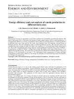
and reliable tool during the last two decades. The structure and principle of commonly used amperometric and
potentiometric microsensors are shown in Fig.1. The spatial resolution of microsensors is about two times the tip
diameter of the sensors as long as analyte consumption by the sensor is negligible and the sensor is small enough
to cause minimum disturbance. The tip diameter of microsensors applied for biofilms and aggregates is about 10
µm (Fig.1), indicating the spatial resolution of about 20 µm. This resolution is good enough to characterize the
concentration gradients across the biofilms, microbial mats and sediments and to calculate the net rates (areal and
volumetric) of production and consumption at a certain depth or of whole microbial community. During the last
decade, microelectrode measurement was nicely combined with FISH to relate microbial community structure and
function of SRB (Ito et al., 2002; Kuhl and Jorgensen, 1992; Okabe et al., 1999b; Okabe et al., 2003; Ramsing et
al., 1993) and nitrifying bacteria (Gieseke et al., 2001; Okabe et al., 1999a; Okabe et al., 2001; Satoh et al., 2003;
Schramm et al., 1996; Schramm et al., 2000) in biofilms. The combination of two methods allows relating in situ
microbial activity directly to occurrence of specific microorganisms within complex microbial consortia.
Microelectrodes, however, measure only net chemical profiles, and the spatial resolution is also above a single-cell
level. To address the question of the higher abundance and activity of SRB in oxic zones of biofilms (Okabe et al.,
1999b; Ito et al., 2002), for example, the resolution of microelectrode measurements is not high enough. In
O
O
2
2
and H
and H
2
2
S microsensors
S microsensors
O
2
-permeable
silicone membrane
Cathode
(Gold)
Liquid ion
exchange
membrane
Electrolyte
(KCl)
Glass
Guard cathode
Reference
electrode
(Ag/AgCl)
Working
electrode
(Pt wire)
Internal
electrolyte
50 m
Working
electrode
(Ag/AgCl)
Charge separation of ions
across a membrane
A potential difference b/w
the working electrode and
the reference
1)
1)
Amperometric
Amperometric
microsensors
microsensors
2)
2)
Potentiometric
Potentiometric
microsensors
microsensors
pH, NO
pH, NO
2
2
-
-
and NO
and NO
3
3
-
-
microsensors
microsensors
O
O
2
2
and H
and H
2
2
S microsensors
S microsensors
O
2
-permeable
silicone membrane
Cathode
(Gold)
Liquid ion
exchange
membrane
Electrolyte
(KCl)
Glass
Guard cathode
Reference
electrode
(Ag/AgCl)
Working
electrode
(Pt wire)
Internal
electrolyte
50 m
Working
electrode
(Ag/AgCl)
O
2
-permeable
silicone membrane
Cathode
(Gold)
Liquid ion
exchange
membrane
Electrolyte
(KCl)
Glass
Guard cathode
Reference
electrode
(Ag/AgCl)
Working
electrode
(Pt wire)
Internal
electrolyte
50 m50 m
Working
electrode
(Ag/AgCl)
Charge separation of ions
across a membrane
A potential difference b/w
the working electrode and
the reference
1)
1)
Amperometric
Amperometric
microsensors
microsensors
2)
2)
Potentiometric
Potentiometric
microsensors
microsensors
pH, NO
pH, NO
2
2
-
-
and NO
and NO
3
3
-
-
microsensors
microsensors
Figure 1. The structure and principle of the amperometric and potentiometric (LIX) microsensors.
Journal of Water and Environment Technology, Vol.2, No.2, 2004
- 67 - - 67 -
Environmental Samples
Cultures
DNA RNA
PCR Products
Genetic Fingerprints Clone Libraries
Sequence Database
Probes
Phylogenetic Tree
Isolation
Nucleic acid extraction
RT-PCR
PCR
DGGE, TGGE or t-RFLP
Cloning
PCR
Sequencing of clones
Sequencing of individual bands
Comparative sequence analysis
Hybridization analysis
Dot blot / Southern
Adaptation of culture conditions
Quantitative
dot blot
Hybridization analysis
Southern
Fluorescent in situ
hybridization (FISH
)
Environmental Samples
CulturesCultures
DNA DNA RNARNA
PCR ProductsPCR Products
Genetic Fingerprints Clone Libraries
Sequence Database
Probes
Phylogenetic Tree
Isolation
Nucleic acid extraction
RT-PCR
PCR
DGGE, TGGE or t-RFLP
Cloning
PCR
Sequencing of clones
Sequencing of individual bands
Comparative sequence analysis
Hybridization analysis
Dot blot / Southern
Adaptation of culture conditions
Quantitative
dot blot
Hybridization analysis
Southern
Fluorescent in situ
hybridization (FISH
)
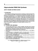
Figure 2. Flow diagram of the different steps in the full- cycle rRNA approach.
addition, when the resources used by an uncultured microorganism are unknown or the abundance of the targeted
microorganism is low in complex and heterogeneous habitats, the chemical profiles and fluxes cannot correlate to
the abundance of the specific bacterial populations. Therefore, an analytical method at a single-cell level that
allows us to more directly correlate the identity (16S rRNA-based phylogeny) and specific metabolic activity of
individual cells has been desired.
16S rRNA Approach
Microbial community structure analysis, primarily based on 16S ribosomal RNA (rRNA) gene sequencing, is
becoming the most powerful tool to study nitrifying bacterial populations present in biofilm reactors (Amann et al.,
1995; Head et al., 1998; Olsen et al., 1986). An overview of a microbial community analysis with 16S rRNA
approach is given in Figure 2. Nucleic acids from an environmental sample including multispecies
microorganisms are extracted, selectively amplified by the polymerase chain reaction (PCR) using a diverse set of
primers and separated by several fingerprinting techniques including denaturing gradient gel electrophoresis
(DGGE) (Muyzer and Smalla, 1998), temperature gradient gel electrophoresis (TGGE) (Muyzer and Smalla, 1998),
and terminal restriction fragment length polymorphisms (t-RFLP) (Liu et al., 1997). The amplified 16S rRNA gene
fragments are “shotgun cloned”, and the different types of cloned rRNA genes are then sorted and are
subsequently subjected to sequence analysis. The retrieved sequences can be phylogenetically analyzed by
comparing with existing 16S rRNA gene database and used for probe and primer designing. The fingerprinting
techniques can be used to monitor microbial community changes in natural environments and bioreactors.
Different hybridization analyses are methods to quantify and identify the RNA genes of different microbial
populations. Fluorescent in situ hybridization (FISH) (Amann, 1995) is used for identification and quantification
of microbial species and to observe the spatial distribution of specific microbial populations in biofilms and
aggregates. This rRNA approach does not include cultivation, which circumvents the biases associated with
culture-dependent techniques. It must be noted that this approach enables identification and quantification of “as
yet unknown and/or uncultivated microorganisms”.
Journal of Water and Environment Technology, Vol.2, No.2, 2004
- 68 - - 68 -
EXPERIMENTAL MATERIALS AND METHODS
Biofilm Samples
Two types of biofilms, a domestic wastewater biofilm and an autotrophic nitrifying biofilm, were studied. Both
biofilms were cultured in partially submerged rotating disk reactors (RDR) consisting of 5
poly-methyl-methacrylate disks. Eight removable slides (1×6 cm) were installed in each disk for sampling
biofilms (Okabe et al., 1996). The autotrophic nitrifying biofilms were first cultured with the primary settling tank
effluent from the Shoseigawa municipal wastewater treatment plant, Sapporo, Japan for a few days and then were
cultured with synthetic nutrient. The nutrient medium was composed of the followings (mM): NH
4
Cl, (3.6);
NaHCO
3
, (17.8); K
2
HPO
4
, (0.03); MgSO
4
· 7H
2
O, (0.41); NaCl, (1.25); pH=7.0±0.2. The reactor volume was
1370 cm
3
, and total biofilm area was 2830 cm
2
. Temperature was maintained at 20°C. Disk rotational speed was
fixed at 14 rpm. Dilution rate in the reactors was kept at 0.2 h
-1
.
Nucleic Acid Extraction and PCR Amplification
Approximately, 1 g of each wet biofilm sample was mixed with 1 ml of AE buffer (20 mM sodium acetate [pH
5.5], 1 mM EDTA) in clean 15-ml tubes. After ultrasonic treatment (10 min) and lysis (5 mg of lysosyme per ml
for 40 min at 37°C and 2 mg of protinase K per ml for 30 at 55°C), bacterial DNA was extracted from biofilm
samples by a combined freeze-thaw (three cycles of freezing in liquid nitrogen and heating at 37°C), 1% (wt/vol)
sodium dodecyl sulfate (SDS) treatment, and hot phenol-chloroform-isoamyl alcohol treatment (Teske et al., 1996).
The 16S rRNA genes (rDNA) from mixed bacterial DNA were amplified by PCR with the primer set of CTO189f
and CTO654r as described by Kowalchuk et al. (1997). For general bacteria, almost-full length bacterial 16S
rDNA fragments were amplified using the primer set of GM3f (Escherichia coli 16S rDNA positions 8 to 24) and
GM4r (E. coli positions 1492 to 1507) as described by Muyzer et al. (1995). To minimize nonspecific annealing of
the primers to nontarget DNA, a hot-start and touch-down PCR program was used for all amplification (Muyzer et
al., 1997). The PCR products were evaluated on a 1 % (w/v) agarose gel.
Cloning of 16S rDNA
One microliter of the amplified bacterial 16S rDNA fragments (465 bp including variable V3 region) was directly
ligated into the pGEM-T vector cloning system (Promega) and transformed into competent cells (high-efficiency E.
coli JM109 [Promega]) as described in the manufacturer’s instruction.
Sequencing and Phylogenetic Analysis
Plasmids were extracted and purified from clones with the Wizard Plus Minipreps DNA purification system
(Promega) in accordance with the manufacturer’s instructions. To avoid redundant sequencing, PCR-amplified
rDNA fragments of all clones were analyzed by RFLP (Restriction fragment length polymorphism) after digestion
with restriction enzymes of cfoI or haeIII as described in the manufacturer’s instruction. The PCR fragments
digested were loaded on a 2.0 % (w/v) agarose gel. Similar fragment migration patterns were defined as identical
recombinants, and one representative of each group of recombinants was selected for comparative sequence
analysis. Partial sequencing (ca. 465 bp) of the 16S rDNA inserts was performed with an automatic sequencer
(HITACHI). All sequences were checked for chimeric artifacts by the CHECH_CHIMERA program in the
Ribosomal Database Project (RDP)(Maidak et al., 1997) and compared with similar sequences of the reference
organisms by BLAST search (Altschul et al., 1990). Sequence data were aligned with the CLUSTAL W package
(Thompson et al., 1994). Phylogenetic trees were constructed by the neighbour-joining method (Saito and Nei,
1987) with Tree Explore. Bootstrap resampling analysis for 100 replicates was performed.
Fixation and Cryosectioning of Biofilm Samples
Biofilm samples were taken at regular time intervals during the periods of the biofilm development. The biofilm
samples were fixed with freshly prepared paraformaldehyde solution (4% in phosphate buffered saline (PBS),
pH=7.2) for 4 to 8 h at 4°C and embedded in Tissue-Tek OCT compound (Miles, Elkhart, IN) overnight to
Journal of Water and Environment Technology, Vol.2, No.2, 2004
- 69 - - 69 -
infiltrate the OCT compound into the biofilm. After rapid freezing at –21°C, 10 to 20-µm-thick vertical slices were
cut with a cryostat (Reichert-Jung Cryocut 1800, Leica) and placed on a gelatin-coated slide (Cell-line, USA,
0.1 % gelatin and 0.01 % chromium potassium sulfate). After airdrying overnight, the slices were dehydrated by
successive passage through 50, 80, and 98 % ethanol washes (for 3 min each), air dried, and stored at room
temperature.
Oligonucleotide Probes
The following oligonucleotide probes were used: Nso190 (Mobarry et al., 1996), NEU (Wagner et al., 1995),
Nsm156 (Mobarry et al., 1996), NmV (Pommerening-Roser et al., 1996), Nsv443 (Mobarry et al., 1996), NIT2
(Wagner et al., 1996), NIT3 (Wagner et al., 1996), Ntspa454 (Hovanec et al., 1998), Ntspa685 (Hovanec et al.,
1998), and Ntspa1026 (Juretschko et al., 1998). Probes were labeled with fluorescein isothiocyanate (FITC) or
tetramethylrhodamine-5-isothiocyanate (TRITC). Unlabelled competitor CNIT3 (Wagner et al., 1996) and CTE
(Schleifer et al., 1992) probes were added to an equimolar amount of NIT3 and NEU probes, respectively.
In Situ Hybridization
The previously published optimal hybridization conditions were used for each probe. All in situ hybridizations
were performed according to the procedure described by Amann (1995) in hybridization buffer (0.9 M NaCl,
20mM Tris hydrochloride (pH=7.2), 0.01% sodium dodecyl sulfate (SDS), x% formamide) at 46°C for 2-3 hours.
The final probe concentration was approximately 5 ng µl
-1
. Subsequently, a stringent wash step was performed at
48°C for 20 min in 50 ml of pre-warmed washing solution (x mM NaCl, 20 mM Tris hydrochloride (pH=7.2),
0.01 % SDS). The stringency of the washing step (at 48°C) was adjusted by lowering the sodium chloride
concentration to achieve the appropriate specificity. The slides were then rinsed briefly with ddH
2
O and allowed to
air dry. Simultaneous hybridization with probes requiring different stringency was performed by a successive
hybridization procedure: hybridization with the probe requiring higher stringency was performed first, and then
hybridization with the probe requiring lower stringency was performed (Wagner et al., 1994). Slides were mounted
in SlowFade
TM
-light antifade kit (Molecular Probes, Eugene, OR).
Microelectrode Preparation and Measurements
For determination of concentration profiles in the biofilms, cathode type oxygen microelectrodes with a tip
diameter of about 10 µm was prepared and calibrated as described previously by Revsbech and Jorgensen (1986).
Liquid ion-exchanging membrane (LIX) microsensors for NH
4
+
, NO
2
-
, and NO
3
-
were prepared according to
deBeer et al. (1997). The LIX microsensors were calibrated in dilution series (10
-3
-10
-6
M) of NH
4
+
, NO
2
-
, and
NO
3
-
in the medium used for the measurements. All measurements were performed as described previously
(deBeer and Heuvel, 1988) in a flow cell reactor at 20°C, with an average liquid velocity of 2-3 cm s
-1
by blowing
air on the liquid surface. The composition of the medium used for microprofile measurements was described
previously by deBeer et al. (1993). The biofilm samples taken from the reactor was acclimated in the medium a
few hours before the measurement, to ensure that steady state profiles were obtained.
Estimation of Consumption and Production Rate Profiles
Net specific consumption and production rates (R; µmol cm
-3
h
-1
) of NH
4
+
, NO
2
-
, and NO
3
-
were estimated from the
measured microprofiles by using the Fick's second law of diffusion as previously described by Lorenzen et al.
(1998). Molecular diffusion coefficients of 1.38×10
-5
cm
2
s
-1
for NH
4
+
, 1.23×10
-5
cm
2
s
-1
for NO
2
-
, and 1.23×
10
-5
cm
2
s
-1
for NO
3
-
at 20°C were used for the calculations (Andrussow, 1969).
RESULTS AND DISCUSSION
Spatial Distributions of Nitrifying Bacteria
In the autotrophic nitrifying biofilm, spherical clusters of densely packed probe Nso190-stained NH
4
+
-oxidizing
bacterial cells were detected throughout the oxic biofilm strata, indicating more or less a homogeneous spatial