Báo cáo khoa học: K182G substitution in DevR or C8G mutation in the Dev box impairs protein–DNA interaction and abrogates DevR-mediated gene induction in Mycobacterium tuberculosis doc
Bạn đang xem bản rút gọn của tài liệu. Xem và tải ngay bản đầy đủ của tài liệu tại đây (428.42 KB, 9 trang )
K182G substitution in DevR or C
8
G mutation in
the Dev box impairs protein–DNA interaction and
abrogates DevR-mediated gene induction in
Mycobacterium tuberculosis
Rajesh Kumar Gupta, Santosh Chauhan* and Jaya Sivaswami Tyagi
Department of Biotechnology, All India Institute of Medical Sciences, New Delhi, India
Introduction
Tuberculosis is the single most prevalent infectious dis-
ease among humans and accounts for one-seventh of
all deaths worldwide. The success of Mycobacte-
rium tuberculosis as a pathogen is closely associated
with its ability to persist in humans for extended peri-
ods without causing disease. It is estimated that one-
third of the global population harbours latent
M. tuberculosis infection which can last for years and
even decades without causing active disease [1,2]. This
enormous reservoir of latent disease greatly compli-
cates efforts aimed at tuberculosis control as it requires
prolonged drug therapy presumably due to persistence
Keywords
DevR (or DosR); DNA–protein interaction;
Mycobacterium tuberculosis
Correspondence
J. S. Tyagi, Department of Biotechnology,
All India Institute of Medical Sciences,
New Delhi-110029, India
Fax: +91 11 2658 8663
Tel: +91 11 2658 8491
E-mail:
*Present address
Department of Cancer Biology, MD
Anderson Cancer Center, Houston, Texas,
USA
(Received 16 November 2010, revised 15
April 2011, accepted 19 April 2011)
doi:10.1111/j.1742-4658.2011.08130.x
The DevR response regulator mediates adaptation of Mycobacterium tuber-
culosis to various signals that are likely to be encountered within the host
such as hypoxia, nitric oxide, carbon monoxide and ascorbic acid. DevR is
proposed as a promising target for developing drugs against dormant bac-
teria. It induces the expression of target genes by interacting with DNA
motifs located in their promoter regions. An understanding of DNA–pro-
tein interactions is expected to facilitate the development of inhibitors tar-
geting DevR. Only three amino acids in DevR, namely Lys179, Lys182 and
Asn183, directly contact nucleotide bases in the DNA motif. The present
study was designed to decipher the contribution of Lys182 in DevR func-
tion. M. tuberculosis fdxA (Rv2007c), a member of the DevR regulon, was
selected for this analysis. Its transcriptional start point was mapped at )1
or )2 with respect to the putative translational start site suggesting that
fdxA is expressed as a leaderless mRNA. DNase I footprinting led to the
discovery of a secondary binding site and induction of the fdxA promoter
is explained by the cooperative binding of DevR to two binding sites.
Mutation of Lys182 lowers the DNA binding affinity of DevR and abro-
gates induction of fdxA and other regulon genes. Mutational analyses also
highlight the singular importance of Lys182–G
13
nucleotide interaction for
DevR binding and regulon induction. Our findings demonstrate that
impairment of Lys182-mediated interactions alone abolishes DevR function
and provide valuable insights for designing molecules that interfere with
DevR-mediated dormancy adaptation.
Abbreviations
EMSA, electromobility shift assay; GFP, green fluorescent protein; qRT-PCR, quantitative real time RT-PCR; TSP, transcription start point;
WT, wild type.
FEBS Journal 278 (2011) 2131–2139 ª 2011 The Authors Journal compilation ª 2011 FEBS 2131
of the dormant tubercle bacilli that are refractory to
current treatment regimens [2,3].
Dormancy adaptation is characterized by the cessa-
tion of active bacterial growth and the transition into
a non-replicative persistent state. An understanding of
the molecular basis of dormancy is a prerequisite for
the identification of novel molecules in dormant organ-
isms that can be targeted by new drugs. In vitro mod-
els have provided valuable insights into the genetic
programmes utilized by M. tuberculosis during dor-
mancy adaptation [4]. Transcription represents the first
and the most crucial step in gene regulation in prok-
aryotes and in vitro exposure of M. tuberculosis to
physiologically relevant stimuli such as hypoxia, NO,
CO and ascorbic acid triggers a dormancy adaptive
response that is initiated by the DevR transcriptional
regulator [5–10]. DevR mediates the rapid upregulation
of 48 M. tuberculosis genes that comprise the DevR
regulon [5,11–13]. This regulator has been proposed as
a key participant in the dormancy programme of
M. tuberculosis and consequently it is potentially
important as a target for novel drug development
[14,15]. This hypothesis is supported by the demonstra-
tion of blocking of the DevR pathway by a small
inhibitor molecule that also prevented hypoxia-induced
bacterial dormancy in vitro [16]. Therefore a fine
understanding of the properties of DevR will undoubt-
edly be invaluable for designing potent inhibitor
molecules.
The analysis of the crystal structure of the DevR
C
-
terminal domain complex with a 20-bp oligonucleotide
representing the consensus binding motif revealed that
a DevR dimer interacts with each DNA motif. A con-
served sequence, G
4
G
5
G
6
A
7
C
8
T
9
, present in each half
palindrome is recognized by a subunit of the DevR
dimeric protein. Only three amino acids per subunit,
namely Lys179, Lys182 and Asn183, contact nucleo-
tide bases in the binding motif [17]. To elucidate the
functionality of DNA–DevR protein interactions, the
present study was designed to decipher the contribu-
tion of Lys182 (K182) to the DNA binding property
of DevR. Lys182 is thought to participate in exclusive
H-bonding interactions with the O
6
and N
7
atoms of
G
13
(complementary to the conserved C
8
base on the
sense strand) and the N
7
atom of A
12
(complementary
to the conserved T
9
base on the sense strand [17]). The
M. tuberculosis fdxA (Rv2007c) promoter was selected
for this analysis as it is a member of the DevR regulon
and harbours a solitary upstream DevR binding motif
[5] containing the consensus C
8
and T
9
nucleotides that
were predicted to interact specifically with Lys182 resi-
due in DevR. fdxA encodes a putative ferredoxin pro-
tein. Ferredoxins are small, acidic proteins containing
iron–sulphur clusters which act as multifunctional elec-
tron carriers in diverse redox systems. It has been sug-
gested that M. tuberculosis FdxA protein may serve
the tubercle bacteria as an electron carrier under
hypoxia [18] or that it may play a role in maintaining
DevS in its reduced functional state [19]. During the
present study, we discovered the presence of a low-
scoring DevR binding site in the fdxA promoter in
addition to the previously predicted site. DevR binds
cooperatively to the second site to induce fdxA pro-
moter transcription. Through mutational analysis of
protein and DNA (in a half-site of the primary binding
motif), we highlight the singular importance of G
13
–
Lys182 and partial importance of A
12
–Lys182 inter-
action for DevR binding and function. Our results
establish that abrogation of interactions mediated by a
single amino acid, namely Lys182, with the primary
binding site is alone sufficient to abolish specific
DNA–protein interactions and downstream gene
induction.
Results
Transcription start point mapping of fdxA
In order to understand the relevance of DevR interac-
tion to transcription, the transcription start point
(TSP) was mapped by primer extension analysis using
RNA isolated from aerobic and hypoxia-induced
M. tuberculosis cultures. fdxA TSP was mapped at )1
or )2 with respect to the putative translational start
site of FdxA under hypoxic conditions (Fig. 1). Based
on previously described consensus sequences [20,21],
SigA- and SigC-like promoter elements were mapped
upstream of the TSP.
fdxA promoter has a conserved architecture of
two DevR binding sites located upstream of its
TSP
The fdxA gene is a member of the DevR regulon. The
members of this regulon often have two or more DevR
binding sites in their upstream regions [5,11–13,22]. In
this context, the fdxA promoter is noteworthy because
only a single upstream DevR binding site was pre-
dicted for this gene [5]. However, DNase I footprinting
analysis of the wild-type (WT) fdxA promoter region
revealed the presence of two binding sites (Fig. 2A),
the previously predicted site P [5] and a newly identi-
fied adjacent site S that was proximal to the TSP and
was not identified previously by in silico analysis.
While both the binding sites were occupied at ‡ 0.5 lm
concentration, binding to a single site was not
Role of Lys182 in DevR function in M. tuberculosis R. K. Gupta et al.
2132 FEBS Journal 278 (2011) 2131–2139 ª 2011 The Authors Journal compilation ª 2011 FEBS
observed at lower protein concentration (not shown).
Four enhanced DNase I cleavage sites were detected
within the DevR-bound region at an apparently
periodic interval (indicated by arrowheads in Fig. 2A).
The results of DNase I footprinting and TSP mapping
indicate that DevR interacts cooperatively with the P
and S sites and that the )35 promoter element
partially overlaps with the secondary DevR binding
site, S.
DevR K182G mutant protein is defective for
interaction with DNA
The phosphorylation and DNA binding properties of
purified WT and K182G mutant DevR proteins were
compared. Both the proteins were phosphorylated with
equivalent efficiency in vitro and therefore a phosphor-
ylation defect in the mutant protein was ruled out
(Fig. 3A). Electromobility shift assay (EMSA) analysis
was performed with phosphorylated WT or K182G
mutant DevR proteins and fdxA promoter DNA. WT
DevR protein bound to fdxA promoter DNA over a
narrow range (< 10-fold) of protein concentration; at
500 nm concentration > 90% saturation of DNA was
observed with WT DevR protein while no binding was
observed with mutant protein up to 1.0 lm concentra-
tion (Fig. 3B, lanes 5 and 12, respectively). Partial
binding of the mutant protein with fdxA promoter
DNA was noted at higher protein concentration (up to
6.0 lm) suggesting that the overall conformation of the
DNA binding domain was preserved relative to the
WT protein. However, the mutant protein failed to
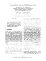
Fig. 1. TSP mapping. The fdxA TSP (shown by arrow) was mapped
using RNA isolated from aerobic (A) and hypoxic (H) cultures and
fdxA tsp primer. DNA sequence of the fdxA promoter region (anti
sense strand). The bent arrow (at T, T) indicates the fdxA TSP
mapped in the present study. The primary, P, and secondary, S,
DevR binding sites that were identified by DNase I footprinting
(Fig. 2) are boxed. Putative )35 and )10 SigC and SigA promoter
consensus elements are indicated below the relevant sequences.
The first boxed GTG codon represents the translational initiation
site annotated in TubercuList (fl.ch). Additional
putative translational initiation codons are boxed.
AB
Fig. 2. DNase I footprinting. (A) DNase I footprint of WT DevR protein (0.5 lM and 1.0 lM concentration) and fdxA promoter DNA containing
WT ⁄ C
8
G ⁄ T
9
A mutant P box. DNA sequencing ladder of the same sequence is shown alongside the footprint. The footprints were analysed
by the lane detection tool and lane profile graphs (red, no protein added; green, with 0.5 l
M DevR; orange, with 1.0 lM DevR) were gener-
ated using
QUANTITYONE software (Bio-Rad). Arrowheads correspond to enhanced DNase I cleavage sites in the DevR binding region. The
sequences of the WT and C
8
GorT
9
A mutant P boxes are shown above the corresponding footprints. (B) DNase I footprint of K182G DevR
mutant protein and WT fdxA promoter DNA.
R. K. Gupta et al. Role of Lys182 in DevR function in M. tuberculosis
FEBS Journal 278 (2011) 2131–2139 ª 2011 The Authors Journal compilation ª 2011 FEBS 2133
bind to the P and S sites in the fdxA promoter (up to
6.0 lm protein concentration) in a DNase I footprint-
ing assay. This property established that the K182G
mutant protein was defective in sequence-specifc inter-
action (Fig. 2B).
M. tuberculosis expressing either K182G or
K182A mutant DevR protein is defective in DevR
regulon response
The effect of K182 mutation on gene activation was
assessed by quantitative real time RT-PCR (qRT-
PCR) analysis of selected DevR regulon genes in iso-
genic M. tuberculosis strains expressing WT or DevR
K182G mutant protein. An induction defect was noted
in the expression of Rv3134c, devR, fdxA and tgs1
genes in the mutant strain under hypoxia (Fig. 3C).
A very feeble ( twofold) induction of hspX was
observed in the mutant strain in contrast to > 80-fold
induction in WT bacteria. However, hypoxic expres-
sion of HspX protein was observed only in M. tubercu-
losis cultures expressing WT DevR protein and not in
those expressing mutant protein (not shown). The
induction defect in DevR K182G-expressing M. tuber-
culosis cultures is attributed to the decreased binding
of mutant protein at target promoters. Moreover,
because DevR expression is under positive autoregula-
tion [11], inducing levels of the regulator are probably
not attained in the mutant strain to overcome the
binding defect of K182G DevR. The functional impor-
tance of K182 residue in gene activation was confirmed
in a second isogenic mutant strain that expresses DevR
K182A version of mutant protein (Fig. 3C). Although
not tested experimentally, a similar mechanism is a
likely explanation for the expression defect in this
mutant strain as well.
C
8
base in the P box is crucial for DevR interaction
and essential for fdxA promoter activation
The experiments described above establish the impor-
tance of K182 residue in the functionality of DevR.
Because K182 residue in DevR was reported to contact
G
13
and A
12
bases in the DNA motif (complementary
to C
8
and T
9
bases, respectively, in the P box [17]), the
relevance of this interaction was confirmed by analy-
sing the binding of WT DevR protein with mutant
fdxA promoter fragments harbouring these mutations
in the P binding site. Comparative EMSA analysis of
the interaction of DevR protein with the fdxA pro-
moter containing either WT or mutated P box
sequences revealed that the C
8
G mutant DNA was
defective in binding. At 0.4 lm DevR concentration,
binding to the C
8
G mutant box was observed to be
substantially reduced and to the T
9
A mutant box
reduced to a lesser extent (Fig. 4A). The double
mutant (C
8
G+T
9
A) was also defective in binding as
expected (not shown). The results of EMSA analysis
were supported by DNase I footprinting analysis and
the C
8
G mutation was observed to be more deleterious
than the T
9
A mutation indicating that the C
8
nucleo-
tide is important for interaction with Lys182 of DevR
(Fig. 2A). A comparison of the footprints and their
profiles shows that the C
8
mutation in the P box abol-
ished the binding of DevR to both the P and S boxes
at 0.5 lm protein concentration. From DNase I foot-
printing and EMSA results, we infer that DevR binds
cooperatively to two sites at the fdxA promoter and
that C
8
base in the P box is crucial for interaction.
The functional relevance of the mutations resulting
in a binding defect was assessed by green fluorescent
protein (GFP) reporter assay using M. tuberculosis
Fig. 3. (A) Phosphorylation of WT and K182G mutant DevR pro-
teins with DevS
201
P (phosphorylated cytoplasmic domain of
DevS). Lane 1, DevSP; lane 2, DevSP and DevR K182G mutant
protein; and lane 3, DevSP and WT DevR protein. The top panel
represents the phosphorimage and the bottom panel the Coomas-
sie stained gel. (B) EMSA with phosphorylated DevR (WT or K182G
mutant) and fdxA WT promoter DNA. Left, lanes 1 to 9 contain 0.1,
0.2, 0.3, 0.4, 0.5, 0.6, 0.7, 0.8, 0.9 l
M of WT DevR protein; lane 10
represents free DNA; lanes 11 and 12 contain 0.5 and 1.0 l
M of
K182G mutant DevR protein. (C) qRT-PCR analysis of DevR regulon
gene expression. RNA was isolated from M. tuberculosis cultures
expressing either WT DevR protein or DevR K182G or DevR K182A
mutant protein and subjected to gene expression analysis.
Mean ± SD fold induction under hypoxia from two to four indepen-
dent cultures is shown.
Role of Lys182 in DevR function in M. tuberculosis R. K. Gupta et al.
2134 FEBS Journal 278 (2011) 2131–2139 ª 2011 The Authors Journal compilation ª 2011 FEBS
strains harbouring the WT and mutant fdxA promot-
ers (C
8
G, T
9
A and C
8
G+T
9
A in the P box).
Although both mutant promoter DNAs were partially
defective in binding to DevR in vitro (Fig. 4A), the
fdxA promoter carrying the C
8
G mutation was com-
pletely defective in the hypoxic induction of promoter
activity while the T
9
A mutation was partially defective
( 50%, Fig. 4B). As expected the doubly mutated
promoter DNA was also completely defective in pro-
moter activation. These results establish that a single
mutation (C
8
G) in one-half of the P box results in a
defect in DevR binding to DNA and abolishes DevR-
regulated gene induction.
Discussion
Only three amino acid residues, namely Lys179,
Lys182 and Asn183, that are located in the a9 helix of
each DevR subunit, directly contact G
4
G
5
G
6
A
7
C
8
T
9
bases in each half-binding site of a DevR
C
–DNA com-
plex [17]. We recently showed that natural substitution
at position G
4
is tolerated while G
5
,G
6
and C
8
nucleo-
tides are well conserved in the interacting boxes of
DevR-dependent promoters [12,13]. The conserved C
8
base does not interact directly with DevR; however,
G
13
in the complementary DNA strand at this position
hydrogen bonds with Lys182. Lys182 also hydrogen
bonds with the A
12
base in the complementary strand.
The precise contribution of individual amino acids in
DevR to its function can be assessed by mutational
studies. It is expected that this type of analysis will
reveal the interaction(s) that are crucial for DevR
function and thereby guide the rational development
of inhibitors to DevR, a target that is believed to play
a key role in the hypoxia-induced bacterial dormancy
programme. In the present study, the role of Lys182
was analysed because it exclusively interacts with only
two bases in each half-site of DNA, namely G
13
and
A
12
, the former being complementary to the highly
conserved C
8
nucleotide. Furthermore, in silico analysis
shows that the binding pocket in the crystal structure
that interacts with a DevR inhibitor contains Lys182
[16]. The importance of Lys182 residue in DevR func-
tion was assessed in vitro and in vivo using DevR
K182G or K182A mutant protein and fdxA promoter
harbouring mutations in C
8
or ⁄ and T
9
base in the pri-
mary DevR binding site, P. The expression of the
DevR regulon genes was severely compromised by
mutation of this amino acid in DevR. The partial
binding defect with the fdxA promoter carrying a C
8
mutant P box was associated with a complete loss in
promoter activity and further established the essential
role of K182 in DevR function. In contrast, a partial
binding defect with the T
9
mutant P box was associ-
ated with 50% promoter activity. The functional
importance of C
8
nucleotide for DevR interaction is
reflected in the positional conservation of C
8
but not
the T
9
nucleotide in binding motifs [5,12,13]. The
mutant proteins analysed in the present study contain
glycine or alanine in place of K182 in the WT protein
wherein the side chain amino group of K182 residue in
each subunit is involved in H-bonding with O
6
and N
7
atoms of G
13
and with the N
7
atom of A
12
in the
DNA strand. In silico analysis of WT versus K182
mutant DevR protein reveals the loss of three K182-
mediated H bonds in the mutant protein which is
apparently sufficient to destabilize the remaining inter-
actions and results in the reduced affinity of mutant
DevR protein for specific DNA sequences that was
observed in the present study.
DNase I footprinting analysis of the fdxA promoter
reveals some important binding properties of DevR. A
Bound
Free
1 2 3 4 5
1 2 3 4 51 2 3 4 5
0 0.2 0.4 0.8 1.0 0 0.2 0.4 0.8 1.0 0 0.2 0.4 0.8 1.0
WT P Box C
8
G P Box T
9
A P Box
DevR~P
(µ
M
)
A
B
Fig. 4. (A) EMSA analysis using WT DevR protein and fdxA
WT ⁄ C
8
G ⁄ T
9
A promoter DNA. A representative result from three
experiments is shown. (B) GFP fluorescence of M. tuberculosis cul-
tures expressing WT DevR protein and harbouring either WT or
mutant fdxA promoter in GFP reporter vector. GFP fluorescence
was assessed in standing cultures in 96-well format and expressed
as relative fluorescence units divided by A (mean ± SD of two inde-
pendent experiments, each in triplicate wells).
R. K. Gupta et al. Role of Lys182 in DevR function in M. tuberculosis
FEBS Journal 278 (2011) 2131–2139 ª 2011 The Authors Journal compilation ª 2011 FEBS 2135
new DevR binding site (designated as S) was identified
downstream and adjacent to the previously assigned P
site. Mutational analysis established that protein bind-
ing to the S site is dependent on its binding to the P
site. In this regard the fdxA promoter displays an
architectural similarity to tgs1 and some other DevR
regulon promoters [12,13]. The presence of prominent
DNase I cleavage sites in the protected region suggests
that bound DevR may induce localized DNA bend-
ing ⁄ distortion and thereby facilitate cooperative pro-
tein–protein interaction at the fdxA promoter. Our
observations are consistent with the bending of DNA
observed in DevR
C
–DNA crystals [17]. Note that we
have analysed DevR binding to a natural target pro-
moter containing a strong and a weak binding site
each while the crystal structure was elucidated using
consensus DNA oligonucleotides. Many DevR regulon
promoters contain a combination of strong and weak
binding sites [12,13]. Taking into consideration the
results of DNase I footprinting analysis and in vivo
assays, it appears that DNA bending ⁄ distortion is cru-
cial for recruiting DevR cooperatively to weak binding
sites and for target promoter induction. Keeping in
mind the overlap of a DevR binding site with the )35
promoter element at target promoters, it is possible
that DevR-induced changes in DNA conformation
may also facilitate interactions between bound DevR
molecules and RNA polymerase.
TSP mapping reveals the presence of a hypoxia-
inducible transcriptional start site at )1or)2 position
with respect to the putative translational start site of
fdxA (as annotated in TubercuList, http://tubercu-
list.epfl.ch) which suggests that the fdxA transcript is a
leaderless mRNA. There are numerous examples
of leaderless mRNA in eubacteria and archaea [23].
A leaderless fdx mRNA encoding ferredoxin was
reported in Halobacterium salinarium where the TSP
mapped at )1 position in relation to the translational
start site [24]. It has been suggested that leaderless
mRNAs may be preferentially translated under
adverse conditions like carbon source downshift,
stationary phase etc. [25]. It is not known whether
translational control of leaderless mRNAs in M. tuber-
culosis is similar to that in Escherichia coli; however,
based on the assumption that similar mechanism(s) are
employed, it is possible that the leaderless fdxA
mRNA is efficiently translated in M. tuberculosis
under conditions of hypoxic stress. Additional
in-frame GTG codons were detected downstream of
the +1 GTG initiation codon and, although no SD-
like sequences were detectable, we cannot exclude the
possibility that any one of them is utilized in the initi-
ation of translation of FdxA.
In conclusion, the results of mutational analysis of
protein and DNA establish the singular importance of
G
13
–Lys182 H-bonding in DevR–DNA interaction and
for downstream gene induction events. It is hoped that
these insights will advance the rational development of
specific inhibitors of DevR.
Materials and methods
Bacterial strains and growth conditions
All M. tuberculosis strains were revived from )80 °C bacte-
rial stocks and grown in Dubos medium containing 0.1%
Tween-80 and 10% (v ⁄ v) albumin dextrose complex (DTA
medium). All cultures were grown at 37 °C in a shaker
incubator (190–220 r.p.m. using an Innova Shaker 4230)
unless mentioned otherwise. Plasmids used in this study are
shown in Table 1.
Overexpression and purification of recombinant
DevR K182G mutant protein
Lysine to glycine or alanine mutation at position 182 of
DevR was introduced by site-directed mutagenesis in plas-
mid pSC1 which expresses WT DevR protein in pGEX4T1
vector using mutagenic primers (Table 2) and Pfu Turbo
DNA polymerase (Stratagene, La Jolla, CA, USA). The
amplified product was digested with DpnI enzyme and then
transformed into E. coli XL-1 Blue. The generation of the
site-specific mutation was confirmed by DNA sequencing.
WT and K182G mutant DevR proteins were purified from
E. coli as described previously [11].
Generation of G
13
,A
12
and double (G
13
+A
12
)
mutant DNA boxes in fdxA promoter
Mutant M. tuberculosis fdxA promoter GFP reporter con-
structs bearing G
13
or A
12
or G
13
+A
12
mutations in the
P box were generated by site-directed mutagenesis in plas-
mid pSG1 (Table 2). All mutations were confirmed by
DNA sequencing.
EMSA
EMSAs were performed as described previously [11].
Briefly,
32
P-labelled fdxA promoter DNA (WT and mutant)
fragments were generated by PCR from M. tuberculosis
H37Rv DNA using oligonucleotide primers fdxA f and
fdxA r (Table 2). DevR protein (WT or K182G mutant)
was purified as described previously [11] and phosphory-
lated DevR was prepared using acetyl phosphate as
described previously [11]. Varying concentrations of phos-
phorylated WT or K182G mutant DevR protein were incu-
bated with 2 ng of the labelled fdxA promoter DNA (WT
Role of Lys182 in DevR function in M. tuberculosis R. K. Gupta et al.
2136 FEBS Journal 278 (2011) 2131–2139 ª 2011 The Authors Journal compilation ª 2011 FEBS
or mutant) on ice for 30 min. DNA–protein complexes
were separated by non-denaturing PAGE and the DNA–
protein complexes were visualized by phosphorimaging.
The fraction of bound DNA was estimated using quantity
one software (Bio-Rad, Hercules, CA, USA).
DNase I footprinting of fdxA promoter
DNase I footprinting assays were performed with phos-
phorylated WT DevR protein and fdxA promoter DNA
variants (WT and mutant) or K182G mutant DevR protein
and WT fdxA promoter DNA as described earlier [11].
TSP mapping
RNA was isolated from M. tuberculosis H37Rv cultures
grown in DTA medium under aerobic shaking and standing
Table 2. Primers used in the study. Underlined bases indicate the
introduced mutations.
Primer name Sequence 5¢fi3¢
fdxA tsp CCAGTAGATCGCCT
fdxA f TGACGGGCTATCGTAAGTTTATG
fdxA r CACGCACTCACTACCGATCACA
K182G f GAAAAGACGGTG
GGGAACTACGTGTCG
K182G r CGACACGTAGTT
CCCCACCGTCTTTTC
K182A f GAAAAGACGGTG
GCGAACTACGTGTCG
K182A r CGACACGTAGTT
CGCCACCGTCTTTTC
fdxA-C8G f TGACGAATAAGGC
GTTTGGTCCTTTCC
fdxA-C8G r GGAAAGGACCAAA
CGCCTTATTCGTCA
fdxA-A9T f TGACGAATAAGGCC
ATTGGTCCTTTCC
fdxA-A9T r GGAAAGGACCAA
TGGCCTTATTCGTCA
fdxA-C8G-A9T f TGACGAATAAGGC
GATTGGTCCTTTCC
fdxA-C8G-A9T r GGAAAGGACCAA
TCGCCTTATTCGTCA
Table 1. Plasmids used in the study.
Plasmid Feature(s) Reference ⁄ source
pSC-DevR pGEX4T1 overexpressing WT DevR with a glutathione S-transferase
N-terminal tag
[11]
pRG-K182G DevR pSC-DevR encoding DevR containing lysine to glycine mutation at amino
acid residue182
This study
pSM P
Operon
devR pJFR19 integrative vector containing WT devR sequences expressed from
its native operon promoter
S. D. Majumdar,
PhD thesis submitted
to AIIMS, 2010
pRG P
Operon
K182G devR pSM P
operon
devR encoding DevR containing lysine to glycine mutation
at amino acid residue182
This study
pRG P
Operon
K182A devR pSM P
operon
devR encoding DevR containing lysine to alanine mutation at
amino acid residue182
This study
pFPV27 E. coli Mycobacterial shuttle plasmid with promoterless gfp;Km
r
[27]
pSG1 pFPV27 containing WT fdxA promoter ()191 to +30) cloned upstream
of gfp
S. Ghosh, M Biotech
dissertation, AIIMS, 2008
pRG1 pSG1 containing C
8
G mutation in P box of fdxA promoter This study
pRG2 pSG1 containing T
9
A mutation in P box of fdxA promoter This study
pRG3 pSG1 containing C
8
G+T
9
A mutation in P box of fdxA promoter This study
Table 3. Strains used in the study.
Strain Feature(s) Reference ⁄ source
M. tuberculosis Mut2 M. tuberculosis H37Rv strain with a 447-bp BalI deletion
in devR coding region
[28]
M. tuberculosis Comp13 Plasmid pSM P
Operon
devR electroporated in
M. tuberculosis Mut2 (expressing WT DevR)
S. D. Majumdar,
PhD thesis submitted
to AIIMS, 2010
M. tuberculosis DevR Mut K182G pRG
Operon
K182G devR electroporated in M. tuberculosis Mut2 This study
M. tuberculosis DevR Mut K182A pRG
Operon
K182A devR electroporated in M. tuberculosis Mut2 This study
M. tuberculosis GFP empty vector Plasmid pFPV27 electroporated in M. tuberculosis H37Rv strain This study
M. tuberculosis fdxA Plasmid pSG1 electroporated in M. tuberculosis H37Rv This study
M. tuberculosis Mut fdxA G13 Plasmid pRG1 electroporated in M. tuberculosis H37Rv This study
M. tuberculosis Mut fdxA A12 Plasmid pRG2 electroporated in M. tuberculosis H37Rv This study
M. tuberculosis Mut fdxA G13 + A12 Plasmid pRG3 electroporated in M. tuberculosis H37Rv This study
R. K. Gupta et al. Role of Lys182 in DevR function in M. tuberculosis
FEBS Journal 278 (2011) 2131–2139 ª 2011 The Authors Journal compilation ª 2011 FEBS 2137
conditions (48 h) as described previously [11]. TSPs were
mapped using
32
P-labelled fdxA tsp primer (Table 2) and
30 lg of RNA from aerobic and standing cultures (twice
using two separate lots of RNA). The reactions were run
alongside the sequence ladder generated using the same pri-
mer and M. tuberculosis H37Rv DNA. The gel was dried
and visualized by phosphorimager (Bio-Rad) as described
previously [11].
GFP reporter assay
M. tuberculosis H37Rv harbouring pSG1, pRG1, pRG2,
pRG3 reporter plasmids carrying WT and mutant fdxA pro-
moter sequences (Table 3) were grown in DTA medium to
mid-logarithmic phase (D
595
0.4) under shaking condi-
tions. The cultures were diluted to D
595
0.025 and
dispensed in 200-lL aliquots per well in 96-well plates. The
plates were incubated for up to 5 days and GFP fluorescence
was measured as described previously [11]. GFP fluorescence
due to promoter activity was calculated by subtracting back-
ground fluorescence of the promoter-less vector and is
expressed as relative fluorescence units divided by D.
Construction of M. tuberculosis strains
expressing DevR K182G or DevR K182A and their
RNA analysis
Plasmid pSM P
Operon
devR containing WT devR sequences
(Table 1) was used as template to generate K182G or K182A
mutation in DevR using K182G f and K182G r or K182A f
and K182A r primers (Table 2). Plasmids expressing mutant
DevR proteins were electroporated into a devR deletion
mutant to generate mutant M. tuberculosis strains in H37Rv
background (Table 3). M. tuberculosis strains were cultured
in DTA medium under aerobic (0 day) and hypoxic (5 days
standing) conditions as described earlier [26]. RNA was
isolated (two separate lots) from the strains expressing WT
or mutant DevR proteins and analysed by qRT-PCR for the
expression of selected DevR regulon genes.
Acknowledgements
J. S. T. is grateful to the Department of Biotechnology
(DBT), Government of India, for research funding and
for a Tata Innovation Fellowship. R. K. G. is grateful
to DBT for a PDF and IYBA fellowship and to DST
for project funding under the Fast Track scheme.
References
1 Corbett EL, Watt CJ, Walker N, Maher D, Williams
BG, Raviglione MC & Dye C (2003) The growing burden
of tuberculosis: global trends and interactions with the
HIV epidemic. Arch Intern Med 163, 1009–1021.
2 Parrish NM, Dick JD & Bishai WR (1998) Mechanisms
of latency in Mycobacterium tuberculosis. Trends Micro-
biol 6, 107–112.
3 Sacchettini JC, Rubin EJ & Freundlich JS (2008) Drugs
versus bugs: in pursuit of the persistent predator Myco-
bacterium tuberculosis. Nat Rev Microbiol 6, 41–52.
4 Shiloh MU & DiGiuseppe Champion PA (2010) To
catch a killer. What can mycobacterial models teach us
about Mycobacterium tuberculosis pathogenesis? Curr
Opin Microbiol 13, 86–92.
5 Park HD, Guinn KM, Harrell MI, Liao R, Voskuil
MI, Tompa M, Schoolnik GK & Sherman DR (2003)
Rv3133c ⁄ dosR is a transcription factor that mediates
the hypoxic response of Mycobacterium tuberculosis.
Mol Microbiol 48, 833–843.
6 Voskuil MI, Schnappinger D, Visconti KC, Harrell MI,
Dolganov GM, Sherman DR & Schoolnik GK (2003)
Inhibition of respiration by nitric oxide induces a
Mycobacterium tuberculosis dormancy program. J Exp
Med 198, 705–713.
7 Ohno H, Zhu G, Mohan VP, Chu D, Kohno S, Jacobs
WR Jr & Chan J (2003) The effects of reactive nitrogen
intermediates on gene expression in Mycobacterium
tuberculosis. Cell Microbiol 5, 637–648.
8 Shiloh MU, Manzanillo P & Cox JS (2008) Mycobacte-
rium tuberculosis senses host-derived carbon monoxide
during macrophage infection. Cell Host Microbe 3,
323–330.
9 Kumar A, Deshane JS, Crossman DK, Bolisetty S, Yan
BS, Kramnik I, Agarwal A & Steyn AJ (2008) Heme
oxygenase-1-derived carbon monoxide induces the
Mycobacterium tuberculosis dormancy regulon. J Biol
Chem 283, 18032–18039.
10 Taneja NK, Dhingra S, Mittal A, Naresh M & Tyagi
JS (2010) Mycobacterium tuberculosis transcriptional
adaptation, growth arrest and dormancy phenotype
development is triggered by vitamin C. PLoS ONE 5,
e10860.
11 Chauhan S & Tyagi JS (2008) Cooperative binding of
phosphorylated DevR to upstream sites is necessary
and sufficient for activation of the Rv3134c-devRS
operon in Mycobacterium tuberculosis: implication in
the induction of DevR target genes. J Bacteriol 190,
4301–4312.
12 Chauhan S & Tyagi JS (2008) Interaction of DevR with
multiple binding sites synergistically activates divergent
transcription of narK2-Rv1738 genes in Mycobacterium
tuberculosis. J Bacteriol 190, 5394–5403.
13 Chauhan S & Tyagi JS (2009) Powerful induction of
divergent tgs1-Rv3131 genes in Mycobacterium tuber-
culosis
is mediated by DevR interaction with a high-
affinity site and an adjacent cryptic low-affinity site.
J Bacteriol 191, 6075–6081.
14 Saini DK & Tyagi JS (2005) High-throughput
microplate phosphorylation assays based on
Role of Lys182 in DevR function in M. tuberculosis R. K. Gupta et al.
2138 FEBS Journal 278 (2011) 2131–2139 ª 2011 The Authors Journal compilation ª 2011 FEBS
DevR-DevS ⁄ Rv2027c 2-component signal transduction
pathway to screen for novel antitubercular compounds.
J Biomol Screen 10, 215–224.
15 Murphy DJ & Brown JR (2007) Identification of gene
targets against dormant phase Mycobacterium tuberculo-
sis infections. BMC Infect Dis 7, 84.
16 Gupta RK, Thakur TS, Desiraju GR & Tyagi JS (2009)
Structure-based design of DevR inhibitor active against
nonreplicating Mycobacterium tuberculosis. J Med Chem
52, 6324–6334.
17 Wisedchaisri G, Wu M, Rice AE, Roberts DM,
Sherman DR & Hol WG (2005) Structures of
Mycobacterium tuberculosis DosR and DosR-DNA
complex involved in gene activation during adaptation
to hypoxic latency. J Mol Biol 354, 630–641.
18 Ricagno S, de Rosa M, Aliverti A, Zanetti G &
Bolognesi M (2007) The crystal structure of FdxA, a
7Fe ferredoxin from Mycobacterium smegmatis.
Biochem Biophysic Res Commun 360, 97–102.
19 Ioanoviciu A, Meharenna YT, Poulos TL & Ortiz de
Montellano PR (2009) DevS oxy complex stability
identifies this heme protein as a gas sensor in Myco-
bacterium tuberculosis dormancy. Biochemistry 48,
5839–5848.
20 Agarwal N & Tyagi AK (2006) Mycobacterial tran-
scriptional signals: requirements for recognition by
RNA polymerase and optimal transcriptional activity.
Nucl Acids Res 34, 4245–4257.
21 Sun R, Converse PJ, Ko C, Tyagi S, Morrison NE &
Bishai WR (2004) Mycobacterium tuberculosis ECF
sigma factor SigC is required for lethality in mice and
for the conditional expression of a defined gene set.
Mol Microbiol 52, 25–38.
22 Florczyk MA, McCue LA, Purkayastha A, Currenti E,
Wolin MJ & McDonough KA (2003) A family of
acr-coregulated Mycobacterium tuberculosis genes shares
a common DNA motif and requires Rv3133c (dosR or
devR) for expression. Infect Immun 71, 5332–5343.
23 Moll I, Grill S, Gualerzi CO & Blasi U (2002) Leaderless
mRNAs in bacteria: surprises in ribosomal recruitment
and translational control. Mol Microbiol 43, 239–246.
24 Pfeifer F, Griffig J & Oesterhelt D (1993) The fdx gene
encoding the [2Fe–2S] ferredoxin of Halobacterium
salinarium (H. halobium). Mol Gen Genet 239, 66–71.
25 Moll I, Hirokawa G, Kiel MC, Kaji A & Blasi U
(2004) Translation initiation with 70S ribosomes: an
alternative pathway for leaderless mRNAs. Nucleic
Acids Res 32, 3354–3363.
26 Majumdar SD, Sharma D, Vashist A, Kaur K, Taneja
NK, Chauhan S, Challu VK, Ramanathan VD, Bala-
sangameshwara V, Kumar P et al. (2010) Co-expression
of DevR and DevR(N)-Aph proteins is associated with
hypoxic adaptation defect and virulence attenuation of
Mycobacterium tuberculosis. PLoS ONE 5, e9448.
27 Valdivia RH, Hromockyj AE, Monack D, Ramakrish-
nan L & Falkow S (1996) Applications for green fluo-
rescent protein (GFP) in the study of host-pathogen
interactions. Gene 173
, 47–52.
28 Parish T, Smith DA, Kendall S, Casali N, Bancroft GJ
& Stoker NG (2003) Deletion of two-component regula-
tory systems increases the virulence of Mycobacterium
tuberculosis. Infect Immun 71, 1134–1140.
R. K. Gupta et al. Role of Lys182 in DevR function in M. tuberculosis
FEBS Journal 278 (2011) 2131–2139 ª 2011 The Authors Journal compilation ª 2011 FEBS 2139