Báo cáo khoa học: Utp25p, a nucleolar Saccharomyces cerevisiae protein, interacts with U3 snoRNP subunits and affects processing of the 35S pre-rRNA docx
Bạn đang xem bản rút gọn của tài liệu. Xem và tải ngay bản đầy đủ của tài liệu tại đây (695.17 KB, 15 trang )
Utp25p, a nucleolar Saccharomyces cerevisiae protein,
interacts with U3 snoRNP subunits and affects
processing of the 35S pre-rRNA
Mauricio B. Goldfeder and Carla C. Oliveira
Department of Biochemistry, University of Sa˜o Paulo, SP, Brazil
Introduction
Ribosome biogenesis is a complex and energy-consum-
ing process in eukaryotic cells that demands tight
regulation between rRNA transcription and process-
ing, r-protein translation and rRNA ⁄ r-protein assem-
bly. Three of the Saccharomyces cerevisiae rRNAs are
transcribed by RNA polymerase I as a polycistronic
35S precursor that undergoes endo- and exonucleolytic
cleavage reactions and nucleotide modifications, before
originating the mature rRNAs 18S, 5.8S and 25S
which will be assembled into the small and large ribo-
somal subunits, respectively. At least 200 factors are
predicted to be involved in pre-rRNA processing in
yeast, and a large number of them are small nucleolar
ribonucleoproteins (snoRNPs) [1,2]. Most snoRNPs
are classified as members of two major families, box
C ⁄ D (that guide 2¢-O-ribose-methylation at specific
Keywords
nucleolus; pre-40S; ribosome synthesis;
rRNA processing; Saccharomyces cerevisiae
Correspondence
C. C. Oliveira, Department of Biochemistry,
Chemistry Institute, University of Sa˜o Paulo,
Av. Prof. Lineu Prestes, 748, Sa˜o Paulo, SP,
Brazil CEP 05508-900
Fax: +55 11 3815 5579
Tel: +55 11 3091 3810; Ext. 208
E-mail:
(Received 19 November 2009, revised 31
March 2010, accepted 28 April 2010)
doi:10.1111/j.1742-4658.2010.07701.x
In eukaryotes, pre-rRNA processing depends on a large number of nonribo-
somal trans-acting factors that form intriguingly organized complexes. Two
intermediate complexes, pre-40S and pre-60S, are formed at the early stages
of 35S pre-rRNA processing and give rise to the mature ribosome subunits.
Each of these complexes contains specific pre-rRNAs, some ribosomal
proteins and processing factors. The novel yeast protein Utp25p has
previously been identified in the nucleolus, an indication that this protein
could be involved in ribosome biogenesis. Here we show that Utp25p
interacts with the SSU processome proteins Sas10p and Mpp10p, and affects
18S rRNA maturation. Depletion of Utp25p leads to accumulation of the
pre-rRNA 35S and the aberrant rRNA 23S, and to a severe reduction in 40S
ribosomal subunit levels. Our results indicate that Utp25p is a novel SSU
processome subunit involved in pre-40S maturation.
Structured digital abstract
l
MINT-7889901: SAS10 (uniprotkb:Q12136) physically interacts (MI:0915) with Utp25p (uni-
protkb:
P40498)bypull down (MI:0096)
l
MINT-7889915: NIP7 (uniprotkb:Q08962) physically interacts (MI:0915) with RRP43 (uni-
protkb:
P25359)bytwo hybrid (MI:0018)
l
MINT-7889852: Utp25p (uniprotkb:P40498) physically interacts (MI:0915) with MPP10
(uniprotkb:
P47083)bytwo hybrid (MI:0018)
l
MINT-7890065: NOP1 (uniprotkb:P15646) and Utp25p (uniprotkb:P40498) colocalize
(
MI:0403)byfluorescence microscopy (MI:0416)
l
MINT-7889865: Utp25p (uniprotkb:P40498) physically interacts (MI:0915) with SAS10 (uni-
protkb:
Q12136)bytwo hybrid (MI:0018)
Abbreviations
GFP, green fluorescent protein; GST, glutathione S-transferase; snoRNP, small nucleolar ribonucleoprotein; SSU, small subunit; UTP,
U three-protein complex; YP, yeast extract–peptone medium.
2838 FEBS Journal 277 (2010) 2838–2852 ª 2010 The Authors Journal compilation ª 2010 FEBS
positions in nascent rRNAs) and box H ⁄ ACA (that
guide pseudouridylation of specific nucleotides in
rRNAs). Some snoRNPs, however, are involved in
endonucleolytic cleavage reactions of the pre-rRNA,
among them the endonuclease MRP (responsible
for the cleavage at site A
3
in ITS1) [3], the box C ⁄ D
snoRNPs U3 and U14, and the box H ⁄ ACA snoRNPs
snR10 and snR30, involved in the cleavage reactions
at sites A
0
,A
1
and A
2
[4–7].
All box C ⁄ D snoRNAs are bound by four core pro-
teins, Nop1p, Nop58p, Nop56p and Snu13p [8]. In
addition to the core proteins, the U3 snoRNP is asso-
ciated with other proteins specific for this snoRNP.
The first proteins to be identified in the U3 snoRNP
complex were Sof1p, Mpp10p, Lcp5p, Imp3p, Imp4p,
Dhr1p and Rrp9p [9–14]. Later experiments have
shown that U3 is associated with at least 28 proteins,
forming a large multisubunit complex also known as
the small subunit (SSU) processome [15]. The mecha-
nism of U3 snoRNP complex assembly in the 90S par-
ticle is unknown. However, recent evidence suggests
that stable subcomplexes bind the nascent 35S
pre-rRNA sequentially [16]. Interestingly, electron
microscopy analyses have shown that early preriboso-
mal particles undergo time-dependent changes in size
and shape upon binding to the primary pre-rRNA pre-
cursor, suggesting that their components are sequen-
tially assembled [17]. Accordingly, recent studies have
revealed the presence of discrete 90S particle subcom-
plexes that have been named U three-protein com-
plexes (UTP) UTP-A ⁄ t-UTP, UTP-B and UTP-C
[18,19]. t-UTP complex binds very early during tran-
scription of the pre-rRNA, followed by the UTP-B
complex, U3 snoRNP and the Mpp10p complex, and
later by Rrp5p and the UTP-C complex [16]. It is pre-
dicted, however, that the SSU processome interacts
with other proteins in order for the cleavage of the
pre-rRNA to occur.
The S. cerevisiae protein coded by the open reading
frame YIL091C had not been previously characterized.
However, analysis of essential yeast proteins had iden-
tified in the YIL091C sequence a domain with low
homology to RNA helicases. These studies have also
shown that this protein is localized to the nucleolus
[20]. Here we show that the protein named Utp25p is
involved in pre-rRNA processing. Its depletion leads
to accumulation of the pre-rRNA 35S and the aber-
rant 23S, and subsequent decrease in the levels of pre-
rRNA 20S and mature 18S rRNA. Consistent with its
subcellular localization and involvement in 18S rRNA
formation, Utp25p interacts with the SSU processome
proteins Sas10p and Mpp10p. Utp25p also co-immu-
noprecipitates U3 snoRNA, which strongly indicates
that it is a novel SSU processome subunit.
Results
Previous global analyses of yeast protein localization
have shown that the 83 kDa protein Utp25p, coded by
the open reading frame YIL091C, localizes to the
nucleolus [20]. In order to confirm the subcellular local-
ization of Utp25p, the UTP25 gene was cloned into a
plasmid, fused to green fluorescent protein (GFP), and
cells were analyzed by fluorescence microscopy. The
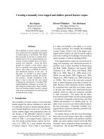
GFP–Utp25p signal is restricted to the nucleus and is
concentrated in the nucleolus (Fig. 1). GFP, by con-
trast, is present throughout the cell. RFP–Nop1p, used
as a control, is restricted to the nucleolus (Fig. 1). The
nucleolar localization of Utp25p suggested that this
protein is involved in ribosome synthesis.
GFP Hoechst GFP + RFPRFP-Nop1
GFP
GFP-
Utp25
GFP + RFP
+ Hoechst
Fig. 1. Subcellular localization of GFP–Utp25p. Yeast strains expressing RFP–Nop1p and either GFP (upper) or a GFP–Utp25p N-terminal
fusion (lower) were analyzed. Hoechst, indicates nuclei stained with the DNA dye Hoechst; GFP, indicates the localization of the green fluo-
rescent protein; RFP, indicates the localization of the red fluorescent protein. GFP + RFP, merging of green and red signals.
GFP + RFP + Hoescht, merging of all signals.
M. B. Goldfeder and C. C. Oliveira Utp25p affects pre-rRNA processing
FEBS Journal 277 (2010) 2838–2852 ª 2010 The Authors Journal compilation ª 2010 FEBS 2839
In order to characterize Utp25p function, we first
obtained a conditional mutant strain. A heterozygous
diploid strain (YIL091C ⁄ yil091c::KanMX4 – Euro-
scarf) was transformed with a plasmid containing a
copy of the UTP25 gene under control of the inducible
promoter GAL1. After sporulation, a haploid deletion
strain was obtained and its genotype was confirmed by
PCR analysis of UTP 25 gene (data not shown). The
conditional Dutp25 ⁄ GAL1::UTP25 strain was then
analyzed for growth in glucose medium, compared
with the otherwise isogenic parental strain, UTP25.
Dutp25 ⁄ GAL1::UTP25 cells are not able to grow on
glucose plates, showing that Utp25p is essential for
growth (Fig. 2A). Dutp25 ⁄ GAL1::UTP25 cells were
transformed with a second plasmid that harbors an
extra copy of UTP25 under the control of a constitu-
tive promoter, which rescues growth of the conditional
mutant on glucose plates (Fig. 2A). As shown here,
the fusion proteins GFP–Utp25p and Gal4AD–Utp25p
(transcription activation domain of Gal4p) are
functional. Growth of the conditional strain was also
analyzed in liquid glucose medium and the results
show that after 5 h in glucose, growth of Dutp25 ⁄
GAL1::UTP25 slows in comparison with the parental
wild-type strain, but the difference in growth rate is
more evident after 14 h in this medium (Fig. 2B).
Based on the Utp25p nucleolar localization and its
possible involvement in ribosome synthesis, we ana-
lyzed the polysome profile of the conditional strain
after depletion of Utp25p. When growing in galactose
medium, Dutp25 ⁄ GAL1::UTP25 cells show a normal
polysome profile on density gradients. However, after
20 h of growth in glucose, it is possible to see a severe
reduction in the relative amounts of the 40S ribosomal
subunit, as well as in 80S ribosomes and polysomes
(Fig. 3A, lower). Accordingly, free 60S accumulate in
the cells, resulting in a large peak that overlaps with
80S ribosomes (Fig. 3A, lower). Analysis of free ribo-
some subunits in the presence of EDTA confirms a
strong decrease in the relative amounts of 40S ribo-
A
B
UTP25
AD-Utp25
GFP-Utp25
–
Glucose
GAL1::UTP2
5
UTP25
h, Glu0 5 10 15 20
Log(OD/OD
0
)
5
4
3
2
1
0
Δ
utp25/
GAL1::UTP25
Fig. 2. UTP25 is an essential S. cerevisiae gene. (A) Tenfold serial
dilution of UTP25 and Dutp25 ⁄ GAL1::UTP25 strains growing on glu-
cose-containing plates. Dutp25 ⁄ GAL1::UTP25 was transformed
with plasmids containing an extra copy of the UTP25 gene under
the control of a constitutive promoter, fused to Gal4AD or GFP.
–, empty plasmid. (B) Growth curve of UTP25 and Dutp25 ⁄
GAL1::UTP25 strains in glucose medium.
A
B
A
254 nm
Polysomes
40S
60S
80S
Polysomes
40S
60S
80S
A
254 nm
Galactose
Polysomes
40S
60S
80S
Glucose
Polysomes
40S
60S
80S
A
254 nm
GAL1::UTP25
UTP25
GAL1::UTP25
Galactose
40S
60S
22.716.3
Glucose
40S
60S
22.3
3.4
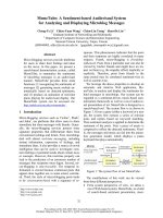
Fig. 3. Analysis of the polysomal profile in strain Dutp25 ⁄
GAL1::UTP25. UTP25 and Dutp25 ⁄ GAL1::UTP25 strains were incu-
bated either in galactose or in glucose medium for 20 h for the
analysis of polysomal profile through sucrose gradient. (A) Upper,
UTP25 strain. Lower, Dutp25 ⁄ GAL1::UTP25 strain shows very low
levels of 40S ribosomal subunit, an accumulation of free 60S sub-
unit, and consequent low number of polysomes. (B) Analysis of
ribosomal subunits through sucrose gradient in the presence of
EDTA. Levels of 40S subunit are strongly decreased upon depletion
of Utp25p. Numbers indicate area quantitation of subunits peaks.
Utp25p affects pre-rRNA processing M. B. Goldfeder and C. C. Oliveira
2840 FEBS Journal 277 (2010) 2838–2852 ª 2010 The Authors Journal compilation ª 2010 FEBS
somal subunits upon depletion of Utp25p (Fig. 3B).
Indeed, estimation of the areas under free subunit
peaks showed a change in the 60S :40S ratio from
1.4, under permissive conditions, to 6.5, after depletion
of Utp25p (Figs 3B and S1).
To investigate the possible association of Utp25p
with ribosomal particles, the sedimentation profile of
endogenous Utp25p on density gradients was analyzed.
Total extracts prepared from wild-type strain UTP25
grown in glucose medium was loaded onto 5–47%
sucrose gradients. Proteins isolated from the gradient
fractions were analyzed by western blot using a poly-
clonal antiserum raised against recombinant Utp25p.
Total RNA isolated from the same fractions was ana-
lyzed by northern blot to detect U3 snoRNA and the
mature rRNAs 25S and 18S. The results show that
endogenous Utp25p is concentrated in the fractions
containing soluble proteins, co-fractionating with free
snoRNA U3, but it is also present in higher molecular
mass fractions (Fig. 4A). As a control, antiserum spe-
cific for large ribosomal subunit protein Rpl5p was
used, showing that it is concentrated in the fractions
containing the 60S ribosomal subunits. Mature rRNAs
25S and 18S were used as controls for large and small
subunit-containing fractions (Fig. 4A).
U3 snoRNA shows a normal sedimentation profile
in these sucrose gradients, being present in the soluble
fractions but concentrated in fractions containing the
90S SSU processome (Fig. 4A and Fig. 4B, upper).
The strong effect of Utp25p depletion on the 40S
subunits levels, and its co-fractionation with free U3
snoRNA suggests that Utp25p is involved in 40S ribo-
somal subunit maturation.
To analyze whether Utp25p depletion might affect
U3 snoRNP association with preribosomes, northern
blot hybridization was performed to detect U3
snoRNA from sucrose gradient fractions. The results
show that, after 20 h of growth in glucose, depletion
of Utp25p leads to a distribution of U3 snoRNA in
two different sets of fractions, those corresponding to
soluble material and in larger complexes (Fig. 4B,
lower). Interestingly, the 35S pre-rRNA distribution in
these gradients is also shifted to larger complexes in
the absence of Utp25p (Fig. 4B).
To assess the possible involvement of Utp25p on
pre-rRNA processing, the effect of its depletion on this
pathway was analyzed by northern hybridization. The
results show that upon depletion of Utp25p there is an
accumulation of the pre-rRNA 35S and the aberrant
23S, and a decrease in pre-rRNA 20S and mature 18S
rRNA (Fig. 5A). The large ribosomal subunit RNAs
25S, 5.8S and 5S are mostly unaffected by the deple-
tion of Utp25p (Figs 5A and S2). The results shown
here indicate the involvement of Utp25p in the early
nucleolar reactions of pre-40S maturation. Pulse-chase
RNA labeling experiments with [
3
H]uracil were also
performed with cells grown in glucose for 20 h. The
results confirm the northern blot data and show that
25
S
40S 60S80S Polysomes
25
S
18
S
18
S
U3
U3
GAL1::UTP25
UTP25
AB
35
S
35
S
40S 60S 80S Polysomes
25S
18S
U3
Utp25p
*
*
Rpl5p
Fig. 4. Analysis of Utp25p and U3 snoRNA sedimentation profile on polysomal gradients. (A) Sedimentation of endogenous Utp25p was
detected by western blot of fractions from the wild-type strain (UTP25) polysomal profile. Total RNA was analyzed using northern blotting to
detect snoRNA U3. The sedimentation of mature rRNAs 25S and 18S were used as controls. Western blot with antiserum against Rpl5p
was performed as a control. (B) The effect of Utp25p depletion on the sedimentation of U3 snoRNA was analyzed by northern hybridization.
(Upper) Extract from UTP25 strain. (Lower) D utp25 ⁄ GAL1::UTP25 growing in glucose medium. Fractions corresponding to peaks of ribosome
subunits are indicated.
M. B. Goldfeder and C. C. Oliveira Utp25p affects pre-rRNA processing
FEBS Journal 277 (2010) 2838–2852 ª 2010 The Authors Journal compilation ª 2010 FEBS 2841
the depletion of Utp25p slows the processing of 35S
pre-rRNA, strongly inhibiting formation of mature
18S rRNA, although little affecting 25S rRNA forma-
tion (Fig. 6).
To gain insight into the effect of Utp25p depletion
on early pre-rRNA cleavage reactions, primer exten-
sion reactions were performed with total RNA
extracted from either wild-type cells or Dutp25 ⁄
GAL1::UTP25 grown in glucose for 16 h. Reaction with
a primer complementary to the 5¢ region of the mature
18S rRNA shows that the early cleavage reactions are
strongly inhibited after the depletion of Utp25p, leading
to an increased concentration of bands corresponding to
the pre-35S rRNA 5¢-end (Fig. 7A). Although the
accumulation of 35S and 23S rRNAs was detected by
northern hybridization, little effect on cleavage at A
1
was observed by primer extension (Fig. 7A). This may
be because of the high stability of mature 18S rRNAs
formed prior to the depletion of Utp25p.
Reaction with primer P
3
, which hybridizes down-
stream of site D in ITS1, also shows the accumulation
of pre-rRNAs, with increased extension bands corre-
sponding to regions in the mature 18S rRNA upon
depletion of Utp25p (Fig. 7B; asterisk). A primer
extension reaction with an oligo complementary to a
region downstream of A
2
shows that depletion of
Utp25p causes a strong inhibition in the cleavage at
this site (Fig. 7), and consequently the accumulation of
extended products that correspond to regions within
the mature 18S rRNA (Fig. 7C, asterisk). These results
further indicate the involvement of Utp25p in the early
steps of processing of pre-40S.
To determine whether Utp25p might associate in vivo
with pre-rRNAs, co-immunoprecipitation experiments
were performed using a ProtA–Utp25p fusion. Total
extract from cells expressing ProtA–Utp25p was sub-
jected to affinity chromatography with IgG–Sepharose
beads. Following co-immunoprecipitation, RNA was
extracted from the different fractions and analyzed by
northern hybridization, compared with RNAs recov-
ered in parallel from the strain expressing only ProtA.
The results show that ProtA–Utp25p co-precipitates
B
A
0 12 16 0 12 16 h, Glu
P1
35S
23S
P6
27S
P7
25S
P2
18S
P3
20S
P5
7S
P4 5.8S
5S
GAL1::UTP25UTP25
A
0
A
1
A
2
A
3
D
B
1L
A
3
→B
1S
B
2
←B
0
B
2
←B
0
C
2
E←C
2
C
2
→C
1
C
2
E←C
2
C
2
→C
1
33S
32S
20S
27SA
2
18S 5.8S
S
25S 5.8S
L
25S
27SA
3
27SB
S
27SB
L
7S
S
7S
L
A1 B2
D
A
2 A3
B1L/B1S E
C
2
C1
5´ETS
A0
ITS1 3´ETSITS2
P
3
P
4
P
5
P
1
P
2
P
7
P
6
P
8
18S
5.8S
25S
35S
or
B0
Fig. 5. Northern blot analysis of pre-rRNA processing. (A) Total RNA (20 lg) extracted from cells incubated in glucose medium for different
periods and hybridized against specific oligonucleotide probes. The relative positions of the probes on the 35S pre-rRNA are indicated in (B).
Bands corresponding to the major intermediates and to the mature rRNAs are indicated on the right-hand side. The lower panel shows
hybridization with a probe against the 5S rRNA, used as an internal control. (B) Structure of the 35S pre-rRNA and major intermediates of
the rRNA processing pathway in S. cerevisiae. The positions of the probes used for northern hybridizations are indicated below the 35S pre-
rRNA. Processing of 35S pre-rRNA starts with endonucleolytic cleavages at sites A
0
and A
1
in the 5¢-ETS, generating 32S pre-rRNA. Subse-
quent cleavage at site A
2
, in ITS1, generates the 20S and 27SA
2
pre-rRNAs. The 20S pre-rRNA is then processed at site D to the mature
18S rRNA. The major processing pathway of the 27SA
2
pre-rRNA involves cleavage at site A
3
, producing 27SA
3
, which is digested quickly
by exonucleases to generate the 27S B short (27SB
s
) pre-rRNA. The subsequent processing step occurs at site B
2
, at the 3¢-end of the
mature 25S rRNA. Processing at sites C
1
and C
2
separates the mature 25S rRNA from the 7S
S
pre-rRNA. This pre-rRNA is subsequently pro-
cessed exonucleolytically to generate the mature 5.8S
S
rRNAs. A fraction of the 27SA
2
pre-rRNA is processed at the 5¢-end by a different
mechanism and, following processing at the remaining sites, gives rise to the 5.8S long (5.8S
L
) rRNA, which is 6-8 nucleotides longer than
the 5.8S
S
rRNA at the 5¢-end.
Utp25p affects pre-rRNA processing M. B. Goldfeder and C. C. Oliveira
2842 FEBS Journal 277 (2010) 2838–2852 ª 2010 The Authors Journal compilation ª 2010 FEBS
the 35S pre-rRNA, the aberrant rRNAs 23S and 22S,
and much less efficiently, the pre-rRNA 20S (Fig. 8).
Mature 18S rRNA was not efficiently co-immunopre-
cipitated with ProtA–Utp25p, further indicating that
this protein is associated only with the early pre-
rRNAs. This is in accordance with the hypothesis of
the involvement of Utp25p in the early cleavages of
the 35S pre-rRNA, ProtA–Utp25p co-immunoprecipi-
tated U3 snoRNA (Fig. 8).
Based on the above results, it seemed likely that
Utp25p might interact with protein subunits of the
SSU processome. To determine whether that interac-
tion could occur, the two-hybrid assay was performed
using Utp25p fused to the lexA DNA-binding domain
and its interaction with Sas10p ⁄ Utp3p, Mpp10p,
Imp3p and Imp4p was investigated [10,12,15]. Expres-
sion of the reporter genes HIS3 and lacZ indicates a
strong interaction of Utp25p with Sas10p ⁄ Utp3p, a
weaker interaction with Mpp10p and no interaction
with Imp3p or Imp4p (Fig. 9A, upper). The direct
interaction between Utp25p and Sas10p was confirmed
after expressing recombinant proteins in Escherichi-
a coli and performing pull-down assays. The results
show that glutathione S-transferase (GST)–Sas10p,
immobilized in glutathione–Sepharose beads pulls
25
S
35
S
18
S
0 3 10 30 60 0 3 10 30 60 min
20
S
27
S
23
S
UTP25
GAL1::UTP25
Fig. 6. Metabolic labeling of rRNA. Pulse-chase labeling with
[
3
H]uracil was performed after incubating Dutp25 ⁄ GAL1::UTP25
and control strain in glucose medium for 20 h. Total RNA (20 lg)
was loaded onto agarose gel after [
3
H]uracil labeling. The figure
shows autoradiograph of RNA transferred to nylon membrane.
Bands corresponding to major intermediates and mature rRNAs are
indicated on the right-hand side.
A
1
A
0
P
2
5’
GATC
0 16 0 16 h, Glu
A
*
P
3
GATC
016016 h, Glu
B
GA TC
016016 h, Glu
C
*
A
2
P
8
GAL1::UTP25
GAL1::UTP25
GAL1::UTP25
Fig. 7. Early cleavage reactions in 35S pre-rRNA were analyzed through primer extension reactions of total RNA extracted from cells grow-
ing in media containing either galactose (0 h) or glucose (16 h). Relative positions of the primers used in the primer extension reactions are
shown in Fig. 5B. (A) Primer extension with the primer P
2
allows the detection of the sites A
0
and A
1
. Processing inhibition in Dutp25 ⁄
GAL1::UTP25 strain allows the detection of the 5¢-end of 35S pre-rRNA. (B) Reaction with primer P
3
that hybridizes between sites D and A
2
shows the accumulation of pre-rRNA after depletion of Utp25p. (C) Primer extension reaction with primer P
8
shows that processing at site
A
2
is inhibited upon depletion of Utp25p. Asterisks indicate longer extensions of the reactions due to inefficient processing.
M. B. Goldfeder and C. C. Oliveira Utp25p affects pre-rRNA processing
FEBS Journal 277 (2010) 2838–2852 ª 2010 The Authors Journal compilation ª 2010 FEBS 2843
down His–Utp25p, whereas the negative control GST
does not (Fig. 9B). These results strongly suggest that
Utp25p is part of the SSU processome, participating in
the early stages of pre-rRNA maturation. In order to
determine the portion of Sas10p that is responsible for
the interaction with Utp25p, two Sas10p truncated
mutants were fused to Gal4-AD and the interaction
with Utp25p was investigated through the two-hybrid
assay. The results show that the N-terminal portion of
Sas10p is sufficient for interaction with Utp25p
(Fig. 9A, lower).
Many of the SSU processome protein subunits are
conserved throughout evolution. In order to identify
possible Utp25p orthologs in other organisms, a
BLAST search was performed. Utp25p homologs are
present in many organisms, including humans
0
10
20
30
40
50
60
70
80
90
100
ProtA
ProtA ProtA-Utp25
TEFT FTWWBB
U3
5S
25S
18S
TE
35S
23S
22S/21
S
20S
Bound/input
ProtA
A-Utp25
35S 23S 22S/
21S
20S 18S 25S 5S U3
23S
A
0
A
1
A
2
A
3
D
22S
A
0
A
1
A
2
A
3
D
21S
A
1
A
2
A
3
D
20S
A
1
A
2
D
A
B
C
Fig. 8. RNA co-immunoprecipitation with ProtA–Utp25p. Total
extracts from cells expressing either ProtA or ProtA–Utp25p were
incubated with IgG–Sepharose beads. (A) After immunoprecipita-
tion, RNA was extracted from fractions of total extract (TE), flow
through (FT), wash (W) and bound material (B), separated by elec-
trophoresis and subjected to northern hybridization with probes
specific for the RNAs indicated on the right. The structures of the
detected pre- and aberrant rRNAs are shown on the left. (B) Pro-
teins isolated from the same fractions were subjected to western
blot for detection of ProtA and ProtA–Utp25p. (C) Quantitation of
the bands obtained from RNA co-ip by phosphorimaging. Ratio of
bound ⁄ input is shown for all RNAs tested.
BD-Utp25
AD-Sas10
AD-Mpp10
AD
L40-61
– His X-Gal
AD-Imp3
AD-Imp4
A
B
FT
1
FT
2
B
GST
+ His-Utp25
GST-Sas10
+ His-Utp25
TE
1
FT
1
BFT
2
TE
1
His-Utp25
GST-Sas10
GST
AD-Sas10
(1–227)
AD-Sas10
AD–Sas10
(226–610)
AD
BD-Utp25
Fig. 9. Utp25p interacts with SSU processome subunits. (A)
Utp25p was fused to lexA DNA-binding domain (BD) and tested for
interaction with Mpp10p, Sas10p, Imp3p and Imp4p, which were
fused to Gal4p transcription activation domain (AD). Sas10p trun-
cated mutants fused to Gal4p-AD are indicated by the amino acid
positions relative to the full-length protein [Sas10(1–227) and
Sas10(226–610)]. Protein interactions were analyzed using the two-
hybrid system, testing for expression of the reporter genes HIS3
(left) and lacZ (right). BD–UTP25 + AD, negative control; strain
L40-61, which harbors plasmids encoding BD–Nip7p and
AD–Rrp43p, was used as a positive control. (B) Western blot for
detection of proteins after pull-down assay. Total extract from cells
expressing either GST or GST–Sas10p (TE
1
) was incubated with a
glutathione–Sepharose resin, the flow-through fraction was
collected (FT
1
) and after washing, total extract of cells expressing
His–Utp25p (TE
2
, not shown) was loaded. The flow-through fraction
was collected again (FT
2
), resin was washed, and bound fraction
obtained (B). His–Utp25p is only pulled-down by GST–Sas10p. His–
Utp25p was detected with monoclonal anti-His IgG2a. GST–Sas10p
and GST were detected with anti-GST serum.
Utp25p affects pre-rRNA processing M. B. Goldfeder and C. C. Oliveira
2844 FEBS Journal 277 (2010) 2838–2852 ª 2010 The Authors Journal compilation ª 2010 FEBS
(Fig. S3). Utp25p and hUtp25p (human Utp25p) show
37% sequence similarity and 28% sequence identity.
Both proteins contain the domain of unknown func-
tion DUF1253, which shows low similarity to DEAD
box helicases [20]. To investigate whether hUtp25p
and Utp25p might perform similar functions in the
cell, the human gene (C1ORF7 ⁄ DEF) was cloned and
expressed in strain Dutp25 ⁄ GAL1::UTP25 under the
control of a constitutive promoter (MET25::GFP-
hUTP25). Expression of the human protein could not
rescue Dutp25 ⁄ GAL1::UTP25 growth under the restric-
tive condition (Fig. 10A). To obtain higher levels of
expression of hUtp25p in yeast cells, the gene was
cloned under control of a stronger constitutive pro-
moter, PGK1, without the GFP tag, but still could not
rescue growth of the conditional strain in glucose med-
ium (Figs 10A and S4), indicating that, although these
proteins show sequence similarity in the C-terminal
portion, divergences in the remaining sequence of the
protein would render hUtp25p nonfunctional and ⁄ or
unstable in yeast.
To analyze the possibility that the C-terminal
DUF1253 domain of Utp25p might be sufficient for
the protein function, truncation mutants were fused to
GFP, cloned in a plasmid under the control of a con-
stitutive promoter and transformed into Dutp25 ⁄ GA-
L1::UTP25 strain. The results show that the DUF1253
domain does not complement growth of the condi-
tional strain (Figs 10A and S4). The GFP-fused dele-
tion mutants were also analyzed by western blot and
the results show that all were expressed in the cell
(Fig. 10B). Sequence analysis also predicted a possible
phosphorylation site in Utp25p. Indeed, high-through-
put analysis showed that Ser196 was phosphorylated
[21]. To investigate whether this modification was
important for function, a point mutation was intro-
duced in Utp25(S196V) that would prevent phosphory-
lation at this specific residue. The more conserved
B
25
40
50
60
80
115
kDa
α-GFP Coomassie
A
GFP
GFP-Utp25
GFP-Utp25Δ243
GFP-Utp25Δ287
GFP-Utp25Δ411
1 721 aa300
DUF1253
Glu
Δ
utp25/GAL1::UTP25
GFP
UTP25
GFP-Utp25
GFP-Utp25 (S196V)
*
GFP-hUtp25
GFP-Utp25 (S198A)
*
hUtp25
Fig. 10. Schematic representation of the
different clones of Utp25p, full-length,
truncated and the human ortholog, that
were tested for complementation of growth
of the conditional strain Dutp25 ⁄ GA-
L1::UTP25 in glucose. (A) Tenfold serial
dilution of UTP25 and Dutp25 ⁄ GAL1::UTP25
strains growing on glucose-containing
plates. Dutp25 ⁄ GAL1::UTP25 was trans-
formed with a plasmid containing an extra
copy of the UTP25 gene, truncated mutants
or hUtp25p under control of a constitutive
promoter, fused to GFP. hUtp25 indicates
PGK1::hUTP25 (without a GFP tag). (B)
Analysis of GFP–Utp25p mutants and
GFP–hUtp25p by western blot with anti-GFP
serum, compared with wild-type
GFP–Utp25p. Arrowheads indicate
full-length proteins. Right, Coomassie
Brilliant Blue-stained poly(vinylidene
difluoride) membrane used in the
immunoblot assay.
M. B. Goldfeder and C. C. Oliveira Utp25p affects pre-rRNA processing
FEBS Journal 277 (2010) 2838–2852 ª 2010 The Authors Journal compilation ª 2010 FEBS 2845
Ser198 was also mutated, originating Utp25(S198A).
Interestingly, cells expressing Utp25(S196V) showed a
growth rate similar to that of the wild-type strain, indi-
cating that phosphorylation at Ser196 of Utp25p is not
essential for function (Figs 10A and S4). Polysomal
profile analysis of Dutp25 ⁄ GAL1::UTP25 ⁄ GFP-
utp25(S196V) strain confirms that this mutant is fully
functional (Fig. S4). Cells expressing Utp25(S198A),
on the other hand, are not able to grow in glucose
medium (Figs 10A and S4).
Discussion
Although various proteins have already been identified
as components of the SSU processome [15,16,22], it is
possible that many subunits remain to be isolated.
Here, we report the characterization of Utp25p as a
novel nucleolar protein required for efficient cleavage
of 35S pre-rRNA at sites A
0
,A
1
and A
2
. Depletion of
Utp25p causes the accumulation of the pre-rRNA 35S
and the aberrant 23S, a consequent decrease in the lev-
els of mature 18S rRNA and strong depletion of 40S
ribosomal subunit. In accordance with its nucleolar
localization and effects on pre-rRNA processing,
Utp25p interacts with the protein subunits of the SSU
processome Sas10p and Mpp10p and co-immunopre-
cipitates U3 snoRNA.
High-throughput assays identified Utp25p in com-
plexes with Mpp10p and Sas10p [23]. Mpp10p has
been characterized as a nucleolar protein that interacts
with the U3 snoRNP, depletion of which causes inhibi-
tion of cleavages at sites A
0
,A
1
and A
2
, leading to
decreased levels of 18S rRNA [10]. Mpp10p is part of
a ternary complex with Imp3p and Imp4p, and these
proteins show interdependence for binding to U3
snoRNA [24]. Because Utp25p showed no interaction
with Imp3p and Imp4p and was not isolated in the
Mpp10p ternary complex, it is possible that its interac-
tion with Mpp10p is transient or might occur in the
context of the assembled SSU processome. Sas10-
p ⁄ Utp3p is also part of the SSU processome, co-immu-
noprecipitates U3 snoRNA and interacts with Mpp10p
[15]. Depletion of Sas10p also causes a severe reduc-
tion in 18S rRNA levels, without affecting 25S rRNA
[15]. Interestingly, individual depletions of either U3
snoRNA or the U3 snoRNP protein subunits Nop1p,
Nop58p, Mpp10p, Imp3p, Imp4p, Sof1p, Lcp5p,
Utp23p, Utp24p and Enp1p all result in accumulation
of the pre-rRNA 35S and the aberrant 23S, and
decreased levels of the 20S pre-rRNA and the mature
18S rRNA, although to different degrees of severity
[10–12,14,25–28]. These results indicate that the SSU
processome must be fully assembled for the cleavage
reactions at sites A
0
,A
1
and A
2
to occur. The observa-
tion that depletion of Utp25p leads to similar pheno-
types and its interaction with U3 snoRNA, Mpp10p
and Sas10p strongly indicate that this is a novel com-
ponent of the SSU processome. The direct interaction
between Utp25p and Sas10p was confirmed through
protein pull-down assays, further indicating that
Utp25p is a subunit of that complex.
As shown here, in addition to interacting with SSU
processome subunits, Utp25p co-immunoprecipitates the
pre-rRNAs 35S, the aberrant rRNAs 23S and 22S,and
much less efficiently the pre-rRNA 20S. Co-immunopre-
cipitation of aberrant rRNAs with SSU processome com-
ponents has been reported previously [29,30]. Utp25p
does not co-immunoprecipitate the mature 18S rRNA,
however, which is in agreement with its involvement in
the early cleavage of the 35S pre-rRNA.
Analysis of endogenous Utp25p sedimentation on
polysomal gradients shows that it is concentrated in
the fractions corresponding to soluble material, frac-
tions that also contain U3 snoRNA, which is consis-
tent with Utp25p being part of U3 snoRNP complex.
SSU processome subunits from different U3 snoRNP
subcomplexes have also been reported to concentrate
in the soluble fractions of polysomal gradients [16,31].
Combined, these results indicate that although Utp25p
interacts with the SSU processome and is involved in
pre-rRNA maturation, its interaction with the complex
may be labile or transient.
The question of whether Utp25p binds directly to
the snoRNA U3 or associates via interaction with
the proteins Sas10p and Mpp10p remains to be
addressed. The fact that no known RNA-binding
motifs can be distinguished in the Utp25p sequence,
however, indicates that the latter is more likely.
Analysis of the Utp25p sequence also shows that this
protein contains the domain DUF1253, which occurs
in several hypothetical eukaryotic proteins of
unknown function and shows remote homology to
DEAD box RNA helicases [20]. Attempts to gain
insight into the role of the DUF1253 domain on
Utp25p function, made by testing the complemen-
tation of growth of the conditional strain Dutp25 ⁄
GAL::UTP25 with deletion mutants expressing only
the DUF1253 domain, gave negative results. Interest-
ingly, Utp25p shows some amino acid residues that
are possible targets for phosphorylation. Indeed,
Utp25p Ser196 has been previously shown to be
phosphorylated [21]. Our data show that a point
mutation in which Ser196 was replaced by a valine
had no effect on Utp25p function. Interestingly, how-
ever, substitution of Ser198 by alanine resulted in a
nonfunctional protein.
Utp25p affects pre-rRNA processing M. B. Goldfeder and C. C. Oliveira
2846 FEBS Journal 277 (2010) 2838–2852 ª 2010 The Authors Journal compilation ª 2010 FEBS
During the final preparation of this article, a study
was published on Utp25p [32]. In that work, a network-
guided genetics approach was used to identify proteins
involved in ribosome biogenesis, and Utp25p was char-
acterized as a nucleolar protein associated with the 40S
ribosomal subunit. Analysis of pre-rRNA processing
also showed that Utp25p depletion causes an accumula-
tion of 35S pre-rRNA. Those results are consistent with
those shown here. Our data complement that study by
showing the direct interaction of Utp25p with SSU pro-
cessome subunits, and the analysis of the 35S pre-rRNA
cleavage reactions that are affected by the depletion of
Utp25p. Furthermore, we show that although a puta-
tive human ortholog of Utp25p was identified, it does
not complement the yeast protein function.
Materials and methods
DNA manipulation and plasmid construction
The plasmids used in this study, described in Table 1, were
constructed according to the cloning techniques described
by Sambrook et al. [33] and sequenced by the Big Dye
method (PerkinElmer, Waltham, MA, USA). Cloning strat-
egies were as follows. UTP25 gene, encoded by the
YIL091C open reading frame, was PCR amplified from
S. cerevisiae genomic DNA using primers specific for
UTP25: 5¢-CCCGGGTGGATCCATGAGTGACAGTTCT
GTGAG-3¢ and 5¢-CTCGAGTTATTTAAATTCATAAAT
TTCCTTTTGTGC-3¢. For the two-hybrid assays, the PCR
product was digested with SmaI and XhoI and cloned into
pBTM116 [34] and pGAD-C2 [35] digested with the same
enzymes, generating pBTM–UTP25 and pGAD–UTP25
(which code for the fusions BD–Utp25p and AD–Utp25p
respectively, where BD refers to the lexA DNA-binding
domain and AD refers to the Gal4p transcription activation
domain). MPP10 and SAS10 genes were PCR amplified, the
products were digested with the enzymes PvuII and SmaI
and cloned into pBTM116 and pGAD-C2 digested with
SmaI. To obtain Sas10p truncation mutants, plasmid
pGAD–SAS10 was cleaved with EcoRI, resulting in a frag-
ment coding for Sas10p amino acid residues 1–226, which
was cloned into pGADC2 generating pGAD–SAS10(1–226).
The plasmid previously digested with EcoRI was religated,
generating pGAD–SAS10(227–610). For the pull-down
assays, BamHI–XhoI fragments of UTP25 and MPP10 genes
were cloned into pET28a (Merck KGaA, Darmstadt, Ger-
many) and pGEX-4T1 (GE Healthcare, Little Chalfont,
UK), respectively. YCp111GAL–UTP25, which carries
UTP25 under the control of GAL1 promoter, was obtained
by inserting an EcoRV–SalI fragment into YCp111-GAL
digested with NdeI (following T4 DNA polymerase treat-
ment) and SalI. To determine the subcellular localization of
yEGFP3–Utp25p by fluorescence microscopy, plasmid
pUG34–UTP25 was constructed by inserting a BamHI–XhoI
fragment into pUG34 (U. Gueldener & J. H. Hegemann,
unpublished) digested with BamHI and SalI. Plasmid pUG36
(U. Gueldener & J. H. Hegemann, unpublished) was used to
Table 1. List of plasmid vectors used.
Plasmid Relevant characteristics Reference
pBTM116 lexA DNA binding domain, TRP1,2lm34
pBTM–UTP25 lexA::UTP25, TRP1,2lm This study
pGAD GAL4 activation domain, LEU2,2lm35
pGAD–MPP10 GAL4::SAS10, LEU2,2lm This study
pGAD–SAS10 GAL4::MPP10, LEU2,2lm This study
pGAD–IMP3 GAL4::IMP3, LEU2,2lm This study
pGAD–IMP4 GAL4::IMP4, LEU2,2lm This study
pET28a–UTP25 His
6
::UTP 25, Kan
R
This study
pGEX4T1–SAS10 GST::SAS10, Amp
R
This study
YCplac33 ARS1, URA3, CEN4 51
YCp33GAL–UTP25 GAL1::UTP25, URA3, CEN4 This study
pUG34 MET25::yEGFP3, CEN6, HIS3 U. Gueldener & J. H. Hegemann,
unpublished
pUG34–UTP25 MET25::yEGFP3-UTP25, HIS3, CEN6 This study
pUG34–hUTP25 MET25::yEGFP3h-UTP25, HIS3, CEN6 This study
pMET25–hUTP25 MET25:: UTP25, HIS3, CEN6 This study
pPGK–hUTP25 PGK1:: UTP25, HIS3, CEN6 This study
pUG36 MET25::yEGFP3, CEN6, URA3 U. Gueldener & J. H. Hegemann,
unpublished
pUG36–DsRed–NOP1 MET25::DsRED-NOP1, CEN6, URA3 This study
YCp33GAL-A GAL1::PROTA, URA3, CEN4 52
YCp33GAL-A–UTP25 GAL1::PROTA-UTP25, URA3, CEN4 This study
M. B. Goldfeder and C. C. Oliveira Utp25p affects pre-rRNA processing
FEBS Journal 277 (2010) 2838–2852 ª 2010 The Authors Journal compilation ª 2010 FEBS 2847
create pUG36–DsRed–NOP1, by substitution of yEGFP3
with DsRed gene (coding a red fluorescent protein from Dis-
cosoma sp., [36]), which was PCR amplified from plasmid
pDsRed–Monomer-C1 (Clontech, Moutain View, CA,
USA). For construction of the Utp25(S196V) mutant, two
fragments of the UTP25 gene were PCR amplified, creating a
SalI site in the gene sequence at the corresponding position.
To obtain Utp25(S198A) mutant, the Quickchange kit (Strat-
agene, La Jolla, CA, USA) was used. The human ortolog
hUTP25 gene (human C1ORF7, a ccession number BC022964)
was P CR amplified from pCMV-SPORT6-C1ORF7 (Imagenes,
Berlin, Germany) using primers: 5¢-GGATCCATGGGC
AAACGCGGGAGCC-3¢ and 5¢-ATCGATGTCGACTCA
TTTTTCTCCAGTAATGAAGAG-3¢. The gene was cloned
in fusion with yEGFP3 using BamHI and SalI sites of
pUG34. This vector was cleaved with XbaI and re-ligated,
generating plasmid pMET25–hUTP25. A XbaI–BamHI frag-
ment containing PGK1 promoter was inserted into the latter
plasmid, generating pPGK–hUTP25.
Yeast maintenance, transformation and
sporulation
Yeast genetic techniques were conducted as described previ-
ously [37]. Strains described in Table 2 were maintained in
yeast extract–peptone (YP) medium or synthetic medium
(YPD) with 2% (w ⁄ v) galactose or glucose as the carbon
source, as indicated, and supplemented with amino acids
when required. Yeast cells were transformed using a lithium
acetate method [38]. A Dutp25 diploid strain (2n,
UTP25 ⁄ UTP25::KanMX4) obtained from Euroscarf, Frank-
furt, Germany was transformed with YCp111GAL–UTP25,
induced to sporulation and tetrad dissection was performed
as described previously [37]. Strains UTP25 and Dutp25 ⁄ GA-
L1::UTP25 were grown in galactose-containing medium to
the stationary phase, cell suspension was 10-fold concen-
trated and plated in glucose containing medium in a 10-fold
serial dilution. For the growth curve in liquid medium, cells
were grown in galactose containing medium until stationary
phase and then shifted to glucose medium for 20 h.
Yeast two-hybrid assays
Fusion proteins with either lexA DNA-binding domain
(BD-protein) or Gal4p transcription activation domain
(AD-protein) were expressed in the host strain L40 [39],
which has two reporter genes for two-hybrid interactions
integrated into the genome: yeast HIS3 and E. coli lacZ.
Transformants were plated in minimal medium lacking
histidine as a first selection and viable clones were further
tested for b-galactosidase activity as follows. Exponen-
tially growing cultures in minimal medium (supplemented
with histidine) were concentrated 10-fold and either trans-
ferred to nitrocellulose membranes and incubated over-
night at 30 °C for the b-galactosidase activity assay [39],
or plated in His
-
medium in a 10-fold serial dilution.
Strain L40-61 [40] was used as a positive control and
strain L40 ⁄ pBTM-NOP53 ⁄ pGAD was used as negative
control for two-hybrid interaction (Table 2).
Generation of Utp25p antiserum
Fusion protein His
6
–Utp25 was expressed in E. coli and puri-
fied by metal-chelating chromatography. Ten BALB-C mice
were injected with 10 lg purified His
6
-Utp25 mixed with com-
plete Freund’s adjuvant (Sigma, St Louis, MO, USA) each.
The mice were injected three more times with 1-week intervals
with 10 lg purified protein with incomplete Freund’s adju-
vant (Sigma). Mice were bled after five weeks; blood was incu-
bated at 37 °C for 30 min, followed by incubation at 4 °C for
18 h. Experiments were approved by the local Comiteˆ de
E
´
tica em Cuidados e Uso Animal and follow NIH guidelines.
Antiserum was isolated through centrifugation, and tested
against purified His
6
–Utp25 and total yeast extract.
Immunoblot analysis
Poly(vinylidene difluoride) membranes (BioRad, Hercules,
CA, USA) were incubated with the antisera diluted in PBS-T
(NaCl ⁄ P
i
buffer plus 0.1% Tween 20) with 0.1% BSA. Sera
were diluted as follows: mouse anti-Utp25, 1 : 1000; rabbit
anti-Rpl5, 1 : 10 000 (kind gift from S.R. Valentini, Sa
˜
o
Table 2. List of yeast strains used.
Strain Relevant features Reference
L40 MATa his3d 200 trp1-901
leu2-3,311 ade2 lys2-801am
URA3::(lexAop)8-lacZ
LYS2::(lexAop)4-HIS3
39
L40-61 L40, pBTM-NIP7, pACT-RRP43 40
YMG-226 L40, pBTM-UTP25, pGAD This study
YMG-227 L40, pBTM-UTP25, pGAD-MPP10 This study
YMG-228 L40, pBTM-UTP25, pGAD-SAS10 This study
YMG-229 L40, pBTM-UTP25, pGAD-IMP3 This study
YMG-230 L40, pBTM-UTP25, pGAD-IMP4 This study
UTP25 MATa, his3D1, leu2D0,
lys2D0, ura3D0
Euroscarf
UTP25 ⁄
UTP25::KAN
R
MATa ⁄ a, his3D1 ⁄ his3D1,
leu2D0 ⁄ leu2D0, lys2D0 ⁄ LYS2,
ura3D0 ⁄ ura3D0, MET15 ⁄ met15D0,
UTP25 ⁄ UTP25::KAN
R
Euroscarf
Dutp25 ⁄
GAL1::UTP25
his3D1, leu2D0, LYS2, met15D0,
ura3D0, Utp25::KAN
R
,
YCp111GAL-UTP25
This study
YMG-231 Dutp25 ⁄ GAL1::UTP25, pUG34,
pUG36-DsRed-NOP1
This study
YMG-232 Dutp25 ⁄ GAL1::UTP25,
pUG34-UTP25,
pUG36-DsRed-NOP1
This study
Utp25p affects pre-rRNA processing M. B. Goldfeder and C. C. Oliveira
2848 FEBS Journal 277 (2010) 2838–2852 ª 2010 The Authors Journal compilation ª 2010 FEBS
Paulo State University-UNESP, Araraquara, SP, Brazil) [41];
rabbit anti-GFP, 1 : 10 000 (kind gift from F. Gueiros-Filho,
University of Sao Paulo, SP, Brazil) [42]; rabbit anti-GST,
1 : 10 000 (Sigma); mouse anti-(poly-histidine), 1 : 3000 (GE
Healthcare); horseradish peroxidase-conjugated anti-rabbit,
1 : 10 000 (GE Healthcare); and anti-(mouse IgG), 1 : 7500
(GE Healthcare). Blots were developed using the enhanced
chemiluminescence (ECL) system (GE Healthcare).
Fluorescence analysis
In order to determine its subcellular localization, Utp25p
was expressed in the strain YMG-231 with an N-terminal
GFP (yEGFP3) tag, encoded by the plasmid pUG34–
UTP25. DNA in the cell nucleus was stained using
Hoechst, and DsRed–Nop1 fluorescence was observed as a
nucleolar marker. Localization of GFP was analyzed in the
strain YMG-232 as a control. Images were obtained on a
Nikon TE300 inverted microscope equipped with a Roper
CoolSnap HQ camera.
RNA analysis
Exponentially growing cultures of yeast strains were shifted
from galactose to glucose medium. At various times, samples
were collected and quickly frozen in a dry ice–ethanol bath.
Total RNA was isolated from yeast cells by a modified hot
phenol method [43]. RNAs were separated by electrophoresis
(20 lg of total RNA was loaded in each lane) on 1.3%
agarose gels, following denaturation with glyoxal [33] and
transferred to Hybond nylon membranes (GE Healthcare).
Membranes were probed with
32
P-labeled oligonucleotides
(Table 3) complementary to specific regions of the 35S pre-
rRNA or to 5S rRNA, using the hybridization conditions
described previously [44] and analyzed in a Phosphorimager
(Molecular Dynamics, Sunnyvale, CA, USA).
Primer extension analysis
Total RNA extracted as described above was used for pri-
mer extension analysis. Reactions were performed by
annealing pmol of
32
P-labeled oligonucleotide to 5 lgof
total RNA. Following annealing, extension was performed
with 100 U of MMLV reverse transcriptase (Invitrogen,
Carlsbad, CA, USA) and dNTPs (0.5 mm) for 30 min at
37 °C. cDNA products were precipitated, resuspended in
H
2
O, treated with Rnase A, denatured and analyzed on 6%
denaturing polyacrylamide gels. Gels were dried and ana-
lyzed in a Phosphorimager. Oligonucleotides used in primer
extension analyses are listed in Table 3.
Polysome profile analysis
For polysome profile analysis cell extracts were generated
from 500 mL cultures grown to A
600
1.0 in YP-Gal (t
0
)or
in YPD for 20 h. Following the addition of cycloheximide
(100 lgÆmL
)1
) to the cultures, cells were harvested by cen-
trifugation and resuspended in breaking buffer A (20 mm
Tris ⁄ HCl pH 7.4, 50 mm NaCl, 10 m m MgCl
2
,1mm
dithiothreitol, 200 lgÆmL
)1
heparin, 100 lgÆmL
)1
cyclohex-
imide, 1 mm phenylmethanesulfonyl fluoride, 1 mm dith-
iothreitol). Polysomes were separated by centrifugation at
190,000 g for 3 h at 4 °C with a Beckman SW41 rotor.
Gradients were fractionated and monitored at 254 nm with
an absorbance monitor (BioRad). Analysis of free ribo-
some subunits was performed as described above, using
breaking buffer B (20 mm Tris ⁄ HCl pH 7.4, 50 mm NaCl,
400 mm EDTA, 1 mm phenylmethanesulfonyl fluoride,
1mm dithiothreitol, 200 lgÆmL
)1
heparin, 100 lgÆmL
)1
cycloheximide, 1 mm phenylmethanesulfonyl fluoride,
1mm dithiothreitol). Area below free ribosomal subunits
peaks was estimated using NIH ImageJ software (W. S.
Rasband, ImageJ, US NIH, Bethesda, MD, USA, http://
rsb.info.nih.gov/ij/). Proteins from each fraction (200 lL)
were precipitated with 15% trichloroacetic acid and ana-
lyzed by western blot. For northern blot experiments, 1.5 mL
of cold ethanol was added to 300 lL of each fraction. Pellets
were suspended in 500 lL of acetate buffer (50 mm NaOAc,
10 mm EDTA, pH 5.0) and RNA was isolated as described
above.
Protein sequence analysis
Protein sequences from different organisms were aligned
using the clustal w algorithm [45], through the Network
Protein Sequence Analysis web server [46].
Acknowledgements
We would like to thank Beatriz A. Castilho for help
and equipment for polysomal profile analyses. This
work was supported by a grant from Fundac¸ a
˜
ode
Amparo a
`
Pesquisa do Estado de Sa
˜
o Paulo – FA-
PESP (07 ⁄ 57096-9 to CCO). MBG was recipient of a
FAPESP postdoctoral fellowship.
Table 3. DNA oligonucleotides used for northern blot hybridization
and primer extension analyses.
Oligo Sequence Ref.
P1 5¢-GGTCTCTCTGCTGCCGGAAATG-3¢ 44
P2 5¢-CATGGCTTAATCTTTGAGAC-3¢ 47
P3 5¢-GCTCTCATGCTCTTGCCAAAAC-3¢ 44
P4 5¢-CGTATCGCATTTCGCTGCGTTC-3¢ 44
P5 5¢-CTCACTACCAAACAGAATGTTTGAGAAGG-3¢ 48
P6 5¢-GTTCGCCTAGACGCTCTCTTC-3¢ 44
P7 5¢-GCCGCTTCACTCGCCGTTACTAAGGC-3¢ 49
P8 5¢-TGTTATCCTCTGGGCCCCG-3¢ 44
anti-U3 5¢-ATGGGGCTCATCAACCAAGTTGG-3¢ 50
M. B. Goldfeder and C. C. Oliveira Utp25p affects pre-rRNA processing
FEBS Journal 277 (2010) 2838–2852 ª 2010 The Authors Journal compilation ª 2010 FEBS 2849
References
1 Kressler D, Linder P & Cruz J (1999) Protein trans-act-
ing factors involved in ribosome biogenesis in Saccharo-
myces cerevisiae. Mol Cell Biol 19, 7897–7912.
2 Venema J & Tollervey D (1999) Ribosome synthesis in
Saccharomyces cerevisiae. Annu Rev Genet 33, 261–311.
3 Lygerou Z, Allmang C, Tollervey D & Seraphin B
(1996) Accurate processing of a eukaryotic precursor
ribosomal RNA by ribonuclease MRP in vitro. Science
272, 268–270.
4 Beltrame M & Tollervey D (1995) Base pairing between
U3 and the pre-ribosomal RNA is required for 18S
rRNA synthesis. EMBO J 14, 4350–4356.
5 Li HD, Zagorski J & Fournier MJ (1990) Depletion of
U14 small nuclear RNA (snR128) disrupts production
of 18S rRNA in Saccharomyces cerevisiae. Mol Cell
Biol 10, 1145–1152.
6 Tollervey D (1987) A yeast small nuclear RNA is
required for normal processing of pre-ribosomal RNA.
EMBO J 6, 4169–4175.
7 Morrissey JP & Tollervey D (1993) Depletion of U14
small nuclear RNA (snR128) disrupts production of
18S rRNA in Saccharomyces cerevisiae. Mol Cell Biol
13, 2469–2477.
8 Reichow SL, Hamma T, Ferre
´
-D’Amare
´
AR & Varani
G (2007) The structure and function of small nucleolar
ribonucleoproteins. Nucleic Acids Res 35, 1452–1464.
9 Colley A, Beggs JD, Tollervey D & Lafontaine DL
(2000) Dhr1p, a putative DEAH-box RNA helicase, is
associated with the box C+D snoRNP U3. Mol Cell
Biol 20, 7238–7246.
10 Dunbar DA, Wormsley S, Agentis TM & Baserga SJ
(1997) Mpp10p, a U3 small nucleolar ribonucleoprotein
component required for pre-18S rRNA processing in
yeast. Mol Cell Biol 17, 5803–5812.
11 Jansen R, Tollervey D & Hurt EC (1993) A U3
snoRNP protein with homology to splicing factor PRP4
and G beta domains is required for ribosomal RNA
processing. EMBO J 12, 2549–2558.
12 Lee SJ & Baserga SJ (1999) Functional separation of
pre-rRNA processing steps revealed by truncation of
the U3 small nucleolar ribonucleoprotein component,
Mpp10. Mol Cell Biol 19, 5441–5452.
13 Venema J, Vos HR, Faber AW, van Venrooij WJ &
Raue
´
HA (2000) Yeast Rrp9p is an evolutionarily con-
served U3 snoRNP protein essential for early pre-rRNA
processing cleavages and requires box C for its associa-
tion. RNA 6, 1660–1671.
14 Wiederkehr T, Pre
´
toˆ t RF & Minvielle-Sebastia L (1998)
Synthetic lethal interactions with conditional poly(A)
polymerase alleles identify LCP5, a gene involved in
18S rRNA maturation. RNA 4, 1357–1372.
15 Dragon F, Gallagher JEG, Compagnone-Post PA,
Mitchell BM, Porwancher KA, Wehner KA, Wormsley
S, Settlage RE, Shabanowitz J, Osheim Y et al. (2002)
A large nucleolar U3 ribonucleoprotein required for
18S ribosomal RNA biogenesis. Nature 417 , 967–970.
16 Pe
´
rez-Ferna
´
ndez J, Roma
´
n A, de las Rivas J, Bustelo
XR & Dosil M (2007) The 90S preribosome is a
multimodular structure that is assembled through a
hierarchical mechanism. Mol Cell Biol 27, 5414–5429.
17 Osheim YN, French KM, Champion EA, Spasov K,
Dragon F, Baserga SJ & Beyer AL (2004) Pre-18S
ribosomal RNA is structurally compacted into the
SSU processome prior to being cleaved from nascent
transcripts in Saccharomyces cerevisiae. Mol Cell 16,
943–954.
18 Gallagher JEG, Dunbar DA, Granneman S, Mitchell
BM, Osheim Y, Beyer AL & Baserga SJ (2004) RNA
polymerase I transcription and pre-rRNA processing
are linked by specific SSU processome components.
Genes Dev 18, 2506–2517.
19 Krogan NJ, Peng WT, Cagney G, Robinson MD, Haw
R, Zhong G, Guo X, Zhang X, Canadien V, Richards
DP et al. (2004) High-definition macromolecular
composition of yeast RNA-processing complexes. Mol
Cell 13, 225–239.
20 Hazbun TR, Malmstro
¨
m L, Anderson S, Graczyk BJ,
Fox B, Riffle M, Sundin BA, Aranda JD, McDonald
WH, Chiu CH et al. (2003) Assigning function to yeast
proteins by integration of technologies. Mol Cell 12,
1353–1365.
21 Ptacek J, Devgan G, Michaud G, Zhu H, Zhu X,
Fasolo J, Guo H, Jona J, Breitkreutz A, Sopko R et al.
(2005) Global analysis of protein phosphorylation in
yeast. Nature 438, 679–684.
22 Grandi P, Rybin V, Bassler J, Petfalski E, Strauss D,
Marzioch M, Scha
¨
fer T, Kuster B, Tschochner H,
Tollervey D et al. (2002) 90S pre-ribosomes include the
35S pre-rRNA, the U3 snoRNP, and 40S subunit
processing factors but predominantly lack 60S synthesis
factors. Mol Cell 10, 105–115.
23 Krogan NJ, Cagney G, Yu H, Zhong G, Guo X,
Ignatchenko A, Li J, Pu S, Datta N, Tikuisis AP
et al.
(2006) Global landscape of protein complexes in the
yeast Saccharomyces cerevisiae. Nature 440, 637–
643.
24 Wehner KA, Gallagher JEG & Baserga SJ (2002)
Components of an interdependent unit within the SSU
processome regulate and mediate its activity. Mol Cell
Biol 22, 7258–7267.
25 Tollervey D, Lehtonen H, Carmo-Fonseca M & Hurt
EC (1991) The small nucleolar RNP protein NOP1
(fibrillarin) is required for pre-rRNA processing in
yeast. EMBO J 10, 573–583.
26 Wu P, Brockenbrough S, Metcalfe AC, Chen S & Aris
JP (1998) Nop5p is a small nucleolar ribonucleoprotein
component required for pre-18S rRNA processing in
yeast. J Biol Chem 273, 16453–16463.
Utp25p affects pre-rRNA processing M. B. Goldfeder and C. C. Oliveira
2850 FEBS Journal 277 (2010) 2838–2852 ª 2010 The Authors Journal compilation ª 2010 FEBS
27 Bleichert F, Granneman S, Osheim YN, Beyer AL &
Baserga SJ (2006) The PINc domain protein Utp24, a
putative nuclease, is required for the early cleavage
steps in 18S rRNA maturation. Proc Natl Acad Sci
USA 103, 9464–9469.
28 Chen W, Bucaria J, Band DA, Sutton A & Sternglanz
R (2003) Enp1, a yeast protein associated with U3 and
U14 snoRNAs, is required for pre-rRNA processing
and 40S subunit synthesis. Nucleic Acids Res 31,
690–699.
29 Dez C, Dlakic M & Tollervey D (2007) Roles of the
HEAT repeat proteins Utp10 and Utp20 in 40S ribo-
some maturation. RNA 13, 1516–1527.
30 Senapin S, Clark-Walker GD, Chen XJ, Seraphin B &
Daugeron MC (2003) RRP20, a component of the 90S
preribosome, is required for pre-18S rRNA processing
in Saccharomyces cerevisiae. Nucleic Acids Res 31,
2524–2533.
31 Billy E, Wegierski T, Nasr F & Filipowicz W (2000)
Rcl1p, the yeast protein similar to the RNA 3¢-phos-
phate cyclase, associates with U3 snoRNP and is
required for 18S rRNA biogenesis. EMBO J 19, 2115–
2126.
32 Li Z, Lee I, Moradi E, Hung NJ, Johnson AW &
Marcotte EM (2009) Rational extension of the
ribosome biogenesis pathway using network-guided
genetics. PLoS Biol 7, e1000213.
33 Sambrook J, Fritsch EF & Maniatis T (1989) Mole-
cular Cloning: A Laboratory Manual, 2nd edn. Cold
Spring Harbor Laboratory Press, Cold Spring Harbor,
NY.
34 Bartel PL & Fields S (1995) Analyzing protein–protein
interactions using two-hybrid system. Methods Enzymol
254, 241–263.
35 James P, Halladay J & Craig EA (1996) Genomic
libraries and a host strain designed for highly efficient
two-hybrid selection in yeast. Genetics 144 , 1425–
1436.
36 Matz MV, Fradkov AF, Labas YA, Savitsky AP,
Zaraisky AG, Markelov ML & Lukyanov SA (1999)
Fluorescent proteins from nonbioluminescent Anthozoa
species. Nat Biotechnol 17, 969–973.
37 Guthrie C & Fink GR (eds) (1991) Guide to Yeast
Genetics and Molecular Biology, Vol. 194. Elsevier
Academic Press, San Diego, CA.
38 Chen DC, Yang BC & Kuo TT (1992) One-step
transformation of yeast in stationary phase. Curr Genet
21, 83–84.
39 Vojtek AB & Hollenberg SM (1995) Ras–Raf
interaction: two-hybrid analysis. Methods Enzymol 255,
331–342.
40 Zanchin NI & Goldfarb DS (1999) Nip7p interacts with
Nop8p, an essential nucleolar protein required for 60S
ribosome biogenesis, and the exosome subunit Rrp43p.
Mol Cell Biol 19, 1518–1525.
41 Zanelli CF, Maragno AL, Gregio AP, Komili S,
Pandolfi JR, Mestriner CA, Lustri WR & Valentini SR
(2006) eIF5A binds to translational machinery compo-
nents and affects translation in yeast. Biochem Biophys
Res Commun 348, 1358–1366.
42 Tavares JR, de Souza RF, Meira GL & Gueiros-Filho
FJ (2008) Cytological characterization of YpsB, a novel
component of the Bacillus subtilis divisome. J Bacteriol
21, 7096–7107.
43 Oliveira CC & McCarthy JE (1995) The relationship
between eukaryotic translation and mRNA stability.
A short upstream open reading frame strongly inhibits
translational initiation and greatly accelerates mRNA
degradation in the yeast Saccharomyces cerevisiae.
J Biol Chem 270, 8936–8943.
44 Zanchin NI, Roberts P, DeSilva A, Sherman F & Gold-
farb DS (1997) Saccharomyces cerevisiae Nip7p is
required for efficient 60S ribosome subunit biogenesis.
Mol Cell Biol 17, 5001–5015.
45 Thompson JD, Higgins DG & Gibson TJ (1994)
clustal w: improving the sensitivity of progressive
multiple sequence alignment through sequence weight-
ing, position-specific gap penalties and weight matrix
choice. Nucleic Acids Res 22, 4673–4680.
46 Combet C, Blanchet C, Geourjon C & Deleage G
(2000) NPS@: network protein sequence analysis.
Trends Biochem Sci 25, 147–150.
47 Fatica A, Cronshaw AD, Dlakic M & Tollervey D
(2002) Ssf1p prevents premature processing of an
early pre-60S ribosomal particle. Mol Cell 9, 341–
351.
48 Oliveira CC, Gonzales FA & Zanchin NI (2002)
Temperature-sensitive mutants of the exosome subunit
Rrp43p show a deficiency in mRNA degradation and
no longer interact with the exosome. Nucleic Acids Res
30, 4186–4198.
49 Gonzales FA, Zanchin NI, Luz JS & Oliveira CC
(2005) Characterization of Saccharomyces cerevisiae
Nop17p, a novel Nop58p-interacting protein that is
involved in pre-rRNA processing. J Mol Biol 346 ,
437–455.
50 Goldfeder MB & Oliveira CC (2008) Cwc24p, a novel
Saccharomyces cerevisiae nuclear ring finger protein,
affects pre-snoRNA U3 splicing. J Biol Chem 283 ,
2644–2653.
51 Gietz RD & Sugino A (1988) New yeast–Escherichia coli
shuttle vectors constructed with in vitro mutagenized
yeast genes lacking six-base pair restriction sites. Gene 74,
527–534.
52 Granato DC, Gonzales FA, Luz JS, Cassiola F,
Machado-Santelli GM & Oliveira CC (2005) Nop53p,
an essential nucleolar protein that interacts with
Nop17p and Nip7p, is required for pre-rRNA
processing in Saccharomyces cerevisiae. FEBS J 272,
4450–4463.
M. B. Goldfeder and C. C. Oliveira Utp25p affects pre-rRNA processing
FEBS Journal 277 (2010) 2838–2852 ª 2010 The Authors Journal compilation ª 2010 FEBS 2851
Supporting information
The following supplementary material is available:
Fig. S1. Area quantitation of peaks obtained in
polysomal profile analysis.
Fig. S2. Quantitation of northern hybridizations.
Fig. S3. Utp25p sequences from different organisms
were aligned using clustalw algorithm.
Fig. S4. Analysis of growth complementation of
Dutp25 ⁄ GAL::UTP25 by Utp25p mutants and
hUtp25p.
Fig. S5. Analysis of pre-rRNA processing through pri-
mer extension.
This supplementary material can be found in the
online version of this article.
Please note: As a service to our authors and readers,
this journal provides supporting information supplied
by the authors. Such materials are peer-reviewed and
may be re-organized for online delivery, but are not
copy-edited or typeset. Technical support issues arising
from supporting information (other than missing files)
should be addressed to the authors.
Utp25p affects pre-rRNA processing M. B. Goldfeder and C. C. Oliveira
2852 FEBS Journal 277 (2010) 2838–2852 ª 2010 The Authors Journal compilation ª 2010 FEBS