Báo cáo khoa học: Sin3 is involved in cell size control at Start in Saccharomyces cerevisiae Octavian Stephan and Christian Koch ppt
Bạn đang xem bản rút gọn của tài liệu. Xem và tải ngay bản đầy đủ của tài liệu tại đây (779.47 KB, 15 trang )
Sin3 is involved in cell size control at Start in
Saccharomyces cerevisiae
Octavian Stephan and Christian Koch
Department of Biology, Friedrich-Alexander-University Erlangen-Nu
¨
rnberg, Germany
Introduction
Most eucaryotic cells regulate their commitment to cell
division at the G
1
to S phase transition. In the bud-
ding yeast Saccharomyces cerevisiae, the events in late
G
1
leading to S-phase entry are collectively referred to
as ‘Start’ [1–3]. During the G
1
phase, yeast cells moni-
tor their size and ensure that they have reached a suffi-
ciently large size for entry into the mitotic cell cycle.
One of the earliest events occurring as cells pass
through Start is the transcriptional activation of a
large set of G
1
⁄ S-specific genes including the G
1
cyclins
CLN1 and CLN2 and S-phase regulators [4–6]. Cln1
and Cln2 with their associated cyclin-dependent kinase
Cdc28 (CDK1) activate the subsequent steps, leading
to the accumulation of Clb5 ⁄ 6–CDK1 activity, DNA
synthesis, budding and spindle pole body duplication.
The periodic expression of G
1
⁄ S-specific RNAs
depends on the two transcription factor complexes
SBF (Swi4 ⁄ Swi6) and MBF (Mbp1 ⁄ Swi6) which share
the common subunit Swi6 but contain different DNA-
binding proteins [7–9]. Swi4 recognizes short cis-acting
sequences called Swi4 ⁄ 6 cell-cycle box (SCB) elements
originally identified in the HO promoter, whereas
Mbp1 binds to MluI cell-cycle box (MCB) elements
found in many S-phase genes, including cyclins CLB5
and CLB6 [7,10–12]. Genes regulated by SBF include
the G
1
cyclins CLN1, CLN2 and PCL1 [13,14]. The
timing of CLN1 and CLN2 transcription is of particu-
lar importance for the control of cell size because their
ectopic expression leads to early entry into the S phase
[3,15]. Inactivation of SWI4 causes a defect in Start-
specific transcription resulting in abnormally large cells
with problems in morphogenesis [13,14,16].
Different cyclins are responsible for regulating
G
1
⁄ S-specific transcription. Whereas repression in G
2
is caused by Clb1–4 ⁄ CDK1 activity and leads to the
dissociation of Swi4 ⁄ Swi6 (SBF) from the promoter,
activation in late G
1
requires Cln3 ⁄ CDK1 activity
[3,15,17–19].
Keywords
G
1
cyclins; histone deacetylase; Rpd3; Swi4;
Swi6
Correspondence
C. Koch, Department of Biology, Chair for
Biochemistry, Friedrich-Alexander-University
Erlangen-Nu
¨
rnberg, Staudtstr. 5, 91058
Erlangen, Germany
Fax: +49 9131 8528254
Tel: +49 9131 8528257
E-mail:
(Received 21 February 2009, revised 7 May
2009, accepted 13 May 2009)
doi:10.1111/j.1742-4658.2009.07095.x
Saccharomyces cerevisiae cells control their cell size at a point in late G
1
called Start. Here, we describe a negative role for the Sin3 ⁄ Rpd3 histone
deacetylase complex in the regulation of cell size at Start. Initiation of
G
1
⁄ S-specific transcription of CLN1, CLN2 and PCL1 in a sin3D strain
occurs at a reduced cell size compared with a wild-type strain. In addition,
inactivation of the transcriptional regulator SIN3 partially suppressed a
cln3D mutant, causing sin3Dcln3D double mutants to start the cell cycle at
wild-type size. Chromatin immunoprecipitation results demonstrate that
Sin3 and Rpd3 are recruited to promoters of SBF (Swi4 ⁄ Swi6)-regulated
genes, and reveal that binding of Sin3 to SBF-specific promoters is cell-
cycle regulated. We observe that transcriptional repression of SBF-depen-
dent genes in early G
1
coincides with the recruitment of Sin3 to specific
promoters, whereas binding of Sin3 is abolished from Swi4 ⁄ Swi6-regulated
promoters when transcription is activated at the G
1
to S phase transition.
We conclude that the Sin3 ⁄ Rpd3 histone deacetylase complex helps to
prevent premature activation of the S phase in daughter cells.
Abbreviations
CDK, cyclin-dependent kinase; ChIP, chromatin immunoprecipitation; MCB, MluI cell cycle box; SCB, Swi4 ⁄ 6 cell cycle box.
3810 FEBS Journal 276 (2009) 3810–3824 ª 2009 The Authors Journal compilation ª 2009 FEBS
In early G
1
, SBF is already bound to the promoter
but does not activate transcription [18,19]. This inac-
tivity is largely because of binding of the Whi5 repres-
sor to SBF [20,21]. Whi5 is thought to be the key
target for the Cln3 ⁄ CDK. Phosphorylation of Whi5
leads to its dissociation from SBF and its subsequent
export from the nucleus [20,21].
This mode of regulation is strikingly similar to the
activation of metazoan E2F transcription factors by
cyclin D ⁄ Cdk4, which phosphorylates and thereby
inactivates the Rb repressor before S phase [22].
In yeast, the G
1
cyclin Cln3 is the key regulator that
integrates signals about cell size and growth rate to
promote cell-cycle progression at Start [1,2,23]. Differ-
ences in Cln3 protein levels and stability have a pro-
found influence on cell size at Start. Activated alleles of
CLN3 lead to smaller cells, whereas a cln3D mutant,
although viable, enters the S phase at a larger cell size
[2]. Consistent with a function as a repressor and
important target for Cln3 ⁄ CDK activity, inactivation
of WHI5 advances cell-cycle entry and largely bypasses
the requirement for CLN3 [20,21]. Studies at the HO
promoter have shown that CDK activation in late G
1
is important for polymerase recruitment, whereas
recruitment of Srb ⁄ mediator complex by SBF occurs
prior to CDK activation [24,25]. A number of addi-
tional regulators were shown to affect the amount and
timing of G
1
⁄ S-specific transcription. These include, in
particular, BCK2, which becomes essential in the
absence of CLN3 [26], CCR4 [27], XBP1 [28], MSA1
[29], NRM1 [30] and STB1 [31]. Despite their similar
architecture, SBF and MBF are not identically regu-
lated. For example, the corepressor Nrm1 specifically
regulates MBF target genes [30]. STB1 was reported to
have different effects on MBF- and SBF-regulated
genes although it binds to the common subunit Swi6
[31]. Deletion of STB1 in a cln3D strain caused a delay
in G
1
⁄ S transcription and the accumulation of large un-
budded G
1
cells [32] suggesting that Stb1 may act as an
activator. Further experiments showed that the interac-
tion of Stb1 with Swi6 is abolished upon phosphoryla-
tion of Stb1 through Cln–Cdc28 kinase complexes
[32,33]. Earlier studies suggested that Stb1 may specifi-
cally act on MCB elements [31], whereas recent chro-
matin immunoprecipitation (ChIP) assays provided
evidence that Stb1 is recruited to both SCB and MCB
elements in the G
1
phase [33]. Stb1 was originally
found to interact with the transcriptional corepressor
Sin3 in a two-hybrid assay [34]. Recent analysis of G
1
-
specific mRNA levels in stb1D and sin3D mutants sug-
gested a role for Sin3 and Stb1 in regulating these genes
[33]. Sin3 and its associated histone deacetylase Rpd3
act together in large multiprotein complexes on tran-
scriptional repression of many genes [35–39]. Through
interaction with DNA-binding proteins, Sin3 recruits
the deacetylase Rpd3 to specific promoters. In particu-
lar, the DNA-binding protein Ume6 was shown to
recruit Sin3 and Rpd3 deacetylase activity to genes
involved in phospholipid biosynthesis, meiosis and
sporulation [37,40–43]. Genome-wide acetylation stud-
ies [44] and genome-wide binding studies for Rpd3 [39]
showed that genes involved in cell growth and cell-cycle
control, including the G
1
-specific gene PCL1, are tar-
geted by the Rpd3 deacetylase.
In this study, we uncover a role for Sin3 and its
associated histone deacetylase Rpd3 in cell size homeo-
stasis at the Start of the cell cycle. We find that SIN3
represses SBF-dependent transcription in early G
1
and
show that Sin3 is bound to promoters in G
1
and
released around the onset of Start transcription. We
conclude that Sin3 is important for the correct timing
of SBF-dependent transcription in G
1
.
Results
Sin3 represses SBF-dependent transcription
Mutations that accelerate cell division relative to cell
growth lead to a reduced cell size at Start [45]. The tim-
ing of Start is mostly determined by the initiation of
G
1
⁄ S-specific cyclin transcription. Activated alleles of
the regulator CLN3 lead to smaller cells, whereas loss of
CLN3 delays CLN1,2 transcription causing cells to start
the cell cycle at a larger size [2]. We exploited this pheno-
type in a screen for novel dose-dependent regulators of
G
1
⁄ S-specific transcription. We transformed cln3D
mutants with a multicopy genomic library derived from
YEplac181 and used centrifugal elutriation to identify
transformants with a reduced cell size in G
1
. Not sur-
prisingly, we found plasmids encoding the known regu-
lators CLN1, CLN2, CLN3 and SWI4 (data not
shown). In addition, we identified a plasmid encoding a
truncated version of SIN3, lacking the C-terminal part
of the coding region (2l SIN3DC) (Fig. 1) that led to a
reduction of cell size in cln3D cells (Fig. 1A). This was
accompanied with increased levels of CLN2 RNA and
an increased budding index (Fig. 1B). The change in cell
size may therefore be the result of increased G
1
cyclin
expression. This was unexpected because Sin3 has been
described as a repressor of transcription [46,47]. We
therefore tested whether the phenotype could be
explained by a dominant-negative effect of the truncated
SIN3 allele on the function of wild-type SIN3 gene.
Sin3D mutants were originally identified because they
allow HO expression in the absence of SWI5 [46,47].
Using a Swi5-dependent HO-ADE2 reporter gene we
O. Stephan and C. Koch Sin3 involvement in cell size control
FEBS Journal 276 (2009) 3810–3824 ª 2009 The Authors Journal compilation ª 2009 FEBS 3811
found that swi5 mutants transformed with the SIN3DC
plasmid expressed HO-ADE2, suggesting that the trun-
cated allele has a dominant-negative effect (data not
shown).
To test directly whether SIN3 has an effect on the
regulation of G
1
⁄ S-specific transcription, we compared
synchronized wild-type and sin3D mutant cells. Because
we were interested in the timing of G
1
cyclin expression,
we analysed small G
1
cells isolated by centrifugal elutri-
ation. The collected G
1
cells were diluted in fresh media
and followed as they progressed through the cell cycle
(Fig. 2). Isolation of small unbudded sin3D cells turned
out to be difficult and yielded populations with a mini-
mum content of 8–9% budded cells. The elutriated
sin3D cells initiated budding at a size of 24–26 fL. This
was smaller than for the congenic wild-type cells, which
initiated budding at 32–35 fL (Fig. 2A). It is unlikely
that the observed difference is caused by a lack of
synchrony in the sin3D culture, because cells from the
elutriated sin3D population were, on average, slightly
smaller than those from the wild-type population
(20.9 fL for sin3D and 21.8 fL for wild-type), although
more sin3D cells had already passed the S phase
(Fig. 2C). To analyse SBF-dependent gene regulation,
the mRNA level of G
1
⁄ S-specific genes was determined
(Fig. 2B). Transcripts of the SBF-regulated genes
CLN2 and PCL1 started accumulating at 23 fL in
sin3D cells compared with 30 fL in the wild-type pop-
ulation, around the time of bud emergence (Fig. 2A,B).
FACS analysis showed that sin3D mutant cells also rep-
licated their DNA at a smaller cell size (Fig. 2C). These
observations suggest that Sin3 is involved in repression
of Start-specific transcription in G
1
and thereby nega-
tively regulates cell-cycle initiation. In most instances,
Sin3 acts together with the histone deacetylase Rpd3
[48]. We therefore analysed gene expression in congenic
rpd3D cells. Rpd3D cells synchronized by elutriation ini-
tiated budding at a size of 24–26 fL, comparable with
the sin3D strain (Fig. 2A). Transcription of SBF-regu-
lated genes in the elutriated cells also started at around
the same cell size as observed for the sin3D mutant
(Fig. 2B). It is therefore likely that Sin3 acts together
with Rpd3 in the regulation of G
1
-specific transcripts.
Because we observed precocious activation of G
1
-spe-
cific transcription in elutriated sin3 mutant cells, we
expected that asynchronously growing mutant cells
would be, on average, smaller than a corresponding
wild-type population. Interestingly, analysis of mean
cell size from asynchronous sin3D cultures showed no
reduced average size compared with a wild-type popula-
tion (Fig. 3A–C). The average cell size of a population,
however, also depends on the time spent in G
2
. Indeed,
we found an increased budding index of 73% in sin3 D
cultures compared with 48% for wild-type cells. We
also observed that log phase sin3D cells had a signifi-
cantly increased percentage of cells that have entered
S phase and replicated their DNA. This may, therefore,
explain why cells from the sin3 population are, on aver-
age, not smaller than the wild-type population.
Inactivation of SIN3 suppresses the CLN3
requirement for Start
When wild-type cells reach a critical cell size, activa-
tion of G
1
-specific transcription by Cln3 ⁄ Cdk1 is rate
A
B
350
300
250
200
150
100
50
0
2.5 3.5 4.5 5.5 6.5 7.5 8.5 9.5
0.7
0.6
0.5
0.4
0.3
0.2
0.1
0
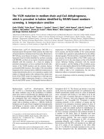
Fig. 1. Multicopy plasmids encoding a trunctated SIN3 allele reduce
the mean cell size of cln3D mutants. (A) Wild-type (CY979) and cln3D
cells (CY2028) were transformed with YEplac181 or a YEplac181
derivative (pCK1509) encoding a truncated SIN3 allele (2 lm SIN3DC).
This truncated Sin3 polypeptide lacks amino acids 811–1536. Trans-
formants were grown at 30 °C to log phase in selective medium lack-
ing leucine. The frequency distribution of cell size was measured with
a CASY
Ò
1 cell counter (Scha
¨
rfe Systems, Innovatis AG, Reutlingen,
Germany). (B) Budding index was determined by counting 250 cells
and the mean cell size of transformants was measured with a
CASY
Ò
1 cell counter. CLN2 RNA levels were determined by northern
blot analysis and quantified with the
BIOCAPT v. 12.3 software.
Sin3 involvement in cell size control O. Stephan and C. Koch
3812 FEBS Journal 276 (2009) 3810–3824 ª 2009 The Authors Journal compilation ª 2009 FEBS
limiting for the further events at Start [3,15]. There-
fore, inactivation of CLN3 results in a large cell
phenotype. To test whether Cln3 is involved in releas-
ing cells from a Sin3-dependent repression of G
1
-spe-
cific transcription, we analysed the consequences of
deleting sin3 in a cln3 mutant. The cell size of
sin3Dcln3D double mutants from a logarithmically
growing culture was compared with that of single
mutants and wild-type cells (Fig. 3A–C). The average
cell size of cln3D cells was reduced to approximately
the size of sin3D in the double mutant, suggesting that
sin3 is partly epistatic to cln3 (Fig. 3A–C). The critical
cell size for the initiation of budding and the activa-
tion of G
1
⁄ S-specific transcription was investigated in
small G
1
cells elutriated from an asynchronous
sin3Dcln3D double-mutant culture (Fig. 3D,E). Similar
to the sin3D population (see above), it proved difficult
to isolate small unbudded sin3Dcln3D cells, suggesting
partial deregulation of cell-cycle entry. As can be seen
from the FACS profile and cell size measurements
15 min after putting cells into fresh medium (Fig. 3F),
inactivation of SIN3 in the cln3D mutant led to a
reduction in cell size at birth. Moreover, although
cln3D mutants started budding at a nearly twice the
size of wild-type cells, sin3Dcln3D double-mutant cells
initiated budding at around the size of wild-type cells
(Fig. 3D) and activated G
1
⁄ S-specific transcription at
a much smaller size than cln3D mutants (Fig. 3E).
Inactivation of SIN3 in a cln3D deletion mutant also
caused the cells to replicate their DNA at a smaller
cell size (Fig. 3F). Hence, inactivation of SIN3
advances Start in a cln3D mutant. These data suggest
that Cln3 is also involved in releasing cells from a
Sin3 dependent repression.
A
Budded cells (%)
Mean volume (%)
B
C
Fig. 2. G
1
⁄ S-specific gene expression in cells synchronized by cen-
trifugal elutriation. (A) Wild-type (strain CY4196), sin3D (strain
CY5538), cln3D (strain CY5713) and sin3Dcln3D double-mutant cells
(strain CY5715) were grown to late log phase in YEPG
al
medium at
25 °C. Cell-cycle times were 240 min (Wt; CY4196), 340 min
(sin3D; CY5538) and 350 min (rpd3D; CY4061). Cells were har-
vested by centrifugation and loaded into an elutriation chamber.
Small unbudded cells were isolated by centrifugal elutriation and
transferred into fresh medium at 25 °C. To determine the budding
index, 250 cells were counted. Cell size during outgrowth was
measured with a CASY
Ò
1 cell counter. Displayed are mean vol-
umes. The peak of the cell size distribution at the time of budding
was at 26 fL for wild-type. For sin3D and rpd3D the peak values
were 19 and 20 fL respectively. (B) RNA levels of CLN2, PCL1 and
TMP1 in samples taken during outgrowth were determined by
northern blot analysis. The transcript levels were normalized in
comparison to the constitutively expressed CMD1 transcript. (C)
DNA content was analysed by flow cytometry. Log, logarithmic
growing cells used for elutriation.
O. Stephan and C. Koch Sin3 involvement in cell size control
FEBS Journal 276 (2009) 3810–3824 ª 2009 The Authors Journal compilation ª 2009 FEBS 3813
B
A
E F
C D
100%
90%
80%
70%
60%
50%
40%
30%
20%
10%
0%
20 30 40 50 60 70
Mean volume (fl)
Budded cells (%)
80 90 100 110 120 130
Fig. 3. Inactivation of SIN3 rescues the cell size of cln3D mutants. (A) Wild-type (strain BY4742 background derived from S288C), congenic
sin3D cells (strain CY5538), cln3D (strain CY5713) and sin3Dcln3D double-mutant cells (strain CY5715) were grown to mid-logarithmic phase
in YEPD medium and analysed by DIC microscopy. Budding index as percentage of cells with bud was determined by counting 250 cells
and was 48% for wild-type, 73% for sin3D, 43% for cln3D and 76% for the sin3Dcln3D double mutant. (B) Cell size analysis of wild-type,
sin3D, cln3D and sin3Dcln3D double mutants. The frequency distribution of cell diameters from asynchronous growing cultures in YEPD was
determined using a CASY
Ò
1 cell counter. (C) Cell size analysis of wild-type, sin3D, cln3D and sin3Dcln3D double mutants. Displayed are the
mean values from 10 independent experiments and their standard error. (D) Wild-type (strain CY4196), cln3D (strain CY5713) and sin3Dcln3D
double-mutant cells (strain CY5715) were grown to late log phase in YEPG
al
medium at 25 °C. Cell-cycle times were 240 min (Wt; CY4196),
290 min (cln3D; CY5713) and 300 min (sin3Dcln3D; CY5715). Small unbudded cells were isolated by centrifugal elutriation and transferred
into fresh medium at 25 °C. Cell size was measured with a CASY
Ò
1 cell counter. Displayed are mean volumes. The peak of the cell size dis-
tribution at the time of budding was at 26 fL for wild-type, 75 fL for cln3D and 22 fL for cln3Dsin3D. (E) RNA levels of CLN2, PCL1 and
TMP1 in samples taken during outgrowth were determined by northern blot analysis and the transcript levels were normalized to the consti-
tutively expressed CMD1 transcript. (F) DNA content of samples was analysed by flow cytometry. Log, logarithmic growing cells used for
elutriation.
Sin3 involvement in cell size control O. Stephan and C. Koch
3814 FEBS Journal 276 (2009) 3810–3824 ª 2009 The Authors Journal compilation ª 2009 FEBS
To elucidate if this repression by Sin3 is dependent
on SBF (Swi4 ⁄ Swi6), the cell size of sin3swi4 double
mutants was determined. Cell size analysis of log-phase
cultures from double mutants revealed that sin3D does
not reduce the cell size of swi4D mutants, and did not
advance transcriptional activation of G
1
⁄ S-specific
genes, but instead increased the average size of swi4D
mutants from 84 to 109 fL (data not shown).
Sin3 is recruited to SBF-specific promoters
The effect of Sin3 on cell size and S-phase entry sug-
gests a role for Sin3 in the timing of CLN1 and CLN2
transcription by SBF. If Sin3 were directly involved in
regulating SBF (Swi4 ⁄ Swi6)-dependent transcription in
late G
1
, it should be present at the relevant promoters.
Sin3 does not directly bind to DNA, but is known to
be recruited to specific promoter regions by other
DNA-binding proteins [40,48]. We therefore asked
whether Sin3 is targeted to promoters of G
1
⁄ S-specific
genes in a Swi4 ⁄ Swi6-dependent manner. For this,
SIN3 was replaced by an epitope-tagged version at the
SIN3 locus. Binding of epitope-tagged Sin3–myc to
G
1
⁄ S-specific promoters was assayed by ChIP experi-
ments. Coprecipitated promoter DNA fragments
encompassing the SBF-binding sites from the promoter
regions of CLN1 and CLN2 were amplified by multi-
plex PCR along with control fragments from their
coding regions and from a nontranscribed region on
chromosome V. As shown in Fig. 4, the promoter
elements of CLN1 and CLN2 were significantly
enriched compared with control fragments from the
coding region and the nontranscribed region of chro-
mosome V. Immunoprecipitations were performed in
triplicate to control for variations in the efficiency of
immunoprecipiation. As an additional control for the
specificity of Sin3 binding, cells not expressing the epi-
tope tag were analysed in parallel. Sin3–myc binding
to the promoter sequences of CLN1 and CLN2 was
strongly reduced in swi4 and swi6 null mutants
(Fig. 4), which further demonstrated the specificity of
the observed interaction. These results suggest that
Sin3 effects G
1
⁄ S transcription directly, and that Sin3
is recruited to G
1
cyclin promoters by SBF or by
factors associated with Swi4 or Swi6.
Sin3 recruitment is regulated in a
cell-cycle-dependent manner
To detect whether the recruitment of Sin3 and its asso-
ciated histone deacetylase Rpd3 to G
1
-specific promot-
ers is regulated during the cell cycle, we tested
promoter occupancy in cells that were arrested at
different stages of the cell cycle. We analysed cdc28-13
cell-cycle mutants arrested in G
1
at 37 °C, as well as
cells that were arrested in G
2
with the microtubule
depolymerizing drug nocodazole. Cdc28-13 mutants
arrest in late G
1
prior to the activation of G
1
⁄ S-specific
transcription. Strong Sin3 binding was detected in such
cells at the CLN1, CLN2 and PCL1 promoter regions
(Fig. 5A,D). The stronger signal in the arrested cultures
is most probably a simple reflection of cell-cycle-depen-
dent binding. Indeed, Sin3 was not associated with the
promoters during G
2
, as we could not significantly
coprecipitate CLN2 or PCL1 promoter elements with
A
B
Fig. 4. Sin3 binds to G
1
⁄ S-specific promoters. Chromatin immuno-
precipitation assays (ChIP) were performed in triplicate using yeast
strains carrying a myc tag at the SIN3 locus (CY5386, swi4D;
CY5387, wt; CY4849, wt; CY5469, swi6D). ChIP assays with
extracts of a strain lacking the myc tag were used as negative con-
trols (CY1617). Crude extracts were prepared from formaldehyde
cross-linked cells and chromatin precipitated with 9E11 antibodies.
Precipitates were analysed by multiplex PCR. Primers for an untran-
scribed region on chromosome V were used as nonspecific control.
These control primers were applied in the same PCR together with
either primers for the amplification of promoter elements or coding
regions of CLN1 and CLN2. PCR results with primers for the coding
region of CLN2 are displayed in comparison to the PCR results of
promoters or the control. Products were analysed on a 2% agarose
gel. WCE, whole cell extract; No Tag, analysis of strains lacking
epitope tagged protein.
O. Stephan and C. Koch Sin3 involvement in cell size control
FEBS Journal 276 (2009) 3810–3824 ª 2009 The Authors Journal compilation ª 2009 FEBS 3815
Sin3–myc from cells arrested with nocodazole
(Fig. 5C,D). To provide further evidence for the spe-
cific binding of Sin3, we analysed the recruitment of
Rpd3, the catalytic component of the Sin3 ⁄ Rpd3 his-
tone deacetylase complex, to G
1
cyclin promoters in G
1
(Fig. 5B). Cdc28-13 mutants expressing an epitope-
tagged RPD3–HA6 were synchronized in late G
1
by
shifting log-phase cultures to 37 °C for 3 h until all
cells were arrested as large unbudded cells. In ChIP
assays with extracts prepared from arrested Rpd3–HA6
cells we observed recruitment of Rpd3–HA6 to SBF-
dependent promoters, although the signal was weaker
than the signal for Sin3–myc (Fig. 5B). The binding of
Sin3 and Rpd3 to promoters of SBF-regulated genes in
G
1
-arrested cells correlates with the transcriptional
repression of CLN1, CLN2 and PCL1 in G
1
.
To analyse if the release of Sin3 from SBF-regulated
genes coincides with transcriptional activation of G
1
cyclins, we performed an arrest–release experiment.
Cdc28-13 mutants were shifted to 37 °C until they were
arrested as unbudded cells in G
1
. The cells were subse-
quently released from cell-cycle arrest by shifting the
culture to 25 °C. RNA levels and Sin3 binding were
analysed from samples taken every 10 min. For the
arrested culture, ChIP analysis demonstrated strong
binding of Sin3–myc to the CLN1 and CLN2 promoter
(Fig. 6A; 0 min). When cells were released from the
cell-cycle block, they synchronously entered the cell
cycle (Fig. 6C). A peak of G
1
⁄ S-specific transcription
was observed between 10 and 20 min after release.
Shortly thereafter, cells entered the S phase (FACS
profile in Fig. 6D) and started budding (Fig. 6C).
A CB
30
25
20
15
10
5
0
log G2 log G2
D
30
25
20
15
10
5
0
log G1 log G1
Fig. 5. Sin3 and Rpd3 are recruited to CLN1, CLN2 and PCL1 promoters in G
1
. Chromatin immunoprecipitation (ChIP) assays were per-
formed in triplicate using cdc28-13 yeast strains (CY239, CY5327 and CY5555). (A) Cells expressing Sin3–myc (CY5327) were grown in
YEPD at 25 °C to a titre of 1 · 10
7
mL
)1
. The culture was subsequently split in two and either arrested in G
1
by shifting to 37 °C for
165 min or kept at 25 °C for the same time. Cells were cross-linked with 1% formaldehyde for 20 min at room temperature. Cell extracts
were subjected to immunoprecipitation with anti-myc (9E11)-coupled Dynabeads. The precipitates were analysed by PCR with primers for
the amplification of promoter elements of CLN1, CLN2 and PCL1 and the coding regions of CLN2. Precipitation of DNA fragments from an
untranscribed region on chromosome V was analysed as a control. PCRs were analysed on 2% gels. ChIP, chromatin immunoprecipitations;
WCE, whole cell extract; No Tag, analysis of strains lacking epitope tagged protein. (B) cdc28-13 cells expressing Rpd3–HA6 (CY5555) were
treated and ChIPs performed as described in (A). (C) Binding of Sin3–myc (CY4849, wt) to the promoters of CLN2 and PCL2 in nocodazole
arrested wild-type cells was analysed by ChIP. Extracts were prepared from cells that were grown in YEPD to an D
600
of 0.4 and subse-
quently arrested with nocodazole at 25 °C for 2.5 h. (D) Precipitated DNA from the ChIP assays shown in (A) and (C) was analysed by real-
time PCR on a Mx3000P thermocycler using the brilliant II QPCR kit, as described by the manufacturer (Stratagene, Heidelberg, Germany).
Values from the untagged control samples were substracted from the signal of the tagged samples. Shown are mean values derived from
three independent experiments with standard deviations.
Sin3 involvement in cell size control O. Stephan and C. Koch
3816 FEBS Journal 276 (2009) 3810–3824 ª 2009 The Authors Journal compilation ª 2009 FEBS
The ChIP signal began to fade 10 min after the cells
were released (Fig. 6A,B). The decrease in promoter
occupancy by Sin3 correlated best with the timing of
transcriptional activation (Fig. 6B). The timing of Sin3
binding is therefore consistent with a role for Sin3 in
repression of CLN transcription in the G
1
phase.
A
B
C
D
E
F
G
Fig. 6. Dissociation of Sin3 from SBF-dependent promoters correlates with activation of G
1
⁄ S-specific transcription. A cdc28-13 mutant
expressing Sin3–myc (CY5327) was grown in YEPD to D
600
= 0.4 and arrested at 37 °C for 180 min. Cells were released from the G
1
arrest
by shifting the culture to 25 °C. Samples were taken at the indicated time points (A–D). (A) Sin3–myc binding was analysed by ChIP as in
Fig. 5. PCRs were analysed on 2% agarose gels and quantified with the
BIOCAPT v. 12.3 software. (B) Northern blot analysis of RNA levels of
CLN2 and PCL1 were analysed in parallel with CMD1 as loading control. (C) Quantified data from ChIP, CLN2 RNA levels and budding index.
The transcript levels of the CLN2 RNA were normalized using the constitutively expressed CMD1 transcript. ChIP signals for the CLN2 pro-
moter region were normalized to the signals of the chromosome V UTR control [(spec. ChIP ⁄ control ChIP) tagged – (spec. ChIP ⁄ control
ChIP) untagged]. (D) DNA content analysis by flow cytometry. (E,F) ChIP of CLN2 and PCL1 promoter elements from small elutriated G
1
cells expressing Sin3–myc. Cells were grown to D
600
= 2.3 in YEPG
al
medium and small G
1
cells were isolated by centrifugal elutriation from
cultures. Unbudded cells were inoculated in fresh medium and incubated at 25 °C. Binding of Sin3–myc to promoters was analysed at
different time points by ChIP. (F) PCL1 and CMD1 RNA levels determined by hybridization of northern blots with radioactive labelled DNA
fragments. (G) DNA content of cells was analysed by flow cytometry at the indicated time points.
O. Stephan and C. Koch Sin3 involvement in cell size control
FEBS Journal 276 (2009) 3810–3824 ª 2009 The Authors Journal compilation ª 2009 FEBS 3817
Because of their abnormally large size, G
1
-arrested
cell-cycle mutants may not accurately reproduce the
situation found in small wild-type daughter cells in the
early G
1
phase. We therefore analysed promoter occu-
pancy of Sin3–myc in elutriated wild-type cells. Small
G
1
cells were isolated by centrifugal elutriation and
allowed to progress through G
1
. The presence of Sin3–
myc at the CLN2 and PCL1 promoter was compared
with cell-cycle progression. Because many cells were
needed for ChIP assays it was not possible to analyse
more than three time points. At each time point, sam-
ples from the culture were analysed by ChIP assay and
the RNA levels of G
1
⁄ S-specific genes and the DNA
content of cells were determined (Fig. 6E–G). The data
confirmed that SBF-specific promoters are occupied by
Sin3 in the G
1
phase. At 160 min, most cells in the cul-
ture had left G
1
and exhibited no Sin3 binding to the
promoter (Fig. 6E). Analysis of G
1
cyclin expression
showed that binding of Sin3 to the SBF-dependent
promoters correlated with repression in G
1
, whereas
disappearance of Sin3 from the promoter elements
coincided with induction of Start-specific transcription
(Fig. 6). To elucidate whether Sin3 leaves the promoter
together with Rpd3, we performed an arrest-release
experiment with cdc28-13 cells expressing Rpd3–HA.
As shown in Fig. 7, the binding of Rpd3 is very
A
D
B
C
0 min 10 min 20 min 30 min 40 min 50 min 60 min 70 min 80 min
1.8
100%
90%
80%
70%
60%
50%
40%
30%
20%
10%
0%
1.6
1.4
1.2
0.8
0.6
0.4
0.2
0
807060504030
Time (min)
Fold enrichment
Realtive RNA CLN2/CMD1 (%)
and budding (%)
20100
1
Fig. 7. Dissociation of Rpd3 from SBF-dependent promoters correlates with activation of G
1
⁄ S-specific transcription. A cdc28-13 mutant
expressing Rpd3–myc (CY5555) was grown in YEPD to a titre of 1 · 10
7
cellsÆmL
)1
and arrested at 37 °C for 180 min. Shifting the culture
to 25 °C released the cells from the G
1
arrest. Samples were taken at the indicated time points (A–D). (A) Rpd3–myc binding was analysed
by ChIP as in Fig. 5. PCRs were analysed on 2% agarose gels and quantified with the
BIOCAPT v. 12.3 software. (B) RNA levels of CLN2 and
PCL1 were analysed by northern blot in parallel with CMD1 as loading control. (C) Quantified data from ChIP, CLN2 RNA levels and budding
index as described in the legend to Fig. 6. The transcript levels of the CLN2 RNA were normalized using the constitutively expressed CMD1
transcript. ChIP signals for the CLN2 promoter region were quantified by normalising band intensities of CLN2 promoter fragments to the
signals of the chromosome V UTR control. (D) DNA content was analysed by flow cytometry at the indicated time points.
Sin3 involvement in cell size control O. Stephan and C. Koch
3818 FEBS Journal 276 (2009) 3810–3824 ª 2009 The Authors Journal compilation ª 2009 FEBS
similar to the kinetics of Sin3 binding (Fig. 6) although
the signal intensities for Rpd3 are generally weaker.
We therefore conclude that Sin3 acts together with
Rpd3 at SBF-dependent promoters (Fig. 7).
Discussion
In this study, we provide evidence that SIN3 is
involved in the correct timing of G
1
cyclin expression
in Saccharomyces cerevisiae at the G
1
–S phase transi-
tion. We have found that inactivation of SIN3 leads to
an advanced induction of Start-specific transcription in
G
1
daughter cells and that budding is initiated at a
smaller cell size. Consistent with a direct role for Sin3
in repressing gene expression prior to Start, we find
that Sin3 is present at the promoters of SBF-regulated
genes in G
1
, but leaves the promoter around the time
cells enter the S phase. Furthermore, inactivation of
Sin3 suppresses the phenotype of cln3 mutants, allow-
ing them to activate G
1
⁄ S-specific transcription at a
smaller cell size (Fig. 3). Such a phenotype would be
expected if Cln3 with its associated Cdc28 kinase were
involved in the inactivation or repression of
Sin3 ⁄ Rpd3-dependent histone deacetylation at the
CLN1,2 promoters. These data raise several questions
concerning the regulation of G
1
cyclin transcription. In
particular, whether CLN3 acts directly on Sin3 ⁄ Rpd3
and how Sin3 is recruited to SBF-regulated genes like
CLN2 in the G
1
phase.
How is Sin3 recruited to SBF-regulated
promoters?
Sin3 does not bind DNA directly, but associates with
transcriptional regulators to bring the Rpd3 histone
deacetylase to specific sites in chromatin [40,49]. There
are different DNA-binding proteins thought to bind to
Sin3. Besides the well-characterized interaction with
Ume6, these include Ash1, Mcm1 and Ssn6 [35,50,51].
At the HO promoter, Sin3 is thought to be recruited
in part by Ash1 [35,51]. Veis et al. reported cell-cycle-
dependent binding of Sin3 to the G
2
⁄ M-specific CLB2
promoter [50]. Their data further showed that the
recruitment of Sin3 is dependent upon an interaction
with Fkh2 and Mcm1. The removal of Sin3 and the
deacetylase complex does not require B-type cyclins
but Cdc28 ⁄ Cln activity [50]. Similar to the recruitment
of Sin3 to G
2
⁄ M-specific promoters by the regulatory
factors Mcm1 and Fkh2, we propose that Sin3 is
recruited to G
1
⁄ S promoters by SBF. The DNA-bind-
ing protein Ume6 was shown to be responsible for
Sin3 ⁄ Rpd3 recruitment at many other sites, for exam-
ple, at SPO13, INO1, IME2 [40,43,52]. We found no
Ume6 consensus sites [48] in the promoter regions of
CLN1 and CLN2. Any one of the proteins present at
the CLN2 promoter in early G
1
could, in principle, be
responsible for recruiting Sin3 to the promoter. These
include Swi4, Swi6, Whi5 and Stb1 [18,19,21,33,35].
Although recruitment of Sin3 to the CLN2 promoter
strongly depends on Swi4 and Swi6 (Fig 4), we found
no significant effects of whi5 or stb1 mutants on the
binding of Sin3 to the CLN2 promoter (data not
shown). In addition, deleting WHI5 in a sin3 mutant
did not reduce the cell size to the level of whi5D single
mutants (data not shown). This makes it unlikely that
Sin3 is recruited to the promoter via Whi5. Because
the absence of Stb1 was observed to increase cell size
of a cln3D mutant [32], it is not likely to mediate Sin3-
dependent repression, although it could be important
for releasing from Sin3-dependent repression later in
the cell cycle. However, the timing of SBF binding
[18,19], which arrives at the promoter as cells exit
mitosis, would be consistent with a direct role as a
Sin3 ⁄ Rpd3 recruiting factor. Earlier ChIP results
showed that the histone deacetylase Rpd3 is associated
with the promoters of cell-cycle genes regulated by
SBF, MBF, Fkh1, Fkh2, Mcm1 and Ndd1, and
showed that SBF affected Rpd3 binding to CDC20
and PCL1, suggesting that Rpd3 can be recruited by
several different transcription factors [39]. This is con-
sistent with our observation that both Sin3 and Rpd3
are recruited to G
1
⁄ S-specific promoters in a cell-cycle-
dependent manner. A situation in which transcrip-
tional activators also directly recruit corepressors is in
fact quite common, for example, in the case of E2F
transcription factors in metazoans [53].
How is Sin3 removed from the promoter in the
S phase?
The observation that deleting SIN3 partly suppresses
the size phenotype of cln3 mutants suggests a possible
role for Cln3 in the inactivation or subsequent removal
of Sin3 ⁄ Rpd3 complexes from the promoter. The only
well-characterized, and presumably critical substrate
for Cln3 ⁄ CDK1 is the repressor Whi5 [20,21].
Removal of Sin3 at the beginning of the S phase is
probably not a consequence of Whi5 inactivation
caused by phosphorylation by Cln3 ⁄ CDK1 [20,21],
because there is no evidence for a direct interaction
between Whi5 and Sin3 or Rpd3.
Alternatively, Sin3 may be a direct target for Cln3
kinase. Sin3 is a phosphoprotein [54] and was found to
coprecipitate with Cln2 in a proteomics study of yeast
CDKs [54]. The timing of Sin3s removal from the
promoter (Fig. 6) would also be compatible with a
O. Stephan and C. Koch Sin3 involvement in cell size control
FEBS Journal 276 (2009) 3810–3824 ª 2009 The Authors Journal compilation ª 2009 FEBS 3819
scenario in which the downstream Cln1 ⁄ 2–CDKs
rather than Cln3 ⁄ CDK are responsible for inactivating
Sin3. Such a mechanism could contribute to a positive
feedback loop of CLN activation [55] assisting in mak-
ing S-phase entry irreversible. A role for Clns in the
removal of Sin3 from promoters was suggested by
Veis et al. in the case of CLB2 [50].
Given that Sin3, together with the Rpd3 histone
deacetylase, is involved in modifying chromatin at
many sites not concerned with cell-cycle control, we
consider it more likely that Sin3 ⁄ Rpd3-dependent
chromatin changes at the CLN2 promoter are regu-
lated by reversible recruitment of Sin3 and or Rpd3,
rather than by regulating Sin3 ⁄ Rpd3 directly.
A good candidate for a factor regulating Sin3 ⁄ Rpd3
binding is Stb1, because it binds to both Swi6 and Sin3
[20,21,32,34]. In fact, Stb1 was originally identified as
an interacting protein of Sin3 in two-hybrid assays
(Stb1 for Sin three binding) [34]. STB1 transcription
was shown to be cell-cycle regulated and peaks in late
G
1
phase [32]. Earlier studies showed that Stb1 binds
only to synthetic MBF promoters [20], but a recent
study provided evidence for in vivo binding to SBF and
MBF promoters in G
1
via an interaction with Swi6
[33]. ChIP assays provided evidence that phosphoryla-
tion of Stb1 coincides with its dissociation from pro-
moters at G
1
⁄ S transition [33]. This study also found
Stb1 to be associated with G
1
⁄ S-specific promoters
until CLN transcription is inactivated [33]. Phosphory-
lation of Stb1 inhibits interaction between Swi6 and
Stb1 [20]. Our finding of cell-cycle-specific promoter
binding by Sin3 is in agreement with the proposal of de
Bruin et al. [33] for a combined role of Stb1 and Sin3
in regulating G
1
⁄ S transcription. We were able to show
that Sin3 binds to promoter sequences prior to tran-
scriptional activation and leaves the promoter around
the time of transcriptional activation. The observation
that Stb1 and Sin3 bind to promoters in G
1
and leave
the promoters at the time of transcriptional activation
suggests that Stb1 and Sin3 may exert effects on
G
1
⁄ S-specific transcription in a concerted manner.
A possible model for the regulation of SBF by
Sin3 ⁄ Rpd3 may be that Sin3 is directly recruited by
SBF in early G
1
and that changes in Stb1 remove Sin3
from the promoter. Data concerning the timing of
Sin3 removal from the promoter are consistent with a
function for both Cln3 and the downstream cyclins
Cln1 and Cln2 in removing Sin3 from the promoter.
The timing is, however, not compatible with a model
in which removal of Sin3 is a simple consequence of
SBF removal from the promoter by Clb-kinase activ-
ity, because Swi6 remains associated with the CLN2
promoter for much longer [19]. Cell-cycle-dependent
binding of Sin3 has also been observed at the CLB2
locus [50]. At the G
2
-specific CLB2 gene we found that
Sin3 is recruited by Fkh2 in G
1
and lost from the pro-
moter after activation of Cln1,2 associated kinases
[50]. Although the timing is clearly different from the
situation at the SBF-regulated genes analysed here, the
inactivation by Cln-kinases may occur by a similar
mechanism.
How important is Sin3 for the regulation of
G
1
⁄
S-specific transcription?
Sin3 mutants are not obviously smaller than wild-type
cells, when the average size in the population is
analysed, although we found a larger proportion of
post-S-phase cells. This, and the fact that Sin3 mutants
are rather pleiotropic, may explain why the deregula-
tion of G
1
⁄ S-specific genes becomes evident only in iso-
lated G
1
cells or in cln3D mutants. In addition, the
effect of ectopically expressing the dominant-negative
allele of SIN3 on cell size was most obvious in cln3
mutants. The importance of Sin3 for regulating CLN2
expression is therefore not so obvious. In the absence
of Cln3, the timing of Start execution becomes quite
variable, whereas once activated, all Start-related
events occur in a coherent fashion [55,56]. It has been
argued that cln3 mutants are particularly dependent on
a positive feedback mechanism for G
1
⁄ S transcription,
i.e. Cln2 and Cln1 have to accumulate to a certain level
before firing the positive feedback loop [55,56]. As a
consequence, every mutation that partly deregulates
CLN2 expression will potentially lower the threshold at
which such a positive loop will fire. This may be one of
the reasons why in cln3 mutants cell size is particularly
sensitive to the inactivation of SIN3 and may make
SIN3 apparently more important for repressing tran-
scription in daughter cells with little Cln3. Remarkably,
Aparicio et al. [57] reported similar effects of Sin3 in
the regulation of S-phase timing. They showed that the
S phase is advanced in the absence of the Sin3 ⁄ Rpd3
histone deacetylase complex. In summary, we have
identified an additional level of control at the G
1
-to
S-phase transition that contributes to the astonishing
precision of transcriptional timing observed in cell cycle
regulated transcription in late G
1
.
Materials and methods
Strains and DNA
Strains used in this study were derived from strains W303,
BY4741 or BY4742 (Table 1). Gene deletions were created by
integrational transformation of PCR cassettes, as described
Sin3 involvement in cell size control O. Stephan and C. Koch
3820 FEBS Journal 276 (2009) 3810–3824 ª 2009 The Authors Journal compilation ª 2009 FEBS
previously [58]. Double mutants were created by mating.
After incubation on sporulation media plates for 2–10 days
at 25 °C, tetrads were dissected with a micromanipulator
(Singer Instruments, Roadwater, UK) and distributed on
YEPD plates. After 4 days at 25 °C the phenotypes were
analysed by replica plating. Genotypes of meiotic segregants
were confirmed by PCR. Epitope tagging of yeast genes at
the C-terminus was performed using a PCR-based strategy to
introduce epitope tags to the chromosomal loci [59]. Deletion
mutant strains used for cell size measurements were obtained
from BIOCAT (BY4741, BY4742, CY5713 and CY5538) and
double mutants (CY5715) were created by mating (Table 1).
The genomic library was a gift from R. Jansen (LMU
Munich, Germany) and was generated by inserting genomic
Sau3A fragments into the multicopy YEplac181 vector.
Plasmid pCK1509 contains a fragment that comprises 488 bp
of the 5¢-UTR and 2436 bp of the SIN3 coding region.
Growth conditions and cell-cycle arrests
Yeast cells were cultivated in YEP-based media with 2%
glucose (YEPD) or 2% galactose (YEPG
al
). Cell-cycle
arrests of temperature-sensitive mutants (cdc28-13) were
performed by growing the cells to a titre of 10
7
cellsÆmL
)1
at 25 °C (YEPD) in a water bath and then shifting the
culture to 37 °C for 165 min. For centrifugal elutriation,
cells were grown to D
600
= 2.0 in YEPG
al
. The elutriation
chamber was loaded with a total of 8000 D
600
cells. Frac-
tions of small cells were collected, pooled and cultivated at
25 °C in fresh media. Samples from the culture were
taken at specific time points and cell size and cell-cycle
progression were monitored. Budding index was determined
by counting 250 cells. Flow cytometry was used to
observe cell-cycle distribution as described previously [19].
Cell size measurements
Cell number and average cell size were analysed by using a
CASY
Ò
1 cell counter model TT from Scha
¨
rfe Systems
(Innovatis AG, Reutlingen, Germany). To determine cell
number and size distribution of yeast cultures, the cell sus-
pensions were diluted in CASY-ton
Ò
isotonic buffer and
sonicated for 30 s before the measurement.
ChIP assays
ChIPs were carried out with modifications as described previ-
ously [24,60]. Crude extracts were prepared from cultures
(55 mL, 2.5 · 10
7
cellsÆmL
)1
) treated with 1% formaldehyde
for 20 min at room temperature before harvesting. After
addition of 135 mm glycine and incubation for 5 min at
room temperature, cells were harvested and washed four
times with 2 mL NaCl ⁄ Tris buffer (20 mm Tris ⁄ HCl pH 7.5,
150 mm NaCl) to remove residual formaldehyde. Cells
were resuspended in 600 lL lysis buffer (50 mm Hepes
KOH pH 7.5, 140 mm NaCl, 1 mm EDTA, 1% Triton
X-100, 0.1% deoxycholic acid) and cell breakage was car-
ried out by addition of glass beads, and usage of a IKA
Ò
Vibrax VXR basic (25 min 2500 rpm, 4 °C). Extracts were
sonicated five times for 30 s using a Bandelin Sonoplus
HD2070 ⁄ SH70G and the debris was removed by
centrifugation (16 000 g 5 min, 4 °C). The supernatant was
Table 1. Yeast strains.
Strain Genotype Source
W303 MATa, ade2-1, trp1-1, can1-100, leu2-3,112, his3-11,15, ura3, GAL, psi+ K. Nasmyth
(Oxford, UK)
CY5450 MATa; his3D1, leu2D0, met15D0, ura3D0
a
By4741 [58]
CY4196 MATalpha; his3D1, leu2D0, lys2D0, ura3D0
a
By4742 [58]
CY239 MAT alpha, cdc28-13 (congenic to W303) K. Nasmyth
CY979 MATa, W303
CY1617 MATa, pep4 :: URA3 (congenic to W303) [61]
CY2028 MATa, cln3::URA3 (congenic to W303) [61]
CY4256 MATa, [YEplac181] CY979
CY4265 MATa, cln3::URA3, [YEplac181] This study
CY4266 MATa, cln3::URA3 [pCK1509] CY2028
CY4849 MATa, trp1-D63, sin3::SIN3-myc9(KLTRP1), pep4::URA3 (congenic to W303) This study
CY5327 MAT alpha, trp1-D63, cdc28-13, sin3::SIN3-myc9(KLTRP1), pep4::URA3 (congenic to W303) This study
CY5386 MATalpha, trp1-D63, swi4::LEU2, sin3::SIN3-myc9(KLTRP1), pep4::URA3 (congenic to W303) This study
CY5387 MATa, trp1-D63, sin3::SIN3-myc9(KLTRP1), pep4::URA3 (congenic to W303) This study
CY5469 MATalpha, trp1-D63, swi6::TRP1, sin3::SIN3-myc9(KLTRP1), pep4::URA3 (congenic to W303) This study
CY5538 MATa, congenic to By4741, except for sin3::KANMX
a
[58]
CY5555 MATa, trp1-D63, cdc28-13, rpd3::RPD3–HA6(KLTRP1), pep4::URA3 (congenic to W303) This study
CY5713 MATalpha, congenic to By4741, except for cln3::KANMX
a
[58]
CY5715 MATalpha, congenic to By4741, except for sin3::KANMX, cln3::KANMX
b
This study
a
Strains were obtained from BIOCAT (Heidelberg, Germany).
b
Strain was created by crossing strain CY5538 with CY5713.
O. Stephan and C. Koch Sin3 involvement in cell size control
FEBS Journal 276 (2009) 3810–3824 ª 2009 The Authors Journal compilation ª 2009 FEBS 3821
applied to 50 lL Dynabeads
Ò
Pan Mouse IgG (Dynal
Ò
Invitrogen (Karlsruhe, Germany); 400 000 beadsÆlL
)1
) that
were loaded with epitope tag-specific antibodies (0.05 lgof
antibodies per lL of bead suspension) and incubated for
3 h at 7 °C on a rotator. Antibodies used for ChIP assays
were anti-myc 9E11 (Dianova, Hamburg, Germany) and
anti-HA 12CA5 (lab preparation). Thereafter, the beads
were washed twice with 1 mL lysis buffer, high salt buffer
(50 mm Hepes KOH pH 7.5, 500 mm NaCl, 1 mm EDTA,
1% Triton X-100, 0.1% deoxycholic acid), washing buffer
(10 mm Tris ⁄ HCl pH 8, 250 mm LiCl, 1 mm EDTA, 0.5%
NP40, 0.5% deoxycholic acid) and TE buffer. Precipitates
were eluted in 50 lL elution buffer (50 mm Tris ⁄ HCl
pH 8, 10 mm EDTA, 1% SDS) at 65 °C for 10 min.
Eluted proteins were analysed by SDS ⁄ PAGE and western
blotting. We added 120 lL 1% SDS ⁄ TE buffer to 30 lL
of the supernatant and incubated for 16 h at 65 °C. The
eluates were treated with 0.5 lgÆlL
)1
proteinase K for 2 h
at 37 °C. Thereafter, coprecipitated DNA was purified by
phenol extraction. DNA fragments of promoters and cod-
ing regions were amplified by multiplex PCR and analysed
on 2% agarose gels. As a control, we coamplified an un-
transcribed region of chromosome V (10562–10699)
together with either the promoter or the coding region of
CLN1, CLN2 and PCL1 in the same PCR. The primers
for amplification of the control region on chromosome V
were CK2229 (CAGTTTAACCCGAAGTTCTG) and
CK2230 (AACAACGCAGCTGCTTTAAC). Primers used
for the amplification of fragments from promoter frag-
ments were:
CLN2, CK2148 (ATCTTTTTCGTATCCTCCGC) and
CK2149 ( AAAGGGCCAACAGTTGTTTC); CLN1, CK2158
(TAGGGTAGCGTGCCACAAAA) and CK2159 (CGTCT
CTTGCAGGCTGAACA); PCL1, CK2364 (GCTAACAA
CTGAGAATGCGA) and CK2366 (ACACAAGAGTTAA
GGACAAG).
The primers used for the amplification of fragments from
the coding regions were:
CLN2, CK1724 (ATAGTGATGCCACTGTAGAC) and
CK1725 (CATGATGGGGTTGATATGGT); CLN1,
CK2254 (TAGTTCACCGCAAAGTACTG) and CK2255
(TATTGTAGAGGCCAGTTGCA); PCL1, CK2348 (CCA
TCCATCGCATTTTCTTG) and CK2349 (CTGTGTTG
TTCGCTATGTTG).
Northern analysis
Yeast cells were harvested and chilled on ice before they
were washed with cold TE buffer. Cell pellets were frozen in
liquid nitrogen and stored at )80 °C. Precipitated cells were
resuspended in RNA buffer 1 (10 mm Tris ⁄ HCl pH 7.5,
300 mm NaCl, 1 mm EDTA, 0.2% SDS) and vortexed
vigorously with phenol ⁄ chloroform ⁄ isoamylalcohol and
glass beads for 15 min. After centrifugation the aqueous
phases were mixed with ethanol (1 : 3 v ⁄ v) and incubated
for 20 min at )20 °C. Tubes were centrifuged for 15 min
at 16 000 g . Pellets were resuspended in RNA buffer 2
(10 mm Tris ⁄ HCl pH 7.5, 1 mm EDTA, 0.2% SDS) and
incubated for 5 min at 65 °C. RNA content was measured
at 260 nm with a Smartspec
Ô
3000 from BioRad (Munich,
Germany). RNA (20 lg per sample) was separated on
1.3% agarose gels containing 1.3% formaldehyde. RNA
was transferred to Gene Screen membranes. Hybridizations
with
32
P-labelled DNA probes were performed at 65 °C for
16 h in Church buffer (0.5 m NaCl ⁄ P
i
pH 7.2, 7% SDS,
10 mm EDTA, 1% BSA). Filters were washed twice for
5 min in 2· NaCl ⁄ Cit ⁄ 0.1% SDS and twice for 10 min in
1· NaCl ⁄ Cit ⁄ 0.1% SDS at 65 °C. CMD1 RNA levels were
determined as internal loading control.
Acknowledgements
We gratefully thank Marlis Dahl, Rosi So
¨
llner, Alex-
ander Schwahn, Uwe Sonnewald and Martin Korn for
their support and helpful discussions. We thank Gus-
tav Ammerer and Kim Nasmyth for strains. We thank
Michael Schwenkert and Christian Kellner for their
help with the FACS analysis.
References
1 Cross FR (1995) Starting the cell cycle: what’s the
point? Curr Opin Cell Biol 7, 790–797.
2 Nash R, Tokiwa G, Anand S, Erickson K & Futcher
AB (1988) The WHI1+ gene of Saccharomyces cerevisi-
ae tethers cell division to cell size and is a cyclin homo-
log. EMBO J 7, 4335–4346.
3 Tyers M, Tokiwa G & Futcher B (1993) Comparison of
the Saccharomyces cerevisiae G1 cyclins: Cln3 may be
an upstream activator of Cln1, Cln2 and other cyclins.
EMBO J 12, 1955–1968.
4 Bahler J (2005) Cell-cycle control of gene expression in
budding and fission yeast. Annu Rev Genet 39, 69–94.
5 Bloom J & Cross FR (2007) Multiple levels of cyclin
specificity in cell-cycle control. Nat Rev Mol Cell Biol 8,
149–160.
6 Mendenhall MD & Hodge AE (1998) Regulation of
Cdc28 cyclin-dependent protein kinase activity during
the cell cycle of the yeast Saccharomyces cerevisiae.
Microbiol Mol Biol Rev 62, 1191–1243.
7 Andrews BJ & Herskowitz I (1989) The yeast SWI4
protein contains a motif present in developmental regu-
lators and is part of a complex involved in cell-cycle-
dependent transcription. Nature 342, 830–833.
8 Breeden L & Nasmyth K (1987) Similarity between cell-
cycle genes of budding yeast and fission yeast and the
Notch gene of Drosophila. Nature 329, 651–654.
9 Koch C, Moll T, Neuberg M, Ahorn H & Nasmyth K
(1993) A role for the transcription factors Mbp1 and
Sin3 involvement in cell size control O. Stephan and C. Koch
3822 FEBS Journal 276 (2009) 3810–3824 ª 2009 The Authors Journal compilation ª 2009 FEBS
Swi4 in progression from G1 to S phase. Science 261,
1551–1557.
10 Iyer VR, Horak CE, Scafe CS, Botstein D, Snyder M &
Brown PO (2001) Genomic binding sites of the yeast
cell-cycle transcription factors SBF and MBF. Nature
409, 533–538.
11 Lowndes NF & Johnston LH (1992) Parallel pathways
of cell cycle-regulated gene expression. Trends Genet 8,
79–81.
12 Pizzagalli A, Valsasnini P, Plevani P & Lucchini G (1988)
DNA polymerase I gene of Saccharomyces cerevisiae:
nucleotide sequence, mapping of a temperature-sensitive
mutation, and protein homology with other DNA
polymerases. Proc Natl Acad Sci USA 85, 3772–3776.
13 Nasmyth K & Dirick L (1991) The role of SWI4 and
SWI6 in the activity of G1 cyclins in yeast. Cell 66,
995–1013.
14 Ogas J, Andrews BJ & Herskowitz I (1991)
Transcriptional activation of CLN1, CLN2, and a
putative new G1 cyclin (HCS26) by SWI4, a positive
regulator of G1-specific transcription. Cell 66, 1015–
1026.
15 Dirick L, Bohm T & Nasmyth K (1995) Roles and regu-
lation of Cln–Cdc28 kinases at the start of the cell cycle
of Saccharomyces cerevisiae. EMBO J 14, 4803–4813.
16 Stern M, Jensen R & Herskowitz I (1984) Five SWI
genes are required for expression of the HO gene in
yeast. J Mol Biol 178, 853–868.
17 Amon A, Tyers M, Futcher B & Nasmyth K (1993)
Mechanisms that help the yeast cell cycle clock tick: G2
cyclins transcriptionally activate G2 cyclins and repress
G1cyclins. Cell 74, 993–1007.
18 Harrington LA & Andrews BJ (1996) Binding to the
yeast SwI4,6-dependent cell cycle box, CACGAAA,
is cell cycle regulated in vivo . Nucleic Acids Res 24,
558–565.
19 Koch C, Schleiffer A, Ammerer G & Nasmyth K
(1996) Switching transcription on and off during the
yeast cell cycle: Cln ⁄ Cdc28 kinases activate bound tran-
scription factor SBF (Swi4 ⁄ Swi6) at start, whereas
Clb ⁄ Cdc28 kinases displace it from the promoter in G2.
Genes Dev 10, 129–141.
20 Costanzo M, Nishikawa JL, Tang X, Millman JS, Schub
O, Breitkreuz K, Dewar D, Rupes I, Andrews B & Tyers
M (2004) CDK activity antagonizes Whi5, an inhibitor of
G1 ⁄ S transcription in yeast. Cell 117, 899–913.
21 de Bruin RA, McDonald WH, Kalashnikova TI, Yates
J III & Wittenberg C (2004) Cln3 activates G1-specific
transcription via phosphorylation of the SBF bound
repressor Whi5. Cell 117, 887–898.
22 Cooper K (2006) Rb, whi it’s not just for metazoans
anymore. Oncogene 25, 5228–5232.
23 Polymenis M & Schmidt EV (1997) Coupling of cell
division to cell growth by translational control of the
G1 cyclin CLN3 in yeast. Genes Dev 11, 2522–2531.
24 Cosma MP, Panizza S & Nasmyth K (2001) Cdk1
triggers association of RNA polymerase to cell cycle
promoters only after recruitment of the mediator by
SBF. Mol Cell 7, 1213–1220.
25 Cosma MP, Tanaka T & Nasmyth K (1999) Ordered
recruitment of transcription and chromatin remodeling
factors to a cell cycle- and developmentally regulated
promoter. Cell 97, 299–311.
26 Wijnen H & Futcher B (1999) Genetic analysis of the
shared role of CLN3 and BCK2 at the G(1)-S transition
in Saccharomyces cerevisiae. Genetics 153, 1131–1143.
27 Manukyan A, Zhang J, Thippeswamy U, Yang J,
Zavala N, Mudannayake MP, Asmussen M, Schneider
C & Schneider BL (2008) Ccr4 alters cell size in yeast
by modulating the timing of CLN1 and CLN2
expression. Genetics 179, 345–357.
28 Mai B & Breeden L (2000) CLN1 and its repression by
Xbp1 are important for efficient sporulation in budding
yeast. Mol Cell Biol 20, 478–487.
29 Ashe M, de Bruin RA, Kalashnikova T, McDonald
WH, Yates JR III & Wittenberg C (2008) The SBF-
and MBF-associated protein Msa1 is required for
proper timing of G1-specific transcription in Saccharo-
myces cerevisiae. J Biol Chem 283, 6040–6049.
30 de Bruin RA, Kalashnikova TI, Chahwan C, McDon-
ald WH, Wohlschlegel J, Yates J III, Russell P & Wit-
tenberg C (2006) Constraining G1-specific transcription
to late G1 phase: the MBF-associated corepressor
Nrm1 acts via negative feedback. Mol Cell 23, 483–496.
31 Costanzo M, Schub O & Andrews B (2003) G1 tran-
scription factors are differentially regulated in Saccharo-
myces cerevisiae by the Swi6-binding protein Stb1. Mol
Cell Biol 23, 5064–5077.
32 Ho Y, Costanzo M, Moore L, Kobayashi R & Andrews
BJ (1999) Regulation of transcription at the Saccharo-
myces cerevisiae start transition by Stb1, a Swi6-binding
protein. Mol Cell Biol 19, 5267–5278.
33 de Bruin RA, Kalashnikova TI & Wittenberg C (2008)
Stb1 collaborates with other regulators to modulate the
G1-specific transcriptional circuit. Mol Cell Biol 28,
6919–6928.
34 Kasten MM & Stillman DJ (1997) Identification of the
Saccharomyces cerevisiae genes STB1–STB5 encoding
Sin3p binding proteins. Mol Gen Genet 256, 376–386.
35 Carrozza MJ, Florens L, Swanson SK, Shia WJ, Ander-
son S, Yates J, Washburn MP & Workman JL (2005)
Stable incorporation of sequence specific repressors
Ash1 and Ume6 into the Rpd3L complex. Biochim
Biophys Acta 1731, 77–87.
36 Carrozza MJ, Li B, Florens L, Suganuma T, Swanson
SK, Lee KK, Shia WJ, Anderson S, Yates J, Washburn
MP et al. (2005) Histone H3 methylation by Set2
directs deacetylation of coding regions by Rpd3S to
suppress spurious intragenic transcription. Cell 123,
581–592.
O. Stephan and C. Koch Sin3 involvement in cell size control
FEBS Journal 276 (2009) 3810–3824 ª 2009 The Authors Journal compilation ª 2009 FEBS 3823
37 Kadosh D & Struhl K (1998) Targeted recruitment of
the Sin3-Rpd3 histone deacetylase complex generates a
highly localized domain of repressed chromatin in vivo.
Mol Cell Biol 18, 5121–5127.
38 Kasten MM, Dorland S & Stillman DJ (1997) A large
protein complex containing the yeast Sin3p and Rpd3p
transcriptional regulators. Mol Cell Biol 17, 4852–4858.
39 Robert F, Pokholok DK, Hannett NM, Rinaldi NJ,
Chandy M, Rolfe A, Workman JL, Gifford DK &
Young RA (2004) Global position and recruitment of
HATs and HDACs in the yeast genome. Mol Cell 16,
199–209.
40 Kadosh D & Struhl K (1997) Repression by Ume6
involves recruitment of a complex containing Sin3 core-
pressor and Rpd3 histone deacetylase to target promot-
ers. Cell 89, 365–371.
41 Kadosh D & Struhl K (1998) Histone deacetylase activ-
ity of Rpd3 is important for transcriptional repression
in vivo. Genes Dev 12, 797–805.
42 Kurdistani SK, Robyr D, Tavazoie S & Grunstein M
(2002) Genome-wide binding map of the histone deacet-
ylase Rpd3 in yeast. Nat Genet 31, 248–254.
43 Rundlett SE, Carmen AA, Suka N, Turner BM &
Grunstein M (1998) Transcriptional repression by
UME6 involves deacetylation of lysine 5 of histone H4
by RPD3. Nature 392, 831–835.
44 Robyr D, Suka Y, Xenarios I, Kurdistani SK, Wang A,
Suka N & Grunstein M (2002) Microarray deacetyla-
tion maps determine genome-wide functions for yeast
histone deacetylases. Cell 109, 437–446.
45 Jorgensen P, Nishikawa JL, Breitkreutz BJ & Tyers M
(2002) Systematic identification of pathways that couple
cell growth and division in yeast. Science 297 , 395–400.
46 Nasmyth K, Stillman D & Kipling D (1987) Both posi-
tive and negative regulators of HO transcription are
required for mother-cell-specific mating-type switching
in yeast. Cell 48, 579–587.
47 Sternberg PW, Stern MJ, Clark I & Herskowitz I
(1987) Activation of the yeast HO gene by release from
multiple negative controls. Cell 48, 567–577.
48 Silverstein RA & Ekwall K (2005) Sin3: a flexible regu-
lator of global gene expression and genome stability.
Curr Genet 47, 1–17.
49 Washburn BK & Esposito RE (2001) Identification of
the Sin3-binding site in Ume6 defines a two-step process
for conversion of Ume6 from a transcriptional repressor
to an activator in yeast. Mol Cell Biol 21, 2057–2069.
50 Veis J, Klug H, Koranda M & Ammerer G (2007)
Activation of the G2 ⁄ M-specific gene CLB2
requires multiple cell cycle signals. Mol Cell Biol 27,
8364–8373.
51 Mitra D, Parnell EJ, Landon JW, Yu Y & Stillman DJ
(2006) SWI ⁄ SNF binding to the HO promoter requires
histone acetylation and stimulates TATA-binding
protein recruitment. Mol Cell Biol 26, 4095–4110.
52 Elkhaimi M, Kaadige MR, Kamath D, Jackson JC,
Biliran H Jr & Lopes JM (2000) Combinatorial regula-
tion of phospholipid biosynthetic gene expression by
the UME6, SIN3 and
RPD3 genes. Nucleic Acids Res
28, 3160–3167.
53 Asp P, Acosta-Alvear D, Tsikitis M, van Oevelen C &
Dynlacht BD (2009) E2f3b plays an essential role in
myogenic differentiation through isoform-specific gene
regulation. Genes Dev 23, 37–53.
54 Ficarro SB, McCleland ML, Stukenberg PT, Burke DJ,
Ross MM, Shabanowitz J, Hunt DF & White FM
(2002) Phosphoproteome analysis by mass spectrometry
and its application to Saccharomyces cerevisiae. Nat
Biotechnol 20, 301–305.
55 Skotheim JM, Di Talia S, Siggia ED & Cross FR
(2008) Positive feedback of G1 cyclins ensures coherent
cell cycle entry. Nature 454, 291–296.
56 Bean JM, Siggia ED & Cross FR (2006) Coherence and
timing of cell cycle start examined at single-cell resolu-
tion. Mol Cell 21, 3–14.
57 Aparicio JG, Viggiani CJ, Gibson DG & Aparicio OM
(2004) The Rpd3-Sin3 histone deacetylase regulates
replication timing and enables intra-S origin control in
Saccharomyces cerevisiae. Mol Cell Biol 24, 4769–4780.
58 Winzeler EA, Shoemaker DD, Astromoff A, Liang H,
Anderson K, Andre B, Bangham R, Benito R, Boeke
JD, Bussey H et al. (1999) Functional characterization
of the S. cerevisiae genome by gene deletion and paral-
lel analysis. Science 285 , 901–906.
59 Knop M, Siegers K, Pereira G, Zachariae W, Winsor
B, Nasmyth K & Schiebel E (1999) Epitope tagging of
yeast genes using a PCR-based strategy: more tags and
improved practical routines. Yeast 15, 963–972.
60 Strahl-Bolsinger S, Hecht A, Luo K & Grunstein M
(1997) SIR2 and SIR4 interactions differ in core and
extended telomeric heterochromatin in yeast. Genes Dev
11, 83–93.
61 Koch C, Wollmann P, Dahl M & Lottspeich F (1999)
A role for Ctr9p and Paf1p in the regulation G1
cyclin expression in yeast. Nucleic Acids Res 27,
2126–2134.
Sin3 involvement in cell size control O. Stephan and C. Koch
3824 FEBS Journal 276 (2009) 3810–3824 ª 2009 The Authors Journal compilation ª 2009 FEBS