Báo cáo khoa học: Llama single-domain antibodies directed against nonconventional epitopes of tumor-associated carcinoembryonic antigen absent from nonspecific cross-reacting antigen doc
Bạn đang xem bản rút gọn của tài liệu. Xem và tải ngay bản đầy đủ của tài liệu tại đây (823.42 KB, 13 trang )
Llama single-domain antibodies directed against
nonconventional epitopes of tumor-associated
carcinoembryonic antigen absent from nonspecific
cross-reacting antigen
Ghislaine Behar
1,2,
*, Patrick Chames
1,2,
, Isabelle Teulon
2,3
, Ame
´
lie Cornillon
1,2
, Faisal Alshoukr
2,4,5
,
Franc¸oise Roquet
2,6
, Martine Pugnie
`
re
2,6
, Jean-Luc Teillaud
2,7,8,9
, Anne Gruaz-Guyon
2,4,5
,
Andre
´
Pe
`
legrin
2,3
and Daniel Baty
1,2,
1 CNRS, Laboratoire d’Inge
´
nierie des Syste
`
mes Macromole
´
culaires, Marseille, France
2 CNRS, Groupement de Recherche Immunociblage des Tumeurs, Marseille, France
3 INSERM, Centre de Recherche en cance
´
rologie de Montpellier, Universite
´
Montpellier, France
4 INSERM, Centre de Recherche Biome
´
dicale Bichat-Beaujon, Paris, France
5 Universite
´
Denis Diderot-Paris 7, France
6 CNRS, Universite
´
Montpellier 1, France
7 INSERM, Centre de Recherche des Cordeliers, Paris, France
8 Universite
´
Pierre et Marie Curie – Paris 6, France
9 Universite
´
Paris Descartes, France
Keywords
carcinoembryonic antigen; CEACAM5;
nonspecific cross-reacting antigen; phage
display; single domain antibodies
Correspondence
D. Baty, INSERM, U624, Stress Cellulaire,
Marseille, France
Fax: +33 4 91 82 60 83
Tel: +33 4 91 82 88 33
E-mail:
Present addresses
*UMR6204, CNRS, Universite
´
de Nantes,
France
INSERM, U624, Stress Cellulaire,
Marseille, France
Database
The nucleotide sequences in this study have
been submitted to the GenBank database
under the accession numbers ABS29543
(C3), ABS29544 (C17), ABS29545 (C25),
ABS29546 (C43) and ABS29547 (C44)
(Received 18 December 2008, revised
20 April 2009, accepted 15 May 2009)
doi:10.1111/j.1742-4658.2009.07101.x
Single-domain antibodies (sdAbs), which occur naturally in camelids, are
endowed with many characteristics that make them attractive candidates as
building blocks to create new antibody-related therapeutic molecules. In
this study, we isolated from an immunized llama several high-affinity
sdAbs directed against human carcinoembryonic antigen (CEA), a heavily
glycosylated tumor-associated molecule expressed in a variety of cancers.
These llama sdAbs bind a different epitope from those defined by current
murine mAbs, as shown by binding competition experiments using immu-
nofluorescence and surface plasmon resonance. Flow cytometry analysis
shows that they bind strongly to CEA-positive tumor cells but show no
cross-reaction toward nonspecific cross-reacting antigen, a highly CEA-
related molecule expressed on human granulocytes. When injected into
mice xenografted with a human CEA-positive tumor, up to 2% of the
injected dose of one of these sdAbs was found in the tumor, despite rapid
clearance of this 15 kDa protein, demonstrating its high potential as a
targeting moiety. The single-domain nature of these new anti-CEA IgG
fragments should facilitate the design of new molecules for immunotherapy
or diagnosis of CEA-positive tumors.
Structured digital abstract
l
MINT-7042030: C3 (genbank_protein_gi:152143600) binds (MI:0407)toCEA (uniprotkb:
P06731)bysurface plasmon resonance (MI:0107)
l
MINT-7045726: C17 (genbank_protein_gi:152143602) binds (MI:0407)toCEA (uniprotkb:
P06731)bysurface plasmon resonance (MI:0107)
l
MINT-7046422: C25 (genbank_protein_gi:152143604) binds (MI:0407)toCEA (uniprotkb:
P06731)bysurface plasmon resonance (MI:0107)
Abbreviations
CDR, complementarity determining region; CEA, carcinoembryonic antigen; FITC-GAM, fluorescein isothiocyanate goat anti-mouse Ig; NCA,
nonspecific cross-reacting antigen; sCEA, soluble carcinoembryonic antigen; sdAb, single-domain antibody; SPR, surface plasmon resonance.
FEBS Journal 276 (2009) 3881–3893 ª 2009 The Authors Journal compilation ª 2009 FEBS 3881
Introduction
Cancer immunotherapy, either active, i.e. based on
the stimulation of specific anti-tumor responses with
tumor-associated antigens ⁄ peptides as the immunizing
materials, or passive, i.e. based on the injection of
mAbs, is now delivering an increasing amount of
encouraging data. Notably, several mAbs have been
approved for therapeutic use over the last decade.
Antibody engineering makes it possible to design new
molecules capable of increasing the efficiency of anti-
body-based therapies. Since the discovery that func-
tional heavy-chain gamma-immunoglobulins lacking
light chains occur naturally in the Camelidae [1], sev-
eral groups have reported the isolation of single-
domain antibodies (sdAbs) consisting of the variable
domain of these heavy chain antibodies, also named
VHH [2]. These minimal antibody domains are
endowed with a large number of properties that make
them very attractive for antibody engineering. Despite
the reduced size of their antigen-binding surface,
VHH domains exhibit affinities similar to those of
conventional mAbs and are also capable of binding
small molecules as haptens [3,4]. Strikingly, they often
use complementarity determining region (CDR) 3
longer than the one of VH domains, which allow
them to bind otherwise difficult-to-reach epitopes
within the cavities on the antigen surface. Conse-
quently, these fragments can recognize epitopes inac-
cessible to conventional antibodies and are a good
source of enzyme inhibitors [5]. Most importantly, the
single-domain nature of VHH permits the amplifica-
tion and subsequent straightforward cloning of the
corresponding genes, without requiring an artificial
linker peptide (as for single-chain Fv fragments) or
bi-cistronic constructs (as for Fab fragments). This
feature allows direct cloning of large sdAb repertoires
from immunized animals, without the need to be con-
cerned by the usual disruption of VH ⁄ VL pairing
faced when generating scFv and Fab fragment
libraries. The sdAb format is also likely responsible
for the high production yield obtained when these
domains or sdAb-based fusion molecules are
expressed. A number of sdAb and sdAb-derived
molecules has been produced in large amounts in
prokaryotic [6] and eukaryotic [7,8] cell lines, and in
plants [9]. Moreover, VHH fragments show exquisite
refolding capabilities and amazing physical stability
[10]. Last, but not least, the genes encoding VHH
show a large degree of homology with the VH3
subset family of human VH genes [11], which might
confer low antigenicity in humans, a very attractive
feature for immunotherapeutic approaches. Taken
together, these data make VHH excellent candidates
to engineer multispecific or multifunctional proteins
for immunotherapy [12].
Carcinoembryonic antigen (CEA or CEACAM5), a
member of the immunoglobulin supergene family, is a
heavily glycosylated protein involved in cell adhesion
and normally produced by fetal gastrointestinal tis-
sues. It was first described by Gold and Freedman in
1965 [13] as a high-molecular mass glycoprotein
( 180 kDa) found in colonic tumors and fetal colon,
but not in normal adult colon; its expression has
since been described in almost all tumors (> 95%)
including rectum, breast, lung, liver, pancreas, stom-
ach, thyroid and ovarian tumors. Nonspecific cross-
reacting antigen (NCA or CEACAM6) is a highly
related member of the same CEACAM family. CEA
and NCA polypeptides have extracellular domains,
some with cysteine-linked loops, that share extensive
amino acid sequence homology ( 78% overall) with
each other and appear similar to other immunoglobu-
lin superfamily members. A major difference between
the two apoproteins is the presence of a single loop-
domain in NCA, compared with three tandemly
repeated loop-domains in CEA. Comparisons between
the extracellular domains of CEA and NCA show
that the N-terminal and adjacent loop-domains of
each apoprotein have high sequence homology (85–
90%). Consequently, many mAbs raised against CEA
also bind with high affinity to NCA. CEA has been
extensively chosen as the target for directed cancer
therapies aimed at selectively destroying cells express-
ing this tumor antigen but sparing normal cells.
Based on a large body of evidence indicating that
CEA is associated with the growth and metastasis of
cancers [14], this tumor marker represents an interest-
ing model target with which to monitor the efficacy
of sdAb-based multifunctional molecules.
As a first step towards the generation of new CEA-
targeted therapeutic molecules, we generated a panel
l
MINT-7046473: C43 (genbank_protein_gi:152143606) binds (MI:0407)toCEA (uniprotkb:
P06731)bysurface plasmon resonance (MI:0107)
l
MINT-7046442: C44 (genbank_protein_gi:152143608) binds (MI:0407)toCEA (uniprotkb:
P06731)bysurface plasmon resonance (MI:0107)
CEA-specific single domain antibodies G. Behar et al.
3882 FEBS Journal 276 (2009) 3881–3893 ª 2009 The Authors Journal compilation ª 2009 FEBS
of llama-derived sdAbs capable of binding with high
affinity to CEA. Importantly, we were able to select
CEA-specific sdAbs showing no cross-reaction with
NCA that is expressed on several normal cell types,
including granulocytes. Moreover, these sdAbs do not
bind to known epitopes recognized by monoclonal
murine anti-CEA IgGs. Thus, these sdAbs represent
versatile tools to generate potent antibody-based
molecules for cancer therapy.
Results
Isolation of sdAbs against CEA
A male llama was immunized subcutaneously five times
with 250 lg recombinant purified soluble CEA (sCEA)
per injection. A library of 10
6
clones was obtained by
RT-PCR amplification and cloning of VHH genes, using
RNA purified from llama peripheral blood cells. A clas-
sic issue with anti-CEA IgGs is their high tendency to
cross-react with the highly related NCA receptor. To
increase the chance of selecting a diverse panel of sdAbs
against CEA, two antigen immobilization methods were
used. Recombinant human sCEA was either directly
immobilized by adsorption onto plastic (immunotubes)
(Method A), or indirectly immobilized on magnetic
beads via a biotin ⁄ streptavidin system (Method B) (see
Material and Methods).
After one round of affinity selection, 48 clones ran-
domly picked from each output were assayed by
phage-ELISA for binding to biotinylated sCEA
immobilized on streptavidin plates. All clones picked
from the output of Method A were positive, whereas
only 61% from Method B were positive. Conse-
quently, two more rounds were performed for Method
B, which ultimately led to 100% of binders by phage
ELISA.
Sequence analysis of the 48 clones picked from
Method A (round 1) revealed that three highly related
antibodies, displaying identical residues in their CDRs,
dominated the population. Sequence analysis of the 48
clones picked at random from method B (rounds 1–3)
revealed that the output included five sdAbs, namely
C3, C17, C25, C43 and C44. C44 was the clone domi-
nating the output of Method A.
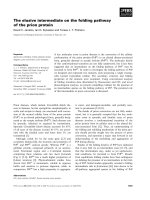
Interestingly, the amino acid alignment (Fig.1)
showed that sdAbs C3, C17, C25 and C43 are likely
clonally related, despite the presence of a relatively
high number of differences scattered all along the gene.
CDR3 of C3, C25 and C43 are very similar, suggesting
use of the same D gene. C17 shows very similar CDR1
and CDR2, but a rather different CDR3. Interestingly,
CDR1 and CDR3 of clone C44 are totally unrelated
to CDR1 and CDR3 of the other sdAbs. In all cases,
the presence of an arginine at position 50 (Fig. 1) con-
firmed the Camelidae nature of these sdAbs. All of
them belong to subfamily VHH2 [15].
Affinity determination by surface plasmon
resonance
To further characterize these sdAbs, the corresponding
cDNA were cloned into the expression vector pPelB55-
PhoA’ [16], allowing efficient production and purifica-
tion of the molecules. SdAbs harboring a hexahistidine
tag at the C-terminus were produced in the periplasm
of Escherichia coli and purified by immobilized ion
metal-affinity chromatography. Final yields were in the
range 5–10 mgÆL culture
)1
for all clones. SDS ⁄ PAGE
analysis demonstrated a satisfying degree of purity
(> 95%, data not shown). Pure sdAbs were then indi-
rectly immobilized on BIAcore sensorchips and their
affinity for soluble CEA determined. As shown in
Table 1, all sdAbs exhibited a good affinity for sCEA,
with a K
D
ranging from 3 to 32 nm.
Specificity analysis by flow cytometry
Flow cytometry was used to determine whether the
selected sdAbs specifically bind to CEA
+
, but not
Fig. 1. Amino acid sequences of
CEA-specific sdAbs. The IMGT numbering
[33] is shown. The localization of
frameworks (FR1 to FR4) and CDRs are
indicated. Dashes indicate sequence
identity.
G. Behar et al. CEA-specific single domain antibodies
FEBS Journal 276 (2009) 3881–3893 ª 2009 The Authors Journal compilation ª 2009 FEBS 3883
CEA
)
, cells and to examine if any cross-reaction with
NCA could be detected.
sdAbs were assayed by flow cytometry for binding
to colon cancer MC38 cells (CEA
)
NCA
)
), or to
transfected MC38 cells expressing either CEA or NCA
(kindly provided by F.J. Primus, Vanderbilt University
Medical Center, Nashville, TN, USA). Cells from the
CEA
+
colon cancer cell line LS174T, as well as from
freshly purified human granulocytes that display NCA
but not CEA, were also tested.
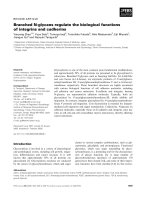
Fig. 2. Flow cytometry analysis of sdAb C17 and C44 binding to MC38 cells expressing CEA or NCA. Purified sdAbs were incubated with
colon cancer MC38 cells (negative control), with transfected CEA
+
or NCA
+
MC38 cells, with CEA
+
colon cancer LS174T cells or with freshly
purified human granulocytes that express NCA but not CEA. Bound sdAbs were detected with a monoclonal anti-c-myc IgG followed by
FITC-labeled F(ab¢)
2
goat anti-mouse IgG (H+L). Mouse mAbs 35A7 (CEA specific) and 192 (binding to an epitope common to CEA and NCA)
were used as controls. C3, C25 and C43 sdAb profiles (not shown) were identical to those of sdAb C17, demonstrating binding to CEA but
not NCA, in contrast to sdAb C44, which binds to both molecules. x-axis, log of fluorescence intensity; y-axis, number of events.
Table 1. Kinetic and affinity constants of the binding of soluble carcinoembryonic antigen (sCEA) to CEA-specific sdAbs immobilized via
monoclonal anti-(c-myc) 9E10 on CM5 microchips. A Langmuir 1 : 1 model was used to fit six different sCEA concentrations. No binding of
sCEA on immobilized 9E10, mass transport or rebinding effect was observed. v
2
, statistical value for describing the closeness of the fit. Val-
ues of v
2
< 10% of the R
max
are usually acceptable. RU, relative units.
Single-domain antibody k
a
· 10
5
(1ÆMs
)1
) k
d
· 10
)3
(1Æs
)1
) K
D
(nM) R
max
(RU) v
2
C44 8.21 ± 0.040 2.64 ± 0.004 3.20 236 3.00
C43 1.78 ± 0.019 1.83 ± 0.002 10.30 284 0.55
C25 1.13 ± 0.014 3.60 ± 0.004 31.7 292 0.30
C17 1.56 ± 0.014 1.30 ± 0.002 8.30 254 0.22
C3 1.24 ± 0.014 1.68 ± 0.002 13.6 254 0.30
CEA-specific single domain antibodies G. Behar et al.
3884 FEBS Journal 276 (2009) 3881–3893 ª 2009 The Authors Journal compilation ª 2009 FEBS
Figure 2 shows that C17 sdAb efficiently binds to
CEA-expressing cells (MC38–CEA
+
and LS174T) but
not to NCA
+
human granulocytes. C3, C25 and C43
sdAb binding profiles were identical to that of C17
sdAb (data not shown). By contrast, sdAb C44 was
also capable of binding to MC38–NCA
+
cells and to
human granulocytes, whereas all other sdAbs did not
show any binding to these cells. C44 sdAb is therefore
recognizing an epitope shared by CEA and NCA, in
contrast to C3, C17, C25 and C43, which are strictly
specific for CEA. Monoclonal antibodies that either
bind both CEA and NCA (mAb 192) or only CEA
(mAb 35A7) were used as controls (Fig. 2).
Competitive inhibition of antibody binding to
LS174T cells
To further characterize the binding properties of sdAbs
C3, C17, C25, C43 and to investigate whether different
CEA epitopes were recognized by these antibodies,
binding competition experiments were performed using
cells from the CEA-expressing cell line LS174T. Cells
were incubated with trace amounts of
125
I-labeled
sdAb C17 (0.4 nm) in the presence of increasing con-
centrations of unlabeled sdAbs. As shown in Fig.3,
sdAbs C25, C3 and C43 were able to compete with
C17, indicating that these sdAbs bind to overlapping
epitopes or to the same epitope. Moreover, sdAb IC
50
is in the nm range for three of the four sdAbs (C3,
1.6 ± 0.4 nm; C17, 7.8 ± 1.3 nm; C43, 5.2 ± 0.3 nm).
Only sdAb C25 exhibits a significantly lower apparent
affinity (59.1 ± 0.6 nm). These values obtained from
cell-binding experiments are in good agreement with
surface plasmon resonance (SPR) data (Table 1),
except for sdAb C3 (K
D
= 13.6 nm). This difference
might be because of a different conformation and ⁄ or
glycosylation of the sdAb epitope on cell-displayed
CEA and on recombinant sCEA immobilized on
sensor chips.
Epitope analysis by surface plasmon resonance
Most CEA-specific mAbs available to date can be clas-
sified into five categories (Gold 1-5) according to their
epitope [17]. To determine if one or more of these epi-
topes was recognized by the CEA-specific sdAbs (C3,
C17, C25, C43), a qualitative SPR-based sandwich
assay was used. sdAbs were first captured via their
c-myc tag on the CM5 sensorchip surface coated with
the monoclonal anti-c-myc IgG 9E10 to favor a good
exposition of the captured sdAbs. Recombinant sCEA
was then injected and captured by the sdAbs. Subse-
quently, one of the five Gold mAbs recognizing one of
the five Gold epitopes was injected. Under these condi-
tions, binding of the Gold mAb indicates that its cor-
responding epitope is not blocked by the capturing
sdAb. All Gold mAbs were tested against all CEA-
specific sdAbs. Sensorgrams obtained with sdAb C17
are shown in Fig. 4A. All sdAb and Gold mAb combi-
nations led to efficient binding of Gold mAb to the
captured sCEA, demonstrating that none of the five
Gold epitopes is recognized by the CEA-specific
sdAbs. All five anti-CEA Gold IgGs were able to bind
captured sCEA, whereas an irrelevant mAb (mouse
anti FccRIII) did not. As a positive of competition, a
version of the sdAb used for CEA capture but devoid
of the c-myc tag was injected instead of Gold mAbs.
No increase in signal was obtained, showing that com-
petition can be efficiently demonstrated between these
molecules in our assay. An irrelevant sdAb [anti-(HIV-
1 NEF) devoid of c-myc tag] injected at the same
concentration was used as a negative control.
Moreover, in a reverse scheme, all sdAbs were able
to bind to recombinant sCEA captured on the chip via
Gold mAbs covalently immobilized on CM5 sensor-
chip. Figure 4B shows the results obtained with the
mAb 35A7 as an example.
Epitope analysis by flow cytometry
The absence of competition between gold mAbs and
sdAbs was confirmed using nonrecombinant cell-
Fig. 3. Competitive inhibition of antibody binding to LS174T cells.
Competition between
125
I-labeled sdAb C17 (0.4 nM) and increasing
amounts of unlabeled sdAb C17 (square), sdAb C25 (triangle), sdAb
C3 (circle) and sdAb C43 (diamond) for binding on LS174T cells
(5 · 10
6
cellsÆmL
)1
). Each point is the mean of triplicate determina-
tions of a representative experiment ± SEM, unless smaller than
the point as plotted. Nonspecific binding was evaluated in the pres-
ence of an excess of unlabeled sdAb C17 (2 · 10
)7
M).
G. Behar et al. CEA-specific single domain antibodies
FEBS Journal 276 (2009) 3881–3893 ª 2009 The Authors Journal compilation ª 2009 FEBS 3885
surface displayed CEA by flow cytometry. CEA-
expressing cells were first incubated with very high
concentrations (up to 2190 nm) of sdAb C17 devoid of
the c-myc tag. After 1 h of incubation, subsaturating
concentrations of either gold mAbs or c-myc-tagged
sdAb C17 or C43 (30-70 nm as determined in a previ-
ous flow cytometry experiment, data not shown) were
added to the wells. After an additional 1 h of incuba-
tion, bound gold mAbs and c-myc-tagged sdAbs were
revealed. As shown in Fig. 5, the presence of a large
excess (more than two orders of magnitude) of untag-
ged sdAb C17 did not interfere with the binding of
gold mAbs, but completely inhibited the binding of
c-myc-tagged sdAb C17 or sdAb C43 that bind to the
same epitope (see above). These results confirmed that
these sdAbs do not bind to any of the epitopes recog-
nized by gold mAbs.
In vivo localization
To analyze the behavior of sdAbs against CEA under
more physiological conditions, immunocompromised
mice were xenografted with LS174T cancer cells. Once
tumors were established (i.e., day 7), radiolabeled
sdAb C17 (displaying the highest affinity as measured
by SPR) was injected and the biodistribution was
monitored.
A fast blood clearance was observed, as assessed by
the low residual blood radioactivity 6 h after injection
[0.30 ± 0.06% of the injected dose per gram %
IDÆg
)1
± SEM)]. Activity uptake was observed pri-
marily in kidneys (7.4 ± 0.4% IDÆg
)1
3 h post injec-
tion and 4.8 ± 0.6% IDÆg
)1
at 6 h post injection).
Three hours after injection, 1.9 ± 0.1% of the injected
dose was localized in the tumor. This was at least two-
fold higher than the radioactivity found in blood, liver
and major organs except kidneys, and was 3.5- and
5-fold higher than the radioactivity found in bones
and leg muscles, respectively (Fig. 6). Six hours after
injection, the increased clearance resulted in higher
tumor-to-normal tissue uptake ratios for all these
organs, reaching a ratio of 10 in the case of muscles.
Discussion
As a first step toward the construction of multispeci-
fic and ⁄ or multivalent molecules aiming at redirecting
immune cells such as T cells or NK cells to tumor
cells, we isolated sdAbs able to bind to CEA (or
CEACAM5), a tumor marker used in cancer diagno-
sis and immunotherapy. We used phage display to
select binders from one sdAb library derived from
peripheral blood mononuclear cells isolated from an
immunized llama. Interestingly, two selection methods
led to strikingly different outputs. Selection by
panning on recombinant sCEA directly adsorbed on
plastic allowed the isolation of a single family of
highly related sdAbs that dominated the selected pop-
ulation only after a single round of selection. How-
Fig. 4. Epitope analysis by surface plasmon resonance (BIAcore).
(A) c-myc-tagged sdAbs were captured on an anti-c-myc IgG-coated
CM5 chip. sCEA was injected (curves a) or not (curves b), followed
by injection of one of the Gold mAbs. The dissociation of sdAbs
from 9E10 was corrected by subtraction of a control flow cell
coated with 9E10 and injected with sdAbs only. Sensorgrams
obtained with sdAb C17 are shown as an example. Irr mAb, irrele-
vant mAb (mouse anti-FccRIII). As a positive control of competition,
sdAb C17 devoid of the c-myc tag was injected after CEA capture.
The absence of binding demonstrates that the epitope of this sdAb
was efficiently blocked by the binding of the immobilized sdAb. An
irrelevant sdAb (anti HIV-1 NEF) injected at the same concentration
was used as a negative control (irr sdAb). (B) sCEA (curves a) or
buffer (curves b) were injected on Gold mAb-coated CM5 chips,
followed by an injection of different sdAbs.
CEA-specific single domain antibodies G. Behar et al.
3886 FEBS Journal 276 (2009) 3881–3893 ª 2009 The Authors Journal compilation ª 2009 FEBS
ever, the epitope recognized by these antibodies is
also present on a highly related molecule, nonspecific
cross-reacting antigen (NCA or CEACAM6) that
shares the same Ig domain-based structure with CEA
and displays a high percentage of sequence homology.
By contrast, a selection based on biotinylated sCEA
captured on streptavidin-coated magnetic beads led to
only 61% binders after a single round and two more
rounds of selection were needed to reach 100% bind-
ers. Unlike the first method, the output of this selec-
tion was more diverse. This method yielded
antibodies belonging to the family selected by pan-
ning on coated sCEA, but also made it possible to
isolate several other clones displaying very similar
CDR1 and CDR2, but more diverse CDR3, suggest-
ing use of the same VHH gene but different D genes.
Flow cytometry analyses demonstrated that these
sdAbs bind specifically to CEA expressed on cancer
cells but do not cross-react with NCA. These anti-
bodies were not present in the output of the panning
method, despite very similar affinities for the antigen,
in the nm range, as determined by SPR. One can
hypothesize that the conformational changes resulting
from the adsorption of CEA on plastic are either
denaturing the epitope recognized by the second
family of sdAbs or are favoring the display of the
epitope recognized by the first family, leading to a
large presence of this latter family during the selec-
tion process.
As expected for llama VHHs, the sequences of the
CEA-specific sdAbs are homologous to human IGHV3
subgroup genes (C3, 79% homology to IGHV3-23;
C17, 68% homology to IGHV3-74; C25, 69% homol-
ogy to IGHV3-48; C43, 69% homology to IGHV3-13).
The most divergent sequences between the four llama
sdAbs and human IGHV3 are localized in the CDRs,
Fig. 5. Epitope analysis by flow cytometry. CEA-expressing cells were preincubated with a large excess of untagged sdAb C17 (competitor).
Subsaturating concentrations (30–70 n
M) of Gold mAbs or c-myc tagged C17 and C43 were then added to the mix. After washing, bound
Gold mAbs or c-myc tagged sdAbs were detected by flow cytometry. Solid black histograms, isotype control. Solid gray histograms indicate
the absence of competitor. Black lines indicate the presence of competitor.
G. Behar et al. CEA-specific single domain antibodies
FEBS Journal 276 (2009) 3881–3893 ª 2009 The Authors Journal compilation ª 2009 FEBS 3887
and in the former VL and CH1 interfaces (residues 40,
42, 49, 50 and residues 15 and 96, respectively). More-
over, the selected llama sdAbs belong to subfamily
VHH2 [15]. As described earlier for VHH belonging to
this family, CDR3 from these four sdAbs do not con-
tain an additional disulfide bond and do not exceed
the mean CDR3 length of classical VH, in contrast to
most camel VHH [18].
Of note is that that none of these antibodies binds
to one of the Gold epitopes. These essentially nonover-
lapping epitopes have been defined by analyzing the
binding specificities of 52 monoclonal anti-CEA IgGs
and define five antigenic regions recognized by murine
mAbs [17]. In this study, the only four mAbs not bind-
ing to these five regions were directed against carbo-
hydrate epitopes, suggesting that the rest of the CEA
surface does not elicit antibodies. Epitope analysis
performed on recombinant sCEA using SPR or on
the surface of living cells demonstrated that the sdAbs
target overlapping epitopes and might even share a
unique epitope, because it could be anticipated by the
high degree of homology of their CDRs. Interestingly,
this is not one of the Gold epitopes because no compe-
tition was observed between Gold mAbs and the
sdAbs, as demonstrated both by SPR on soluble CEA
and flow cytometry experiments on cell-displayed
CEA. This new epitope, not found on NCA, is there-
fore not easily detected by murine mAbs. This finding
supports a previous study showing that sdAbs have a
tendency to bind to epitopes usually invisible to other
mAbs, such as cavities, and are a rich source of
enzyme inhibitors [19]. In the case of CEA, a heavily
glycosylated molecule, one can also hypothesize that
the oligosaccharide chains can hinder the access of
some regions of the polypeptide to large molecules
such as mAbs (150 kDa) but not to very compact
sdAb (13 kDa). However, it should be reminded that
the sdAbs were selected as phage–sdAbs, which implies
that the large phage particle did not prevent binding
of the sdAbs to this epitope.
IC
50
values calculated from cell-surface competition
experiments and K
D
values measured by SPR are in
the nm range for sdAbs C17, C3 and C43, demonstrat-
ing that these sdAbs, selected against a recombinant
ectodomain of CEA can efficiently bind their antigen
when displayed at the cell surface of human tumor
cells. The high affinities of the selected sdAbs, which
compares favorably with conventional mAbs despite
their monovalency, should allow an efficient in vivo
targeting of tumor cells expressing CEA. To verify this
hypothesis, we conducted an in vivo localization experi-
ment in LS174T-xenografted nude mice with sdAb
C17, which binds to CEA with the highest affinity as
measured by SPR. Blood clearance of this sdAb was
fast because low blood radioactivity was observed as
early as 6 h after injection. This result is in agreement
with the sdAb blood half-life that has been estimated
to be 20–40 min in mice [20,21]. Despite this rapid
blood clearance and the monovalent nature of the
sdAb excluding an avidity effect, almost 2% of the
injected dose accumulates in tumor tissues (fivefold
higher than in muscle tissues), which compares well
with the results of Cortez-Retamozo et al. [22]. These
authors injected LS174T-xenografted mice with a
CEA-specific sdAb fused to beta-lactamase and 2.8%
of the total injected dose was found in the tumor 6 h
after injection, despite a blood half-life expected to be
significantly higher for this 45 kDa construct than for
sdAb C17 (13 kDa). In this study, 3 h after injection,
7% IDÆg
)1
were found in kidneys, which decreased to
5% at 6 h post injection. This renal accumulation
is expected for very small molecules such as sdAbs
(13-15 kDa). Ultrafiltration of low molecular mass
proteins and subsequent uptake by proximal tubular
cells followed by lysosomal degradation leads to the
intracellular accumulation of radioactivity. It is
Fig. 6. Biodistribution of sdAb against CEA C17 injected in xeno-
grafted mice. Nude mice subcutaneously xenografted with tumor
cells LS174T were injected in the tail vein with 10 pmol of
125
I-
labeled sdAb C17. After 3 h (black bars) and 6 h (open bars), mice
were anesthetized and killed. Blood, organs and tumor masses
were weighed and the radioactivity counted. Results are expressed
as the ratio between tumor uptake and organ uptake (mean ± -
SEM, n = 3). Injected doses were corrected by subtraction of non-
injected and subcutaneously injected material. Bl, blood; Lu, lung;
Li, liver; Sp, spleen; Si, small intestine; Co, colon; Ki, kidney; Mu,
muscle; Bo, bone.
CEA-specific single domain antibodies G. Behar et al.
3888 FEBS Journal 276 (2009) 3881–3893 ª 2009 The Authors Journal compilation ª 2009 FEBS
expected that systemic administration of basic amino
acids may reduce renal retention of radioiodinated
sdAbs because it is efficient in lowering kidney uptake
of antibody fragments [22a]. The low activity accre-
tion observed in other organs led to tumor-to-organ
radioactivity uptake ratios of at least 2 (range 2–5) 3 h
after injection and 3 (range 3–9) at 6 h after injection.
Overall, sdAb C17 showed an expected biodistribu-
tion profile, demonstrating its utility as a CEA
+
tumor-targeting molecule.
In conclusion, the specificity, affinity and single-
domain structure of the new CEA-specific sdAbs
isolated here make them very attractive candidates to
build, together with sdAbs targeting receptors such as
CD16 (FccRIII) [23] or other activating receptors or
radiolabeled haptens [4], new multivalent and ⁄ or
multispecific molecules with superior characteristics for
immunotherapy or radioimmunotherapy.
Material and methods
Llama immunization
A young adult male llama (Lama glama) was immunized
subcutaneously at days 1, 30, 60, 90 and 120 with 250 lg
recombinant human soluble CEA extracellular domain
(sCEA) produced as previously described [24]. Sera were
collected 15 days prior to each injection to follow the
immune response against the immunogen.
VHH library construction
Blood samples (100 mL) were taken 15 days after each of the
three latest immunizations and peripheral blood mononu-
clear cells were isolated by Ficoll-Histopaque-1077 (Sigma-
Aldrich, St. Louis, MO, USA) discontinuous gradient
centrifugation. Total RNA was isolated by acid guanidinium
thiocyanate ⁄ phenol ⁄ chloroform extraction [25] and synthesis
of the cDNA was performed with Superscript II reverse
transcriptase (GibcoBRL, Gaithersburg, MD, USA) using
primer CH2FORTA4 [26]. A first PCR was performed using
an equimolar mixture of four backward primers originally
designed to anneal on human VH genes (5¢ VH1–Sfi:
5¢-CATGCCATGACTCGCGGCCCAGCCGGCCATGGC
CCAGGTGCAGCTGGTGCAGTCTGG-3¢;5¢ VH2–Sfi:
5¢-CATGCCATGACTCGCGGCCCA GCCGGCCATGGC
CCAGGTCACCTTGAAGGAGTCTGG-3¢;5¢ VH3–Sfi:
5¢-CATGCCATGACTCGCGGCCCA GCCGGCCATGGC
CGAGGTGCAGCTGGTGGAGTCTGG-3¢;5¢ VH4–Sfi:
5¢-CATGCCATGACTCGCGGCCCA GCCGGCCATGGC
CCAGGTGCAGCTGCAGGAGTCGGG-3¢) and one for-
ward primer (CH2FORTA4). These primers allow the ampli-
fication of two bands corresponding two the VH + CH1 +
hinge + part of CH2 gene fragment of traditional antibodies
or the VHH + hinge + part of CH2 gene fragment of
HcAbs. Using the gel-purified (Qiaquick gel extraction kit;
Qiagen, Hilden, Germany) lower band as the template, VHH
genes were re-amplified using an equimolar mixture of the
four backward primers (5¢ VH1 to 4-Sfi) and 3¢ VHH–Not
primer (5¢-CCACGATTCTGCGGCCGCTGAGGAGACR
GTGACCTGGGTCC-3¢) containing SfiI and NotI restric-
tion enzyme sites. Resulting VHH fragments were purified
from 1% agarose gel, digested with SfiI and NotI and ligated
into pHEN1 phagemid [27] digested with SfiI and NotI. The
ligated material was transformed into TG1 E. coli electro-
poration-competent cells (Stratagen, Miami, FL, USA). Cells
were plated on 2YT ⁄ ampicillin (100 lgÆmL
)1
) ⁄ glucose (2%)
agar plates. Colonies (10
6
) were scraped from the plates with
2YT ⁄ ampicillin (100 lgÆmL
)1
) ⁄ glucose (2%), and stored at
)80 °C in the presence of 20% glycerol. Because llamas were
hyperimmunized, a library containing a million of different
clone can be considered as representative.
Selection of phage–sdAbs
Selections were performed as described previously [28].
Briefly, 10 lL of the library was grown in 50 mL of
2YT ⁄ ampicillin (100 lgÆmL
)1
) ⁄ glucose (2%) at 37 ° Cto
an D
600
of 0.5. Five milliliters of the culture were then
infected with 2 · 10
10
M13KO7 helper phage for 30 min
at 37 °C without shaking. The culture was centrifuged for
10 min at 3000 g. The bacterial pellet was resuspended
in 25 mL of 2YT ⁄ ampicillin (100 lgÆmL
)1
) ⁄ kanamycine
(25 lgÆmL
)1
), and incubated for 16 h at 30 °C with
shaking (270 rpm). The culture was then centrifuged for
20 min at 3000 g and one-fifth of the volume of 20%
PEG 6000, 2.5 m NaCl was added to the supernatant and
incubated for 1 h on ice to precipitate phage particles.
The solution was then centrifuged for 15 min at 3000 g at
4°C and the phage-containing pellet was re-suspended with
1 mL of NaCl ⁄ P
i
.
Phage were selected using either immunotubes coated with
recombinant sCEA [24] (10 lgÆmL
)1
in NaCl ⁄ P
i
, overnight
4 °C) or biotinylated sCEA and streptavidin-coated para-
magnetic beads (Dynabeads M-280; Dynal Biotech, Oslo,
Norway). Recombinant sCEA was biotinylated using a bio-
tin protein-labeling kit according to the manufacturer’s
instructions (Roche, Basel, Switzerland). Two hundreds
microliters of beads were mixed with 1 mL NaCl ⁄ P
i
contain-
ing 2% skimmed milk powder (NaCl ⁄ P
i
⁄ 2% milk) for
45 min at room temperature in a siliconized Eppendorf tube.
Beads were washed with NaCl ⁄ P
i
⁄ 2% milk using a magnetic
particle concentrator and resuspended with 250 lL
NaCl ⁄ P
i
⁄ 2% milk. We added 200 lL of biotinylated sCEA
and the solution was gently rotated for 30 min at room tem-
perature; 150, 75 and 25 nm of biotinylated sCEA were used
for the first, second and third rounds of selection, respec-
tively. We then added 450 lL of the phage preparation
(10
12
pfu), preincubated for 1 h in 500 lL NaCl ⁄ P
i
⁄ 2% milk.
G. Behar et al. CEA-specific single domain antibodies
FEBS Journal 276 (2009) 3881–3893 ª 2009 The Authors Journal compilation ª 2009 FEBS 3889
The mixture was rotated for 3 h at room temperature and
washed five times with 800 lL NaCl ⁄ P
i
⁄ 4% milk, five times
with 800 lL NaCl ⁄ P
i
containing 0.1% Tween and five times
with 800 lL NaCl ⁄ P
i
. Every five washes, the mixture was
transferred to a new siliconized tube. Phage fixed on sCEA-
coated beads were resuspended with 200 lL NaCl ⁄ P
i
and
incubated without shaking with 1 mL of log-phase TG1 cells
and plated on 2YT ⁄ ampicillin (100 l g ÆmL
)1
) ⁄ glucose (2%)
in 243 · 243 mm dishes (Nalgene Nunc, Roskilde, Den-
mark). Some isolated colonies were grown overnight in mi-
crotiter plate containing 200 lL 2YT ⁄ ampicillin
(100 lgÆmL
)1
) ⁄ glucose (2%) and stored at )80 °C after the
addition of 15% glycerol (masterplates). The remaining colo-
nies were harvested from the plates, suspended in 2 mL
2YT ⁄ ampicillin (100 lgÆmL
)1
) ⁄ glucose (2%) and used for
phage production for the next round of selection.
ELISA screening of phage–sdAb
A 96-well plate replicator was used to replicate the mas-
terplates in 120 lL of fresh broth. Colonies were grown
for 2 h at 37 °C under shaking (400 rpm) and 35 lL
2YT ⁄ ampicillin (100 lgÆmL
)1
) ⁄ glucose (2%) containing
2 · 10
9
M13KO7 helper phage were added to each well
and incubated for 30 min at 37 °C without shaking. The
plate was centrifuged for 10 min at 1200 g and the bacte-
rial pellet was suspended in 150 lL 2YT ⁄ ampicillin
(100 lgÆmL
)1
) ⁄ kanamycine (25 lgÆmL
)1
) and grown for
16 h at 30 °C under shaking (400 rpm). Phage-containing
supernatants were tested for binding to sCEA by ELISA.
Briefly, biotinylated sCEA (5 lgÆ mL
)1
) was coated on
streptavidin 96-well microplates (BioBind assembly strepta-
vidin coated; Thermo Fischer Scientific, Waltham, MA,
USA) saturated with NaCl ⁄ P
i
⁄ 2% milk. Fifty microliters
of phage supernatant were added to 50 lL NaCl ⁄ P
i
⁄ 4%
milk and incubated for 2 h at room temperature in the
ELISA microplate. Bound phage were detected with a per-
oxidase-conjugated monoclonal anti-M13 mouse IgG (GE
Healthcare, Munich, Germany). Reading was performed
at A
405
. DNA of positive phage (A
405
three times above
the blank) was sequenced using abi prismÒ bigdyeÔ
Terminators (Applied Biosystems, Foster City, CA, USA).
SdAb production and purification
Selected clones were sequenced and amplified by PCR using
primers 5¢ pJF–VH3–Sfi (CTTTACTATTCTCAC
GGCCA
TGGCGGCCGAGGTGCAGCTGGTGG) and 3¢ c-myc–
6His ⁄ HindIII (CCGCGCGCGC CAAGACCC
AAGCTTG
GGCTARTGRTGRTGRTGRTGRTGTGCGGCCCCAT
TCAGATC) to add the HindIII site for further cloning, a
hexahistidine tag for purification and the c-myc tag for
detection. For production of clones without the c-myc tag,
the PCR amplification was performed using primers
5¢ pJF–VH3–Sfi and 3¢ 6His ⁄ HindIII (CCGCGCGCGCC
AAGACCC
AAGCTTGGGCTACTAGCTCCCGTGGTG
ATGGTGGTGATGTGAGGAGACAGTGACCTG).
PCR fragments were cloned into the pPelB55PhoA¢ [16]
vector between the Sfi I and HindIII sites. E. coli K12 strain
TG1 was used to produce the sdAb-tagged fragments. An
inoculum was grown overnight at 30 °C in 2YT medium sup-
plemented with 100 lgÆmL
)1
ampicillin and 2% glucose.
Four hundred milliliters of fresh medium were inoculated to
obtain an D
600
of 0.1, and bacteria were grown at 30 °Cto
an D
600
of 0.5–0.7 and induced with 100 lm isopropyl thio-b-
d-galactoside for 16 h. The cells were harvested by centrifu-
gation at 4200 g for 10 min at 4 ° C. The cell pellet was
suspended in 4 mL of cold TES buffer (0.2 m Tris ⁄ HCl, pH
8.0; 0.5 mm EDTA; 0.5 m sucrose), and 160 lL lysozyme
(10 mgÆmL
)1
in TES buffer) was added. The cells were then
subjected to osmotic shock by the addition of 16 mL of cold
TES diluted 1 : 2 with cold H
2
O. After incubation for 30 min
on ice, the suspension was centrifuged at 4200 g for 40 min
at 4 °C. The supernatant was incubated with 150 lL DNase
I (10 mgÆmL
)1
) and MgCl
2
(5 mm final) for 30 min at room
temperature. The solution was dialyzed against 50 mm
sodium acetate pH 7.0, 0.1 m NaCl, for 16 h at 4 °C. sdAbs
were purified by TALON metal-affinity chromatography
(Clontech, Mountain View, CA, USA) and concentrated by
ultrafiltration with Amicon Ultra 5000 MWCO (Millipore,
Billerica, MA, USA). The protein concentration was deter-
mined spectrophotometrically using a protein assay kit (Bio-
Rad Laboratories, Hercules, CA, USA).
Affinity measurements
Kinetic parameters were determined by real-time SPR
using a BIACORE 3000 apparatus. Monoclonal anti-c-
myc IgG 9E10 was covalently immobilized (3300 RU) on
a flow cell of CM5 sensor chip (Biacore AB, Uppsala,
Sweden) with EDC ⁄ NHS activation according to the
manufacturer’s instructions. A control flow cell surface
was prepared with the same treatment but without anti-
body. All analyses were performed at 25 °C, at a flow
rate of 30 lgÆmL
)1
and using HBS-EP (Biacore AB;
10 mm Hepes pH 7.4, 150 mm NaCl, 3.4 mm EDTA and
0.005% BiacoreÔ surfactant) as running buffer. Each
sdAb was injected (90 lL) at a concentration of
50 lgÆmL
)1
in HBS-EP over 3 min and followed by a
90 lL injection of sCEA at six different concentrations
(0.19–6.2 lgÆmL
)1
). A 400 s dissociation step was applied
before a pulse of 5 mm HCl to regenerate the flow cell
surfaces between each run. The absence of direct sCEA
binding to 9E10 was assessed. The control sensorgram
obtained by injection of sdAb only on the 9E10 flow cell
was subtracted from all other sensorgrams to compensate
for sdAb dissociation from 9E10 mAb. Resulting senso-
grams were fitted to a Langmuir 1 : 1 binding isotherm
model and errors on k
a
and k
d
were calculated using
biaevaluation 3.2 software.
CEA-specific single domain antibodies G. Behar et al.
3890 FEBS Journal 276 (2009) 3881–3893 ª 2009 The Authors Journal compilation ª 2009 FEBS
Immunofluorescence assays
The CEA-positive human colon carcinoma LS174T cell line
was obtained from the American Type Culture Collection
(Rockville, MD, USA). The murine colon carcinoma MC38
cells either transfected with human CEA (MC38–CEA cell
line) or with human NCA (MC38–NCA cell line) were
kindly provided by F.J. Primus (Vanderbilt University
Medical Center, Nashville, TN, USA) [29]. These cells were
cultured in Dulbecco’s modified Eagle’s medium (Gibco
Laboratories, Lyon, France) supplemented with 10%
heat-inactivated fetal bovine serum (Gibco Laboratories),
l-glutamine (300 lgÆmL
)1
), fungizone (0.25 lgÆmL
)1
), strep-
tomycin (100 lgÆmL
)1
), penicillin G (100 UnitsÆmL
)1
) and
geneticin (0.5 mgÆmL
)1
). These cells are adherent and grow
as monolayers at 37 °C in a humidified 5% CO
2
incubator.
Immunofluorescence assays were performed by incubating
5 · 10
5
indicator cells with sdAbs (10 lgÆmL
)1
) for 30 min
on ice. sdAbs binding to MC38, MC38–CEA, MC38–NCA,
LS174T cell lines and human granulocytes were then
revealed by incubation with the monoclonal anti-c-myc
9E10 IgG (10 lgÆmL
)1
) followed by incubation with
fluorescein isothiocyanate (FITC)-labeled F(ab)¢
2
goat
anti-mouse IgG (H+L) (FITC-GAM) antibodies (Jackson
ImmunoResearch Laboratories, West Grove, PA, USA).
Human granulocytes were purified as described previously
[30].
sdAb iodination
sdAb C17 (0.5 nmol in 50 lL NaCl ⁄ P
i
) was iodinated with
Na
125
I (18.5 MBq) using iodogen [31] for 20 min at 4 °C.
Ten microliters of 1 mmdl-tyrosine pH 7.4 was added to
the solution and the mix was incubated for a further 5 min.
The iodinated antibody was purified by gel-permeation
chromatography on a PD 10 column (Sephadex G-25, GE
Healthcare, Waukesha, WI, USA).
Cell-binding experiments
Cell-binding experiments were performed on cells from the
LS174T colon carcinoma cell line (ATCC). We incubated
150 lLof
125
I-labeled sdAb C17 (4 · 10
)10
m final concen-
tration, specific activity: 5 · 10
17
cpmÆmol
)1
) with 100 lL
of cell suspension (5 · 10
6
cellsÆmL
)1
final) in binding med-
ium [modified Eagle’s medium with Earle’s salts (GIBCO-
Invitrogen-France), 0.2% BSA] in the presence of increas-
ing concentrations of unlabeled sdAb (100 lL in binding
medium). After 2.5 h under shaking, 100 lL of the suspen-
sions were centrifuged in triplicate for 30 s through a phth-
alate mixture [32]. An aliquot of supernatant and the cell
pellet from each tube were counted (three experiments,
each in triplicate). The non-specific binding was evaluated
in the presence of an excess of unlabelled sdAb C17
(2 · 10
)7
m).
IC
50
values and statistics were calculated with graphpad
prism
Ò
(GraphPad Software, Inc. San Diego, CA, USA)
using a one-site competition nonlinear regression analysis.
Epitope mapping by SPR and flow cytometry
In a first set of experiments, epitope mapping of sdAbs and
Gold mAbs (B17, CE25, 35A7, B93, 192) [17] was carried
out by SPR at a flow rate of 20 l g ÆmL
)1
. First, each sdAb
(50 lgÆmL
)1
) was injected on 9E10 mAb immobilized
(11 000 RU) on a CM5 sensorchip. Second, 60 lL of sCEA
antigen (25 lgÆmL
)1
) were injected and, third, one of the
Gold mAbs was injected (10 lgÆmL
)1
). An irrelevant sdAb
devoid of c-myc tag [anti-(HIV-1 NEF)] was also used as
negative control. As a competition control, the sdAb used
for CEA capture was produced without a c-myc tag and
tested for its ability to bind to the captured CEA. The
9E10-coupled surface was regenerated with 10 lLof5mm
HCl and the process was repeated to test the ability of each
Gold mAb to bind sCEA once this molecule had been
bound to a given sdAb. The absence of binding of the dif-
ferent Gold mAbs to the 9E10 mAb alone or to the sdAbs
in absence of sCEA, as well as the absence of binding of an
irrelevant antibody to the captured sCEA (mouse anti-
FccRIII) was verified.
The absence of competition between the sdAbs and
Gold mAbs for binding to CEA in its normal environ-
ment was also assessed by flow cytometry. Briefly, MC38–
CEA cells (5 · 10
5
Æwell
)1
) were preincubated with various
concentrations (from 2 l m to 3 nm) of sdAb C17 (devoid
of c-myc tag) for 1 h on ice in NaCl ⁄ P
i
+ 1% BSA.
Gold mAbs or sdAb C17 or sdAb C43 were then directly
added in the wells at subsaturating concentrations (30–
70 nm, determined in a previous experiment) and cells
were incubated on ice for an additional hour. After
washing, bound Gold mAbs were stained using FITC–
GAM (10 lgÆmL
)1
) and sdAb C17 (with c-myc tag) was
stained using mAb 9E10 (10 lgÆmL
)1
) followed by FITC–
GAM.
In vivo localization
All in vivo experiments were performed in compliance with
the French guidelines for experimental animal studies.
Female HSD athymic nude-Foxn1
nu
8–9 weeks old (Har-
lan, Gannat, France) were engrafted by subcutaneous
injection of 2 · 10
6
LS174T human colorectal carcinoma
cells in the flank. Biodistribution studies were performed
13–15 days later. Mice were injected intravenously in the
tail vein with
125
I-labeled sdAb C17 (10 pmol in 100 lL
NaCl ⁄ P
i
0.2% BSA) and killed at 3 and 6 h post injection.
Blood, organs and tumors were collected, the two latter
were weighted and radioactivity in the samples was deter-
mined. Injected doses were corrected for losses by subtrac-
tion of non-injected and subcutaneously injected material
G. Behar et al. CEA-specific single domain antibodies
FEBS Journal 276 (2009) 3881–3893 ª 2009 The Authors Journal compilation ª 2009 FEBS 3891
(remaining in the animal tail) from the total dose. All stud-
ies were performed with groups of three mice. Results were
expressed as the mean percentage of injected dose per gram
of tissue ± SEM.
Acknowledgements
We would like to thank Martine Chartier, Jallane
Abdelhak and Sandra Mendes for their excellent
technical assistance, Sophie Sibe
´
ril and Agne
`
s Grou-
let for preliminary work. We are grateful to Dr J.
Barbet for fruitful discussions. We also thank Chris-
tiane and Bernard Guidicelli for generously providing
a llama for immunization. This work was supported
by CNRS, INSERM, the Association pour la
Recherche sur le Cancer (ARC), the Cance
´
ropole
Ile-de-France and by the GDR N°2352 CNRS
‘Tumor immuno-targeting’.
References
1 Hamers-Casterman C, Atarhouch T, Muyldermans S,
Robinson G, Hamers C, Songa EB, Bendahman N &
Hamers R (1993) Naturally occurring antibodies
devoid of light chains. Nature 363, 446–448.
2 Muyldermans S (2001) Single domain camel antibod-
ies: current status. J Biotechnol 74, 277–302.
3 Spinelli S, Frenken LG, Hermans P, Verrips T, Brown
K, Tegoni M & Cambillau C (2000) Camelid heavy-
chain variable domains provide efficient combining
sites to haptens. Biochemistry 39, 1217–1222.
4 Alvarez-Rueda N, Behar G, Ferre V, Pugniere M,
Roquet F, Gastinel L, Jacquot C, Aubry J, Baty D,
Barbet J et al. (2007) Generation of llama single-
domain antibodies against methotrexate, a prototypi-
cal hapten. Mol Immunol 44, 1680–1690.
5 Lauwereys M, Arbabi Ghahroudi M, Desmyter A,
Kinne J, Holzer W, De Genst E, Wyns L & Muylder-
mans S (1998) Potent enzyme inhibitors derived from
dromedary heavy-chain antibodies. EMBO J 17, 3512–
3520.
6 Pant N, Hultberg A, Zhao Y, Svensson L, Pan-Ham-
marstrom Q, Johansen K, Pouwels PH, Ruggeri FM,
Hermans P, Frenken L et al. (2006) Lactobacilli
expressing variable domain of llama heavy-chain anti-
body fragments (lactobodies) confer protection against
rotavirus-induced diarrhea. J Infect Dis 194, 1580–1588.
7 Frenken LG, van der Linden RH, Hermans PW, Bos
JW, Ruuls RC, de Geus B & Verrips CT (2000) Isola-
tion of antigen specific llama VHH antibody fragments
and their high level secretion by Saccharomyces cerevisi-
ae. J Biotechnol 78, 11–21.
8 Bazl MR, Rasaee MJ, Foruzandeh M, Rahimpour A,
Kiani J, Rahbarizadeh F, Alirezapour B & Moham-
madi M (2007) Production of chimeric recombinant
single domain antibody–green fluorescent fusion
protein in Chinese hamster ovary cells. Hybridoma
(Larchmt) 26, 1–9.
9 Ismaili A, Jalali-Javaran M, Rasaee MJ, Rahbarizadeh
F, Forouzandeh-Moghadam M & Memari HR (2007)
Production and characterization of anti-(mucin
MUC1) single-domain antibody in tobacco (Nicotiana
tabacum cultivar Xanthi). Biotechnol Appl Biochem 47,
11–19.
10 Dumoulin M, Conrath K, Van Meirhaeghe A, Meers-
man F, Heremans K, Frenken LG, Muyldermans S,
Wyns L & Matagne A (2002) Single-domain antibody
fragments with high conformational stability. Protein
Sci 11, 500–515.
11 Su C, Nguyen VK & Nei M (2002) Adaptive evolution
of variable region genes encoding an unusual type of
immunoglobulin in camelids. Mol Biol Evol 19, 205–
215.
12 Conrath KE, Lauwereys M, Galleni M, Matagne A,
Frere JM, Kinne J, Wyns L & Muyldermans S (2001)
Beta-lactamase inhibitors derived from single-domain
antibody fragments elicited in the camelidae. Antimic-
rob Agents Chemother 45, 2807–2812.
13 Gold P & Freedman SO (1965) Specific carcinoembry-
onic antigens of the human digestive system. J Exp
Med 122, 467–481.
14 Hammarstrom S (1999) The carcinoembryonic antigen
(CEA) family: structures, suggested functions and
expression in normal and malignant tissues. Semin
Cancer Biol 9, 67–81.
15 Harmsen MM, Ruuls RC, Nijman IJ, Niewold TA,
Frenken LG & de Geus B (2000) Llama heavy-chain
V regions consist of at least four distinct subfamilies
revealing novel sequence features. Mol Immunol 37,
579–590.
16 Le Calvez H, Green JM & Baty D (1996) Increased
efficiency of alkaline phosphatase production levels in
Escherichia coli using a degenerate PelB signal
sequence. Gene 170, 51–55.
17 Hammarstrom S, Shively JE, Paxton RJ, Beatty BG,
Larsson A, Ghosh R, Bormer O, Buchegger F, Mach
JP, Burtin P et al. (1989) Antigenic sites in carcino-
embryonic antigen. Cancer Res 49, 4852–4858.
18 Muyldermans S, Atarhouch T, Saldanha J, Barbosa
JA & Hamers R (1994) Sequence and structure of VH
domain from naturally occurring camel heavy chain
immunoglobulins lacking light chains. Protein Eng 7,
1129–1135.
19 De Genst E, Silence K, Decanniere K, Conrath K,
Loris R, Kinne J, Muyldermans S & Wyns L (2006)
Molecular basis for the preferential cleft recognition
by dromedary heavy-chain antibodies. Proc Natl Acad
Sci USA 103, 4586–4591.
CEA-specific single domain antibodies G. Behar et al.
3892 FEBS Journal 276 (2009) 3881–3893 ª 2009 The Authors Journal compilation ª 2009 FEBS
20 Holliger P & Hudson PJ (2005) Engineered antibody
fragments and the rise of single domains. Nat Biotech-
nol 23, 1126–1136.
21 Coppieters K, Dreier T, Silence K, de Haard H,
Lauwereys M, Casteels P, Beirnaert E, Jonckheere
H, Van de Wiele C, Staelens L et al. (2006) Format-
ted anti-tumor necrosis factor alpha VHH proteins
derived from camelids show superior potency and
targeting to inflamed joints in a murine model of
collagen-induced arthritis. Arthritis Rheum 54, 1856–
1866.
22 Cortez-Retamozo V, Backmann N, Senter PD, Wer-
nery U, De Baetselier P, Muyldermans S & Revets H
(2004) Efficient cancer therapy with a nanobody-based
conjugate. Cancer Res 64, 2853–2857.
22a Behr TM, Goldenberg DM & Becker W (1998) Redu-
cing the renal uptake of radiolabeled antibody frag-
ments and peptides for diagnosis and therapy: present
status, future prospects and limitations. Eur J Nucl
Med 25, 201–212.
23 Behar G, Siberil S, Groulet A, Chames P, Pugniere M,
Boix C, Sautes-Fridman C, Teillaud JL & Baty D
(2008) Isolation and characterization of anti-Fc-
{gamma}RIII (CD16) llama single-domain antibodies
that activate natural killer cells. Protein Eng Des Sel
21, 1–10.
24 Terskikh A, Mach JP & Pelegrin A (1993) Marked
increase in the secretion of a fully antigenic recombi-
nant carcinoembryonic antigen obtained by deletion of
its hydrophobic tail. Mol Immunol 30, 921–927.
25 Chomczynski P & Sacchi N (1987) Single-step
method of RNA isolation by acid guanidinium thio-
cyanate–phenol–chloroform extraction. Anal Biochem
162, 156–159.
26 Arbabi Ghahroudi M, Desmyter A, Wyns L, Hamers
R & Muyldermans S (1997) Selection and identifica-
tion of single domain antibody fragments from camel
heavy-chain antibodies. FEBS Lett 414, 521–526.
27 Hoogenboom HR, Griffiths AD, Johnson KS, Chi-
swell DJ, Hudson P & Winter G (1991) Multi-subunit
proteins on the surface of filamentous phage: method-
ologies for displaying antibody (Fab) heavy and light
chains. Nucleic Acids Res 19, 4133–4137.
28 Chames P, Hoogenboom HR & Henderikx P (2002)
Selection of antibodies against biotinylated antigens.
Methods Mol Biol 178, 147–157.
29 Mizobata S, Tompkins K, Simpson JF, Shyr Y &
Primus FJ (2000) Induction of cytotoxic T cells and
their antitumor activity in mice transgenic for carcino-
embryonic antigen. Cancer Immunol Immunother 49,
285–295.
30 Vely F, Gruel N, Moncuit J, Cochet O, Rouard H,
Dare S, Galon J, Sautes C, Fridman WH & Teillaud
JL (1997) A new set of monoclonal antibodies against
human Fc gamma RII (CD32) and Fc gamma RIII
(CD16): characterization and use in various assays.
Hybridoma 16, 519–528.
31 Salacinski PR, McLean C, Sykes JE, Clement-Jones
VV & Lowry PJ (1981) Iodination of proteins, glyco-
proteins, and peptides using a solid-phase oxidizing
agent, 1,3,4,6-tetrachloro-3 alpha, 6 alpha-diphenyl
glycoluril (Iodogen). Anal Biochem 117, 136–146.
32 Dower SK & Segal DM (1981) C1q binding to anti-
body-coated cells: predictions from a simple multiva-
lent binding model. Mol Immunol 18, 823–829.
33 Lefranc MP (2003) IMGT, the international
ImMunoGeneTics database. Nucleic Acids Res 31,
307–310.
G. Behar et al. CEA-specific single domain antibodies
FEBS Journal 276 (2009) 3881–3893 ª 2009 The Authors Journal compilation ª 2009 FEBS 3893