Báo cáo khoa học: Novel isoenzyme of 2-oxoglutarate dehydrogenase is identified in brain, but not in heart potx
Bạn đang xem bản rút gọn của tài liệu. Xem và tải ngay bản đầy đủ của tài liệu tại đây (379.42 KB, 17 trang )
Novel isoenzyme of 2-oxoglutarate dehydrogenase is
identified in brain, but not in heart
Victoria Bunik
1,2
, Thilo Kaehne
3
, Dmitry Degtyarev
1
, Tatiana Shcherbakova
2
and Georg Reiser
4
1 Bioengineering and Bioinformatics Department, Lomonosov Moscow State University, Russia
2 Belozersky Institute of Physico-Chemical Biology, Lomonosov Moscow State University, Russia
3 Institute of Experimental Internal Medicine, Otto-von-Guericke University Magdeburg, Germany
4 Institute of Neurobiochemistry, Medical Faculty, Otto-von-Guericke University Magdeburg, Germany
The 2-oxoglutarate dehydrogenase complex (OGDHC)
is a key regulator of a branch point in the tricarboxylic
acid cycle. It belongs to the family of 2-oxo acid dehy-
drogenase complexes which comprise multiple copies
of the three catalytic enzyme components: E1, thia-
mine diphosphate (ThDP)-dependent 2-oxo acid dehy-
drogenase (in OGDHC it is E1o); E2, dihydrolipoyl
acyltransferase with the covalently bound lipoic acid
Keywords
2-oxoglutarate dehydrogenase isoenzyme;
mitochondrial membrane; multienzyme
complex; thiamine; tricarboxylic acid cycle
Correspondence
V. Bunik, Belozersky Institute of Physico-
Chemical Biology, Lomonosov Moscow
State University, Moscow 119992, Russia
Fax: +7 495 939 31 81
Tel: +7 495 939 44 84
E-mail:
G. Reiser, Institut fu
¨
r Neurobiochemie,
Medizinische Fakulta
¨
t, Otto-von-Guericke-
Universita
¨
t Magdeburg, Leipziger Straße 44,
39120 Magdeburg, Germany
Fax: +49 391 67 13097
Tel:+49 391 67 13088
E-mail:
(Received 17 April 2008, revised 5 July
2008, accepted 8 August 2008)
doi:10.1111/j.1742-4658.2008.06632.x
2-Oxoglutarate dehydrogenase (OGDH) is the first and rate-limiting com-
ponent of the multienzyme OGDH complex (OGDHC) whose malfunction
is associated with neurodegeneration. The essential role of this complex in
the degradation of glucose and glutamate, which have specific significance
in brain, raises questions about the existence of brain-specific OGDHC iso-
enzyme(s). We purified OGDHC from extracts of brain or heart mitochon-
dria using the same procedure of poly(ethylene glycol) fractionation,
followed by size-exclusion chromatography. Chromatographic behavior
and the insufficiency of mitochondrial disruption to solubilize OGDHC
revealed functionally significant binding of the complex to membrane.
Components of OGDHC from brain and heart were identified using nano-
high performance liquid chromatography electrospray tandem mass spec-
trometry after trypsinolysis of the electrophoretically separated proteins. In
contrast to the heart complex, where only the known OGDH was deter-
mined, the band corresponding to the brain OGDH component was found
to also include the novel 2-oxoglutarate dehydrogenase-like (OGDHL) pro-
tein. The ratio of identified peptides characteristic of OGDH and OGDHL
was preserved during purification and indicated comparable quantities of
the two proteins in brain. Brain OGDHC also differed from the heart com-
plex in the abundance of the components, lower apparent molecular mass
and decreased stability upon size-exclusion chromatography. The func-
tional competence of the novel brain isoenzyme and different regulation of
OGDH and OGDHL by 2-oxoglutarate are inferred from the biphasic
dependence of the overall reaction rate versus 2-oxoglutarate concentra-
tion. OGDHL may thus participate in brain-specific control of 2-oxogluta-
rate distribution between energy production and synthesis of the
neurotransmitter glutamate.
Abbreviations
E1, 2-oxo acid dehydrogenase; E2, dihydrolipoyl acyl transferase; E3, dihydrolipoyl dehydrogenase; nanoLC-MS ⁄ MS, nano-high performance
liquid chromatography–electrospray tandem mass spectrometry; OGDH (E1o), 2-oxoglutarate dehydrogenase; OGDHC, 2-oxoglutarate
dehydrogenase complex; OGDHL, 2-oxoglutarate dehydrogenase-like protein; ROS, reactive oxygen species; ThDP, thiamin diphosphate.
4990 FEBS Journal 275 (2008) 4990–5006 ª 2008 The Authors Journal compilation ª 2008 FEBS
residue (in OGDHC it is E2o); and the terminal com-
ponent E3, FAD-dependent dihydrolipoyl dehydroge-
nase, which is common to all complexes. The
consecutive action of these components within the
multienzyme complex provides for the multistep pro-
cess of oxidative decarboxylation of a 2-oxo acid
(R = -CH
2
-CH
2
-COOH for 2-oxoglutarate; R =
-CH
3
for pyruvate):
According to reaction (1), oxidative decarboxylation
of 2-oxoglutarate produces energy in the form of
NADH and a macroergic acyl thioester bond of succi-
nyl-CoA. Essential for aerobic energy production in all
tissues, the reaction also involves the important
branch-point metabolites 2-oxoglutarate and succinyl-
CoA and may thus be subject to differential regulation
according to the tissue-specific metabolic network. In
particular, succinyl-CoA, which in mammalian mito-
chondria may be used for the substrate-level phosphor-
ylation of GDP or ADP, is preferentially transformed
into ATP in brain [1]. 2-Oxoglutarate is generated both
within the tricarboxylic acid cycle and through gluta-
mate transamination and oxidative deamination. The
ensuing role of OGDHC in the degradation of gluta-
mate, which is neurotoxic in excess, is in accordance
with the known association between reduced OGDHC
activity and neurodegeneration, both age-related [2]
and inborn [3,4]. Furthermore, 2-oxoglutarate takes
part in metabolic signaling [5–10], and therefore its
degradation by OGDHC may affect signal transduc-
tion. Regulated by thioredoxin, OGDHC is at the
intercept of not only energy production and glutamate
turnover, but also mitochondrial production ⁄ scaveng-
ing of reactive oxygen species (ROS) [11]. To tune
these pathways to the specific demands of the brain,
the featured integration of OGDHC into the cell-
specific metabolic network is required. This may be
achieved through the expression of isoenzymes, their
structural differences providing for specificity in both
regulation and protein–protein interactions. However,
no tissue-specific isoenzymes of the OGDHC compo-
nents have been isolated to date. Moreover, the insta-
bility of brain OGDHC during purification interferes
with obtaining the brain complex in a homogeneous
state [12]. In addition to general problems known to
arise upon enzyme purification from fat-rich brain tis-
sue, the isolation of functional 2-oxo acid dehydro-
genase multienzyme complexes poses additional
challenges regarding the preservation of non-covalent
protein–protein interactions which determine the native
structure of such megadalton systems. In this study,
we therefore aimed at structural characterization of
brain OGDHC using approaches that do not require
the complex to be purified to homogeneity. In parti-
cular, MS analysis is used to identify the individual
proteins and their relative abundance in complex pro-
tein mixtures [13–15]. Using this technique, we ana-
lyzed a preparation of brain OGDHC which was
purified to an extent that enabled kinetic study of the
complex. As a result, the structure and function of
brain OGDHC were characterized under conditions
that preserved the native state of the complex. Specific
features of brain OGDHC were revealed by compari-
son with OGDHC from heart. We show that, in
contrast to heart, the brain preparation comprises
comparable amounts of both the known 2-oxogluta-
rate dehydrogenase and its novel isoenzyme, a hith-
erto hypothetical 2-oxoglutarate dehydrogenase-like
(OGDHL) protein, with the isoenzyme ratio preserved
during the purification of OGDHC by different proce-
dures. Although the existence of OGDHL has been
inferred from nucleic acid data, with recent structure–
function analysis predicting it to be a novel OGDH
isoenzyme [16], the protein has not been reported in
mammalian mitochondrial proteomes [17–19]. We
show that the presence in brain of the novel isoenzyme
of the first component of OGDHC is accompanied by
a different supramolecular organization and stability
of the complex. Our kinetic study corroborates the cat-
alytic competence of the novel isoenzyme in the overall
OGDHC reaction predicted previously [16], and also
reveals specific regulation of the two isoenzymes by
2-oxoglutarate, which may have implications for brain
glutamate metabolism.
Results
Solubilization and partial purification of OGDHC
from rat brain and heart mitochondria
The 2-oxo acid dehydrogenase complexes are presumed
to be enzymes of the mitochondrial matrix. Accordingly,
given that the mitochondria were disrupted, their purifi-
cation was carried out without detergents [20,21]. Later,
it was found that detergents may improve the solubiliza-
tion of both the pyruvate and 2-oxoglutarate dehydro-
genase complexes from mammalian tissues at different
stages of purification [22–25], although the mecha-
nism(s) of their solubilizing action on the complexes
have not been systematically studied. In order to better
preserve native enzyme regulation and protein–protein
interactions, we attempted to obtain detergent-free
OGDHC from isolated brain mitochondria using soni-
cation only. Solubilization was controlled by following
V. Bunik et al. Novel 2-oxoglutarate dehydrogenase
FEBS Journal 275 (2008) 4990–5006 ª 2008 The Authors Journal compilation ª 2008 FEBS 4991
the distribution of OGDHC activity between the
supernatant and the detergent extract of the broken
mitochondria pellet. Mitochondrial disruption with the
probe sonicator did not reproducibly solubilize
OGDHC activity. Although disruption was evident
from the appearance in the supernatant of the activity of
the third component of the mitochondrial 2-oxo acid
dehydrogenase complexes, dihydrolipoyl dehydroge-
nase, overall OGDHC activity (Reaction 1) remained in
the broken mitochondria pellet and was solubilized from
the pellet only in the presence of detergent (6%Tri-
ton X-100 or 1% Chaps). By contrast, sonication using
‘Bioruptor’ enabled reproducible solubilization of the
majority (90%) of OGDHC activity from brain
mitochondria without detergents. This preparation is
further referred to as ‘soluble’ OGDHC. A similar
procedure with heart mitochondria left significant
amounts of OGDHC in the pellet. Hence, 1% Chaps
was used to fully solubilize the heart complex from the
pellet. Detergent extraction was also used for brain
OGDHC when its solubilization by sonication was not
efficient or complete. Such preparations are further
called ‘detergent-extracted’ OGDHC. Independent of
the OGDHC extraction details, the majority of the
complex from the two tissues solubilized together with
the integral membrane proteins, such as mitochondrial
ADP ⁄ ATP translocase and other transporters (voltage-
dependent anion channel, tricarboxylate, 2-oxoglutarate
and phosphate carriers). These membrane proteins were
identified by nanoLC-MS ⁄ MS in bands 8 and 9 of
Fig. 1. Thus, our data on the solubilization of OGDHC
activity and the accompanying proteins indicate that in
mitochondria from both brain and heart OGDHC inter-
acts rather strongly with the membrane fraction.
Unlike the heart complex [23], OGDHC from brain
was much more prone to lose its activity under gel-
filtration conditions which fully resolved it from the
pyruvate dehydrogenase complex. Because of this, rela-
tively rapid gel-filtration on Sephacryl HR300 16 ⁄ 60 or
Sephacryl S300 12 ⁄ 30 columns was used to purify the
OGDHC-enriched fraction of the 2-oxo acid dehydro-
genase complexes (molecular mass in the range 10
6
–
10
7
Da) from the proteins of a lower molecular mass
(10
5
–10
6
Da). Although this fraction contained compo-
nents of the pyruvate dehydrogenase complex, as
shown below, the activity peak of the latter complex
was shifted to lower elution volumes compared with
OGDHC. Being rather low even at its peak, the pyru-
vate dehydrogenase reaction rate in the OGDHC-
enriched fraction did not exceed 10% of the rate of
the 2-oxoglutarate dehydrogenase reaction. Impor-
tantly, the elution profile of the common E3 compo-
nent of the two complexes coincided with the elution
of OGDHC, indicating that there was no significant
contribution of the pyruvate dehydrogenase complex-
bound E3 to the E3 content of our OGDHC-enriched
preparation. The latter fraction also lacked the
branched chain 2-oxo acid dehydrogenase complex, as
neither component of the complex was identified by
MS analysis, nor was the activity with 2-oxoisovaleric
acid detected. Components of the glycine cleavage
system, which also includes E3, were not identified in
the OGDHC-enriched fraction. No co-elution of the
glycine cleavage system in the high molecular mass
fraction comprising the pyruvate and 2-oxoglutarate
dehydrogenase complexes was expected, as this com-
plex is much smaller and dissociates easily into its
components [26].
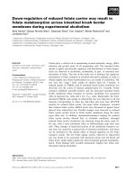
ABC
Fig. 1. Comparison of the SDS electrophoretic patterns of OGDHC preparations from brain and heart mitochondria upon separation on 10%
(A, C) and 7% (B) gels. Molecular mass markers (kDa) are indicated on the right, lane numbers are given in the upper row, protein bands
are numbered on the left. (A) Brain OGDHC solubilized using ‘Bioruptor’ sonication (lane 1); 1% Chaps extract of the pellet from ‘Bioruptor’-
sonicated mitochondria (lane 2); heart OGDHC solubilized by 1% Chaps after ‘Bioruptor’ sonication (lane 3); markers (lane 4). (B) Heart
OGDHC solubilized by 1% Chaps after ‘Bioruptor’ sonication (lane 1); brain OGDHC solubilized by ‘Bioruptor’ sonication (lane 2); markers
(lane 3). (C) Brain OGDHC solubilized by the probe sonicator (lane 1); markers (lane 3).
Novel 2-oxoglutarate dehydrogenase V. Bunik et al.
4992 FEBS Journal 275 (2008) 4990–5006 ª 2008 The Authors Journal compilation ª 2008 FEBS
A comparison of the SDS electrophoretic patterns of
partially purified heart and brain complexes is shown
in Fig. 1A,B. Varying the concentration of the separat-
ing gel (10% in Fig. 1A and 7% in Fig. 1B) allowed
for a better resolution of some proteins, in particular,
those in band 6. The SDS electrophoretic pattern of
our preparation from rat heart mitochondria (Fig. 1A,
lane 3; Fig. 1B, lane 1) agrees with the known mobility
of the components of bovine heart complexes isolated
from total heart extract [23]. According to the molecu-
lar mass values for the mature proteins, the components
of the 2-oxoglutarate and pyruvate dehydrogenase
complexes were ascribed to the major protein bands of
our preparation as follows: E1o (band 1), E2p (band 2),
E3 (band 3), E2o and the E3-binding component of the
pyruvate dehydrogenase complex (protein X; band 4),
E1pa (band 6), E1pb (band 8). This was confirmed by
nanoLC-MS ⁄ MS identification of the components in the
protein bands (Table 1). Our study also showed that
there are two isoenzymes of pyruvate dehydrogenase
kinases in brain (band 6a). Isoenzymes 2 and 3 were
distinguished by three and five specific peptides out of
four and six total peptides identified, respectively
(Table 1).
Interaction of OGDHC with membraneous
proteo-lipid particles and its functional significance
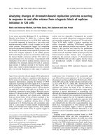
With the sonication parameters fixed, later elution on
size-exclusion chromatography on a Sephacryl HR300
column was observed for OGDHC extracted using
detergent compared with OGDHC solubilized by soni-
cation only. V
e
decreased reproducibly, from 47 to
44 mL for brain OGDHC and from 44 to 42 mL for
heart OGDHC, with standard deviations in V
e
between different chromatographies of a certain prepa-
ration type of < 1 mL. Concomitantly, the shift in V
e
was observed for the high molecular mass opalescent
peak eluted between the column void volume
(V
0
= 38 mL) and the OGDHC activity peak (V
e
between 42 and 47 mL) (Fig. 2A). Elution near the
void volume of the column (Fig. 2A), high opalescence
at a relatively low protein level and the dependence of
V
e
on both the detergent and the sonication mode sug-
gest that this peak comprises membraneous particles.
Membrane vesicles that form spontaneously during
homogenization are known as the microsomal fraction
[27]. A strong dependence of the elution volume of
OGDHC on the elution volume of the opalescent peak
(Fig. 2B, correlation coefficient 1.13) points to OG-
DHC binding to these membrane particles, with their
complex disrupted by the chromatography-accom-
plished trapping of the dissociated intermediates.
Table 2 shows that the better the separation of
OGDHC from microsomes, the more E1o and E3
dissociate from the complex, accompanied by a loss of
total OGDHC activity when subjected to chromatogra-
phy. Increasing dissociation was obvious from the
appearance of the well-defined peak for the compo-
nent activities (DV
e
„ 0; Table 2), which follows the
Table 1. MS identification of known components of the 2-oxo acid dehydrogenase complexes from brain. Proteins of the bands shown in
Fig. 1 were identified through an NCBI search using
MASCOT as described in Experimental procedures. The data for a representative experi-
ment are given. Components of the 2-oxoglutarate dehydrogenase complex were also identified in heart. Unless indicated otherwise,
matches to rat sequences were found. Molecular mass corresponds to the precursor proteins as given in NCBI. NCBI-provided molecular
mass of dihydrolipoyllysine acetyltransferase refers to an incomplete sequence, therefore the true molecular mass from the Expasy data-
base, which corresponds to that in the SDS-electrophoresis (Fig.1), is added (marked by asterisk). NA, not analyzed.
Band
in Fig. 1
Component of the 2-oxo acid dehydrogenase
complexes
NCBI
identifier
Molecular
mass (Da)
Brain Heart
Protein
score
No.
peptides
matched
Protein
score
No.
peptides
matched
1 2-Oxoglutarate dehydrogenase (E1o) 62945278 117 419 1131 28 1647 60
2 Dihydrolipoyllysine acetyltransferase (E2p) 220838 57 645
67 166*
443 16 NA
3 Dihydrolipoyl dehydrogenase (E3) 40786469 54 574 579 12 975 36
4 Dihydrolipoyl succinyl transferase (E2o) 55742725 49 236 400 7 709 28
4 Component X 28201978 mus 54 250 126 2 279 7
6a Pyruvate dehydrogenase kinase, isoenzyme 3 21704122 mus 48 064 196 6 NA
6a Pyruvate dehydrogenase kinase 2 subunit
variant p45
8895958 44 198 151 4 NA
6b Pyruvate dehydrogenase alpha subunit (E1pa) 57657 43 853 716 20 NA
8 Pyruvate dehydrogenase beta subunit (E1pb) 56090293 39 299 519 26 NA
V. Bunik et al. Novel 2-oxoglutarate dehydrogenase
FEBS Journal 275 (2008) 4990–5006 ª 2008 The Authors Journal compilation ª 2008 FEBS 4993
overall OGDHC activity peak, and an increased ratio
of dissociated to complex-bound activities for E3 and
E1o at the corresponding elution volumes. Importantly,
the chromatography-induced dissociation into compo-
nents and the accompanying loss of total OGDHC
activity were dependent on the separation from micro-
somes rather than on the protein applied (Table 2;
experiment N 1 versus 3). Because of the higher analy-
tical sensitivity of the E3-catalyzed NAD
+
reduction
compared with ferricyanide reduction by E1o, the
E3-catalyzed reaction allowed a better comparison of
the significantly different levels of the component activ-
ities obtained in these experiments. However, a similar
trend was observed for the two components (Table 2),
in good agreement with the known formation of the
E1o–E3 subcomplex upon OGDHC dissociation [28].
Separation from microsomes decreases both the total
and the specific (lmolÆmin
)1
Æmg
)1
of protein) activity
of OGDHC in the peak. Table 2 shows that purifica-
tion of OGDHC by chromatography led to a 30-fold
increase in specific activity with a low degree of
separation from microsomes (experiment 1), but full
separation (experiment 3) resulted in no increase in spe-
cific activity, despite the OGDHC fractions containing
fewer contaminant proteins. Thus, disruption of the
interaction between OGDHC and the microsomal frac-
tion during chromatography destabilizes the complex
structure and function.
At a comparable protein concentration in the
column eluate, the fraction of applied OGDHC activ-
ity found in the eluate differed dramatically for heart
(70%) and brain (10%) complexes. The greater loss of
brain OGDHC activity (90%) compared with that
from heart complex (30%) was not due to a higher
degree of purification, because more proteins co-eluted
with OGDHC from brain. This was evident from the
additional bands on SDS electrophoresis (bands 6a, 7,
8a in Fig. 1) and the greater heterogeneity indicated by
nanoLC-MS ⁄ MS analysis of common bands 1, 3, 4, 5.
The tissue specificity of the heterogeneity was mostly
due to synaptosomal proteins in the brain preparation,
Fig. 2. Gel filtration of brain OGDHC on a Sephacryl HR300 16 ⁄ 60
column. (A) Elution profile, showing attenuance at 280 nm (D
280
)
and the OGDHC activity in arbitrary units (A). (B) Dependence of V
e
of OGDHC on V
e
of membraneous fraction, the line is drawn
according to the equation: y=1.13x ) 3.05.
Table 2. Dependence of the OGDHC activity yield on the separation of OGDHC from microsomes. Partially purified from ‘Bioruptor’-soni-
cated mitochondria, OGDHC (40–60 mgÆmL
)1
) was applied to the 12 ⁄ 30 column with Sephacryl S-300. The separation varied due to the dif-
ferences in the sample volume and ⁄ or relative content of the microsomes. The interference of the elution volumes of OGDHC and
microsomes, I, was calculated from the elution profiles as the percentage of the microsome-including OGDHC fractions to the total number
of the OGDHC-containing fractions. Separation of E3 or E1o from the complex upon chromatography was characterized by the difference
between the elution volumes, DV
e
, of the peaks of E3 or E1o and OGDHC and the ratio of the component activities at these V
e
(A
non-bound
E3 ⁄ E1o
⁄ A
bound E3 ⁄ E1o
). The OGDHC activity yield is the ratio of the total activity of OGDHC in the eluate to the total activity of the OGDHC
applied to the column. ND, not determined.
No.
Total
protein
applied
(mg)
Separation
of OGDHC
and microsomes,
(100 ) I ) (%)
Dissociation of E1o
from OGDHC
Dissociation of E3
from OGDHC
Total
OGDHC
activity
yield (%)
Specific
OGDHC
activity
increase (%)
DV
e
A
non-bound E1o
A
bound E1o
DV
e
A
non-bound E3
A
bound E3
1 80 25 0 0.3 0 0.8 66 3000
2 40 56 3 0.9 3 1.9 24 300
3 80 100 ND ND 3 2.7 9 100
Novel 2-oxoglutarate dehydrogenase V. Bunik et al.
4994 FEBS Journal 275 (2008) 4990–5006 ª 2008 The Authors Journal compilation ª 2008 FEBS
pointing to the presence of synaptosome-derived
microsomes in the membraneous fraction accompany-
ing brain OGDHC.
Structural differences between OGDHC from
brain and heart
An essential difference between brain and heart
OGDHC was revealed by nanoLC-MS ⁄ MS analysis of
band 1. In the brain preparation, this band contained
both OGDH and OGDHL, a hypothetical isoenzyme
of OGDH predicted from the nucleic acid data [16].
Our analysis of 10 band 1 samples from 9 different
brain preparations identified the structures of 10–17
peptides which were specific for OGDH and 5–10 pep-
tides specific for OGDHL (Table 3, Fig. 3). Although
direct quantification of proteins from the nanoLC-
MS ⁄ MS peak intensities is difficult, there is a general
correlation between the number of protein peptides
identified and the amount of protein present in the
mixture, if protein size is normalized [13–15]. For
OGDH and OGDHL, which have similar molecular
masses, the ratio of identified peptides may be taken as
an estimate of the relative abundance of the isoen-
zymes in the analyzed sample. We calculated this ratio
for OGDH and OGDHL, using either the number of
all peptides identified or only those specific for the
sequences and that were non-redundant (when peptides
with the same primary sequence were counted as one).
The latter excludes a possible bias due to common
peptides, and is thus a better measure of the specific
sequence coverage. However, with the high sequence
coverage for each of the isoenzymes (Fig. 3), both cal-
culations give a similar ratio. The ratio points to a
comparable amount of the two isoenzymes in the brain
preparation ( 60% OGDH and 40% OGDHL;
Table 3). No reproducible enrichment of OGDHC
with one of the isoenzymes could be detected in the
different OGDHC preparations, for example, isolated
with or without detergents, before or after gel-filtra-
tion, precipitated by either poly(ethylene glycol) or
pH, and collected from different pools of column elu-
ate, which may vary in the OGDHC saturation by
peripheral components E1o and E3 (Table 3). It is
worth noting that the same isoenzyme ratio was
observed in both the crude poly(ethylene glycol) frac-
tion of the mitochondrial extract and the chromatogra-
phy-purified OGDHC (Table 3). Co-purification of the
novel isoenzyme with the high molecular mass
OGDHC fraction points to OGDHL being the com-
plex component, in good agreement with predictions
based on the structural analysis [16].
Table 3. Ratio of the peptides characteristic of OGDH and OGDHL isoenzymes in different preparations of brain OGDHC. Samples isolated
under the indicated conditions (details in Experimental procedures) were subjected to SDS electrophoresis, and the OGDH ⁄ OGDHL band of
110 kDa was analyzed using nanoLC-MS ⁄ MS. The indicated number of specific peptides refers to the non-redundant peptides only, i.e. the
same peptide modified or of a reduced length was not counted. The total number of peptides found by
MASCOT search, as described in
Experimental procedures is given in parentheses.
Isolation conditions
OGDH-specific
(total) peptides
OGDHL-specific
(total) peptides
Specific (total)
peptide ratio (%
OGDH : OGDHL)
‘Bioruptor’ + PEG before chromatography 10 (17) 6 (11) 60 : 40 (60 : 40)
‘Bioruptor’ + PEG
V
e
= 43–45 mL 17 (27) 5 (10) 80 : 20 (70 : 30)
V
e
= 43–46 mL 14 (23) 6 (13) 70 : 30 (60 : 40)
V
e
= 43–48 mL 17 (28) 7 (14) 70 : 30 (70 : 30)
‘Bioruptor’ + pH
V
e
= 44–48 mL
12 (20) 10 (16) 50 : 50 (55 : 45)
‘Bandelin’ + PEG
V
e
= 43–46 mL
8 (17) 8 (15) 50 : 50 (50 : 50)
OGDHC solubilized by sonication only 8–17 (17–28)
average 13 (22)
5–10
average 7 (13)
Average 65 : 35 (60 : 40)
(50 : 50 to 80 : 20)
‘Bandelin’, 6% Triton X-100 extract + pH
V
e
= 42–48 mL
12 (23) 9 (19) 60 : 40 (55 : 45)
‘Bioruptor’, 1% Chaps extract + pH
V
e
= 45–48 mL
13 (21) 6 (13) 70 : 30 (60 : 40)
‘Bioruptor’, 1% Chaps extract + PEG
V
e
= 45–48 mL
11 (17) 7 (12) 60 : 40 (60 : 40)
Detergent-solubilized OGDHC 11–13 (17–23)
average 12 (20)
6–9 (12–19)
average 7 (15)
Average 60 : 40 (60 : 40)
(60 : 40 to 70 : 30)
V. Bunik et al. Novel 2-oxoglutarate dehydrogenase
FEBS Journal 275 (2008) 4990–5006 ª 2008 The Authors Journal compilation ª 2008 FEBS 4995
OGDHL_rat_h MSQLRLLLFRLGP QARKLLATRDIAAFG GRRRSSGPPTTIPRSRGGVSPSYVEEMYFAWLENPQSVHKSWDNFF 74
OGDH_rat_h MFHLRTCAAKLRPLTASQTVKTFSQNKPAAIRTFQQIRCYSAPVAAEPFLSGTSSNYVEEMYCAWLENPKSVHK SWDIFF 80
OGDH_rat_b MFHLRTCAAKLRPLTASQTVKTFSQNKPAAIRTFQQIRCYSAPVAAEPFLSGTSSNYVEEMYCAWLENPKSVHK SWDIFF 80
OGDHL_rat_b MSQLRLLLFRLGP QARKLLATRDIAAFG GRRRSSGPPTTIPRSRGGVSPSYVEEMYFAWLENPQSVHKSWDNFF 74
OGDHL_rat_h QRATKEASVGPAQPQPP AVIQESRASVSSCTKTSKLVEDHLAVQSLIRAYQIRGHHVAQLDPLGILDADLDSF 147
OGDH_rat_h RNTNAGAPPGTAYQSPLSLSRSSLATMAHAQSLVEAQPNVDKLVEDHLAVQSLIRAYQIRGHHVAQLDPLGILDADLDSS 160
OGDH_rat_b RNTNAGAPPGTAYQSPLSLSRSSLATMAHAQSLVEAQPNVDKLVEDHLAVQSLIRAYQIRGHHVAQLDPLGILDADLDSS 160
OGDHL_rat_b QRATKEASVGPAQPQPP AVIQESRASVSSCTKTSKLVEDHLAVQSLIRAYQIRGHHVAQLDPLGILDADLDSF 147
OGDHL_rat_h VPSDLITTIDKLAFYDLQEADLDKEFRLPTTTFIGGSENTLSLREIIRRLESTYCQHIGLEFMFINDVEQCQWIRQKFET 227
OGDH_rat_h VPADIISSTDKLGFYGLHESDLDKVFHLPTTTFIGGQEPALPLREIIRRLEMAYCQHIGVEFMFINDLEQCQWIR QKFET 240
OGDH_rat_b VPADIISSTDKLGFYGLHESDLDKVFHLPTTTFIGGQEPALPLREIIRRLEMAYCQHIGVEFMFINDLEQCQWIRQKFET 240
OGDHL_rat_b VPSDLITTIDKLAFYDLQEADLDKEFRLPTTTFIGGSENTLSLREIIRRLESTYCQHIGLEFMFINDVEQCQWIRQKFET 227
OGDHL_rat_h PGVMKFSIEEKRTLLARLVRSMRFEDFLARKWSSEKRFGLEGCEVMIPALKTIIDKSSEMGVENVILGMPHRGR LNVLAN 307
OGDH_rat_h PGIMQFTNEEKRTLLARLVRSTRFEEFLQRKWSSEKRFGLEGCEVLIPALKTIIDMSSANGVDYVIMGMPHRGRLNVLAN 320
OGDH_rat_b PGIMQFTNEEKRTLLARLVRSTRFEEFLQRKWSSEKRFGLEGCEVLIPALKTIIDMSSANGVDYVIMGMPHRGRLNVLAN 320
OGDHL_rat_b PGVMKFSIEEKRTLLARLVRSMRFEDFLARKWSSEKRFGLEGCEVMIPALKTIIDKSSEMGVENVILGMPHRGRLNVLAN 307
OGDHL_rat_h VIRKDLEQIFCQFDPKLEAADEGSGDVKYHLGMYHERINRVTNRNITLSLVANPSHLEAVDPVVQGKTKAEQFYRGDAQG 387
OGDH_rat_h VIRKELEQIFCQFDSKLEAADEGSGDMKYHLGMYHRRINRVTDRNITLSLVANPSHLEAADPVVMGKTKAEQFYCGDTEG 400
OGDH_rat_b VIRKELEQIFCQFDSKLEAADEGSGDMKYHLGMYHRRINRVTDRNITLSLVANPSHLEAADPVVMGKTK AEQFYCGDTEG 400
OGDHL_rat_b VIRKDLEQIFCQFDPKLEAADEGSGDVKYHLGMYHERINRVTNRNITLSLVANPSHLEAVDPVVQGKTKAEQFYRGDAQG 387
OGDHL_rat_h RKVMSILVHGDAAFAGQGVVYETFHLSDLPSYTTNGTVHVVVNNQIGFTTDPRMAR SSPYPTDVARVVNAPIFHVNADDP 467
OGDH_rat_h KKVMSILLHGDAAFAGQGIVYETFHLSDLPSYTTHGTVHVVVNNQIGFTTDPRMAR SSPYPTDVARVVNAPIFHVNSDDP 480
OGDH_rat_b
KKVMSILLHGDAAFAGQGIVYETFHLSDLPSYTTHGTVHVVVNNQIGFTTDPRMARSSPYPTDVARVVNAPIFHVNSDDP 480
OGDHL_rat_b RKVMSILVHGDAAFAGQGVVYETFHLSDLPSYTTNGTVHVVVNNQIGFTTDPRMAR SSPYPTDVARVVNAPIFHVNADDP 467
OGDHL_rat_h EAVIYVCSVAAEWRNTFNKDVVVDLVCYRRRGHNEMDEPMFTQPLMYKQIHKQVPVLKKYADKLIAEGTVTLQEFEEEIA 547
OGDH_rat_h EAVMYVCKVAAEWRNTFHKDVVVDLVCYRRNGHNEMDEPMFTQPLMYKQIRKQKPVLQKYAELLVSQGVVNQPEYEEEIS 560
OGDH_rat_b EAVMYVCKVAAEWRNTFHKDVVVDLVCYRRNGHNEMDEPMFTQPLMYKQIRKQKPVLQKYAELLVSQGVVNQPEYEEEIS 560
OGDHL_rat_b EAVIYVCSVAAEWRNTFNKDVVVDLVCYRRRGHNEMDEPMFTQPLMYKQIHKQVPVLKKYADKLIAEGTVTLQEFEEEIA 547
OGDHL_rat_h KYDRICEEAYGRSKDKKILHIKHWLDSPWPGFFNVDGEPKSMTYPTTGIPEDTLSHIGNVASSVPLEDFKIHTGLSRILR 627
OGDH_rat_h KYDKICEEAFTRSKDEKILHIKHWLDSPWPGFFTLDGQPRSMTCPSTGLEEDILTHIGNVASSVPVENFTIHGGLSRILK 640
OGDH_rat_b KYDKICEEAFTRSKDEKILHIKHWLDSPWPGFFTLDGQPRSMTCPSTGLEEDILTHIGNVASSVPVENFTIHGGLSRILK 640
OGDHL_rat_b KYDRICEEAYGRSKDKKILHIKHWLDSPWPGFFNVDGEPKSMTYPTTGIPEDTLSHIGNVASSVPLEDFKIHTGLSRILR 627
OGDHL_rat_h GRADMTKKRTVDWALAEYMAFGSLLKEGIHVRLSGQDVERGTFSHRHHVLHDQDVDRRTCVPMNHLWPDQAPYTVCNSSL 707
OGDH_rat_h TRRELVTNRTVDWALAEYMAFGSLLKEGIHVRLSGQDVERGTFSHRHHVLHDQNVDKRTCIPMNHLWPNQAPYTVCNSSL 720
OGDH_rat_b TRRELVTNRTVDWALAEYMAFGSLLKEGIHVRLSGQDVERGTFSHRHHVLHDQNVDKRTCIPMNHLWPNQAPYTVCNSSL 720
OGDHL_rat_b GRADMTKKRTVDWALAEYMAFGSLLKEGIHVRLSGQDVERGTFSHRHHVLHDQDVDRRTCVPMNHLWPDQAPYTVCNSSL 707
OGDHL_rat_h SEYGVLGFELGYAMASPNALVLWEAQFGDFHNTAQCIIDQFISTGQAKWVRHNGIVLLLPHGMEGMGPEHSSARPERFLQ 787
OGDH_rat_h SEYGVLGFELGFAMASPNALVLWEAQFGDFNNMAQCIIDQFICPGQAKWVRQNGIVLLLPHGMEGMGPEHSSARPERFLQ 800
OGDH_rat_b SEYGVLGFELGFAMASPNALVLWEAQFGDFNNMAQCIIDQFICPGQAKWVRQNGIVLLLPHGMEGMGPEHSSARPERFLQ 800
OGDHL_rat_b SEYGVLGFELGYAMASPNALVLWEAQFGDFHNTAQCIIDQFISTGQAKWVRHNGIVLLLPHGMEGMGPEHSSARPERFLQ 787
OGDHL_rat_h MSNDDSDAYP-VFTEDFEVSQLYDCNWIVVNCSTPASYFHVLRRQVLLPFR KPLIVFTPKSLLRHPDAKSSFDQMVSGTS 866
OGDH_rat_h MCNDDPDVLPNLQEENFDISQLYDCNWIVVNCSTPGNFFHVLRRQILLPFRKPLIVFTPKSLLRHPEARTSFDEMLPGTH 880
OGDH_rat_b MCNDDPDVLPNLQEENFDISQLYDCNWIVVNCSTPGNFFHVLRRQILLPFR KPLIVFTPKSLLRHPEARTSFDEMLPGTH 880
OGDHL_rat_b MSNDDSDAYP-VFTEDFEVSQLYDCNWIVVNCSTPASYFHVLRRQVLLPFR KPLIVFTPKSLLRHPDAKSSFDQMVSGTS 866
OGDHL_rat_h FQRMIPEDGPAAQSPERVERLIFCTGKVYYDLVKERSSQGLEKQVAITRLEQISPFPFDLIMREAEKYSGAELVWCQEEH 946
OGDH_rat_h FQRVIPEDGPAAQNPDKVKRLLFCTGKVYYDLTRERKARDMAEEVAITRIEQLSPFPFDLLLKEAQKYPNAELAWCQEEH 960
OGDH_rat_b FQRVIPEDGPAAQNPDKVKRLLFCTGKVYYDLTRERKARDMAEEVAITRIEQLSPFPFDLLLKEAQKYPNAELAWCQEEH 960
OGDHL_rat_b FQRMIPEDGPAAQSPERVERLIFCTGKVYYDLVKERSSQGLEKQVAITRLEQISPFPFDLIMREAEKYSGAELVWCQEEH 946
OGDHL_rat_h KNMGYYDYISPRFMTLLGHSRPIWYVGREPAAAPATGNKNTHLVSLRKFLDTAFNLKAFEGKTF 1010
OGDH_rat_h
KNQGYYDYVKPRLRTTIDRAKPVWYAGRDPAAAPATGNKKTHLTELQRFLDTAFDLDAFKK
FS- 1023
OGDH_rat_b KNQGYYDYVKPRLRTTIDRAKPVWYAGRDPAAAPATGNKKTHLTELQRFLDTAFDLDAFKKFS- 1023
OGDHL_rat_b KNMGYYDYISPRFMTLLGHSRPIWYVGREPAAAPATGNKNTHLVSLRKFLDTAFNLKAFEGKTF 1010
*
Fig. 3. Sequence alignment of rat OGDH and OGDHL showing (in color) the peptides identified by nanoLC-MS ⁄ MS in the OGDHC prepara-
tion from heart (two upper sequences marked by ‘h’) and brain (two lower sequences marked by ‘b’). Common peptides for the two
sequences are shown in red. The sequence-specific peptides are in bold: pink for the OGDH and blue for the OGDHL. The N-terminal cleav-
age site, as determined by the sequencing of the truncated bovine E1o [35], is marked by an asterisk above the alignment.
Novel 2-oxoglutarate dehydrogenase V. Bunik et al.
4996 FEBS Journal 275 (2008) 4990–5006 ª 2008 The Authors Journal compilation ª 2008 FEBS
In contrast to brain OGDHC, no peptide specific
for OGDHL was identified in the heart complex,
despite the higher protein load and the purity of the
E1o band (band 1, lane 3 versus lane 1; Fig. 1A),
which resulted in an increase in the sequence coverage
(26–36 non-redundant or 45–60 total peptides in inde-
pendent determinations). As shown in Fig. 3, heart
preparation exhibits either OGDH-specific peptides
(pink) or peptides common to the two proteins (red),
but OGDHL-specific peptides (blue) were found in the
brain preparation only. Thus, whereas only the known
OGDH component coded by chromosome 7 in
humans [29,30] was identified by nanoLC-MS ⁄ MS in
OGDHC from heart, brain complex, purified using
the same procedure, contained comparable amounts
of both OGDH (chromosome 7) and OGDHL
(human chromosome 10) [31–33] proteins (Table 3,
Fig. 3), which were identified even at lower purifica-
tion yields.
Another structural feature of brain 2-oxo acid dehy-
drogenase complexes is seen from SDS electrophoresis.
The E3 component (band 3), the majority of which is
associated with OGDHC as shown above, is hardly
visible in the brain preparation (Fig. 1A, lanes 1–2)
compared with the heart preparation (Fig. 1A, lane 3).
Despite the low E3 level, under standard assay condi-
tions we did not observe any activation of brain
OGDHC in the presence of or following preincubation
with at least a 10-fold protein excess of E3 (commer-
cial bovine enzyme). Thus, even the low levels of E3
seen in the brain preparation were able to support
maximal OGDHC reaction rates. This is in accordance
with published data on the rate-limiting role of the
E1o component in Reaction (1) catalyzed by the com-
plex [34]. It is known that the binding of E3 to OG-
DHC is mediated by E1o, with the proteolytic removal
of a small N-terminal fragment of E1o impairing bind-
ing [28,35]. However, the lower E3 level in brain
OGDHC was not due to E1o proteolysis, because
several peptides preceding the cleavage site (marked by
asterisk in Fig. 3) were identified in both isoenzymes
by MS analysis. This was in good agreement with the
mobility of the E1o band on SDS electrophoresis
(Fig. 1), which corresponded to the molecular mass of
non-proteolysed E1o (110 kDa), being higher than that
of truncated E1o with an apparent molecular mass of
94 kDa [28,35]. Because full extraction of the OGDHC
activity from heart mitochondria required 1% Chaps,
we checked whether the E3 deficiency of brain
OGDHC could be due to the membrane binding of its
E3. Figure 1A shows that 1% Chaps extract of the
pellet fraction (lane 2) obtained after removal of
E3-deficient OGDHC (lane 1) did not contain E3. By
contrast, when the activity of E3 and OGDHC was
followed in parallel upon sonication, a significant
portion of the E3 activity solubilized before the overall
activity of OGDHC. Taken together, these findings
indicate that the E3 deficiency of brain 2-oxo acid
dehydrogenase complexes (Fig. 1) is not due to mem-
brane binding of the E3 component. Compared with
heart complexes, easier dissociation of this component
appears to occur upon sonication of brain mitochon-
dria. Indeed, E3 was better presented in complexes
that were detergent-extracted after a less efficient soni-
cation (Fig. 1C). Sonication by ‘Bioruptor’ (Fig. 1A,B)
was nevertheless preferred for the isolation, because it
gave reproducible results and did not lead to the high
molecular mass aggregates (150–300 kDa) observed in
the SDS electrophoresis of OGDHC solubilized with
the probe sonicator (Fig. 1C).
A different supramolecular organization for OG-
DHC from brain and heart was further supported by
size-exclusion chromatography, in which proteins of a
higher molecular mass are eluted more rapidly, i.e. at
a lower elution volume V
e
. As mentioned above, under
the same sonication conditions the activity peak of
OGDHC from brain eluted later than that of OGDHC
from heart: 44 versus 42 mL for soluble OGDHC and
47 versus 44 mL for Chaps-extracted OGDHC. The
later elution corresponds to a lower molecular mass
for the purified brain complex, which agrees with its
lower saturation with peripheral E3 component, as dis-
cussed above. As inferred from both SDS electropho-
resis (Fig. 1A,B) and size-exclusion chromatography,
the different supramolecular organization of heart and
brain OGDHC was further supported by the
MS-based estimate of the relative abundance of
the complex components in the preparation (Table 4).
Abundance coefficients were calculated as described in
Experimental procedures according to the previously
developed approach of comparative proteomics [13–
15]. As indicated by the standard deviation values for
the preparations from one tissue, these ratios showed
good agreement in different experiments. However, the
values were clearly different for OGDHC from heart
and brain. Table 4 shows that in OGDHC from brain
the E2o ⁄ E1o and E3 ⁄ E1o ratios (40 and 70%, respec-
tively) were no more than half those in the heart com-
plex (120 and 140%, respectively). Because the heart
preparation did not possess OGDHL, we also com-
pared the abundance coefficients for brain OGDHC
when based on the OGDH content only. The brain
ratios remained lower than those of heart (Table 4).
Thus, compared with the heart complex, OGDHC
isolated from brain showed an excess of the first
component over the second and third. The decrease in
V. Bunik et al. Novel 2-oxoglutarate dehydrogenase
FEBS Journal 275 (2008) 4990–5006 ª 2008 The Authors Journal compilation ª 2008 FEBS 4997
the MS-based abundance of E2o and E3 components
in brain OGDHC correlated with the low intensity of
the E3 band in SDS electrophoresis and a lower
molecular mass of OGDHC from brain versus heart
upon size-exclusion chromatography. Thus, the data
obtained using the three independent approaches sug-
gest differences in the supramolecular organization of
OGDHC isolated from heart and brain.
Saturation of brain OGDHC with 2-oxoglutarate
Kinetic analysis of the dependence of the overall activ-
ity of brain OGDHC on the saturation with 2-oxoglut-
arate agrees with the presence in the preparation of
two isoenzymes of 2-oxoglutarate dehydrogenase
which are functionally competent in Reaction (1). Sol-
ubilized with or without detergents, brain OGDHC
did not exhibit standard Michaelis–Menten kinetics
(Fig. 4). That is, simulations using the parameters
yielded by the double reciprocal linearization of the
experimental data showed a systematic shift in the the-
oretical curves to lower rates at high 2-oxoglutarate
saturation (Fig. 4A). In view of the identification of
the second isoenzyme of OGDH by nanoLC-MS ⁄ MS,
we introduced a second saturation function into the
equation. As shown in Fig. 4B, this abolished the
inconsistencies between the experiment and the simula-
tion, resulting in a satisfactory description of the sys-
tem behavior at both low and high substrate
saturation. The better correspondence between the
experimental data and the two-saturation model is
obvious not only from visual inspection of the coinci-
dence between the experimental points and theoretical
curves in Fig. 4B compared with Fig. 4A, but also
from an increase in the correlation coefficients (from
0.804 and 0.918 in Fig. 4A to 0.986 and 0.997 in
Fig. 4B). The biphasic saturation parameters provided
in the legend to Fig. 4 show that K
m,1
and K
m,2
values,
as well as the contributions of V
1
and V
2
to the
maximal reaction rate (V=V
1
+ V
2
), were similar
for soluble and Chaps-extracted OGDHC. Based
on three independent experiments, the following
parameters were obtained: K
m,1
= 0.07 ± 0.02 mm;
K
m,2
=0.40 ± 0.07 mm; V
1
⁄ (V
1
+ V
2
) = 45 ± 4%;
V
2
⁄ (V
1
+ V
2
) = 55 ± 4%. It is worth noting that the
simulation-derived partial contributions of V
1
and V
2
to the overall V value are close to the relative abun-
dance of the isoenzymes as determined by nanoLC-
MS ⁄ MS (35–40% of OGDHL and 60–65% of OGDH;
Table 3). Moreover, detergents are known to desensi-
tize cooperative and allosteric enzymes to effectors,
but they do not significantly change the kinetic param-
eters of brain OGDHC (Fig. 4), in accordance with
the lack of change in the isoenzyme ratio caused by
detergents (Table 3). Thus, the parameters obtained by
simulation of the v(S) dependence according to the
model suggested by the MS identification of the two
isoenzymes are reproducible and in a good agreement
with the MS-based abundance of the isoenzymes in the
OGDHC preparation. Taken together, the kinetic and
MS data support functional competence of the novel
isoenzyme in the overall OGDHC reaction and differ-
ent saturation of the two isoenzymes of OGDH with
2-oxoglutarate.
Discussion
Identification of novel OGDH isoform and its
implication in brain metabolism
Distinguishing proteins with highly similar primary
structures, such as the products of alternative splic-
ing or of different genes (isoforms or isoenzymes),
represents one of the challenges in characterizing the
cellular proteome [15]. The modern development of
MS analysis provides strong advantages over immu-
nological approaches to address this challenge,
because determination of isoform-specific peptides
distinguishes unambiguously between isoforms which
may show cross-reactivity to antibodies [36]. In this
study, we successfully applied nanoLC-MS ⁄ MS to
identify both the known OGDH and the hypotheti-
cal OGDHL in OGDHC partially purified from
brain mitochondria. At the same time, only the
Table 4. Relative abundance of the OGDHC components in the preparations. Abundance index, A, corresponds to the number of peptides
detected by nanoLC-MS ⁄ MS, normalized to the molecular mass of the OGDHC component (see Experimental procedures). The E2o and E3
abundance indexes were related to that of either E1o (the sum of OGDH + OGDHL) or OGDH taken as 100%. The data are presented as
the average values ± SD.
Tissue
E1o (OGDH + OGDHL) OGDH E2o E3
A % E1o A % OGDH A % E1o ⁄ %OGDH A % E1o ⁄ %OGDH
Brain 0.34 ± 0.03 100 0.22 ± 0.04 100 0.14 ± 0.05 40 ⁄ 60 0.23 ± 0.01 70 ⁄ 100
Heart 0.48 ± 0.1 100 0.57 ± 0.01 120 0.66 ± 0.18 140
Novel 2-oxoglutarate dehydrogenase V. Bunik et al.
4998 FEBS Journal 275 (2008) 4990–5006 ª 2008 The Authors Journal compilation ª 2008 FEBS
known OGDH was determined in a similar prepara-
tion of the complex from heart. Expression of the novel
OGDHL component of OGDHC in brain is in accord
with the isolation of OGDHL cDNA from brain tissue
[31–33], whereas earlier cloning of the OGDH gene
used a fetal liver cDNA library [29]. Thus, apart from
housekeeping OGDH, OGDHL is synthesized in
brain. Integration of the OGDHL isoenzyme into the
complex, which was predicted by our structure–
function analysis [16], is evident from the constant
ratio of OGDH and OGDHL during the purification
of brain OGDHC (Table 1), elution of OGDHL in
the high molecular mass fraction corresponding to
OGDHC, and biphasic saturation with 2-oxoglutarate
(Fig. 4), indicative of a functional competence of
the two isoenzymes in Reaction (1) catalyzed by the
complex.
Identification of the two isoenzymes of OGDHC by
MS was taken into account in the kinetic modeling of
the dependence of the overall reaction rate of brain
OGDHC on the 2-oxoglutarate concentration (Fig. 4).
Indeed, the dependence can be better described by the
sum of two saturation processes than by standard
Michaelis–Menten kinetics (Fig. 4), which is in accord
with the contribution to the overall reaction rate of
the two isoenzymes having different affinities to 2-oxo-
glutarate. Moreover, simulation of this model revealed
that partial contributions of each of the isoenzymes,
V
1
and V
2
into the overall reaction rate V are in a
good agreement with the MS-based relative abundance
of the isoenzymes (Table 3), suggesting that OGDH
and OGDHL have similar catalytic rates. The compat-
ibility of parameters derived from kinetic modeling
and MS analysis strongly supports the plausibility of
the model assuming two isoenzymes for interpretation
of the kinetic data. High correlation between the simu-
lated dependence and experimental data within this
model (Fig. 4B) did not justify further refinement of
the model. Thus, kinetic analysis of OGDHC from
brain provides experimental evidence in support of an
earlier prediction from genome data that OGDHL is a
functionally active isoenzyme of OGDH [16]. Further-
more, the kinetics is indicative of an approximately
sixfold difference between K
m,1
and K
m,2
characterizing
saturation of the two isoenzymes with 2-oxoglutarate.
Compared with OGDHC from heart and adrenal
glands, which are half-saturated with 2-oxoglutarate at
0.2 mm [37–39], OGDHC from brain requires higher
concentrations for full saturation (K
m,2
= 0.40
± 0.06 mm), being sensitive to lower concentrations of
2-oxoglutarate (K
m,1
= 0.07 ± 0.02 mm). Possessing
the two isoenzymes which provide the different K
m
values, brain OGDHC may thus respond to an
expanded interval in the 2-oxoglutarate levels. The
differential regulation of brain OGDH isoenzymes by
the substrate may also address the physiological needs
of brain tissue to establish different steady-state
concentrations of 2-oxoglutarate, depending on cellular
conditions, compartment or type. Compared with
other tissues, physiological concentrations of glutamate
in brain differ not only between regions and cell types,
Fig. 4. Kinetic analysis of brain OGDHC saturation with 2-oxogluta-
rate. Hollow circles, soluble OGDHC; filled circles, detergent-
extracted OGDHC. Dependence of the reaction rate v (arbitrary
units of the fluorescence change dFÆmin
)1
Æmg
)1
of protein) on the
2-oxoglutarate concentration ([S]) was approximated by a single
Michaelis–Menten curve v=170*[S] ⁄ (0.07 + [S]), r
2
= 0.804 for
soluble OGDHC and v=480*[S] ⁄ (0.09 + [S]) for detergent-
extracted OGDHC, r
2
= 0.918 (A) or the sum of the two Michaelis–
Menten curves v=110*[S] ⁄ (0.07 + [S]) + 170*[S] ⁄ (0.47 + [S]),
r
2
= 0.986 for the soluble OGDHC and v=350*[S] ⁄ (0.09
+ [S]) + 320*[S] ⁄ (0.42 + [S]), r
2
= 0.997 for detergent-extracted
OGDHC (B). Details of the simulation procedure are given in Experi-
mental procedures.
V. Bunik et al. Novel 2-oxoglutarate dehydrogenase
FEBS Journal 275 (2008) 4990–5006 ª 2008 The Authors Journal compilation ª 2008 FEBS 4999
but also during behavioral responses [40]. Because of
this, the distribution of 2-oxoglutarate between irre-
versible degradation by OGDHC and transformation
to glutamate must be subject to more diverse regula-
tion in the brain. OGDH isoenzymes with different
affinities to 2-oxoglutarate extend regulatory means to
control the glutamate ⁄ 2-oxoglutarate ratio, which is
governed by the brain-specific isoenzymes of glutamate
dehydrogenase and isoforms of mitochondrial gluta-
mate carrier [41].
Differences in supramolecular organization of
brain and heart OGDHC
In this study, we characterized the relative abundance
of the OGDHC components in partially purified
complexes from brain and heart (Table 4) by using
MS-based estimates of protein abundance. This semi-
quantitative approach developed for comparative pro-
teomics studies of (sub)cellular proteomes [13–15]
was very useful in our comparative structural charac-
terization of non-homogeneous OGDHC from heart
and brain, because it enabled us to study the com-
plex which is prone to dissociation and inactivation
upon purification. Applying this approach to partially
purified OGDHC from heart, we also showed that
the component ratio determined by MS analysis in
this study is in a good agreement with the ratio
established previously by alternative approaches using
highly purified preparations of heart OGDHC. That
is, the stoichiometry of the enzymatic components
determined through the content of cofactors bound
to the highly purified OGDHC from heart was
shown to be 1 : 1 : 1.5 [42]. Taking into account that
each subunit of the complex components binds one
cofactor, this ratio is in good agreement with our
MS-based estimation of the relative abundance of the
components in partially purified OGDHC from heart,
which is 1 : 1.2 : 1.4 (Table 4). In another study on
homogeneous OGDHC from heart, which used deter-
gents and a dissociation–association procedure to
estimate the molar ratio of the components in the
complex, the ratio was identified as 1 : 2 : 1 [22]. The
lower content of the peripheral components in this
preparation compared with the earlier estimation of
the same research group 1 : 1 : 1.5 [42] may well be
due to the partial dissociation and ⁄ or lost ability to
reassociate upon purification and resolution of the
complex components in the presence of detergents.
Thus, the abundance coefficients determined in our
study for heart OGDHC by MS (Table 4) are in rea-
sonable agreement with the published data on heart
OGDHC [22,42], confirming the applicability of MS
to estimate the relative abundance of OGDHC com-
ponents in our partially purified preparations. The
agreement also shows that the common E3 compo-
nent of the pyruvate dehydrogenase complex does
not contribute greatly to the abundance coefficients
determined for the OGDHC-enriched fraction from
heart. Regarding our brain OGDHC preparation,
this is independently supported by < 10% of the
pyruvate dehydrogenase complex activity relative to
that of OGDHC, and co-elution of E3 with OGDHC
or E1o, whereas the activity peak of the pyruvate
dehydrogenase complex with a higher molecular mass
is shifted to the lower elution volume. Together with
the elution beyond the void volume of the column
(Fig. 2), these findings suggest that our OGDHC-
enriched preparation does not contain significant
amounts of E3-saturated pyruvate dehydrogenase
complex. This is in a good agreement with the E3
deficiency of the rat brain pyruvate dehydrogenase
complex, which was observed with purified complex
[43]. Independently, these and our results indicate
that E3 saturation of the 2-oxo acid dehydrogenase
complexes is lower in brain than in heart. Even if
the MS-based abundance of brain OGDHC compo-
nents (Table 4) is distorted by a loss of brain E3
and E2o during electrophoresis and ⁄ or peptide
extraction from gel, the tissue specificity of the abun-
dance (Table 4) would indicate structural differences
between heart and brain E2o and E3. Because these
enzymes are encoded by the same genes in the two
tissues, their structural differences may be due to
alternative splicing or post-translational modification.
In general, these modifications may affect protein
solubility and peptide extraction upon electrophoresis
per se and ⁄ or through membrane binding of the
modified protein [44]. However, the same electropho-
retic mobility of the E2o and E3 components from
heart and brain (Fig. 1) does not support significant
peptide loss due to alternative splicing or post-trans-
lational modification. Besides, we showed that E3
from brain mitochondria is not membrane bound,
and it appears unlikely that the major fraction of
brain E2o molecules is. That is, if all E2os were
membrane bound, the known assembly into the mul-
tienzyme complex would be compromised much more
than is suggested by the relatively mild differences
between the heart and brain complexes upon size-
exclusion chromatography. Thus, although tissue-
specific structural changes in a fraction of the E2o
and ⁄ or E3 molecules can not be excluded, the com-
bined results of SDS electrophoresis, size-exclusion
chromatography and MS analysis do not support the
idea that such changes are responsible for differences
Novel 2-oxoglutarate dehydrogenase V. Bunik et al.
5000 FEBS Journal 275 (2008) 4990–5006 ª 2008 The Authors Journal compilation ª 2008 FEBS
in the MS-based abundance of the OGDHC compo-
nents (Table 4). By contrast, all these approaches are
consistent with the supramolecular structure of brain
OGDHC differing from that of the heart complex.
Established differences in the supramolecular struc-
ture and stability of brain versus heart OGDHC cor-
relate with the presence in the brain complex of the
novel isoenzyme, OGDHL, agree with previous struc-
ture–function analysis of protein–protein interactions
in OGDHC and provide important insights into phys-
iologically relevant issues. A more pronounced disso-
ciation of the E3 component from brain versus heart
OGDHC, as suggested by SDS electrophoresis
(Fig. 1) and a lower abundance of E3 estimated by
MS analysis (Table 4), is not due to E1o proteolysis
which is known to impair E3 binding to OGDHC
[28,35]. As shown above, this is evident from the
non-proteolysed structures of OGDH and OGDHL
according to both MS (Fig. 3) and SDS electrophore-
sis (Fig. 1). However, the impaired E3 binding agrees
with the pre-existing structural difference critical for
binding the N-terminal domain ( 10 kDa) which has
several deletions in OGDHL compared with OGDH
(Fig. 3). The reduced affinity of brain OGDHC to E3
may provide additional means to regulate the 2-oxo-
glutarate plus CoA-dependent production of ROS in
brain mitochondria because complex-bound E3 is
needed for this side reaction of OGDHC to occur
[11]. Furthermore, structural differences between the
otherwise highly similar (85% overall sequence simi-
larity) OGDHL and OGDH are mostly confined to
the N- and C-termini of the proteins [16]. These
regions are known to control the homo- and heterol-
ogous protein–protein interactions of the 2-oxo acid
dehydrogenases [28,45–48]. Hence, isoenzyme-specific
protein–protein interactions may also lead to the
over-representation of E1o over E2o in brain
OGDHC (Table 4). For example, OGDHL may form
tetramers bound to the E2o-formed core, which is
known for some 2-oxoglutarate dehydrogenases [49],
whereas in heart complex, OGDH dimers are bound
to the core [22]. The different structure may, in par-
ticular, contribute to the difference in the observed
K
m
value of OGDHC for 2-oxoglutarate, because K
m
is known to be affected by the catalytic steps of the
overall reaction, being different from the dissociation
constant (K
S
) of 2-oxoglutarate binding to E1o [50].
In view of the rate-limiting status of E1o [34], its
allosteric responses to a number of effectors [51] and
its essential role in the regulation of ROS production
by OGDHC [11], its over-representation in brain OG-
DHC may further extend regulatory opportunities of
the overall reaction.
Interaction with proteo-lipid particles stabilizes
the structure and function of OGDHC
Chromatographic purification of OGDHC pointed to
its interaction with the membraneous fraction (Fig. 2),
which is further supported by OGDHC solubilization
concomitant with integral membrane proteins identi-
fied in sonication-solubilized OGDHC by nanoLC-
MS ⁄ MS. The membrane binding of OGDHC is also
inferred from our data on differential solubilization
from sonicated brain mitochondria of the overall activ-
ity of the complex and the activity of the lipoyl dehy-
drogenase component. On the one hand, under
sonication conditions when the overall activity
remained membrane bound, the component activity
was solubilized. This indicates that the mitochondrial
disruption detected by the component activity is not
sufficient for solubilization of the whole complex, and
this is in accordance with an earlier study of kidney
OGDHC [24]. On the other hand, the lipoyl dehydro-
genase component was not abundant in the detergent-
extracted OGDHC fraction (Fig. 1). Hence, it is not
E3, but E1o and⁄ or E2o which bind the complex to
the membrane. The membrane binding of E1o and
E2o agrees with the proteomic data, which revealed an
accumulation of E1o in the mitochondrial outer mem-
brane [52] and an abundance of E2o in the presynaptic
membrane [53].
The interaction of OGDHC with membrane is of
functional significance, as their separation destabilizes
the complex structure and function (Table 2). It is
worth noting, however, that the presence of synaptoso-
mal membranes in the microsomal fraction from brain
correlates with a greater loss of OGDHC activity upon
gel-filtration for the brain versus the heart preparation.
In view of the known abundance of E2o in synaptic
membrane [53], the synaptosomal fraction probably
acts as a sink for E2o, thus promoting the dissociation
of brain OGDHC into components during chromatog-
raphy-induced separation of OGDHC from the mito-
chondrial membrane. Taken together, the data suggest
that interaction with the mitochondrial membrane sta-
bilizes brain OGDHC, whereas substitution of this
interaction for that with synaptic membrane interferes
with the integrity of the complex. This may explain the
observed difference in the stability of brain and heart
OGDHC over the course of purification.
A strong interaction between OGDHC and the
membrane fraction deserves attention in view of the
developing concept of membrane-including microdo-
mains, which can be organized and scaffolded by olig-
omeric proteins [54–56], and the known scaffolding
properties of the E2 components of 2-oxo acid
V. Bunik et al. Novel 2-oxoglutarate dehydrogenase
FEBS Journal 275 (2008) 4990–5006 ª 2008 The Authors Journal compilation ª 2008 FEBS 5001
dehydrogenase complexes [57,58]. This may have
important physiological implications due to the known
pro-oxidant role of lipoic acid. On the one hand,
2-oxo acid dehydrogenase complexes are known as
mitochondrial microcompartments of lipoic acid, and
may generate ROS in response to a distorted ratio of
the substrates of Reaction (1) [11]. On the other hand,
the pro-oxidant action of lipoic acid was shown to
stimulate mitochondrial permeability transition and
apoptosis through the proteins of the mitochondrial
contact sites, the voltage-dependent anion channel and
ATP ⁄ ADP translocase [59,60]. According to our MS
analysis, both of these proteins were expressed in our
preparation enriched with OGDHC. Because under
physiological conditions lipoic acid is not freely avail-
able outside the complexes, the vicinity of the lipoic
acid microcompartments to the mitochondrial contact
sites, suggested by our results, may underlie the known
lipoate-dependent signal transduction involving ROS
and mitochondrial permeability [59,60].
Thus, unraveling some structural and functional fea-
tures of brain OGDHC, the results obtained in this
study indicate potential implications of these features
into adaptation of the 2-oxoglutarate dehydrogenase
reaction to organization of the brain-specific pathways.
Experimental procedures
Materials
Salts and Chaps were from Roth (Karlsruhe, Germany),
leupeptine, pepstatin and aprotinin were from Biomol
(Hamburg, Germany), Pefabloc SC was from Fluka (Seelze,
Germany), CoA from Gerbu (Gaiburg, Germany), trypsin
(modified, sequencing grade) was from Roche Diagnostics
(Mannheim, Germany), and all other reagents were from
Sigma (Munich, Germany).
Isolation and disruption of mitochondria for the
OGDHC purification
Rats (8–12 weeks old) were decapitated and the brains and
hearts were taken and washed in the ice-cold distilled water.
All animal procedures have been approved by the ethics
committee of the German federal country of Sachsen-Anhalt
and are in accordance with the European Communities
Council Directive 86 ⁄ 609 ⁄ EEC. Mitochondria were isolated
from tissues that were either fresh or stored frozen at )80°C
using differential centrifugation, blade or Potter homogeniz-
ers and Sorvall centrifuge (SS-34 rotor). All operations were
carried out at 4 °C. The isolation medium for brain mito-
chondria included 0.32 m sucrose, 10 mm Tris ⁄ HCl buffer
pH 7.4, 0.5 mm EGTA, 0.5 mm EDTA and 0.5% bovine
serum albumin. Approximately 20 mL of the medium was
used per brain, with between three and eight brains taken
per isolation. Cell debris and nuclei were removed by 10 min
centrifugation at 2000 g. Centrifugation was repeated with
the supernatant, and the mitochondria were pelleted from
the second supernatant by 20 min centrifugation at
20 000 g. The pellet was washed in medium without bovine
serum albumin (30 mL per brain), and the mitochondria
precipitation step was repeated (20 min centrifugation at
20 000 g). Heart mitochondria were isolated in medium
including 0.25 m sucrose, 20 mm Mops buffer pH 7.4, 1 mm
EGTA and 0.1% bovine serum albumin. Approximately
8 mL of medium was used per heart, with 10 hearts taken
per isolation. Cell debris and nuclei were removed by 10 min
centrifugation at 600 g. The pellet was homogenized in the
same volume of isolation medium, and the centrifugation
was repeated (10 min at 600 g). Mitochondria were pelleted
from the combined supernatants of the two centrifugations
by 10 min centrifugation at 9000 g. The mitochondrial pellet
was washed with the medium without bovine serum albumin
(10 mL per heart), and the mitochondria precipitation step
was repeated (10 min centrifugation at 9000 g).
Pellets of the washed mitochondria from brain or heart
were suspended in mitochondria sonication buffer (1–2 mL
per brain or heart). The sonication buffer was composed of
buffer 1 complemented with 20% glycerol. Buffer 1
included 0.05 m potassium phosphate pH 7.0, 1 mm MgCl
2
,
1mm CaCl
2
,1mm dithiothreitol, 1 mm ThDP and the
mammalian protease inhibitor cocktail (leupeptine
1 lgÆmL
)1
, pepstatin A 1 lgÆmL
)1
, aprotinin 10 lgÆmL
)1
,
Pefablock SC 0.2 mm and benzamidine 1 mm). Sonication
was performed under ice cooling using a high-power water-
bath sonicator ‘Bioruptor’ (Diagenode, Liege, Belgium) or
the traditional probe sonicator (Bandelin, Berlin,
Germany). Unlike the latter, ‘Bioruptor’ does not require
contact between the sample and the metal probe, performing
efficient sonication by uniform distribution of the high-
power ultrasound (frequency of 20 kHz) through the water
bath. OGDHC solubilization was controlled by the distri-
bution of OGDHC activity between the pellet and the
supernatant of the sonicated mitochondria. ‘Bioruptor’
sonication was performed by repeated sonication cycles of
0.5 min sonication at high power followed by a 1.5 min
pause. Supernatant was collected after three to four sonica-
tion cycles by 20 min centrifugation at 40 000 g, the pellet
was resuspended in the minimal volume of the sonication
buffer. After two additional sonication cycles the suspen-
sion was centrifuged for 20 min at 40 000 g. Supernatants
of the sonicated mitochondria were combined and used for
the OGDHC purification. Alternatively, the mitochondria
were disrupted using between five and six cycles of 0.5 min
sonication at the middle power of the probe sonicator
(Bandelin). After five to six sonication cycles the pellet was
extracted with buffer 1 complemented with 1% Chaps and
centrifuged for 40 min at 40 000 g. The supernatant was
used for the OGDHC purification.
Novel 2-oxoglutarate dehydrogenase V. Bunik et al.
5002 FEBS Journal 275 (2008) 4990–5006 ª 2008 The Authors Journal compilation ª 2008 FEBS
OGDHC purification
2-Oxoglutarate dehydrogenase complex was purified from
mitochondrial extracts of heart or brain, obtained with or
without Chaps as described above. The extract was
adjusted to pH 6.2 with 10% acetic acid, and the protein
precipitated with 0.15 vol. of 35% poly(ethylene glycol)
6000. The resulting suspension was incubated with gentle
mixing for 30–40 min in the cold room. The pellet was col-
lected by 40 min centrifugation at 40 000 g, suspended in a
minimal volume of buffer 1 and clarified by 10 min centri-
fugation in an Eppendorf centrifuge at 10 000 g. The super-
natant was subject to size-exclusion chromatography.
Slower chromatography on the sorbents providing for a
good resolution of the pyruvate and 2-oxoglutarate dehy-
drogenase complexes led to inactivation of OGDHC.
Owing to this, we used relatively fast chromatography on
Sephacryl HR300 column (Pharmacia, Uppsala, Sweden)
eluted with 0.1 m potassium phosphate containing 1 mm
MgCl
2
. Although this chromatography did not efficiently
resolve the pyruvate and 2-oxoglutarate dehydrogenase
complexes, it provided for a significant purification of
active OGDHC. Fractions with the high OGDHC activity
were collected, supplemented with the protease inhibitor
cocktail indicated above and 1 mm ThDP, and concen-
trated in a Millipore centrifugal device (cut-off M
r
10 000)
according to the recommendations of the manufacturer.
Enzyme assays
Assay media for determination of the OGDHC and compo-
nent activities were as described previously [25]. NADH
production was followed fluorimetrically using totally black
96-well Cellstar Greiner bio-one plates and the plate reader
TECAN GENious Plus (Crailsheim, Germany) in the man-
ual gain mode (gain 80) with the excitation ⁄ emission at
360 ⁄ 465 nm. Reaction rates are given as arbitrary units
(au) corresponding to the change in fluorescence in 100 lL
of the medium per min per mg of protein. No reaction was
observed when any of the substrates was omitted, or when
oxidation of NADH by our preparation was tested. Thus,
the control experiments revealed no activity interfering with
the OGDHC assay, which was confirmed independently by
identification of the contaminant proteins by nanoLC-
MS ⁄ MS.
Enzyme kinetics
Dependence of the overall OGDHC activity on the 2-oxo-
glutarate concentrations was measured at 0.005–2 mm
2-oxoglutarate and saturating concentrations of CoA
(0.1 mm) and NAD
+
(2.5 mm) under the conditions
described previously [25]. The experimental dependencies
were approximated by a single v=V*[S]) ⁄ (K
m
+[S]) or
biphasic v=V
1
*[S]) ⁄ (K
m,1
+[S]) + V
2
*[S]) ⁄ (K
m,2
+[S])
saturation, where v, V,[S] and K
m
are the reaction rate,
maximal reaction rate, concentration of 2-oxoglutarate and
Michaelis–Menten constant for 2-oxoglutarate, correspond-
ingly. Parameters of the biphasic curves were found at fixed
values of K
m,1
determined from the double reciprocal line-
arization of the experimental data. Simulations were carried
out using sigmaplot. Protein determination was performed
with Bio-Rad reagent, using bovine serum albumin as a
calibration standard.
SDS-electrophoresis was performed according to Laemmli
[61] with 4% stacking gel and 10% or 7% resolving gel run
in a Mini Protean II cell (Bio-Rad, Munich, Germany).
Alignment of the 2-oxoglutarate dehydrogenase OGDH
and 2-oxoglutarate dehydrogenase-like hypothetical protein
OGDHL was performed by clustal w [.
uk/Tools/clustalw/index.html] under default parameters.
In-gel digestion
SDS ⁄ PAGE-separated and Coomassie Brilliant Blue-stained
protein bands of interest were excised and in-gel digested in
an adapted manner according to Shevchenko et al. [62].
Gel pieces were washed twice in 0.1 m NH
4
HCO
3
exchanged with acetonitrile, followed by drying in a vac-
uum centrifuge. The proteins were reduced by rehydrating
the gel pieces in 10 mm dithiothreitol for 45 min at 56 °C.
The thiol groups of the cysteine side chains were subse-
quently alkylated by adding 55 mm iodacetamide for
30 min at room temperature. Gel pieces were again washed,
dried, rehydrated using a freshly prepared digestion buffer
containing 50 mm NH
4
HCO
3
and 12.5 ngÆlL
)1
of trypsin
(Roche Diagnostics, modified, sequencing grade) and incu-
bated at 37 °C overnight. Generated tryptic peptides were
extracted from the gel by repeated addition of a sufficient
volume of 25 mm NH
4
HCO
3
exchanged with acetonitrile.
Sonication (Bandelin Sonorex RK 156, 15 min, 4 °C) was
used to increase the extraction. All extracts were pooled
and dried in a vacuum centrifuge. The peptides were redis-
solved in 5 lL 0.1% trifluoroacetic acid and purified on a
250 nL reversed-phase (C
18
, Poros R2) nanocolumn.
Peptides were eluted in 7 lL 70% (v ⁄ v) acetonitrile and
subsequently dried in a vacuum centrifuge.
MS protein identification
For MS analysis, samples were redissolved in 10 lL2%
acetonitrile, 0.05% trifluoroacetic acid and subjected to an
Ultimate ⁄ Swichos Nano-HPLC (Dionex, Idstein, Germany).
Samples were trapped on a 1 mm PepMap-trapping column
for 10 min at 30 lLÆmin
)1
and subsequently subjected to a
75 lm ID, 5 cm PepMap C
18
column (Dionex). Peptide
separation was performed by an acetonitrile gradient at
150 nLÆmin
)1
using the following conditions: 0–40 min,
2–50% acetonitrile; 40–50 min, 50–90% acetonitrile;
50–55 min, 90% acetonitrile; and 55–70 min, 2% acetonitrile.
V. Bunik et al. Novel 2-oxoglutarate dehydrogenase
FEBS Journal 275 (2008) 4990–5006 ª 2008 The Authors Journal compilation ª 2008 FEBS 5003
The nano-HPLC was coupled online via a nano-spray
source (Bruker, Bremen, Germany) to an Esquire HCT ion-
trap mass spectrometer (Bruker). Mass spectra were
acquired in the positive mode, tuned for tryptic peptides.
Processing of the spectra was performed by the use of data
analysis software package and biotools software from
Bruker. A peptide mass tolerance of ± 2 Da, a fragment
mass tolerance of ± 1 Da and one maximal missed cleav-
age side were accepted. The carbamidomethylation of cyste-
ine and oxidation of methionine were permitted as variable
modifications. A database search was carried out using
mascot software and both the non-redundant NCBI and
in-house protein databases. The latter included, among oth-
ers, the sequences of rat OGDH and OGDHL as identified
previously [16]. Using the in-house database increased the
sequence coverage upon the isoenzyme identification. In
view of the species-specific structural differences, the search
against the NCBI database, which found only the human
OGDHL sequence, resulted in the reduced number of the
rat OGDHL-specific peptides identified.
Identification of the relative abundance of the
OGDHC components
Abundance coefficients for the OGDHC components in the
partially purified preparation were determined according to
the semi-quantitative approach developed for comparative
proteomics studies [13–15]. The approach assumes that the
protein abundance in a sample is proportional to the num-
ber of its identified peptides, normalized to the protein
mass. Normalization is required because larger proteins can
give rise to more peptides. Accordingly, abundance coeffi-
cients for the OGDHC components in a preparation were
calculated as the number of peptides identified in the elec-
trophoretically separated protein bands of E1o, E2o and
E3, related to the protein mass. For each electrophoretic
separation, the abundance of E1o was taken as an internal
standard (100%), to which the abundance of E2o and E3
were related.
Acknowledgements
This study was supported by the Alexander von Hum-
boldt Foundation (program RUS ⁄ 1003594), Russian
Foundation of Basic Research (grant 06-08-01441),
and Bundesministerium fu
¨
r Bildung und Forschung
(BMBF, grants 01ZZ0407 and RUS 04 ⁄ 04).
References
1 Lambeth DO, Tews KN, Adkins S, Frohlich D & Mila-
vetz BI (2004) Expression of two succinyl-CoA synthe-
tases with different nucleotide specificities in
mammalian tissues. J Biol Chem 279, 36621–36624.
2 Gibson GE, Blass JP, Beal MF & Bunik V (2005) The
a-ketoglutarate dehydrogenase complex: a mediator
between mitochondria and oxidative stress in neurode-
generation. Mol Neurobiol 31, 43–63.
3 Dunckelmann RJ, Ebinger F, Schulze A, Wanders
RJA, Rating D & Mayatepek E (2000) Dehydrogenase
deficiency with intermittent 2-ketoglutaric aciduria.
Neuropediatrics 31, 35–38.
4 Amsterdam A, Nissen RM, Sun Z, Swindell EC, Far-
rington S & Hopkins N (2004) Identification of 315
genes essential for early zebrafish development. Proc
Natl Acad Sci USA 101, 12792–12797.
5 Scaduto RC Jr (1994) Calcium and 2-oxoglutarate-med-
iated control of aspartate formation by rat heart mito-
chondria. Eur J Biochem 223, 751–758.
6 O’Donnell JM, Doumen C, LaNoue KF, White LT, Yu
X, Alpert NM & Lewandowski ED (1998) Dehydrogenase
regulation of metabolite oxidation and efflux from mito-
chondria in intact hearts. Am J Physiol 274, H467–H476.
7 Rubi B, del Arco A, Bartley C, Satrustegui J & Maech-
ler P (2004) The malate–aspartate NADH shuttle mem-
ber Aralar1 determines glucose metabolic fate,
mitochondrial activity, and insulin secretion in beta
cells. J Biol Chem 279, 55659–55666.
8 Pardo B, Contreras L, Serrano A, Ramos M, Kobayashi
K, Iijima M, Saheki T & Satru´ stegui J (2006) Essential
role of aralar in the transduction of small Ca
2+
signals
to neuronal mitochondria. J Biol Chem 281, 1039–1047.
9 Butow RA & Avadhani NG (2004) Mitochondrial sig-
naling: the retrograde response. Mol Cell 14, 1–15.
10 MacKenzie ED, Selak MA, Tennant DA, Payne LJ,
Crosby S, Frederiksen CM, Watson DG & Gottlieb E
(2007) Cell-permeating alpha-ketoglutarate derivatives
alleviate pseudohypoxia in succinate dehydrogenase-
deficient cells. Mol Cell Biol 27, 3282–3289.
11 Bunik V (2003) 2-Oxo acid dehydrogenase complexes in
redox regulation. Role of the lipoate residues and thior-
edoxin. Eur J Biochem 270, 1036–1042.
12 Bunik VI, Denton TT, Xu H, Thompson CM, Cooper
AJ & Gibson GE (2005) Phosphonate analogues of
alpha-ketoglutarate inhibit the activity of the alpha-keto-
glutarate dehydrogenase complex isolated from brain
and in cultured cells. Biochemistry 44, 10552–10561.
13 Gilchrist A, Au CE, Hiding J, Bell AW, Fernandez-
Rodriguez J, Lesimple S, Nagaya H, Roy L, Gosline
SJ, Hallett M et al. (2006) Quantitative proteomics
analysis of the secretory pathway. Cell 127, 1265–1281.
14 Peng J, Kim MJ, Cheng D, Duong DM, Gygi SP &
Sheng M (2004) Semiquantitative proteomic analysis of
rat forebrain postsynaptic density fractions by mass
spectrometry. J Biol Chem 279, 21003–21011.
15 Rappsilber J, Ryder U, Lamond AI & Mann M (2002)
Large-scale proteomic analysis of the human spliceo-
some. Genome Res 12, 1231–1245.
Novel 2-oxoglutarate dehydrogenase V. Bunik et al.
5004 FEBS Journal 275 (2008) 4990–5006 ª 2008 The Authors Journal compilation ª 2008 FEBS
16 Bunik V & Degtyarev D (2007) Structure–function rela-
tionships in the 2-oxo acid dehydrogenase family: sub-
strate-specific signatures and functional predictions for
the 2-oxoglutarate dehydrogenase-like proteins. Proteins
71, 874–890.
17 Mootha VK, Bunkenborg J, Olsen JV, Hjerrild M, Wis-
niewski JR, Stahl E, Bolouri MS, Ray HN, Sihag S,
Kamal M et al. (2003) Integrated analysis of protein
composition, tissue diversity, and gene regulation in
mouse mitochondria. Cell 115, 629–640.
18 Forner F, Foster LJ, Campanaro S, Valle G & Mann
M (2006) Quantitative proteomic comparison of rat
mitochondria from muscle, heart, and liver. Mol Cell
Proteomics 5, 608–619.
19 Reifschneider NH, Goto S, Nakamoto H, Takahashi R,
Sugawa M, Dencher NA & Krause F (2006) Defining
the mitochondrial proteomes from five rat organs in a
physiologically significant context using 2D blue-nati-
ve ⁄ SDS–PAGE. J Proteome Res 5, 1117–1132.
20 Massey V. (1960) The composition of the ketoglutarate
dehydrogenase complex. Biochim Biophys Acta 38, 447–
460.
21 Ishikawa E, Oliver RM & Reed LJ. (1966) Alpha-keto
acid dehydrogenase complexes, V. Macromolecular
organization of pyruvate and alpha-ketoglutarate dehy-
drogenase complexes isolated from beef kidney mito-
chondria. Proc Natl Acad Sci USA 56, 534–541.
22 Tanaka N, Koike K, Hamada M, Otsuka KI, Suematsu
T & Koike M (1972) Mammalian keto acid dehydroge-
nase complexes. VII. Resolution and reconstitution of
the pig heart 2-oxoglutarate dehydrogenase complex.
J Biol Chem 247, 4043–4049.
23 Stanley CJ & Perham RN (1980) Purification of 2-oxo
acid dehydrogenase multienzyme complexes from ox
heart by a new method. Biochem J 191, 147–154.
24 Maas E & Bisswanger H (1990) Localization of the
alpha-oxoacid dehydrogenase multienzyme complexes
within the mitochondrion. FEBS Lett 277, 189–190.
25 Bunik VI, Raddatz G, Wanders RJ & Reiser G (2006)
Brain pyruvate and 2-oxoglutarate dehydrogenase com-
plexes are mitochondrial targets of the CoA ester of the
Refsum disease marker phytanic acid. FEBS Lett 580,
3551–3557.
26 Perham RN (2000) Swinging arms and swinging
domains in multifunctional enzymes: catalytic machines
for multistep reactions. Annu Rev Biochem 69, 961–
1004.
27 Dreger M (2003) Proteome analysis at the level of sub-
cellular structures. Eur J Biochem 270, 589–599.
28 MacCartney RG, Rice JE, Sanderson S, Bunik VI,
Lindsay H & Lindsay JG (1998) Subunit interactions in
the mammalian a-ketoglutarate dehydrogenase complex:
evidence for direct association of the a-ketoglutarate
dehydrogenase (E1) and dihydrolipoamide dehydroge-
nase (E3) components. J Biol Chem 273, 24158–24164.
29 Koike K (1998) Cloning, structure, chromosomal locali-
zation and promoter analysis of human 2-oxoglutarate
dehydrogenase gene. Biochim Biophys Acta 1385, 373–
384.
30 Szabo P, Cai X, Ali G & Blass JP (1994) Localization
of the gene (OGDH) coding for the E1k component of
the alpha-ketoglutarate dehydrogenase complex to chro-
mosome 7p13–p11.2. Genomics 20, 324–326.
31 Strausberg RL, Feingold EA, Grouse LH, Derge JG,
Klausner RD, Collins FS, Wagner L, Shenmen CM,
Schuler GD, Altschul SF et al. (2002) Mammalian Gene
Collection Program Team. Generation and initial analy-
sis of more than 15,000 full-length human and mouse
cDNA sequences. Proc Natl Acad Sci USA 99 , 16899–
16903.
32 Ota T, Suzuki Y, Nishikawa T, Otsuki T, Sugiyama T,
Irie R, Wakamatsu A, Hayashi K, Sato H, Nagai K
et al. (2004) Complete sequencing and characterization
of 21,243 full-length human cDNAs. Nat Genet 36,
40–45.
33 Nagase T, Ishikawa K, Kikuno R, Hirosawa M, Nom-
ura N & Ohara O (1999) Prediction of the coding
sequences of unidentified human genes. XV. The com-
plete sequences of 100 new cDNA clones from brain
which code for large proteins in vitro. DNA Res 6, 337–
345.
34 Waskiewicz DE & Hammes GG (1984) Elementary
steps in the reaction mechanism of the alpha-ketogluta-
rate dehydrogenase multienzyme complex from Escheri-
chia coli: kinetics of succinylation and desuccinylation.
Biochemistry 23, 3136–3143.
35 Rice JE, Dunbar B & Lindsay JG. (1992) Sequences
directing dihydrolipoamide dehydrogenase (E3) binding
are located on the 2-oxoglutarate dehydrogenase (E1)
component of the mammalian 2-oxoglutarate dehydro-
genase multienzyme complex. EMBO J 11, 3229–3235.
36 Yamamoto T, Yamada A, Watanabe M, Yoshimura Y,
Yamazaki N, Yoshimura Y, Yamauchi T, Kataoka M,
Nagata T, Terada H et al. (2006) VDAC1, having a
shorter N-terminus than VDAC2 but showing the same
migration in an SDS–polyacrylamide gel, is the predom-
inant form expressed in mitochondria of various tissues.
J Proteome Res 5 , 3336–3344.
37 McMinn CL & Ottaway JH (1977) Studies on the
mechanism and kinetics of the 2-oxoglutarate dehydro-
genase system from pig heart. Biochem J 161, 569–581.
38 Hamada M, Koike K, Nakaula Y, Hiraoka T & Koike
M (1975) A kinetic study of the alpha-keto acid dehy-
drogenase complexes from pig heart mitochondria.
J Biochem (Tokyo) 77, 1047–1056.
39 Strumilo SA, Taranda NI, Senkevich SB & Vinogradov
VV (1981) 2-Oxoglutarate dehydrogenase complex from
bovine-adrenal-cortex mitochondria. Purification and
partial characterization. Acta Biol Med Ger 40, 257–
264.
V. Bunik et al. Novel 2-oxoglutarate dehydrogenase
FEBS Journal 275 (2008) 4990–5006 ª 2008 The Authors Journal compilation ª 2008 FEBS 5005
40 Rutherford EC, Pomerleau F, Huettl P, Stro
¨
mberg I &
Gerhardt GA (2007) Chronic second-by-second mea-
sures of l-glutamate in the central nervous system of
freely moving rats. J Neurochem 102, 712–722.
41 Choi MM, Kim EA, Yang SJ, Choi SY, Cho SW &
Huh JW (2007) Amino acid changes within antenna
helix are responsible for different regulatory preferences
of human glutamate dehydrogenase isozymes. J Biol
Chem 282, 19510–19517.
42 Hirashima M, Hayakawa T & Koike M (1967) Mam-
malian alpha-keto acid dehydrogenase complexes. II.
An improved procedure for the preparation of 2-oxo-
glutarate dehydrogenase complex from pig heart mus-
cle. J Biol Chem 242, 902–907.
43 Koeplin R, Ulrich H & Bisswanger H. (1991) Pyruvate
dehydrogenase complex from rat brain: characteriza-
tion, reaction with enantiomers of lipoic acid and distri-
bution in the cerebellum as determined by monoclonal
antibodies. In Biochemistry and Physiology of Thiamin
Diphosphate Enzymes (Bisswanger H & Ullrich H, eds),
pp. 242–250. VCH, Weinheim.
44 Babu GJ, Wheeler D, Alzate O & Periasamy M (2004)
Solubilization of membrane proteins for two-dimensional
gel electrophoresis: identification of sarcoplasmic reticu-
lum membrane proteins. Anal Biochem 325, 121–125.
45 Hengeveld AF & de Kok A (2002) Identification of the
E2-binding residues in the N-terminal domain of E1 of
a prokaryotic pyruvate dehydrogenase complex. FEBS
Lett 522, 173–176.
46 Aevarsson A, Chuang JL, Wynn RM, Turley S,
Chuang DT & Hol WG (2000) Crystal structure of
human branched-chain alpha-ketoacid dehydrogenase
and the molecular basis of multienzyme complex
deficiency in maple syrup urine disease. Structure 8,
277–291.
47 Arjunan P, Nemeria N, Brunskill A, Chandrasekhar K,
Sax M, Yan Y, Jordan F, Guest JR & Furey W (2002)
Structure of the pyruvate dehydrogenase multienzyme
complex E1 component from Escherichia coli at 1.85 A
˚
resolution. Biochemistry 41, 5213–5221.
48 Park YH, Wei W, Zhou L, Nemeria N & Jordan F
(2004) Amino-terminal residues 1–45 of the Escherichi-
a coli pyruvate dehydrogenase complex E1 subunit
interact with the E2 subunit and are required for activ-
ity of the complex but not for reductive acetylation of
the E2 subunit. Biochemistry 43, 14037–14046.
49 De Kok A, Kornfeld S, Benziman M & Milner Y
(1980) Subunit composition and partial reactions of the
2-oxoglutarate dehydrogenase complex of Acetobacter
xylinum. Eur J Biochem 106, 49–58.
50 Bunik V, Westphal AH & DeKok A (2000) Kinetic
properties of the 2-oxoglutarate dehydrogenase complex
from Azotobacter vinelandii. Evidence for the formation
of a precatalytic complex with 2-oxoglutarate. Eur J
Biochem 267, 3583–3591.
51 Strumilo S (2005) Short-term regulation of the alpha-
ketoglutarate dehydrogenase complex by energy-linked
and some other effectors. Biochemistry (Moscow) 70,
726–729.
52 Zahedi RP, Sickmann A, Boehm AM, Winkler C,
Zufall N, Scho
¨
nfisch B, Guiard B, Pfanner N & Meisinger
C (2006) Proteomic analysis of the yeast mitochondrial
outer membrane reveals accumulation of a subclass of
preproteins. Mol Biol Cell 17, 1436–1450.
53 Li KW, Hornshaw MP, Van Der Schors RC, Watson
R, Tate S, Casetta B, Jimenez C R, Gouwenberg Y,
Gundelfinger ED, Smalla KH et al. (2004) Proteomics
analysis of rat brain postsynaptic density. Implications
of the diverse protein functional groups for the integra-
tion of synaptic physiology. J Biol Chem 279, 987–1002.
54 Langhorst MF, Solis GP, Hannbeck S, Plattner H &
Stuermer CA (2007) Linking membrane microdomains
to the cytoskeleton: regulation of the lateral mobility of
reggie-1 ⁄ flotillin-2 by interaction with actin. FEBS Lett
581, 4697–4703.
55 Serulle Y, Sugimori M & Llina
´
s RR (2007) Imaging syn-
aptosomal calcium concentration microdomains and ves-
icle fusion by using total internal reflection fluorescent
microscopy. Proc Natl Acad Sci USA 104, 1697–1702.
56 Huang G, Kim JY, Dehoff M, Mizuno Y, Kamm KE,
Worley PF, Muallem S & Zeng W (2007) Ca
2+
signal-
ing in microdomains: Homer1 mediates the interaction
between RyR2 and Cav1.2 to regulate excitation–con-
traction coupling. J Biol Chem 282, 14283–14290.
57 Domingo GJ, Orru’ S & Perham RN (2001) Multiple
display of peptides and proteins on a macromolecular
scaffold derived from a multienzyme complex. J Mol
Biol 305, 259–267.
58 De Berardinis P, Sartorius R, Caivano A, Mascolo D,
Domingo GJ, Del Pozzo G, Gaubin M, Perham RN,
Piatier-Tonneau D & Guardiola J (2003) Use of fusion
proteins and procaryotic display systems for delivery of
HIV-1 antigens: development of novel vaccines for
HIV-1 infection. Curr HIV Res 1, 441–446.
59 Morkunaite-Haimi S, Kruglov AG, Teplova VV, Stolze
K, Gille L, Nohl H & Saris NE (2003) Reactive oxygen
species are involved in the stimulation of the mitochon-
drial permeability transition by dihydrolipoate. Biochem
Pharmacol 65, 43–49.
60 Aoyama S, Okimura Y, Fujita H, Sato EF, Umegaki T,
Abe K, Inoue M, Utsumi K & Sasaki J (2006) Stimula-
tion of membrane permeability transition by alpha-
lipoic acid and its biochemical characteristics. Physiol
Chem Phys Med NMR 38, 1–20.
61 Laemmli UK (1970) Cleavage of structural proteins
during the assembly of the head of bacteriophage T4.
Nature 227, 680–685.
62 Shevchenko A, Wilm M, Vorm O & Mann M (1996)
Mass spectrometric sequencing of proteins from silver
stained polyacrylamide gels. Anal Chem 68, 850–858.
Novel 2-oxoglutarate dehydrogenase V. Bunik et al.
5006 FEBS Journal 275 (2008) 4990–5006 ª 2008 The Authors Journal compilation ª 2008 FEBS