Báo cáo khoa học: Activation of PMCA by calmodulin or ethanol in plasma membrane vesicles from rat brain involves dissociation of the acetylated tubulin/PMCA complex pdf
Bạn đang xem bản rút gọn của tài liệu. Xem và tải ngay bản đầy đủ của tài liệu tại đây (423.64 KB, 13 trang )
Activation of PMCA by calmodulin or ethanol in plasma
membrane vesicles from rat brain involves dissociation
of the acetylated tubulin/PMCA complex
Noelia E. Monesterolo
1
, Vero
´
nica S. Santander
1
, Alexis N. Campetelli
1
, Carlos A. Arce
2
,
He
´
ctor S. Barra
2
and Cesar H. Casale
1
1 Departamento de Biologı
´
a Molecular, Universidad Nacional de Rı
´
o Cuarto, Argentina
2 Centro de Investigaciones en Quı
´
mica Biolo
´
gica de Co
´
rdoba (CIQUIBIC), Universidad Nacional de Co
´
rdoba, Argentina
Tubulin, the main protein constituent of microtubules,
is a soluble cytosolic protein which has also been
found associated with membranes. In neural and non-
neural cells, the membrane localization of tubulin was
previously reported to be due, at least in part, to its
association with Na
+
,K
+
-ATPase [1–4]. In Saccharo-
myces cerevisiae, membrane tubulin is bound to H
+
-
ATPase [5]. In all cases, the association of tubulin with
ATPase results in inhibition of the enzyme activity.
Conversely, when the ATPase ⁄ tubulin complex is dis-
sociated, ATPase activity is restored. For example,
treatment of cells with Na
+
,K
+
-ATPase activators
induced dissociation of the complex and stimulated
enzyme activity [3,4]. A similar effect was observed in
S. cerevisiae using glucose as an activator of H
+
-AT-
Pase [5].
When tubulin is bound to the ATPase, it behaves as
a hydrophobic compound and is found in the deter-
gent phase after partition with Triton X-114. By con-
trast, free tubulin is recovered in the hydrophilic phase
[6]. These observations were the basis for a method
used in our previous studies (and this study) to
estimate the amount of acetylated tubulin ⁄ ATPase
complex by measuring the amount of acetylated
Keywords
acetylated tubulin; calmodulin; ethanol;
plasma membrane Ca
2+
-ATPase; P-type
ATPase
Correspondence
C. H. Casale, Departamento de Biologı
´
a
Molecular, Facultad de Ciencias Exactas,
Fı
´
sico-Quı
´
micas y Naturales, Universidad
Nacional de Rı
´
o Cuarto, Rı
´
o Cuarto, 5800
Co
´
rdoba, Argentina
Fax: +54 358 467 6232
Tel: +54 358 467 6422
E-mail:
(Received 11 April 2008, revised 6 May
2008, accepted 12 May 2008)
doi:10.1111/j.1742-4658.2008.06502.x
We have recently shown that acetylated tubulin interacts with plasma mem-
brane Na
+
,K
+
-ATPase and inhibits its enzyme activity in several types of
cells. H
+
-ATPase of Saccharomyces cerevisiae is similarly inhibited by
interaction with acetylated tubulin. The activities of both these ATPases
are restored upon dissociation of the acetylated tubulin ⁄ ATPase complex.
Here, we report that in plasma membrane vesicles isolated from brain syn-
aptosomes, another P-type ATPase, plasma membrane Ca
2+
-ATPase
(PMCA), undergoes enzyme activity regulation by its association ⁄ dissocia-
tion with acetylated tubulin. The presence of acetylated tubulin ⁄ PMCA
complex in membrane vesicles was demonstrated by analyzing the behavior
of acetylated tubulin in a detergent partition, and by immunoprecipitation
experiments. PMCA is known to be stimulated by ethanol and calmodulin
at physiological concentrations. We found that treatment of plasma mem-
brane vesicles with these reagents induced dissociation of the complex, with
a concomitant restoration of enzyme activity. Conversely, incubation of
vesicles with exogenous tubulin induced the association of acetylated tubu-
lin with PMCA, and the inhibition of enzyme activity. These findings indi-
cate that activation of synaptosomal PMCA by ethanol and calmodulin
involves dissociation of the acetylated tubulin ⁄ PMCA complex. This regu-
latory mechanism was shown to also operate in living cells.
Abbreviations
PMCA, plasma membrane Ca
2+
-ATPase; PMV, plasma membrane vesicle; TSA, Trichostatin A.
FEBS Journal 275 (2008) 3567–3579 ª 2008 The Authors Journal compilation ª 2008 FEBS 3567
tubulin present in the detergent phase. Immunoprecipi-
tation was also a useful technique to characterize and
quantify the complex under different circumstances.
The presence of an acetyl group on Lys40 of the alpha
chain has recently been shown to be an absolute
requirement for tubulin to associate with Na
+
,
K
+
-ATPase [7].
Na
+
,K
+
-ATPase and H
+
-ATPase are both mem-
bers of the P-type ATPase family, and plasma mem-
brane Ca
2+
-ATPase (PMCA), a calmodulin-regulated
P-type ATPase that also belongs to this family has a
key role in the control of intracellular Ca
2+
[8]. Like
the other P-type pumps, PMCA contains 10 trans-
membrane segments, including both terminal ends,
which are exposed to the cytosol. PMCA is encoded
by at least four different genes resulting in four basic
isoforms, one of which, isoform 4 (PCMA4), has been
found in rat synaptosomes [9]. The central portion of
the PMCA molecule contains the catalytic domain,
which is homologous with those of other family mem-
bers [8]. We investigated the possible presence of an
acetylated tubulin ⁄ PMCA complex in the plasma
membrane. Here, we report the regulation of PMCA
by acetylated tubulin, and the effects of ethanol and
calmodulin, which have previously been described as
stimulators of PMCA activity, on the association ⁄
dissociation of acetylated tubulin ⁄ PMCA complex.
Results
Presence of acetylated tubulin ⁄ PMCA complex in
plasma membrane vesicles
A plasma membrane vesicle (PMV) preparation was
analyzed by western blotting using staining with anti-
bodies to total PMCA, PMCA4 isoform and acety-
lated tubulin. These proteins were present in PMVs
(Fig. 1A, ‘PMV’). After partition in Triton X-114,
total PMCA and PMCA4 isoform were detected
uniquely in the detergent phase (Fig. 1A, ‘Deterg’),
whereas most acetylated tubulin was found in the
detergent phase with a small fraction in the aqueous
phase (Fig. 1A, ‘Aqueous’). To investigate the possible
association of acetylated tubulin with PMCA, we per-
formed immunoprecipitation experiments using the
corresponding antibodies linked to Sepharose beads.
ABC
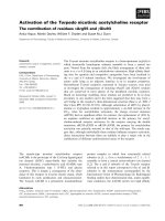
Fig. 1. Acetylated tubulin ⁄ PMCA complex
is present in PMVs from rat brain. (A) PMVs
were analyzed by western blotting by stain-
ing on separate lanes with 5F10 Ig, anti-
PMCA4 Ig and 6-11B-1 Ig (Ac-tubulin).
Another sample of the same membrane
preparation was partitioned in Triton X-114
to determine hydrophobic acetylated tubulin
and hydrophilic tubulin, as described in
Experimental procedures. Aliquots of the
detergent phase (Deterg) and hydrophilic
phase (Aqueous) were subjected to western
blotting and stained with 5F10 Ig (upper),
anti-PMCA4 Ig (middle) and 6-11B-1 Ig
(lower). (B) PMVs solubilized with 0.5% Tri-
ton X-100 were immunoprecipitated with
Sepharose beads linked to 6-11B-1 Ig
(lane 2), 5F10 Ig (lane 3) or anti-(phosphati-
dylinositol 3-kinase) Ig (lane 4). Immuno-
precipiated materials were analyzed. Lane 1,
detergent-solubilized PMVs prior to immuno-
precipitation. (C) Supernatant fractions of
the immunoprecipitation experiment
described in (B). In all cases, volumes of the
analysed samples were calculated to be
representative of the same amount of
PMVs.
Interaction of PMCA with acetylated tubulin N. E. Monesterolo et al.
3568 FEBS Journal 275 (2008) 3567–3579 ª 2008 The Authors Journal compilation ª 2008 FEBS
As a control of immunoprecipitation specificity, phos-
phatidylinositol 3-kinase mAb (an irrelevant antibody)
bound to Sepharose beads was also used. Figure 1B
(lane 1) shows that PMCA, PMCA4 isoform and acet-
ylated tubulin are present in solubilized membranes
prior to immunoprecipitation. When solubilized
membranes were immunoprecipitated with 6-11B-1 Ig
bound to Sepharose beads, total PMCA, PMCA4
isoform and acetylated tubulin were detected in the
precipitated material (Fig. 1B, lane 2). These proteins
were also detected by immunoprecipitation with 5F10
Ig (Fig. 1B, lane 3). However, none of these proteins
was precipitated with antibody to phosphatidylinosi-
tol 3-kinase bound to Sepharose beads (lane 4). The
supernatant fractions of the immunoprecipitation
experiments were also investigated. As shown in
Fig. 1C (lane 2), when anti-(acetylated tubulin)
Ig–Sepharose beads were used as the precipitant, part
of the PMCA isoform and total PMCA remained in
the soluble state, although acetylated tubulin did not.
By contrast, when anti-PMCA Ig–Sepharose beads
were used (lane 3), neither PMCA isoform nor total
PMCA was detected in the supernatant fractions,
although part of the acetylated tubulin was. Because
samples loaded in each lane were represented the same
amount of solubilized membrane, we are able to calcu-
late (from densitometric scanning of three independent
experiments) that 35% of the PMCA isoform (and
total PMCA) and 50% of the acetylated tubulin pres-
ent in PMVs are not part of the acetylated tubu-
lin ⁄ PMCA complex. These results indicate that
acetylated tubulin and PMCA form part of the same
complex inserted into PMVs and that PMCA4 is one
of the isoforms present in the complex.
The influence of ethanol and calmodulin on the
acetylated tubulin ⁄ PMCA complex
Ethanol and calmodulin have been reported to acti-
vate PMCA in rat brain synaptosomes [10–12].
P-ATPases (Na
+
,K
+
-ATPase and H
+
-ATPase) are
known to be activated by effectors that disrupt the
corresponding acetylated tubulin ⁄ ATPase complex.
We investigated whether ethanol and ⁄ or calmodulin
induce activation of Ca
2+
-ATPase via the mechanism
observed for H
+
- and Na
+
,K
+
-ATPases, that is,
dissociation of the tubulin ⁄ Ca
2+
-ATPase complex.
For this purpose, we exposed PMVs to various con-
centrations of ethanol and calmodulin (using experi-
mental conditions described by other authors) [10,13]
and determined the amount of hydrophobic acety-
lated tubulin by measuring the tubulin ⁄ PMCA com-
plex. Using partitioning in Triton X-114 and western
blotting we also determined PMCA activity by mea-
suring its
45
Ca
2+
uptake. The amount of acetylated
tubulin in the detergent phase in PMVs decreased
gradually as the concentration of ethanol or calmod-
ulin increased (Fig. 2A,B). The immunoblots in
AB
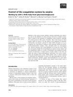
Fig. 2. Effect of ethanol and calmodulin on the quantity of acetylated tubulin ⁄ PMCA complex and PMCA activity. PMVs (0.25 mg
proteinÆmL
)1
) were incubated in transport buffer for 20 min at 37 °C in the presence of various amounts of (A) ethanol or (B) calmodulin.
The amount of hydrophobic acetylated tubulin, as a measure of the tubulin ⁄ PMCA complex, was then determined by the Triton X-114 parti-
tion method and subsequent western blot analysis; PMCA activity was determined by measuring
45
Ca
2+
uptake (see Experimental proce-
dures). Immunoblots of aliquots of the detergent and aqueous phases representative of the same amount of PMVs and stained with
anti-(acetylated tubulin) Ig are shown in the upper panels. Acetylated tubulin bands corresponding to detergent phases were scanned.
Absorbance values and PMCA activities are shown in the lower panels. Values are mean ± SD from three independent experiments.
N. E. Monesterolo et al. Interaction of PMCA with acetylated tubulin
FEBS Journal 275 (2008) 3567–3579 ª 2008 The Authors Journal compilation ª 2008 FEBS 3569
Fig. 2 (upper) show that the decreased intensity of
the acetylated tubulin bands in the detergent phase
is not an artifact but is due to a consistent increase
in the amount of this protein in the aqueous phase.
At 0.6% ethanol or 60 nm calmodulin 30% of
acetylated tubulin remained in the membranes. The
observed diminution in hydrophobic acetylated tubu-
lin was assumed to be due to dissociation of the
acetylated tubulin ⁄ PMCA complex which renders
hydrophilic tubulin. This was confirmed by immuno-
precipitation experiments (see below). However,
PMCA activity gradually increased to > 190% of
the control value in response to ethanol or calmodu-
lin treatment. Thus, similar to our previous results
with other P-ATPases, Ca
2+
-ATPase activity
increased gradually as the acetylated tubulin ⁄ PMCA
complex dissociated, suggesting that ethanol or cal-
modulin stimulates enzyme activity via dissociation
of the complex. That the observed decrease in acety-
lated tubulin in the detergent phase as ethanol or
calmodulin concentration increased was due to modi-
fication of the acetylation pattern or a change in
antibody affinity, rather than dissociation of the
complex, was ruled out because incubation of PMVs
with 0.8% ethanol or 80 nm calmodulin followed by
western blotting of these vesicles (without Triton
X-114 partition) produced no change in the intensity
of the acetylated tubulin bands (results not shown).
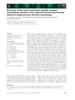
Physical dissociation of the acetylated tubu-
lin ⁄ PMCA complex by ethanol or calmodulin treat-
ment was also assessed by immunoprecipitation. PMVs
were treated with 0.6% ethanol, washed by sedimenta-
tion ⁄ resuspension, solubilized with detergent and
immunoprecipitated with anti-(acetylated tubulin)
Ig–Sepharose beads or anti-PMCA Ig–Sepharose
beads. The precipitated and soluble fractions were
immunoblotted and revealed using anti-PMCA Ig and
anti-(acetylated tubulin) Ig. Comparison of the absor-
bance value for PMCA in the input material (Fig. 3A,
lane 1) with that for PMCA precipitated by anti-(acet-
ylated tubulin) Ig–Sepharose beads (Fig. 3A, lane 3)
reveals that 66 ± 15% of the PMCA in the mem-
brane is associated with acetylated tubulin
(mean ± SD from three independent experiments).
The presence of an acetylated tubulin band in the solu-
ble fraction after immunoprecipitation with anti-
PMCA Ig–Sepharose beads (Fig. 3A, lane 4), even
when no PMCA remains in that fraction (Fig. 3A,
lane 4), indicates that part of the acetylated tubulin in
membranes is not associated with PMCA. Importantly,
ethanol treatment led to less PMCA being precipitated
with anti-(acetylated tubulin) Ig. This can be seen by
comparing PMCA bands precipitated from membranes
treated (Fig. 3B, lane 3) and not treated (Fig. 3A,
lane 3) with ethanol. This suggests that ethanol treat-
ment induced dissociation of the PMCA ⁄ acetylated
tubulin complex. The same conclusion can be drawn
by comparing the amounts of acetylated tubulin pre-
cipitated by anti-PMCA Ig from membranes that were
treated (Fig. 3B, lane 5) or not treated (Fig. 3A,
lane 5) with ethanol. From densitometric scanning of
those bands, it can be calculated that ethanol induced
the dissociation of 60 ± 8% of the complex
(mean ± SD from three independent experiments).
When membranes pre-treated with various concen-
trations of calmodulin were solubilized with detergent
and immunoprecipitated with anti-(acetylated tubulin)
Ig bound to Sepharose beads, the amount of PMCA
precipitated decreased as the calmodulin concentration
increased (Fig. 4). At between 40 and 50 nm calmo-
dulin, 50% of the complex was dissociated (lower).
This is consistent with the percentage activation of
PMCA determined at 50 nm calmodulin (Fig. 2B),
reinforcing the idea that calmodulin induces disso-
ciation of the complex and consequent activation of
ATPase activity.
A
B
Fig. 3. Effect of ethanol on the interaction between PMCA and
acetylated tubulin in PMVs. PMVs were incubated in the absence
(A) or presence (B) of 0.6% ethanol for 20 min at 37 °C, and
washed by centrifugation. Pelleted vesicles were dissolved in Tri-
ton X-100 and immunoprecipitated with anti-(acetylated tubulin) Ig
(Anti ac-tub–Sepharose) or anti-PMCA Ig (anti-PMCA–Sepharose) as
described in Experimental procedures. Input (IN), supernatant (S)
and pellet (P) fractions were analyzed by SDS ⁄ PAGE followed by
immunoblotting with an antibody against acetylated a-tubulin or
PMCA. In all cases, volumes of analyzed samples were calculated
to be representative of the same amount of PMVs. Only areas cor-
responding to relevant bands from a typical experiment are shown.
Interaction of PMCA with acetylated tubulin N. E. Monesterolo et al.
3570 FEBS Journal 275 (2008) 3567–3579 ª 2008 The Authors Journal compilation ª 2008 FEBS
Effect of calmodulin on PMCA activity and
acetylated tubulin ⁄ PMCA complex after ethanol
treatment
Ethanol and calmodulin are known to have additive
stimulatory effects on PMCA [9,12,13]. From a mecha-
nistic point of view, it is of interest to determine
whether the decrease in the quantity of acetylated
tubulin ⁄ PMCA complex determined separately for
each effector was additive (Fig. 2). PMVs were treated
with 0 or 0.6% ethanol and aliquots were incubated in
the presence of increasing concentrations of calmodu-
lin, followed by the immediate determination of
PMCA activity and the amount of acetylated tubulin
remaining in the membrane (by measuring the acety-
lated tubulin ⁄ PMCA complex). The stimulatory effects
of calmodulin and ethanol on PMCA activity were
additive (Fig. 5A). Acetylated tubulin bands corre-
sponding to the complex quantified under each experi-
mental condition and the densitometric values for
these bands are shown in Figs 5B,C, respectively.
When PMVs were treated with both effectors, the
amount of complex was significantly less than when
the effectors were tested separately. Treatment with
individual effectors (0.6% ethanol or 72 nm calmo-
dulin) resulted in 37% non-dissociated complex
A
B
Fig. 4. Dissociation of acetylated tubulin ⁄ PMCA complex by cal-
modulin. PMVs were incubated in the presence of the indicated
concentrations of calmodulin for 20 min at 37 °C and sub-
sequently solubilized (without prior washing of membranes) by
the addition of Triton X-100 (see Experimental procedures). Aliqu-
ots were immunoprecipitated with anti-(acetylated tubulin) Ig
bound to Sepharose beads. (A) Typical immunoblots of precipi-
tated (P) and soluble (S) fractions were revealed with 5F10 Ig
(upper) or 6-11B-1 Ig (lower). (B) Absorbance values correspond-
ing to PMCA and acetylated tubulin detected in the precipitated
fractions shown in (A). Values are mean ± SD from three inde-
pendent experiments.
A
B
C
Fig. 5. Additive effects of ethanol and calmodulin. PMVs (70 lg
protein in 275 l L final volume) were incubated in the presence or
absence of 0.6% ethanol for 5 min at 37 ° C. Calmodulin was then
added to the indicated concentrations, incubation continued for
10 min and PMVs were washed by centrifugation ⁄ resuspension.
Aliquots were separated for PMCA activity assay (A) and for wes-
tern blot analysis with anti-(acetylated tubulin) Ig (B). (C) Densitom-
etry values (mean ± SD) of immunoblots from three independent
experiments similar to that in (B). The absorbance value for the
acetylated tubulin band corresponding to PMVs treated with 0%
ethanol and 0% calmodulin was arbitrarily set at 100%.
N. E. Monesterolo et al. Interaction of PMCA with acetylated tubulin
FEBS Journal 275 (2008) 3567–3579 ª 2008 The Authors Journal compilation ª 2008 FEBS 3571
(Fig. 5C) and 90% stimulation of PMCA activity
(Fig. 5A). After the addition of calmodulin to ethanol-
treated PMVs, PMCA activity was stimulated to a
higher degree than expected based on the degree of
dissociation of the complex. Treatment with 0.6% eth-
anol and subsequently with 72 nm calmodulin resulted
in a reduction of complex quantity to 9% of control
values (Fig. 5C), and stimulation of 300% relative
to the PMCA activity of untreated PMVs (Fig. 5A).
This stimulation cannot be explained solely by dissoci-
ation of the complex. As mentioned above, PMCA
that does not form part of the complex makes up
33% of total (Fig. 1). If this 33% represents enzyme
in the active state, then maximum stimulation could be
achieved when the remaining 67% molecules were dis-
sociated. This would be a maximal stimulation of
200%. Therefore, the observed stimulation of 300%
resulting from ethanol and calmodulin treatment
would involve an additional activating mechanism.
PMCA activity in PMVs is inhibited by exogenous
acetylated tubulin
Because dissociation of the acetylated tubulin ⁄ PMCA
complex results in stimulation of PMCA activity
(Fig. 2A,B), active PMCA may be inhibited by the
addition of acetylated tubulin. To test this possibility,
we incubated PMVs with 0.6% ethanol to dissociate
the complex and washed the PMVs to eliminate
released acetylated tubulin. Tubulin-free PMVs were
then incubated with purified brain tubulin containing
two different proportions (differing by a factor of
four) of the acetylated isoform. As shown in Fig. 6B,
PMCA activity decreased as PMVs were incubated
with increasing concentrations of exogenous tubulin.
The proportion of acetylated tubulin correlated
directly with the degree of inhibition, indicating that
acetylated tubulin is the isoform that interacts with the
enzyme. After eliminating excess exogenous tubulin by
centrifugation, estimation of the acetylated tubu-
lin ⁄ PMCA complex was performed by solubilizing
membranes with detergent followed by immunoprecipi-
tation with 6-11B-1 Ig bound to Sepharose beads.
Western blots of precipitated and soluble fractions
revealed with 5F10 Ig and 6-11B-1 Ig showed that, in
the absence of exogenous tubulin, PMCA was mostly
not associated with acetylated tubulin because PMCA
remained in the soluble fraction following immunopre-
cipitation (Fig. 6A). This was expected because the
acetylated tubulin ⁄ PMCA complex was dissociated
when PMVs were previously treated with ethanol.
Figure 6A also shows that as PMVs were incubated
with increasing concentrations of exogenous tubulin,
the amount of PMCA increased in the precipitates
with a corresponding decrease in the soluble fractions.
Taken together, these results indicate that association
A
B
C
Fig. 6. Effect of exogenous tubulin on PMCA activity and on tubu-
lin ⁄ PMCA complex. PMVs (0.25 mg protein) pretreated with 0.6%
ethanol and washed by centrifugation ⁄ resuspension to eliminate
tubulin dissociated from the complex were incubated for 30 min at
37 °C in a final volume of 1 mL transport buffer, in the presence of
various amounts of purified tubulin preparations containing a low or
high proportion of the acetylated isotype. We checked that under
these incubation conditions tubulin is not assembled into microtu-
bules. After incubation, samples of PMVs that were incubated with
preparations containing a low proportion of acetylated tubulin were
centrifuged at 100 000 g for 20 min at 37 °C to eliminate excess
exogenous tubulin and the pellets resuspended in the original
volume with NaCl ⁄ Tris containing 0.5% Triton X-100 and immuno-
precipitated with anti-(acetylated tubulin) Ig bound to Sepharose
beads. Precipitated (P) and supernatant (S) fractions were immu-
noblotted and revealed with 5F10 Ig and 6-11B-1 Ig (A). Samples
incubated with tubulin containing low (s) and high (d) proportions
of acetylated tubulin were processed to determine PMCA activity
(B). PMCA activity in the absence of exogenous tubulin was
40.6 ± 2 pmol Ca
2+
Æmin
)1
Æmg
)1
protein. Values are mean ± SD
from three experiments. (C) Amount of PMCA in brain membranes
(Control) and in 50 lg of purified tubulin preparation containing low
proportion of the acetylated isotype.
Interaction of PMCA with acetylated tubulin N. E. Monesterolo et al.
3572 FEBS Journal 275 (2008) 3567–3579 ª 2008 The Authors Journal compilation ª 2008 FEBS
of acetylated tubulin with PMCA leads to inhibition of
the enzyme activity. Western blot analysis of purified
tubulin (containing a low proportion of the acetylated
isotype) used in this experiment showed it to be free of
PMCA (Fig. 6C).
Effect of tubulin-interacting drugs on PMCA
activity and the quantity of acetylated
tubulin/PMCA complex
Tubulin is the structural monomer of microtubules.
We examined the effects of taxol and nocodazole on
PMCA activity and the acetylated tubulin ⁄ PMCA
complex because these compounds are known to stabi-
lize or disintegrate microtubules. PMVs were incubated
in the presence or absence of taxol or nocodazole,
followed by determination of PMCA activity as
45
Ca transport activity; the amount of acetylated tubu-
lin complex in the detergent phase after partition in
Triton X-114 was also measured. Taxol decreased the
amount of complex and stimulated PMCA activity
(Table 1). Interestingly, following treatment of PMVs
with taxol, calmodulin treatment did not stimulate
PMCA activity further (data not shown). Nocodazole
partially dissociated the tubulin ⁄ PMCA complex and
inhibited PMCA activity. Inhibition was presumed to
result from an intrinsic property of nocodazole sepa-
rate from its complex-dissociating capacity. This was
tested by determining the effect of nocodazole on
PMCA activity in PMVs pretreated with ethanol or
calmodulin. Treatment of PMCA with ethanol or cal-
modulin for 30 min resulted in an 90% increase in
enzyme activity. By contrast, when nocodazole was
added following ethanol or calmodulin treatment,
PMCA activity measured after 30 min incubation was
reduced to 20–27% (Table 2). Thus, nocodazole is
effectively an inhibitor of PMCA.
Effect of ethanol treatment on the calcium-
pumping activity of PMCA in living cells
To study the physiological relevance of PMCA activity
regulation based on association ⁄ dissociation of the
PMCA ⁄ tubulin complex, we used the Fura-2 method
to estimate the Ca
2+
concentration in living cells and
how the concentration varied following ethanol treat-
ment. We used CAD cells from a mouse brain tumor
that proliferate in serum-containing medium, but stop
dividing and differentiate into neurons when placed in
serum-free medium. These cells contain little or no
acetylated tubulin [7]. It is known that treatment of
these cells with Trichostatin A (TSA; a non-specific
inhibitor of deacetylases) leads to a significant increase
in acetylated tubulin [7]. We suspected that this incres-
ase in tubulin acetylation correlates with acetylated
tubulin ⁄ PMCA complex formation and inhibition of
enzymatic activity, because a similar effect on another
P-type ATPase has been described previously [7].
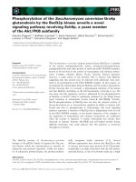
We therefore measured PMCA activity (as calcium-
pumping activity) in cells treated and not treated with
TSA. The acetylated microtubule content of TSA-trea-
ted CAD cells increased significantly (Fig. 7A). In cells
not treated with TSA, acetylated tubulin was absent
and only PMCA was precipitated by anti-PMCA Ig
bound to Sepharose beads, regardless of whether cells
were treated with ethanol (Fig. 7B). In TSA-treated
cells, PMCA and acetylated tubulin were both precipi-
tated, indicating the presence of PMCA⁄ acetylated
tubulin complex in the membrane. When these cells
were treated with ethanol, acetylated tubulin was not
Table 1. Effect of tubulin-interacting drugs on plasma membrane
Ca
2+
-ATPase (PMCA) activity and quantity of acetylated tubu-
lin ⁄ PMCA complex. Plasma membrane vesicles (PMVs) (120 lg
protein) were incubated in 500 lL transport buffer in the absence
(control) or presence of 50 l
M nocodazole or 5 lM taxol. PMVs
were incubated for 30 min at 37 °C and washed twice by centrifu-
gation ⁄ resuspension to eliminate tubulin that was dissociated by
the effectors. Aliquots (200 lL) were processed to quantify tubu-
lin ⁄ PMCA complex by partition with Triton X-114, and to determine
PMCA activity as described in Experimental procedures. Data are
mean ± SD from three independent experiments.
Treatment
of PMVs
Tubulin ⁄ PMCA
complex (% of control)
PMCA activity
(% of control)
None (control) 100 100
+Nocodazole 33 ± 13 17 ± 11
+Taxol 34 ± 6 189 ± 20
Table 2. Inhibitory activity of nocodazol on plasma membrane
Ca
2+
-ATPase (PMCA) activity. Plasma membrane vesicles (PMVs)
(120 lg protein) were incubated in 500 lL transport buffer in the
absence (control) or presence of 72 n
M calmodulin or 0.6% ethanol
for 30 min at 37 °C. Aliquots (200 lL) were processed to determine
PMCA activity as described in Experimental procedures. In other
samples, after 30 min incubation with calmodulin or ethanol, noco-
dazole (50 l
M, final concentration) was added and incubation con-
tinued for 30 min, followed by PMCA activity assay. Data are
mean ± SD from three independent experiments.
Condition PMCA activity (% of control)
Control (no treatment) 100
+ calmodulin 190 ± 13
+ ethanol 185 ± 22
+ calmodulin + nocodazole 27 ± 09
+ ethanol + nocodazole 20 ± 12
N. E. Monesterolo et al. Interaction of PMCA with acetylated tubulin
FEBS Journal 275 (2008) 3567–3579 ª 2008 The Authors Journal compilation ª 2008 FEBS 3573
found in the precipitate, indicating dissociation of the
complex (Fig. 7B).
TSA-treated and non-treated cells were analyzed for
internal Ca
2+
concentration and its variation after the
addition of effectors. To determine whether the varia-
tion in Ca
2+
concentration was due to P-type
ATPases, measurements were carried out in the pres-
ence or absence of sodium vanadate (a potent inhibitor
of P-type ATPases). The Ca
2+
concentration was
lower in cells lacking acetylated tubulin ()TSA) than
in cells containing acetylated tubulin (Fig. 7C; com-
pare )TSA and +TSA, time zero). We ascribe this dif-
ference to a higher calcium-pumping activity in cells in
which PMCA was not inhibited (due to the absence of
acetylated tubulin). Addition of A23187 (a calcium
ionophore) did not modify the Ca
2+
concentration in
cells not treated with TSA in the absence of vanadate
(continuous line). This seemingly unexpected result
may be explained by the high PMCA activity in these
cells (as it is not associated with acetylated tubulin)
which counteracts the influx of calcium due to the ion-
ophore. This was supported by the finding that when
PMCA was inhibited by vanadate (scattered points in
Fig. 7C, )TSA), addition of A23187 increased the
internal Ca
2+
concentration. Subsequent addition of
0.6% ethanol did not modify the Ca
2+
concentration.
This is compatible with the observation that no tubu-
lin ⁄ PMCA complex is present in cells not treated with
TSA (Fig. 7B). In TSA-treated cells, addition of
A23187 resulted in an increased internal Ca
2+
concen-
tration even in the absence of vanadate (Fig. 7C,
+TSA), presumably because PMCA was inhibited by
its association with acetylated tubulin. This high cyto-
plasmic Ca
2+
concentration decreased abruptly upon
the addition of ethanol in the absence of vanadate. By
contrast, the addition of ethanol had no effect in the
presence of vanadate, indicating that a P-type ATPase
was involved in the decrease in Ca
2+
concentration.
Even when, due to the complexity of living cells and
the non-specificity of vanadate, other explanations can
be drawn, these results coincide exactly with our pre-
sumption that ethanol induces dissociation of the acet-
ylated tubulin ⁄ PMCA complex with a consequent
activation of PMCA.
ATPase activities are crucial in the reception ⁄ trans-
mission of signals at the membrane level. Endogenous
activators of these cation pumps, for example adducin
in the sodium pump [14] and calmodulin for PMCA,
are therefore important factors in the regulation of
signaling pathways. In this context, acetylated tubulin
(or acetylated microtubules?) is the first described
endogenous ATPase inhibitor.
Discussion
The plasma membrane Ca
2+
pump removes Ca
2+
from the cell during intracellular signaling. Calcium,
an early-response second messenger, plays a key role
in a number of physiological processes including cell
A
B
C
Fig. 7. Effect of ethanol on PMCA activity and acetylated tubu-
lin ⁄ PMCA complex in CAD cells in culture. (A) CAD cells were
grown to 70% confluence on coverslips and treated for 6 h with
(+TSA) or without ()TSA) 5 l
M TSA, and acetylated microtubules
were visualized by immunofluorescence using mAb 6-11B-1. (B)
CAD cells were grown on 10 cm dishes, treated (or not) with TSA
as described in (A), and incubated for 20 min in transport buffer
containing (+) or not ()) 0.6% ethanol. After elimination of incuba-
tion buffer, cells were solubilized with NaCl ⁄ Tris-Triton and immu-
noprecipitated with anti-PMCA Ig bound to Sepharose beads as
described in Experimental procedures. Typical immunoblots of pre-
cipitated materials revealed with anti-PMCA Ig or anti-(acetylated
tubulin) Ig (Ac-tub) are shown. (C) Intracellular calcium as a function
of incubation time was estimated in CAD cells (±TSA) as relative
fluorescence intensity, using Fura-2AM as the indicator (for details
see Experimental procedures). Calcium ionophore A23187 (final
concentration 3 l
M) and ethanol (final concentration 0.6%) were
added at the times indicated by arrows. This experiment was per-
formed in the absence (continuous lines) or presence (scattered
points) of 2 m
M sodium vanadate, a potent inhibitor of P-type ATP-
ases, added at time zero.
Interaction of PMCA with acetylated tubulin N. E. Monesterolo et al.
3574 FEBS Journal 275 (2008) 3567–3579 ª 2008 The Authors Journal compilation ª 2008 FEBS
proliferation, differentiation and apoptosis [15–17].
PMCA can be activated by several factors, including
acidic phospholipids, proteolysis, calmodulin and etha-
nol. Our findings show that PMCA is partially associ-
ated with acetylated tubulin and this association
results in inhibition of its activity, as estimated by
Ca
2+
-transport. Several pieces of evidence support the
existence of an acetylated tubulin ⁄ PMCA complex in
the membrane. (a) PMCA and acetylated tubulin are
present in isolated membranes. (b) When PMVs are
solubilized in detergent and partitioned in Triton
X-114, PMCA and acetylated tubulin partition to the
detergent phase even though acetylated tubulin is a
hydrophilic protein that partitions in the aqueous
phase. (c) When membranes are solubilized with
Triton X-100 and subsequently immunoprecipitated
with 6-11B-1 Ig bound to Sepharose beads, PMCA
precipitates in addition to acetylated tubulin. PMCA
does not precipitate under these conditions if Sepha-
rose beads are bound to an irrelevant antibody; this
rules out the possibility that PMCA was detected in
the precipitate as an artifact or because it was bound
to a sedimentable structure rather than to acetylated
tubulin. (d) When membranes are solubilized with
Triton X-100 and subsequently immunoprecipitated
with 5F10 Ig bound to Sepharose beads, acetylated
tubulin precipitates in addition to PMCA. (e) When
acetylated tubulin-depleted membranes (by ethanol
treatment) are solubilized with detergent and immuno-
precipitated with anti-(acetylated tubulin) Ig bound to
Sepharose beads, PMCA does not precipitate. How-
ever, when membranes have been incubated previously
with purified exogenous tubulin (Fig. 6), PMCA does
precipitate. The need for the presence of exogenous
tubulin for PMCA to sediment indicates that a
complex forms between PMCA and tubulin. (f) The
complex is not present in membranes from cells lack-
ing acetylated tubulin, however, it appears when the
cells have acetylated tubulin (Fig. 7B). (h) Other
P-type ATPases (sodium and proton pumps) have been
shown to interact with acetylated tubulin [2,3,5].
Considering that in a classical PMV preparation
40% of the vesicles are of the inside-out type, the
increase in complex formation after the addition of
exogenous tubulin should be smaller (Fig. 6) and the
ability of calmodulin to induce complex dissociation
should be less pronounced (Figs 2 and 5). It is possible
that the percentage of inside-out vesicles obtained by
other authors is smaller than that obtained by us
because of differences in the experimental conditions
used. Another possibility is that almost all inside-out
recircularized vesicles have acetylated tubulin, whereas
inside-in circularized vesicles do not; in fact, there
are no proteomic studies about different recircularized
vesicle populations.
It should be noted that it is not clear from these
experiments whether PMCA interacts directly with
tubulin or via intermediary compounds. We use the
term ‘tubulin ⁄ PMCA complex’ for simplicity. Prelimin-
ary results from our laboratory in relation to the
sodium pump indicate that acetylated tubulin interacts
directly with a cytoplasmic fragment of the ATPase
(G. G. Zampar, M. E. Chesta, N. L. Chanaday, N. M.
Dı
´
az, A. Carbajal, C. H. Casale & C. A. Arce, unpub-
lished data). This may also be the case for PMCA.
When the tubulin ⁄ PMCA complex is dissociated,
tubulin no longer partitions into the detergent phase,
but rather into the aqueous phase. Two lines of evi-
dence support the conclusion that the decrease in acet-
ylated tubulin in the detergent phase upon incubation
of PMVs with ethanol or calmodulin (Fig. 2A,B) is
due to dissociation of the tubulin ⁄ PMCA complex.
First, acetylated tubulin partitions into the detergent
phase when bound to Na
+
,K
+
-ATPase and into the
aqueous phase when dissociated from the complex
[1,2]. Second, the decrease in acetylated tubulin in the
detergent phase was correlated with a reduction in the
amount of complex in the immunoprecipitate. That
dissociation of this complex (decrease in acetylated
tubulin in the detergent phase) by ethanol or calmo-
dulin results in stimulation of PMCA activity
(Fig. 2A,B) indicates both that the interaction of tubu-
lin with PMCA inhibits enzyme activity and that the
hydrophobic behavior of acetylated tubulin is due to
its association with a hydrophobic compound (PMCA
or a hydrophobic complex containing PMCA). Disso-
ciation of the complex by ethanol and calmodulin was
confirmed by immunoprecipitation experiments (Figs 3
and 4). Again, although these experiments show that
acetylated tubulin forms part of the same complex as
PMCA, we have no direct evidence that the two mole-
cules interact directly with each other. PMCA has been
shown to interact with various membrane proteins,
forming complex arrays [18–20]. Our results suggest
that acetylated tubulin is a typical cytoplasmic compo-
nent capable of interacting with such a membrane
complex, and that association ⁄ dissociation of the com-
plex regulates PMCA calcium-transporting activity.
Two other findings support the view that PMCA is
associated with acetylated tubulin, and is thereby inac-
tived: (a) when the complex was dissociated by taxol
(Table 1), PMCA activity was stimulated; and (b) the
exogenous addition of acetylated tubulin inhibited
PMCA activity in PMVs (Fig. 6). Stimulation of
PMCA by taxol seems to proceed via a mechanism
similar to that for calmodulin, because the subsequent
N. E. Monesterolo et al. Interaction of PMCA with acetylated tubulin
FEBS Journal 275 (2008) 3567–3579 ª 2008 The Authors Journal compilation ª 2008 FEBS 3575
treatment of PMVs with 72 nm calmodulin had no
additional effect (data not shown).
Most experiments in this study indicated that the
interaction of acetylated tubulin with PMCA results in
the inhibition of enzyme activity, and that dissociation
of the complex reactivates the enzyme. A possible alter-
native explanation is that calmodulin or ethanol dissoci-
ates the complex, and that PMCA activation results, not
from dissociation of the complex, but from the direct
influence of these effectors on the PMCA molecule.
However, the fact that three different chemicals (etha-
nol, calmodulin and taxol) dissociate the tubu-
lin ⁄ PMCA complex, and coincidentally activate PMCA,
supports the idea that activation proceeds via dissocia-
tion of the tubulin ⁄ PMCA complex. The inhibition of
PMCA activity seen when the complex was dissociated
by nocodazole (Table 1) seems to contradict this. How-
ever, the inhibition seen in this case was shown to be
due to an inhibitory effect of nocodazole itself (Table 2).
Even when activation of purified PMCA by calmod-
ulin independent of other proteins has been reported
[21], it should be noted that the systems we used in
this study, PMVs and whole cells, are more complex
than purified PMCA. In effect, PMVs could contain,
in addition to PMCA, multiple components such as
membrane proteins or tubulin (or microtubules) and
others cytoplasmic elements. Therefore, it is possible
that calmodulin exerts a double activating effect on
PMCA by dissociating the acetylated tubulin ⁄ PMCA
complex and directly activating PMCA. This is in line
with the apparently excessive stimulation of PMCA
activity (300%) in relation to the amount of acetylated
tubulin ⁄ PMCA complex dissociated when PMVs were
treated with ethanol and calmodulin (Fig. 5). Another
interesting possibility is that calmodulin exerts its stim-
ulating effect directly on PMCA and that the ‘acti-
vated state’ of PMCA is not favorable for interaction
(low affinity) with acetylated tubulin, so that this tubu-
lin species dissociates from the complex.
The association of acetylated tubulin with PMCA
and the simultaneous inhibition of the enzyme, or con-
versely the stimulation of enzyme activity due to disso-
ciation of the complex, represents a novel mechanism
for regulating PMCA activity and consequently the
calcium concentration of the cell. The microtubular
system (tubulin and ⁄ or microtubules) is clearly
involved in this mechanism because nocodazole and
taxol affect the integrity of the PMCA ⁄ acetylated
tubulin complex and the ATPase activity (Tables 1
and 2). However, additional studies are needed to
obtain a detailed description of this scenario.
The association ⁄ dissociation of PMCA and acety-
lated tubulin and the corresponding inhibition ⁄ stimula-
tion of PMCA activity seem to operate in living cells. In
effect, the cytoplasmic calcium concentration in CAD
cells was altered by ethanol treatment accompanied by
events consistent with the proposed mechanism.
According to this, cells lacking acetylated tubulin could
not form the PMCA ⁄ tubulin complex and this was seen
to be the case (Fig. 7A,B). However, the complex was
shown to be present in the membranes of cells contain-
ing acetylated tubulin (Fig. 7A,B). Furthermore, in cells
in which PMCA was in an inhibited state due to its
interaction with acetylated tubulin, the cytoplasmic
calcium concentration (Fig. 7C, zero time) was higher
than in cells having PMCA in an non-inhibited state
(because it is not interacting with acetylated tubulin). In
the presence of sodium vanadate, the A23187 iono-
phore was able to increase the calcium concentration
and further treatment with ethanol did not result in a
change in the concentration (Fig. 7C, )TSA). This was
expected on the basis that there was no PMCA to acti-
vate because it is not interacting with acetylated tubu-
lin. In cells with the complex (Fig. 7C, +TSA),
following the increase in calcium concentration by
A23187, ethanol treatment induced an abrupt decrease
in concentration, coincident with enzyme activation due
to dissociation of the complex. The blockade of this
decrease in calcium concentration by vanadate demon-
strates that the rapid efflux of calcium was due to a
P-type ATPase. It is clear that treatment of cells with
TSA led to increased tubulin acetylation which corre-
lated with the inhibition of PMCA, but TSA, as a non-
specific inhibitor of deacetylases, is also able to induce
changes in transcriptional activities and therefore, the
observed effects could be indirect.
We have previously shown that the acetylated isotype
of tubulin is required for interaction with the sodium
pump [7]. Here, we demonstrated that the same tubulin
isotype is necessary for interaction with the calcium
pump and that the isoform PMCA4 is involved in the
complex (Fig. 1). It is possible that acetylated tubulin
interacts and regulates the activity of all P-ATPases.
With this aim, we are currently trying to identify the
isoforms that form complexes for each of the P-ATPases,
to characterize more precisely the ‘ATPase ⁄ acetylated
tubulin complex’, and to identify the interacting
domains of the ATPase and acetylated tubulin.
Experimental procedures
Materials
Triton X-114, ATP, anti-mouse IgG conjugated with perox-
idase, calmodulin, mouse mAb (ascites fluid) 6-11B-1
specific for acetylated tubulin, mAb 5F10 specific for
Interaction of PMCA with acetylated tubulin N. E. Monesterolo et al.
3576 FEBS Journal 275 (2008) 3567–3579 ª 2008 The Authors Journal compilation ª 2008 FEBS
PMCA and mAb JA9 specific for PMCA4 were from
Sigma Chemical Co (St Lousi, MO, USA) and mAb
PI3-kinase p110 (D-4) was from Santa Cruz Biochnology
(Santa Cruz, CA, USA).
45
CaCl
2
(12 mCiÆmg
)1
) was from
Perkin-Elmer Life Science (Wellesley, MA, USA) and Fura-
2AM was from Molecular Probes (Eugene, OR, USA).
Cell culture
CAD cells were cultured in Dulbecco’s modified Eagle’s
medium ⁄ F12 (1 : 1; Sigma) supplemented with 10% fetal
bovine serum (Carlsbad, CA, USA) at 37 °C in an air ⁄ CO
2
(19 : 1) incubator. The culture medium was renewed every
48 h.
Immunofluorescence
CAD cells were grown on coverslips and fixed with anhy-
drous methanol at )20 °C. Samples were rehydrated, incu-
bated with 2% BSA in NACl ⁄ P
i
for 60 min, and stained by
indirect immunofluorescence using mouse mAb 6-11B-1
(dilution 1 : 600) in NaCl ⁄ P
i
containing 1% BSA. Fluores-
cein-conjugated anti-mouse IgG (dilution 1 : 400) was used
as secondary antibody. Coverslips were mounted on Fluor-
Save and observed for epifluorescence with a confocal Zeiss
LSM microscope.
Isolation of brain plasma membrane vesicles
Isolation of plasma membrane vesicles from rat brain was
based on the method of Michaelis et al. [22], and modified
for optimal results. All procedures and treatments for hand-
ling animals were reviewed and approved by the Comite
´
de
Etica of CONICET (res. number 1806 ⁄ 04). Seven rat brains
( 10 g) were homogenized at 4 °C in 10 vol of 10 mm
Hepes ⁄ KOH, pH 7.4, 0.32 m sucrose, 0.5 mm MgSO
4
,
0.1 mm phenylmethanesulfonyl fluoride, 2 mm 2-mercapto-
ethanol. The homogenate was centrifuged at 1500 g for
10 min and the supernatant was centrifuged at 20 000 g for
20 min. The resulting pellet was resuspended in homogeniza-
tion buffer to obtain a protein concentration of 10 mgÆmL
)1
in 6 mL. Samples of 1 mL were layered onto a discontinu-
ous gradient containing 3 mL of 40% (w ⁄ v) sucrose and
3 mL of 20% sucrose and centrifuged at 63 000 g for 45 min
at 2–4 °C. The synaptosome fraction was obtained at the
interface, washed in 25 vol of 10 m m Hepes ⁄ KOH, pH 7.4,
centrifuged at 20 000 g for 30 min, collected in the pellet and
resuspended in 10 mm Hepes ⁄ KOH, pH 7.4 to give a protein
concentration of 14 mgÆmL
)1
. An aliquot (1 mL) of the
synaptosome fraction was incubated for 40 min at 4 °C, in
100 vol of lysis buffer (10 mm Hepes ⁄ KOH, pH 7.4, 1 mm
EDTA, 2 mm 2-mercaptoethanol) with continuous stirring.
The lysate was centrifuged at 20 000 g for 30 min to give a
pellet containing PMVs. This fraction was resuspended in
2mLof10mm Hepes ⁄ KOH, pH 7.4 (final protein concen-
tration 5 mgÆmL
)1
) and stored at )70 °C until use.
PMCA activity assay
PMCA activity was determined using the
45
Ca
2+
transport
assay as described previously [8]. The reaction mixture con-
tained PMVs (50–60 lg protein) in 300 lL transport buffer
[50 mm Tris ⁄ HCl, pH 7.3, 100 mm KCl, 95 lm EGTA,
5mm NaN
3
, 400 nm thapsigargin, 20 mm sodium phos-
phate, 2.5 mm MgCl
2
, and
45
CaCl
2
(1 · 10
5
dpmÆnmol
)1
)to
obtain a free Ca
2+
concentration of 10 lm]. Free Mg
2+
and Ca
2+
concentrations were calculated using the program
described by Fabiato and Fabiato [23]. After preincubation
for 5 min at 37 °C, the reaction was initiated by the addi-
tion of 2 mm ATP. After 5 min, the reaction was stopped
by filtering the samples through a 0.45 lm filter, and
45
Ca
2+
taken up by the vesicles was determined using a
liquid scintillation counter. PMCA-mediated
45
Ca
2+
uptake
was calculated as the difference in
45
Ca
2+
uptake between
samples incubated in the presence versus absence of ATP.
Determination of calcium concentration in the
cytoplasm of living cells
This was performed using the Fura-2 method of Jeremic
et al. [24] with some modification. CAD cells were grown
as described above and resuspended in 5 mm Hepes,
pH 7.4, containing 135 mm NaCl, 5.4 mm KCl, 1.8 mm
MgCl
2
,10mmd-glucose (buffer A). Cells were washed
twice with buffer A by centrifugation (1000 g for 5 min at
4 °C) and resuspended to a density of 1 · 10
9
cellsÆmL
)1
in
buffer A containing 10 lm Fura-2AM. The suspension was
incubated for 30 min at 30 °C with mild agitation. Cells
were then washed twice with ice-cold buffer A (without
Fura-2AM) and resuspended at a density of 1 · 10
9
cell-
sÆmL
)1
. For fluorescence measurements, each suspension
was loaded into a cuvette (final density 5 · 10
7
cellsÆmL
)1
)
in buffer A containing 140 lm CaCl
2
, 100 lm EGTA and
1 lm thapsigargin, and placed in a thermostated (30 °C)
Fluoromax-3 Jobin Yvon-Horiva spectrofluorimeter. Exci-
tation was at 340 and 380 nm, and emission was measured
at 510 nm. The Fura-2 fluorescence response to intra-
cellular calcium concentration ([Ca
2+
]
i
) was calibrated from
the ratio of 340 ⁄ 380 nm fluorescence values after subtrac-
tion of background fluorescence of the cells at 340 and
380 nm, as described by Grynkiewicz et al. [25].
Isolation and determination of acetylated
tubulin/PMCA complex
Acetylated tubulin ⁄ PMCA complex was isolated into Tri-
ton X-114 phase as described previously with slight modifi-
cation [26]. Briefly, PMVs (50 lg protein) were washed once
N. E. Monesterolo et al. Interaction of PMCA with acetylated tubulin
FEBS Journal 275 (2008) 3567–3579 ª 2008 The Authors Journal compilation ª 2008 FEBS 3577
with NaCl ⁄ Tris (50 mm Tris ⁄ HCl buffer, pH 7.4, containing
150 mm NaCl) and immediately solubilized in 1 mL NaCl ⁄
Tris containing 1% Triton X-100. After 30 min at 0 °C, the
preparation was centrifuged at 100 000 g for 15 min and
Triton X-114 was added to the supernatant fraction (1%
final concentration). For phase separation, the preparation
was warmed for 5 min at 37 °C and centrifuged at 600 g for
5 min. The detergent-rich lower phase containing acetylated
tubulin ⁄ PMCA complex was washed once with NaCl ⁄ Tris.
Aliquots were subjected to electrophoresis and immunoblot-
ting to determine acetylated and total tubulin.
Electrophoresis and immunoblotting
Proteins were separated by SDS ⁄ PAGE on 10% polyacryl-
amide slab gels [27], transferred to nitrocellulose and
reacted with mouse mAb 6-11B-1 (dilution 1 : 1000) to
determine acetylated tubulin [28], mouse mAb 5F10 (dilu-
tion 1 : 300) to determine PMCA [29] or mouse mAb JA9
(dilution 1 : 1000) to determine PMCA4 isoform [9]. The
nitrocellulose sheet was reacted with anti-mouse IgG conju-
gated with peroxidase. Intensities of tubulin bands were
quantified by scion imaging software.
Tubulin preparation
Two brain tubulin preparations containing different propor-
tions of the acetylated isotype were obtained as described
previously [2]. These preparations, referred to as tubulin of
low and high acetylated isotype content, differ approximately
fourfold in the proportion of acetylated tubulin.
Preparation of antibody linked to Sepharose
Monoclonal antibodies 6-11B-1, 5F10 and phosphatidylino-
sitol 3-kinase p110 were covalently bound to CNBr-acti-
vated Sepharose 4B as described by Hubbert et al. [30], with
slight modification. Sepharose beads were washed with a
100 vol excess of 0.001 m HCl at 21 °C. The resulting
packed beads (1 mL) were mixed with 2.5 mg protein of
ascites fluid containing 6-11B-1 antibody (or another anti-
body) in 1 mL coupling buffer (0.5 m NaCl containing
0.2 m NaHCO
3
, pH 8.2). The mixture was agitated on a
platform rocker for 4 h at 21 °C and loaded into a small
chromatographic column. Unbound antibodies were
removed by washing with 5 mL coupling buffer. Antibody–
Sepharose beads were transferred to a beaker and suspended
in 1 mL of coupling buffer containing 0.2 m glycine to block
unreacted Sepharose sites. The mixture was agitated for 2 h
at 21 °C and unbound glycine was removed by washing the
beads with 10 mL of coupling buffer. The resulting anti-
body-coupled Sepharose was washed with 1.5 mL of 0.01 m
Tris ⁄ HCl, pH 8, containing 0.14 m NaCl and 0.025%
NaN
3
, and stored at 4 °C until use (maximum 2 days).
Immunoprecipitation procedure
PMVs (300 lL, 5 mg proteinÆmL
)1
) were solubilized with
NaCl ⁄ Tris containing 0.5% Triton X-100 (NaCl ⁄ Tris-
Triton) and centrifuged to eliminate residual insoluble
material. Aliquots (0.3 mL) were mixed with 0.15 mL of
packed antibody [anti-(acetylated tubulin) Ig or anti-PMCA
Ig–Sepharose beads] and incubated for 4 h at 20 °C. Sam-
ples were centrifuged and the precipitated material was
washed five times with NaCl ⁄ Tris-Triton. Fractions (50 lL)
of packed beads were resuspended in 50 lL Laemmli sam-
ple buffer, heated at 50 °C for 15 min and centrifuged.
Aliquots (20 lL) of soluble fractions were subjected to
SDS ⁄ PAGE. A control was run in parallel using mAb
phosphatidylinositol 3-kinase–Sepharose instead of mAb
6-11B-1 or 5F10 antibody–Sepharose.
Protein determination
Protein concentration was determined by the method of
Bradford [31].
Acknowledgements
We thank Dr S. Anderson for the English editing. This
work was supported by grants from Agencia Nacional
de Promocio
´
n Cientı
´
fica y Tecnolo
´
gica de la Secretarı
´
a
de Ciencia y Tecnologı
´
a del Ministerio de Cultura y
Educacio
´
n en el marco del Programa de Moderniza-
cio
´
n Tecnolo
´
gica (BID 802 OC ⁄ AR), Consejo Nacion-
al de Investigaciones Cientı
´
ficas y Te
´
cnicas (Conicet),
Agencia Co
´
rdoba Ciencia (Gobierno de la Provincia
de Co
´
rdoba), Secretarı
´
a de Ciencia y Te
´
cnica de la
Universidad Nacional de Co
´
rdoba y Secretarı
´
ade
Ciencias de la Universidad Nacional de Rı
´
o Cuarto.
References
1 Alonso AC, Nun
˜
ez-Fernandez M, Beltramo DM,
Casale CH & Barra HS (1998) Na
+
,K
+
-ATPase was
found to be the membrane component responsible for
the hydrophobic behaviour of the brain membrane
tubulin. Biochem Biophys Res Commun 253, 824–927
[Erratum: 257, 642].
2 Casale CH, Alonso AC & Barra HS (2001) Brain
plasma membrane Na
+
,K
+
-ATPase is inhibited by
acetylated tubulin. Mol Cell Biochem 216, 85–92.
3 Casale CH, Previtali G & Barra HS (2003) Involvement
of acetylated tubulin in the regulation of Na
+
,K
+
-AT-
Pase activity in cultured astrocytes. FEBS Lett 534,
115–118.
4 Casale CH, Previtali G, Serafino JJ, Arce CA & Barra
HS (2005) Regulation of acetylated tubulin ⁄ Na
+
,
Interaction of PMCA with acetylated tubulin N. E. Monesterolo et al.
3578 FEBS Journal 275 (2008) 3567–3579 ª 2008 The Authors Journal compilation ª 2008 FEBS
K
+
-ATPase interaction by l-glutamate in non-neural
cells: involvement of microtubules. Biochim Biophys
Acta 1721, 185–192.
5 Campetelli AN, Previtali G, Arce CA, Barra HS &
Casale CH (2005) Activation of the plasma membrane
H
+
-ATPase of Saccharomyces cerevisiae by glucose is
mediated by dissociation of the H
+
-ATPase–acetylated
tubulin complex. FEBS J 272, 5742–5752.
6 Beltramo DM, Nun
˜
ez M, Alonso AC & Barra HS (1994)
The relationship of hydrophobic tubulin with mem-
branes in neural tissue. Mol Cell Biochem 141, 57–63.
7 Santander VS, Bisig CG, Purro SA, Casale CH, Arce
CA & Barra HS (2006) Tubulin must be acetylated in
order to form a complex with membrane Na
+
,K
+
-
ATPase and to inhibit its enzyme activity. Mol Cell
Biochem 291, 167–174.
8 Adamo HP & Grimaldi ME (1998) Functional conse-
quences of relocating the C-terminal calmodulin-binding
autoinhibitory domains of the plasma membrane Ca
2+
pump near the N-terminus. Biochem J 331, 763–766.
9 Sepu´ lveda MR & Mata AM (2004) The interaction
of ethanol with reconstituted synaptosomal plasma
membrane Ca
2+
-ATPase. Biochim Biophys Acta 1665,
75–80.
10 Jarret HW & Penniston J (1977) Partial purification of
the Ca
2+
–Mg
2+
ATPase activator from human erythro-
cytes: its similarity to the activator of 3¢:5¢-cyclic nucleo-
tide phosphodiesterase. Biochem Biophys Res Commun
77, 1210–1216.
11 Gopinath RM & Vicenzi FF (1979) (Ca
2+
,Mg
2+
)-AT-
Pase activity of sickle cell membranes: decreased activa-
tion by red blood cell cytoplasmic activator. Am J
Hematol 7, 303–312.
12 Benaim G, Cervino V, Lopez-Estran
˜
o C & Weitzman C
(1994) Ethanol stimulates the plasma membrane cal-
cium pump from human erythrocytes. Biochim Biophys
Acta 1195, 141–148.
13 Cervino V, Benaim G, Carafoli E & Guerini D (1998)
The effect of ethanol on the plasma membrane calcium
pump is isoform-specific. J Biol Chem 273, 29811–29815.
14 Ferrandi M, Salardi S, Tripodi G, Barassi P, Rivera R,
Manunta P, Goldshleger R, Ferrari P, Bianchi G &
Karlish SJ (1999) Evidence for an interaction between
adducin and Na
+
,K
+
-ATPase: relation to genetic
hypertension. Am J Physiol 277, H1338–H1349.
15 Van Breemen C & Saida K (1989) Cellular mechanisms
regulating [Ca
2+
]
i
smooth muscle. Annu Rev Physiol 51,
315–329.
16 Trump BF & Berezesky IK (1995) Calcium-mediated
cell injury and cell death. FASEB J 9, 219–228.
17 McConkey DJ (1996) The role of calcium in the regula-
tion of apoptosis. Scanning Microsc 10, 777–793.
18 Enyedi A, Vorherr T, James P, MacCormick DJ, Filo-
teo AG, Carafoli E & Penniston JT (1989) The calmod-
ulin binding domain of the plasma membrane Ca
2+
pump interacts both with calmodulin and with another
part of the pump. J Biol Chem 264, 12313–12321.
19 Sgambato-Faure V, Xiong Y, Berke JD, Hyman SE &
Strehler EE (2006) The Homer-1 protein Ania-3 inter-
acts with the plasma membrane calcium pump. Biochem
Biophys Res Commun 343, 630–637.
20 Williams JC, Armesilla AL, Mohamed TM, Hagarty
CL, McIntyre FH, Schomburg S, Zaki AO, Oceandy
D, Cartwright EJ, Buch MH et al. (2006) The sarcolem-
mal calcium pump, alpha-1 syntrophin, and neuronal
nitric-oxide synthase are parts of a macromolecular pro-
tein complex. J Biol Chem 281, 23341–23348.
21 Niggli V, Penniston JT & Carafoli E (1979) Purification
of the (Ca
2+
–Mg
2+
) ATPase from human erythrocyte
membranes using a calmodulin affinity column. J Biol
Chem 254, 9955–9958.
22 Michaelis EK, Michaelis ML, Chang HN & Kitos TE
(1983) High affinity Ca
2+
-stimulated Mg
2+
-dependent
ATPase in rat brain synaptosomes, synaptic mem-
branes, and microsomes. J Biol Chem 258, 6101–6108.
23 Fabiato A & Fabiato F (1979) Calculator programs for
computing the composition of the solutions containing
multiple metals and ligands used for experiments in
skinned muscle cells. J Physiol 75, 463–505.
24 Jeremic A, Jeftinija D, Stevanovic J, Glavaski A &
Jeftinija S (2001) ATP stimulates calcium-dependent
glutamate release from cultured astrocytes. J Neurochem
77, 664–675.
25 Grynkiewicz G, Poenie M & Tsien RY (1985) A new
generation of Ca
2+
indicators with greatly improved
fluorescence properties. J Biol Chem 260, 3440–3450.
26 Nun
˜
ez-Fernandez M, Beltramo DM, Alonso AC &
Barra HS (1997) Conversion of hydrophilic tubulin into
a hydrophobic compound. Evidence for the involvement
of membrane proteins. Mol Cell Biochem 170, 91–98.
27 Laemmli UK (1970) Cleavage of structural proteins
during the assembly of the head of bacteriophage T4.
Nature 227, 680–685.
28 Piperno G & Fuller MT (1985) Monoclonal antibodies
specific for an acetylated form of alpha-tubulin recog-
nize the antigen in cilia and flagella from a variety of
organisms. J Cell Biol 101, 2085–2094.
29 Caride AJ, Filoteo AG, Enyedi A, Verma AK & Penn-
iston JT (1996) Detection of isoform 4 of the plasma
membrane calcium pump in human tissues by using
isoform-specific monoclonal antibodies. Biochem J
316, 353–359.
30 Hubert JJ, Schenk DB, Skeelli H & Leffert HL (1986)
Rat hepatic Na
+
,K
+
ATPase: alpha-subunit isolation
by inmunoaffinity chromatography and structural anal-
ysis by peptide mapping. Biochemistry 5, 4156–4163.
31 Bradford MM (1976) A rapid and sensitive method for
the quantitation of microgram quantities of protein
utilizing the principle of protein–dye binding. Anal
Biochem 72, 248–254.
N. E. Monesterolo et al. Interaction of PMCA with acetylated tubulin
FEBS Journal 275 (2008) 3567–3579 ª 2008 The Authors Journal compilation ª 2008 FEBS 3579