Báo cáo khoa học: PACSIN 1 forms tetramers via its N-terminal F-BAR domain pdf
Bạn đang xem bản rút gọn của tài liệu. Xem và tải ngay bản đầy đủ của tài liệu tại đây (701.39 KB, 10 trang )
PACSIN 1 forms tetramers via its N-terminal F-BAR domain
Arndt Halbach
1
, Matthias Mo
¨
rgelin
2
, Maria Baumgarten
2
, Mark Milbrandt
1
, Mats Paulsson
1
and Markus Plomann
1
1 Center for Biochemistry and Center for Molecular Medicine (CMMC), Medical Faculty, University of Cologne, Germany
2 Department of Clinical Sciences, Section for Clinical and Experimental Infectious Medicine, University of Lund, Sweden
In eukaryotic cells, complex regulatory mechanisms
involving numerous proteins must operate to ensure
the temporal and spatial specificity of intracellular
membrane-trafficking pathways. We and others have
identified three members of the protein kinase C and
casein kinase 2 substrate in neurons (PACSIN) protein
family, also named syndapin and focal adhesion pro-
tein 52 (FAP52), which participate in rearrangements
of actin networks during endocytosis [1–5]. In contrast
to the neuron-specific PACSIN 1, other members of
the PACSIN protein family show a broader tissue dis-
tribution [2,4,6]. Via their C-terminal Src homology 3
(SH3) domains, PACSIN proteins bind to proline-rich
domains of dynamin, synapsin and synaptojanin,
three proteins also involved in vesicle endocytosis, as
well as to neural Wiskott–Aldrich syndrome protein
Keywords
F-BAR domain; membrane; oligomerization;
PACSIN 1; syndapin 1
Correspondence
M. Plomann, Center for Biochemistry,
Medical Faculty, University of Cologne,
Joseph-Stelzmann-Str. 52, D-50931
Cologne, Germany
Fax: +49 221 478 6977
Tel: +49 221 478 6944
E-mail:
(Received 13 October 2006, revised
23 November 2006, accepted 4 December
2006)
doi:10.1111/j.1742-4658.2006.05622.x
The ability of protein kinase C and casein kinase 2 substrate in neurons
(PACSIN) ⁄ syndapin proteins to self-polymerize is crucial for the simulta-
neous interactions with more than one Src homology 3 domain-binding
partner or with lipid membranes. The assembly of this network has pro-
found effects on the neural Wiskott–Aldrich syndrome protein-mediated
attachment of the actin polymerization machinery to vesicle membranes as
well as on the movement of the corresponding vesicles. Also, the sensing of
vesicle membranes and ⁄ or the induction of membrane curvature are more
easily facilitated in the presence of larger PACSIN complexes. The N-ter-
minal Fes-CIP homology and Bin-Amphiphysin-Rvs (F-BAR) domains of
several PACSIN-related proteins have been shown to mediate self-inter-
actions, whereas studies using deletion mutants derived from closely related
proteins led to the view that oligomerization depends on the formation of
a trimeric complex via a coiled-coil region present in these molecules. To
address whether the model of trimeric complex formation is applicable to
PACSIN 1, the protein was recombinantly expressed and tested in four
different assays for homologous interactions. The results showed that
PACSIN 1 forms tetramers of about 240 kDa, with the self-interaction
having a K
D
of 6.4 · 10
)8
m. Ultrastructural analysis of these oligomers
after negative staining showed that laterally arranged PACSIN molecules
bind to each other via a large globular domain and form a barrel-like
structure. Together, these results demonstrate that the N-terminal F-BAR
domain of PACSIN 1 forms the contact site for a tetrameric structure,
which is able to simultanously interact with multiple Src homology 3 bind-
ing partners.
Abbreviations
ADAM, a disintegrin and metalloprotease; BAR, Bin-Amphiphysin-Rvs; BS
3
, bis[sulfosuccinimidyl]suberate; FAP52, focal adhesion protein 52;
F-BAR, Fes-CIP4 homology and Bin-Amphiphysin-Rvs; FCH, Fes-CIP4 homology; GST, glutathione S-transferase; NMDA, N-methyl-
D-
aspartate; NOSTRIN, eNOS-trafficking inducer; N-WASP, neural Wiskott–Aldrich syndrome protein; PACSIN, protein kinase C and casein
kinase 2 substrate in neurons; PACSIN 1-CS, PACSIN 1 carrying a C-terminal Strep II tag; PCH, pombe CDC15 homology; PSTPIP, proline,
serine, threonine phosphatase-interacting protein; SH3, Src homology 3; Sulfo-EGS, ethylene glycol bis[sulfosuccinimidylsuccinate].
FEBS Journal 274 (2007) 773–782 ª 2007 The Authors Journal compilation ª 2007 FEBS 773
(N-WASP), a stimulator of actin-related protein
2 ⁄ 3-induced actin polymerization [4,6]. Accordingly,
the PACSIN proteins have been implicated not only in
vesicle endocytosis at the plasma membrane but in a
variety of membrane traffic events, most of which
occur at membranes of intracellular sorting compart-
ments [7,8].
PACSIN proteins also interact with specific trans-
membrane proteins, such as ADAM (a disintegrin and
metalloprotease) metalloproteinases [9,10], the CD95
ligand [11] and, in the case of PACSIN 1, the phos-
phodiesterase 6c [12] and the N-methyl-d-aspartate
(NMDA) receptor chain NR3A [13]. These interac-
tions, most of which also involve the SH3 domains,
indicate a role of PACSIN proteins in the regulation
of the surface expression of some transmembrane
molecules by endocytosis. For NR3A, we were able
to demonstrate an activity-dependent mechanism by
which PACSIN 1 regulates NMDA receptor expres-
sion at synapses during development [13].
PACSIN proteins represent a subgroup within a lar-
ger protein family, named pombe CDC15 homology
(PCH), displaying a similar arrangement of domains,
including at least one C-terminal SH3 domain and a
conserved N-terminal region (Fig. 1A). The latter was
originally defined as the CDC15-N-terminal (CDC15-
NT) domain, spanning about 250 amino acids in PAC-
SIN proteins [4]. Later, others distinguished between a
region covering the N-terminal circa 100 amino acids,
named the Fes-CIP4 homology (FCH) domain, and
the adjacent a-helical stretch, which is believed to form
a coiled-coil structure [14]. So far, no function has
been reported for the FCH domain, whereas the
a-helical region has been shown to be responsible for
oligomerization of PCH proteins [15–17]. Recently,
the whole N-terminal region, corresponding to the
CDC15-NT domain and renamed the FCH and Bin-
Amphiphysin-Rvs (BAR) (F-BAR) domain, has been
characterized in more detail [18]. Like classical BAR
domains, this related domain is able to bind to lipid
bilayers. Moreover, the PCH proteins tested, including
PACSIN ⁄ syndapin 1, bind to liposomes containing
phosphatidylserine and phosphoinositides, and are
alone sufficient to deform them into tubules [18]. In
agreement with this, PACSIN 1 was recently identified
as the key interaction partner of dynamin 1 in synaptic
vesicle endocytosis [19]. In this process, PACSIN 1 is
thought to bind to the synaptic plasma membrane,
and induce a curvature in the membrane and neck for-
mation prior to vesicle fission through the action of
dynamin 1.
Another proposed role for PACSIN proteins is in
attachment of the actin polymerization machinery to
vesicles after endocytosis. They are believed to act as
linkers between the endocytic protein dynamin and
N-WASP, thereby directing the actin propulsion
machinery to the site of vesicle fission [4,6]. As several
important interaction partners, including dynamin and
N-WASP, interact with PACSIN’s single SH3 domain
[4], only PACSIN oligomers would be able to act as
linkers. Previously, we showed that, in vitro, all three
PACSIN proteins are able to to bind to each other
and might exist as homo-oligomers and ⁄ or hetero-
oligomers [4]. Recently, the interconnecting function of
PACSIN oligomers was shown to be essential for
PACSIN-mediated cytoskeletal rearrangements and
A
BC
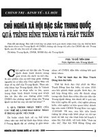
Fig. 1. Domain structure and purification of
recombinant PACSIN 1. (A) Domain struc-
ture of PACSIN 1 with an N-terminal F-BAR
domain and a C-terminal SH3 domain. The
F-BAR domain was formerly known as the
CDC15-NT domain, and includes a con-
served region, also named the FCH domain,
and an a-helical polypeptide stretch postula-
ted to act as a coiled-coil domain. Addition-
ally, PACSIN 1 contains two asparagine-
proline-phenylalanine (NPF) motifs.
(B) Fractions eluted from a StrepTactin
Sepharose column were purified by an
additional ultracentrifugation step and tested
for purity by SDS ⁄ PAGE under reducing
conditions. C, Coomassie Brilliant Blue; S,
silver nitrate. The arrow marks the
recombinant PACSIN 1-CS. (C) MALDI-TOF
MS analysis of the same purified PACSIN 1
sample.
PACSIN 1 oligomerization A. Halbach et al.
774 FEBS Journal 274 (2007) 773–782 ª 2007 The Authors Journal compilation ª 2007 FEBS
endocytosis [20]. We report here that purified PAC-
SIN 1 exists as a 230–240 kDa tetrameric complex in
solution. Although it shares many structural and func-
tional properties with other members of the PCH pro-
tein family, PACSIN 1 is distinct in that it forms
barrel-like homotetramers in vitro, held together by the
N-terminal F-BAR domain.
Results
Purification and characterization of PACSIN 1
PACSIN 1, carrying a C-terminal Strep II tag (PAC-
SIN 1-CS) and expressed in HEK293 cells, was puri-
fied by a single affinity chromatography step. The
homogeneity of the preparation was demonstrated by
reducing SDS ⁄ PAGE followed either by staining with
Coomassie Brilliant Blue and silver nitrate (Fig. 1B)
or by immunoblotting (data not shown). MALDI-
TOF MS analysis confirmed the purity (Fig. 1C).
Secondary structure and thermal stability
of PACSIN 1
In order to analyze the conformation and thermal sta-
bility of PACSIN 1, the purified protein was studied
by CD spectroscopy. Aliquots of PACSIN 1-CS
(100 lgÆmL
)1
)in5mm Tris ⁄ HCl (pH 7.4) were incu-
bated at 20 °C, 37 °C, or 60 °C, and CD spectra were
recorded. At the two lower temperatures, PACSIN 1
showed similar CD spectra with characteristic minima
in the 208–223 nm region, indicative of a high a-helical
content (Fig. 2A). When PACSIN 1 CD spectra were
recorded at 60 °C, changes were observed, reflecting a
loss of a-helical structure (Fig. 2A). This heat denatur-
ation was irreversible, as a subsequent decrease in tem-
perature was unable to restore the original structure
(results not shown). Furthermore, calculation of the
relative proportions of different secondary structure
elements by three different algorithms revealed that, at
37 °C, PACSIN 1 contains about one-third a-helix, a
portion of which is lost upon heating, accompanied by
an increase in b-structure (supplementary Table S1). A
melting point of 44 °C was determined from the mid-
point of the transition at 221 nm (Fig. 2B).
Oligomeric structure of PACSIN 1 complexes
To determine the oligomerization state of PACSIN 1,
purified PACSIN 1-CS was analyzed by gel filtration.
In a KCl-containing buffer at cytosolic ion strength,
PACSIN 1 eluted as a single peak between the marker
proteins aldolase (158 kDa) and ferritin (440 kDa)
(Fig. 3A). The presence of PACSIN 1 in individual
fractions was confirmed by immunoblotting (results
not shown). For the calculation of the molecular mass
of the PACSIN protein complex, the K
av
values of the
marker proteins used as a standard were plotted
against log M
r
. With this method, the molecular mass
of the native PACSIN complex was determined as
being 234 kDa, corresponding to an oligomerization
state of about 4.5 (Fig. 3B, supplementary Table S2A).
Furthermore, performing the same experiment with a
bis(sulfosuccinimidyl)suberate (BS
3
)-crosslinked sample
led to comparable results (M
r
¼ 240 kDa; oligomeriza-
tion state 4.6), showing that crosslinking with this rea-
gent captures PACSIN complexes in a native state
(Fig. 3C, supplementary Table S2B).
We next used protein crosslinking to study intermo-
lecular interactions between PACSIN 1 subunits. Three
homobifunctional imidoester reagents, disuccinimidyl
suberate (DSS), BS
3
and ethylene glycol bis(sulfosuc-
Fig. 2. CD spectra of PACSIN 1. (A) PACSIN 1-CS protein (100 lgÆ
mL
)1
in 5 mM Tris ⁄ HCl, pH 7.4) was measured at 20 °C (solid line),
37 °C (dotted line) and 60 °C (dashed line). Changes in the content
of a-helix and b-structure were observed between 37 °C and 60 °C.
(B) For the determination of the melting temperature, recordings
were performed at 221 nm with a linear temperature gradient from
20 °Cto80°C. The midpoint of the conformational transition was
at 44 °C.
A. Halbach et al. PACSIN 1 oligomerization
FEBS Journal 274 (2007) 773–782 ª 2007 The Authors Journal compilation ª 2007 FEBS 775
cinimidylsuccinate) (Sulfo-EGS) were tested for their
ability to covalently link recombinantly expressed
PACSIN 1-CS molecules. All reagents crosslinked
PACSIN 1 to dimers (Fig. 4A, lanes 2 and 3 for BS
3
,
and lane 4 for Sulfo-EGS) or, at higher crosslinker
concentrations, to tetramers (Fig. 4A, lanes 4–7 for
BS
3
, and lanes 5–7 for Sulfo-EGS). Sulfo-EGS differs
from DSS and BS
3
by having a slightly longer spacer
arm (16.1 A
˚
versus 11.4 A
˚
), and was less efficient in
crosslinking PACSIN 1 to tetramers. Adducts larger
than tetramers were only occasionally observed at
higher protein concentrations (50 lg versus 20 lg),
corroborating the tetrameric structure indicated by the
gel filtration experiments.
MS analysis of crosslinked fractions confirmed the
presence of dimers, but ionization was insufficient for
the detection of higher oligomers (Fig. 4B,C). The mod-
erate increase in mass of PACSIN 1 monomers resulted
from bound crosslinker molecules. The actual presence
of tetrameric complexes in a BS
3
-crosslinked sample
was confirmed by size exclusion chromatography, which
resulted in a single symmetrical peak comparable to the
previously analyzed native sample (Fig. 3C).
As oligomerization is a prerequisite for the proposed
function of PACSIN as a linking protein, surface plas-
mon resonance was used to further support the pres-
ence of a self-interaction between PACSIN 1 subunits
and to determine the strength of this binding (Fig. 5).
High-affinity binding between PACSIN 1 monomers
could indeed be detected (Fig. 5), and a k
a
of 1.44 ·
10
5
m
)1
Æs
)1
,ak
D
of 4.3 · 10
)3
and a K
D
of 6.4 · 10
)8
m
were calculated (supplementary Table S3).
Purified full-length PACSIN 1-CS (Fig. 1) was also
submitted to electron microscopy after negative stain-
ing with uranyl formate (Fig. 6). The protein particles
were heterogeneous in size, and closer examination
revealed that both monomers and, predominantly,
higher aggregates were present in the sample (Fig. 6A).
Most of the monomers formed elongated curved struc-
tures, but fully extended 7–8 nm monomeric particles
could occasionally be seen (Fig. 6B). Dimers showed a
lateral alignment of PACSIN molecules joined at one
end. Tetramers displayed a barrel-like structure, often
with a more heavily stained hole in the middle and
most mass at the periphery (Fig. 6C,D). The average
diameter of the tetramers was 8 nm. Occasionally, par-
ticles were oriented to give a top view (Fig. 6D, top
panel). We never observed a waist-like structure, which
would have indicated that a rod-like coiled-coil a-helix
might assemble the higher-order structure. Electron
microscopy of negatively stained recombinant F-BAR
domains of PACSIN 1 again showed monomeric
(Fig. 6E, upper row) and dimeric (Fig. 6E, lower row)
Fig. 3. Analysis of PACSIN 1 oligomers by gel filtration. (A) Elution
profile of PACSIN 1-CS from size exclusion chromatography using a
Superdex 200 column (solid line). The elution profile of a mixture of
thyroglobin (669 kDa), ferritin (440 kDa), aldolase (158 kDa), ovalbu-
min (43 kDa) and ribonuclease A (13.7 kDa) is shown as a dotted
line. Numbers at the peaks represent the molecular masses of the
corresponding marker proteins. The Y-axes show the relative fluor-
escence intensities at 280 nm, with the left axis corresponding to
the marker proteins, and the right axis to PACSIN 1-CS. AU ¼ arbi-
trary units. (B) Plot of K
av
values of marker proteins against log M
r
.
Numbers at the open squares represent the molecular masses.
The relative mass of the native PACSIN complex was determined
as being about 234 kDa (closed square), corresponding to an olig-
omerization state of 4.5. (C) Analysis of BS
3
-crosslinked PACSIN 1
gave comparable results, with a molecular mass of 240 kDa and an
oligomerization state of 4.6 (closed square). Numbers at the open
squares represent the molecular masses of the corresponding mar-
ker proteins.
PACSIN 1 oligomerization A. Halbach et al.
776 FEBS Journal 274 (2007) 773–782 ª 2007 The Authors Journal compilation ª 2007 FEBS
particles, demonstrating that this domain is sufficient
for oligomerization.
Discussion
The present characterization of the neurospecific repre-
sentative of the PACSIN proteins, PACSIN 1 [1,3],
supports the hypothesis that PACSINs act as linking
molecules in vesicular trafficking. It was previously
shown that PACSIN proteins bind to both dynamin
and N-WASP, and that impairment of these inter-
actions leads to changes in actin dynamics, block of
endocytosis, and mislocalization of involved proteins
[4,6,17,20–22]. As both binding partners are recognized
by the single C-terminal SH3 domain of PACSINs,
multiple simultaneous interactions are only possible if
PACSINs form oligomers. Members of the PCH fam-
ily contain at least one a-helical polypeptide stretch,
which is assumed to form a coiled-coil and thereby
enable oligomerization. Several studies on individual
members of this protein family have shown their abil-
ity to homo-oligomerize to dimers [proline, serine,
threonine phosphatase-interacting protein (PSTPIP)
and PSTPIP 2 [15]], or trimers [FAP52 [16] and
(endothelial nitric oxide synthase) eNOS-trafficking
inducer (NOSTRIN) [17]]. We previously observed
that all PACSIN isoforms are able to interact with
each other in two-hybrid assays [4], and recently
another study confirmed the ability of PACSIN
proteins to self-associate [20]. To determine the
A
B
C
Fig. 4. Crosslinking of PACSIN 1. (A)
PACSIN 1 was incubated at 50 lgÆmL
)1
with 0.5 lM–10 mM BS
3
(left panel) or at
20 lgÆmL
)1
with 0–5 mM Sulfo-EGS cross-
linker (right panel) and analyzed by reducing
SDS ⁄ PAGE on 5–15% gels. The crosslinked
products are labeled. (B, C) The products of
PACSIN 1 crosslinked with 25 l
M (*, B) and
2.5 m
M Sulfo-EGS (#, C) were analyzed by
MALDI-TOF MS.
A. Halbach et al. PACSIN 1 oligomerization
FEBS Journal 274 (2007) 773–782 ª 2007 The Authors Journal compilation ª 2007 FEBS 777
stoichiometry of PACSIN 1 oligomers, we expressed
recombinant PACSIN 1 in eukaryotic cells and, by use
of CD spectroscopy, confirmed that the protein was
correctly folded. The content of a-helix in the full-
length protein was found to be 33.7–37.9%, depending
on the software used, which is significantly higher than
the 25.8% calculated for the closely related PACSIN 2
ortholog FAP52 [16]. Heat treatment leads to an irre-
versible loss of a high proportion of these helices and,
interestingly, to an increase of b-structure (supplement-
ary Table S1).
When subjected to size exclusion chromatography,
PACSIN 1 eluted as a complex of about 234 kDa,
indicating that oligomers are formed in solution. This
mass slightly exceeds that of a tetramer, which may
result from the shape of the PACSIN 1 complexes
when compared to the standard proteins. A similar
oligomerization has been shown for a recombinantly
produced GST–NOSTRIN fragment [17], but here gel
filtration indicated a trimerization. In a recent publi-
cation, it was suggested that PACSIN 1 predomin-
antly forms dimers in vivo [20]. This was concluded
from crosslinking studies in brain and cell extracts,
but less well-resolved higher molecular weight com-
plexes were also observed [20]. To address the appar-
ent discrepancy with our gel filtration results, we used
increasing concentrations of three different crossl-
inkers with varying spacer arm lengths, and clearly
detected the preferential formation of PACSIN 1
dimers and tetramers. In contrast to the other studies,
we avoided the use of cell lysates in which potential
exogenous interaction partners might be present and
Fig. 5. Surface plasmon resonance binding curves obtained for
the PACSIN 1 self-interaction. Antibodies to GST were coupled
(15 000 RU) to a CM5 sensor chip and saturated with GST–
PACSIN 1 or GST as a control. PACSIN 1-CS was injected at differ-
ent concentrations. The binding curves shown have been corrected
by subtracting the values obtained with GST alone.
A
B
C
D
E
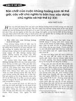
Fig. 6. Electron microscopy of negatively stained recombinant
PACSIN 1-CS and the PACSIN 1 F-BAR domain. The overview (A)
shows aggregates as well as monomeric PACSIN 1 molecules; the
lower panels show selected monomeric (B), dimeric (C) and tetra-
meric (D) particles. The bottom panel (E) shows PACSIN 1 F-BAR
monomers (upper row) and dimers (lower row). The picture at the
lower right includes a monomer located next to the dimer. The bars
correspond to 10 nm.
PACSIN 1 oligomerization A. Halbach et al.
778 FEBS Journal 274 (2007) 773–782 ª 2007 The Authors Journal compilation ª 2007 FEBS
in which complexes may include proteins other than
PACSIN. The analysis of PACSIN 1 self-association
by real-time surface plasmon resonance gave a K
D
of
6.4 · 10
)8
m, which is comparable to the K
D
of
4.7 · 10
)9
m calculated for FAP52 [16]. Although clo-
sely related to PACSIN 2, FAP52 appears to partici-
pate in different processes. In chicken embryo heart
fibroblasts, it localizes to focal adhesion contacts [5],
and this was never observed for any of the three
PACSIN isoforms.
To complement our biochemical analysis, we also
employed electron microscopy of negatively stained
PACSIN 1 complexes; this confirmed the formation
of dimers and tetramers observed in solution. The
PACSIN tetramers form a barrel-like structure in the
absence of any lipids or other proteins. Taken together,
our results suggest that tetramers are the highest oligo-
mers formed by PACSIN 1, and that dimers may be
intermediates in the assembly. The proportions of
dimers and tetramers detected may depend on the ana-
lytical method used. None of the particles seen with
electron microscopy showed a waist-like structure,
which would have been indicative of assembly via a
coiled-coil a-helix. Instead, subunit contacts appear to
be mediated by domains of globular shape. These may
be the PACSIN 1 F-BAR domains, particularly as elec-
tron microscopy of isolated PACSIN 1 F-BAR
domains also shows oligomerization (Fig. 6E). The
importance of this region for self-assembly has been
confirmed for PACSIN 1 [20], and also been reported
for other F-BAR domain-containing proteins [16,17].
The exact role of PACSINs is controversial, especi-
ally with regard to when and where PACSIN mole-
cules contribute to vesicle formation and removal.
PACSIN proteins have been proposed to play a role in
the regulation of transferrin endocytosis. However,
these findings were based on overexpression of either
isolated SH3 domains [23] or full-length proteins [4],
and may reflect an impairment of proper dynamin
localization, as overexpression of other dynamin-bind-
ing SH3 domains has comparable effects [22–24]. A
recent study demonstrated that F-BAR domains are
able to bind to phospholipids, in particular to mem-
branes containing phosphatidylserine, and that they
are able to cause membranes to form tubules in vitro
[18]. This suggests an involvement of PACSIN proteins
early in vesicle formation at donor membranes, which
has recently been confirmed for PACSIN 1 at nerve
terminals [19]. Here, the phosphorylation-dependent
interaction of PACSIN 1 with dynamin 1 is essential
for synaptic vesicle endocytosis. The authors propose a
new model in which PACSIN 1 induces membrane
curvature and ⁄ or formation of a neck at endocytic
sites before dynamin 1 facilitates vesicle fission inde-
pendently of the actin cytoskeleton. PACSIN 1
F-BAR domain oligomers might be required for this
function, as the related BAR domains need to dimerize
to be active [25]. Also, PACSIN 1 tetramers localized
around a vesicle neck could provide multiple docking
sites for dynamin molecules.
However, biochemical analysis revealed that PAC-
SIN 1 is present in microsomal and cytosolic fractions
from brain ([26]; unpublished results) and can only
occasionally be detected at the plasma membrane
[3,4,27]. Immunofluorescence microscopy clearly shows
that most endogenous PACSIN 1 molecules are distri-
buted throughout the neuron, including synapses, pro-
cesses and cell bodies [3,4]. Also, proteomic studies of
the composition of clathrin-coated vesicles [28], postsy-
naptic densities [29] and brain plasma membranes [30]
failed to identify PACSIN 1. Recently, we demonstra-
ted a postsynaptic role for PACSIN 1 in regulating
NR3A endocytosis [13], which may represent an exam-
ple of PACSIN 1 acting as a linker molecule on moving
in neurons. It has previously been shown that dynamin
remains attached to vesicle membranes after scission,
and serves as an anchoring site for actin tails [31,32].
The proline-rich region of dynamin is essential for the
formation of actin comet tails, indicating that interac-
tion partners that bind via their SH3 domains to this
region, such as PACSIN proteins, may also be neces-
sary. The role of PACSIN proteins in connecting the
GTPase dynamin with N-WASP through oligomeriza-
tion has recently been confirmed [20]. However,
tetramerization of PACSIN proteins provides a more
efficient mode of interconnection.
The results presented here show that PACSIN forms
tetramers via its F-BAR domain. Such tetramers may
participate in synaptic vesicle endocytosis by deform-
ing the corresponding membrane, and ⁄ or in the assem-
bly of the vesicle docking site for actin-mediated
propulsion [19,20]. The increasing number of trans-
membrane molecules identified as PACSIN-binding
partners may represent the cargo molecules transpor-
ted by these vesicles, and may provide the specificity of
the PACSIN–vesicle association.
Experimental procedures
Expression and purification of PACSIN 1
A full-length murine PACSIN 1 cDNA clone in pBlue-
script [1] was used as template for PCR using AmpliTaq
DNA Polymerase (Perkin Elmer, Wellesley, MA, USA)
and specific primers (sense, 5¢-AAG CTT GCC ACC
ATG TCT GGC TCC TAC GAT GAG GCC-3¢; antisense,
A. Halbach et al. PACSIN 1 oligomerization
FEBS Journal 274 (2007) 773–782 ª 2007 The Authors Journal compilation ª 2007 FEBS 779
5¢-GCG GCC GCT ATA GCC TCA ACG TAG TTG G-3¢).
The a mplified DNA fragment was cloned into the pCR2.1
vector (Invitrogen, Karlsruhe, Germany), and after sequence
confirmation was digested with HindIII and NotI and cloned
into the HindIII–NotI-digested expression vector pCEP-
puBM40-cStrep [33]. This produced a fusion protein in which
a Strep II tag was placed in frame with the PACSIN 1 coding
region. The plasmid was transfected into 293-EBNA cells by
electroporation, and the cells were subsequently selected for
puromycin resistance. Cell pellets were lysed in NaCl ⁄ P
i
(pH 7.5) containing 0.25 mm sucrose and 1 mm phenyl-
methanesulfonyl fluoride by sonification, and centrifuged at
20 000 g for 15 min at 4 °C (Beckman ultracentrifuge L7-55,
SW41 Ti rotor), and finally at 180 000 g for 2 h at 4 °C.
Supernatants containing Strep II-tagged PACSIN 1 were
loaded on a StrepTactin Sepharose column (IBA, Gottingen,
Germany) at a flow rate of 0.5 mLÆmin
)1
. After being washed
with 10 column volumes of 100 mm Tris ⁄ HCl (pH 8.0) con-
taining 1 mm EDTA and 1 mm phenylmethanesulfonyl fluor-
ide, the proteins were eluted with the same buffer containing
2.5 mm desthiobiotin. The protein samples were resolved by
SDS ⁄ PAGE, and analyzed either by Coomassie or silver
staining of the gel, or transferred to a poly(vinylidene difluo-
ride) membrane and detected with antibodies against PAC-
SIN 1 [1].
Glutathione S-transferase (GST) fusion proteins of
PACSIN 1 were produced by cloning cDNAs correspond-
ing to either the complete coding region of PACSIN 1 or
the F-BAR domain (amino acids 1–285) into the pGEX-6P
vector (Amersham Pharmacia Biotech, Freiburg, Germany)
and then expressing in Escherichia coli (BL21). The fusion
proteins were purified by affinity chromatography on gluta-
thione–Sepharose 4B, and GST was removed by cleavage
with Precission protease (Amersham Pharmacia Biotech)
for some applications.
Gel filtration analysis
For the size determination of purified PACSIN complexes,
freshly purified recombinant PACSIN 1-CS was dialyzed
against 10 mm Pipes ⁄ KOH (pH 7.4) containing 100 mm
KCl, 3 mm NaCl and 3.5 mm MgCl
2
. The sample (50 lgÆ
mL
)1
) was applied to a Pharmacia SMART Superdex 200
column and analyzed at a flow rate of 10 lLÆmin
)1
. For
size calculation, the standard proteins ribonuclease A
(13.7 kDa), ovalbumin (43 kDa), aldolase (158 kDa), fer-
ritin (440 kDa) and thyroglobin (669 kDa) were treated
equally and analyzed. The eluted fractions were monitored
at 280 nm by UV photometry.
CD measurements
CD spectra were recorded in a Jasco (Gross-Umstadt,
Germany) J-715 spectropolarimeter. PACSIN 1-CS was
dialyzed against 5 mm Tris ⁄ HCl (pH 7.5), at a concentra-
tion of 100 lgÆmL
)1
.
Crosslinking assays
Crosslinking assays were carried out using the three lysine
side-chain-reactive crosslinkers (Pierce, Rockford, IL,
USA), BS
3
, DSS and Sulfo-EGS. The PACSIN 1-CS was
added at a concentration of 20 or 50 lgÆmL
)1
. The reaction
was carried out in a final volume of 40 lL in NaCl ⁄ P
i
(pH 7.4) for 1 h at 4 °C and was stopped by the addition
of 10 lLof1m Tris ⁄ HCl (pH 8.0).
MALDI-TOF MS
For MALDI-TOF MS analysis, the samples were dissolved
in 5 l L of 0.1% aqueous trifluoroacetic acid. MS was
carried out in linear mode on a Bruker Reflex IV equipped
with a video system, a nitrogen UV laser (O
max
¼ 337 nm)
and a HiMass detector (Bruker, Bremen, Germany). One
microliter of the sample solution was placed on the target,
and 1 lL of a freshly prepared saturated solution of sinapi-
nic acid in acetonitrile ⁄ H
2
O (2 : 1) with 0.1% trifluoroace-
tic acid was added. The spot was then recrystallized by
addition of another 1 lL of acetonitrile ⁄ H
2
O (2 : 1), which
resulted in a fine crystalline matrix. For recording of spec-
tra, an acceleration voltage of 20 kV was used, and the
detector voltage was adjusted to 1.9 kV. About 500 single
laser shots were summed into an accumulated spectrum.
Calibration was carried out using the single and double
protonated ion signal of BSA for external calibration.
Surface plasmon resonance binding assays
Assays were performed using a Biacore 2000 (BIAcore AB).
Coupling of antibodies to GST (BIAcore, Freiburg, Ger-
many) to the CM5 chip was performed in 10 mm sodium
acetate (pH 5.0), at a flow rate of 5 lLÆmin
)1
. A 6 min
pulse of 0.05 mm N-hydroxysuccinimide ⁄ 0.2 m N-ethyl-N¢-
dimethylaminopropyl carbodiimide was used to activate the
surface. The antibodies to GST (30 lgÆ mL
)1
) were injected
for 7 min in 10 mm sodium acetate (pH 5.0), until the
desired amount was coupled (15 000 RU), and excess
reactive groups were deactivated by a 7 min pulse of 1 m
ethanolamine hydrochloride (pH 8.5). The antibodies were
saturated with GST–PACSIN 1 (100 lgÆmL
)1
), or, as a
control, GST alone until saturation. Measurements were
carried out in NaCl ⁄ P
i
(pH 7.4) containing 2.5 mm desthio-
biotin at a flow rate of 30 lLÆmin
)1
. The injection of 90 lL
of the PACSIN 1-CS solution (0.1–2 lm) and the 180 s
association was followed by a 180 s dissociation. Each ana-
lysis was carried out a minimum of four times with two
parallel samples. After subtraction of the data obtained for
GST, they were analyzed with biaevaluation software 3.0,
PACSIN 1 oligomerization A. Halbach et al.
780 FEBS Journal 274 (2007) 773–782 ª 2007 The Authors Journal compilation ª 2007 FEBS
according to the Langmuir model for 1 to 1 binding. All
binding curves could be fitted with an accuracy of
v
2
< 0.5.
Electron microscopy
Purified PACSIN 1-CS (10 lgÆmL
)1
) or a purified recom-
binant PACSIN 1 fragment containing the F-BAR domain
was adsorbed onto a 400-mesh carbon-coated copper grid,
which was rendered hydrophilic by glow discharge at low
pressure in air. The grid was immediately washed with
two drops of water, and stained with 0.75% uranyl for-
mate for 15 s. Specimens were observed in a Jeol JEM
1230 transmission electron microscope (Jeol, Tokyo,
Japan) operated at 60 kV accelerating voltage. The images
were recorded with a Gatan Multiscan 791 CCD camera
(Gatan, Munich, Germany). Evaluation of the data from
electron micrographs was done as described previously
[34].
Acknowledgements
We would like to thank the Bioanalytical Laboratory
of the Center for Molecular Medicine Cologne for
the MS analysis. This work was supported by Deut-
sche Forschungsgemeinschaft grant PL233 ⁄ 1-2 (to
M. Plomann) and by a grant from the Ko
¨
ln Fortune
program of the Medical Faculty of the University of
Cologne.
References
1 Plomann M, Lange R, Vopper G, Cremer H, Heinlein
UAO, Scheff S, Baldwin SA, Leitges M, Cramer M,
Paulsson M et al. (1998) PACSIN, a brain protein that
is upregulated upon differentiation into neuronal cells.
Eur J Biochem 256, 201–211.
2 Ritter B, Modregger J, Paulsson M & Plomann M
(1999) PACSIN2, a novel member of cytoplasmic adap-
ter proteins. FEBS Lett 454, 356–362.
3 Qualmann B, Roos J, DiGregorio PJ & Kelly RB
(1999) Syndapin I, a synaptic dynamin-binding protein
that associates with the neural Wiskott–Aldrich syn-
drome protein. Mol Biol Cell 10, 501–513.
4 Modregger J, Ritter B, Witter B, Paulsson M & Plo-
mann M (2000) All three PACSIN isoforms bind to
endocytic proteins and inhibit endocytosis. J Cell Sci
113, 4511–4521.
5 Merila
¨
inen J, Lehto VP & Wasenius VM (1997) FAP52,
a novel, SH3 domain-containing focal adhesion protein.
J Biol Chem 272, 23278–23284.
6 Qualmann B & Kelly RB (2000) Syndapin isoforms par-
ticipate in receptor-mediated endocytosis and actin
organization. J Cell Biol 148, 1047–1062.
7 Braun A, Pinyol R, Dahlhaus R, Koch D, Fonarev P,
Grant BD, Kessels MM & Qualmann B (2005) EHD
proteins associate with syndapin I and II and such inter-
actions play a crucial role in endosomal recycling. Mol
Biol Cell 16, 3642–3658.
8 Kessels MM, Dong J, Leibig W, Westermann P &
Qualmann B (2006) Complexes of syndapin II with
dynamin II promote vesicle formation at the trans-Golgi
network. J Cell Sci 119, 1504–1516.
9 Cousin H, Gaultier A, Bleux C, Darribere T & Alfan-
dari D (2000) PACSIN 2 is a regulator of the
metalloprotease ⁄ disintegrin ADAM13. Dev Biol 227,
197–210.
10 Mori S, Tanaka M, Nanba D, Nishiwaki E, Ishiguro H,
Higashiyama S & Matsuura N (2003) PACSIN3 binds
ADAM12 ⁄ meltrin alpha and up-regulates ect domain
shedding of heparin-binding epidermal growth factor-
like growth factor. J Biol Chem 278, 46029–46034.
11 Ghadimi MP, Sanzenbacher R, Thiede B, Wenzel J,
Jing Q, Plomann M, Borkhardt A, Kabelitz D & Jans-
sen O (2002) Identification of interaction partners of the
cytosolic polyproline region of CD95 ligand (CD178).
FEBS Lett 519, 50–58.
12 Houdart F, Girard-Nau N, Morin F, Voisin P &
Vannier B (2006) The regulatory subunit of PDE6
interacts with PACSIN in photoreceptors. Mol Vis 11,
1061–1070
13 Pe
´
rez-Otan
˜
o I, Lujan R, Tavalin SJ, Plomann M, Mod-
regger J, Liu XB, Jones EG, Heinemann SF, Lo DC &
Ehlers MD (2006) Endocytosis and synaptic removal
of NR3A-containing NMDA receptors by PACSIN1 ⁄
syndapin1. Nat Neurosci 9, 611–621.
14 Aspenstro
¨
m P (1997) A Cdc42 target protein with
homology to the non-kinase domain of FER has a
potential role in regulating the actin cytoskeleton. Curr
Biol 7, 479–487.
15 Wu Y, Dowbenko D & Lasky LA (1998) PSTPIP 2, a
second tyrosine phosphorylated, cytoskeletal-associated
protein that binds a PEST-type protein-tyrosine phos-
phatase. J Biol Chem 273, 30487–30496.
16 Nikki M, Merila
¨
inen J & Lehto VP (2002) Focal adhe-
sion protein FAP52 self-associates through a sequence
conserved among the members of the PCH family pro-
teins. Biochemistry 41, 6320–6329.
17 Icking A, Matt S, Opitz N, Wiesenthal A, Mu
¨
ller-Esterl
W & Schilling K (2005) NOSTRIN functions as a
homotrimeric adaptor protein facilitating internalization
of eNOS. J Cell Sci 118, 5059–5069.
18 Itoh T, Erdmann KS, Roux A, Habermann B, Werner H
& De Camilli P (2005) Dynamin and the actin cytoskele-
ton cooperatively regulate plasma membrane invagina-
tion by BAR and F-BAR proteins. Dev Cell 9, 791–804.
19 Anggono V, Smillie KJ, Graham ME, Valova VA,
Cousin MA & Robinson PJ (2006) Syndapin I is the
A. Halbach et al. PACSIN 1 oligomerization
FEBS Journal 274 (2007) 773–782 ª 2007 The Authors Journal compilation ª 2007 FEBS 781
phosphorylation-regulated dynamin I partner in synap-
tic vesicle endocytosis. Nat Neurosci 9, 752–760.
20 Kessels MM & Qualmann B (2006) Syndapin oligomers
interconnect the machineries for endocytic vesicle for-
mation and actin polymerization. J Biol Chem 281,
13285–13299.
21 Kessels MM & Qualmann B (2002) Syndapins integrate
N-WASP in receptor-mediated endocytosis. EMBO J
21, 6083–6094.
22 Shupliakov O, Lo
¨
w P, Grabs D, Gad H, Chen H,
David C, Takei K, De Camilli P & Brodin L (1997)
Synaptic vesicle endocytosis impaired by disruption of
dynamin–SH3 domain interactions. Science 276, 259–
263.
23 Simpson F, Hussain NK, Qualmann B, Kelly RB, Kay
BK, McPherson PS & Schmid SL (1999) SH3-domain-
containing proteins function at distinct steps in clathrin-
coated vesicle formation. Nat Cell Biol 1, 119–124.
24 Wigge P, Vallis Y & McMahon HT (1997) Inhibition of
receptor-mediated endocytosis by the amphiphysin SH3
domain. Curr Biol 7, 554–560.
25 Peter BJ, Kent HM, Mills IG, Vallis Y, Butler PJ,
Evans PR & McMahon HT (2004) BAR domains as
sensors of membrane curvature: the amphiphysin BAR
structure. Science 303, 495–499.
26 Wasiak S, Quinn CC, Ritter B, de Heuvel E, Baranes
D, Plomann M & McPherson PS (2001) The Ras ⁄ Rac
guanine nucleotide exchange factor mammalian Son-of-
sevenless interacts with PACSIN 1 ⁄ syndapin I, a regula-
tor of endocytosis and the actin cytoskeleton. J Biol
Chem 276, 26622–26628.
27 Modregger J, DiProspero NA, Charles V, Tagle DA &
Plomann M (2002) PACSIN 1 interacts with huntingtin
and is absent from synaptic varicosities in presympto-
matic Huntington’s disease brains. Hum Mol Genet 11,
2547–2558.
28 Blondeau F, Ritter B, Allaire PD, Wasiak S, Girard M,
Hussain NK, Angers A, Legendre-Guillemin V, Roy L,
Boismenu D et al. (2004) Tandem MS analysis of brain
clathrin-coated vesicles reveals their critical involvement
in synaptic vesicle recycling. Proc Natl Acad Sci USA
101, 3833–3838.
29 Li B, Otsu Y, Murphy TH & Raymond LA (2003)
Developmental decrease in NMDA receptor desensitiza-
tion associated with shift to synapse and interaction with
postsynaptic density-95. J Neurosci 23, 11244–11254.
30 Nielsen PA, Olsen JV, Podtelejnikov AV, Andersen JR,
Mann M & Wisniewski JR (2005) Proteomic mapping
of brain plasma membrane proteins. Mol Cell Proteo-
mics 4, 402–408.
31 Lee E & De Camilli P (2002) Dynamin at actin tails.
Proc Natl Acad Sci USA 99, 161–166.
32 Orth JD, Krueger EW, Cao H & McNiven MA (2002)
The large GTPase dynamin regulates actin comet for-
mation and movement in living cells. Proc Natl Acad
Sci USA 99, 167–172.
33 Sardy M, Odenthal U, Karpati S, Paulsson M & Smyth
N (1999) Recombinant human tissue transglutaminase
ELISA for the diagnosis of gluten-sensitive enteropathy.
Clin Chem 45, 2142–2149.
34 Engel J & Furthmayr H (1987) Electron microscopy
and other physical methods for the characterization of
extracellular matrix components: laminin, fibronectin,
collagen IV, collagen VI, and proteoglycans. Methods
Enzymol
145, 3–78.
Supplementary material
The following supplementary material is available
online:
Table S1. PACSIN 1 secondary structure at different
temperatures.
Table S2. Calculation of K
av
values for native (A) and
crosslinked (B) PACSIN 1 complexes.
Table S3. Surface plasmon resonance analysis of
PACSIN 1 self-interaction.
This material is available as part of the online article
from
Please note: Blackwell Publishing is not responsible
for the content or functionality of any supplementary
materials supplied by the authors. Any queries (other
than missing material) should be directed to the corres-
ponding author for the article.
PACSIN 1 oligomerization A. Halbach et al.
782 FEBS Journal 274 (2007) 773–782 ª 2007 The Authors Journal compilation ª 2007 FEBS