Báo cáo khoa học: Hepatocyte-specific interplay of transcription factors at the far-upstream enhancer of the carbamoylphosphate synthetase gene upon glucocorticoid induction doc
Bạn đang xem bản rút gọn của tài liệu. Xem và tải ngay bản đầy đủ của tài liệu tại đây (741.3 KB, 9 trang )
Hepatocyte-specific interplay of transcription factors at
the far-upstream enhancer of the carbamoylphosphate
synthetase gene upon glucocorticoid induction
Maarten Hoogenkamp
1
, Ingrid C. Gaemers
1
, Onard J. L. M. Schoneveld
1
, Atze T. Das
1
,
Thierry Grange
2
and Wouter H. Lamers
1
1 AMC Liver Center, Academic Medical Center, University of Amsterdam, the Netherlands
2 Institut Jacques Monod du CNRS, Universites Paris 6-7, Paris, France
Many of the metabolic functions of the liver are divi-
ded in a complementary fashion among the periportal
and pericentral hepatocytes. The expression of enzymes
involved in amino acid degradation and gluconeogene-
sis is largely confined to the periportal hepatocytes and
regulated by intracellular cAMP levels, in combination
Keywords
carbamoylphosphate synthetase-I; FoxA;
glucocorticoid receptor; in vivo footprinting;
liver
Correspondence
W. H. Lamers, AMC Liver Center, Academic
Medical Center, University of Amsterdam,
Meibergdreef 69-71, 1105 BK, Amsterdam,
the Netherlands
Fax: +31 205669190
Tel: +31 205665405
E-mail:
(Received 26 July 2006, revised 12 October
2006, accepted 27 October 2006)
doi:10.1111/j.1742-4658.2006.05561.x
Carbamoylphosphate synthetase-I is the flux-determining enzyme of the
ornithine cycle, and neutralizes toxic ammonia by converting it to urea. An
80 bp glucocorticoid response unit located 6.3 kb upstream of the trans-
cription start site mediates hormone responsiveness and liver-specific
expression of carbamoylphosphate synthetase-I. The glucocorticoid
response unit consists of response elements for the glucocorticoid receptor,
forkhead box A, CCAAT ⁄ enhancer-binding protein, and an unidentified
protein. With only four transcription factor response elements, the car-
bamoylphosphate synthetase-I glucocorticoid response unit is a relatively
simple unit. The relationship between carbamoylphosphate synthetase-I
expression and in vivo occupancy of the response elements was examined
by comparing a carbamoylphosphate synthetase-I-expressing hepatoma cell
line with a carbamoylphosphate synthetase-I-negative fibroblast cell line.
DNaseI hypersensitivity assays revealed an open chromatin configuration
of the carbamoylphosphate synthetase-I enhancer in hepatoma cells only.
In vivo footprinting assays showed that the accessory transcription factors
of the glucocorticoid response unit bound to their response elements in car-
bamoylphosphate synthetase-I-positive cells, irrespective of whether car-
bamoylphosphate synthetase-I expression was induced with hormones. In
contrast, the binding of glucocorticoid receptor to the carbamoylphosphate
synthetase-I glucocorticoid response unit was dependent on treatment of
the cells with glucocorticoids. Only forkhead box A was exclusively present
in hepatoma cells, and therefore appears to be an important determinant
of the observed tissue specificity of carbamoylphosphate synthetase-I
expression. As the glucocorticoid receptor is the only DNA-binding protein
specifically recruited to the glucocorticoid response unit upon stimulation
by glucocorticoids, it is likely to be directly responsible for the transcrip-
tional activation mediated by the glucocorticoid response unit.
Abbreviations
C ⁄ EBP, CCAAT ⁄ enhancer-binding protein; CPS, carbamoylphosphate synthetase-I; CRU, cAMP response unit; FoxA, forkhead box A;
GR, glucocorticoid receptor; GRE, glucocorticoid receptor response element; GRU, glucocorticoid response unit; LM, ligation-mediated;
PEPCK, phosphoenolpyruvate carboxykinase; PFK-2, 6-phosphofructo-2-kinase; PKA, cAMP-dependent protein kinase; TAT, tyrosine
aminotransferase.
FEBS Journal 274 (2007) 37–45 ª 2006 The Authors Journal compilation ª 2006 FEBS 37
with glucocorticoids. One of these enzymes is car-
bamoylphosphate synthetase-I (CPS; EC 6.3.4.16),
which mediates the rate-determining step of the orni-
thine cycle that converts ammonia into urea [1].
Hepatocyte-specific expression of CPS is regulated
by a distal enhancer, located 6.3 kb upstream of the
transcription start site, in combination with the pro-
moter region [2]. The distal enhancer is composed of
two functional units, i.e. an upstream cAMP-response
unit (CRU), covering 150–200 bp, and 100 bp further
downstream, a glucocorticoid response unit (GRU) of
approximately 80 bp. Transient transfection experi-
ments have shown that the functions of these two units
are well separated. The CPS CRU is the sole mediator
of cAMP-dependent transcriptional activity (O. J. L. M.
Schoneveld, M. Hoogenkamp, J. M. P. Stallen, I. C.
Gaemers & W. H. Lamers, unpublished results). On
the other hand, constructs containing the CPS GRU
and the elements at the promoter show approximately
70% of the maximal induction of reporter gene activity
after addition of dexamethasone alone. The combina-
tion of dexamethasone and cAMP induces the con-
struct maximally, whereas such a construct is not
sensitive to cAMP alone [3].
The GRU consists of a response element for the
ubiquitously expressed glucocorticoid receptor (GR)
and three accessory factors. These accessory factors
are the liver-enriched transcription factors forkhead
box A (FoxA) and CCAAT ⁄ enhancer-binding protein
(C ⁄ EBP), whereas the third factor is an unidentified
75 kDa protein denoted P3 [3,4]. The importance of
each individual factor has been investigated extensively
in transient transfection experiments [2,4]. Mutation
analyses have shown that the presence of each of the
four elements is essential for the glucocorticoid
response.
Although there does not seem to be a general rule
for how a GRU is organized, the CPS GRU and the
GRUs of gluconeogenic genes that are expressed in
hepatocytes all contain binding sites for FoxA and
C ⁄ EBP [5]. Mutation analyses of the CPS GRU have
shown that each of the four elements is essential for
the glucocorticoid response and that changes in spa-
cing, order or orientation of the elements all cause a
strong reduction in gene inducibility [2,4]. Neverthe-
less, it is unknown what rules determine their activity.
In particular, it is unknown to what extent DNA
accessibility to transcription factors and the sequence
in which the transcription factors bind play a role.
Furthermore, it remains unclear whether accessory fac-
tors have to bind first to the GRU, thereby allowing
stable binding of GR, or whether it is binding of GR
that allows access for the accessory factors [6,7].
With only four transcription factor response ele-
ments located within close proximity of each other, the
CPS GRU is a relatively simple unit. Therefore, this
GRU is an ideal target with which to establish which
factors are constitutively bound and which function as
the trigger to initiate liver-specific gene expression. In
order to investigate the in vivo occupancy of the GRU
response elements, we compared CPS-positive FTO-2B
hepatoma cells with CPS-negative Rat-1 fibroblasts.
We show that the CPS enhancer is in an open confi-
guration in FTO-2B cells, whereas the chromatin is
not accessible in Rat-1 cells. We further show by
in vivo footprinting assays that the accessory factors
bind constitutively to their response elements in the
CPS-expressing hepatoma cells, but not in CPS-negat-
ive fibroblasts. Similarly, GR solely binds to its
response element in CPS-expressing cells, but does so
only after activation by its ligand.
Results
Because the expression of CPS in FTO-2B hepatoma
cells has previously been shown to be responsive to the
hormonal stimuli relevant in vivo [2], these cells were
used as a paradigm for CPS-expressing cells, whereas
Rat-1 fibroblasts served as CPS-negative control cells
(Fig. 1A) [8]. Western blot analysis showed that the
GR is expressed at a comparable level in both cell lines
(Fig. 1A). C⁄ EBPa DNA-binding activity was only
present in nuclear extract from FTO-2B cells, whereas
C ⁄ EBPb DNA-binding activity was present in nuclear
extracts of both cell lines (Fig. 1B). FoxA1 and FoxA2
DNA-binding activities were found only in FTO-2B
nuclear extracts, whereas FoxA3 DNA-binding activity
could not be detected in either cell line. The P3 protein
is ubiquitously expressed [3].
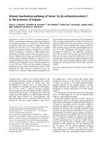
Local chromatin accessibility at the CPS enhancer
was determined by DNaseI hypersensitivity analysis.
Figure 2A shows the position of both SstI restriction
sites that were used for digestion, as well as the posi-
tion of the probe, directly upstream of the ) 5.3 kb
SstI site. Untreated samples showed the expected
7.6 kb SstI fragment and an additional, much longer,
band, presumably resulting from incomplete SstI diges-
tion (Fig. 2B, lanes 1, 5, 9 and 13). Both in untreated
and in dexamethasone ⁄ cAMP-treated FTO-2B cells,
fragments of 0.7–1.2 kb could be identified at inter-
mediate DNaseI concentrations (Fig. 2B, lanes 7 and
15). Thus, chromatin appears to be accessible at the
CPS GRU irrespective of hormonal activation. In con-
trast, Rat-1 fibroblasts did not exhibit such a hypersen-
sitive area, regardless of hormone treatment. The lack
of accessibility of the GRU enhancer in Rat-1 cells
Transcription factors at the CPS GRU M. Hoogenkamp et al.
38 FEBS Journal 274 (2007) 37–45 ª 2006 The Authors Journal compilation ª 2006 FEBS
therefore corresponds with the lack of CPS expression
in these cells, as shown in Fig. 1A.
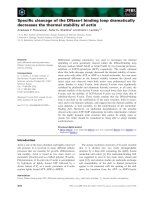
We analyzed the binding of transcription factors
to the CPS GRU in Rat-1 fibroblasts and FTO-2B
hepatoma cells that were or were not treated with dex-
amethasone ⁄ cAMP. Alterations in the accessibility of
DNA sequences were visualized by ligation-mediated
PCR (LM-PCR). Previously, we subjected the CPS
enhancer to in vitro footprinting using an end-labeled
DNA fragment. The transcription factors producing
the footprints in these assays were identified by com-
parison with footprints produced by purified proteins
[3]. To compare and validate the in vivo footprinting,
we also analyzed in vitro DNaseI-treated DNA by
LM-PCR. Linearized plasmid containing the CPS
enhancer was incubated with either BSA or rat liver
nuclear extract prior to DNaseI treatment. Analysis of
the in vitro footprinted samples for both strands of the
GRU region revealed three clear footprints (Fig. 3,
compare lane 1 with lane 2, and lane 7 with lane 8), in
agreement with our earlier observations [3]. Very simi-
lar results were observed when the in vivo footprinted
FTO-2B samples were compared with the Rat-1 sam-
ples (Fig. 3). The footprint due to C ⁄ EBP binding was
observed only in the FTO-2B samples at positions
322–343. FoxA binding was apparent in the FTO-2B
samples at positions 343–357, and highlighted by a
characteristic DNaseI hypersensitivity at position 350
on the upper strand and position 347 on the lower
strand [7]. Both the in vitro and in vivo experiments
showed that this FoxA-specific hypersensitivity was
consistently more prominent on the upper strand than
1+
3.5-
tsS I
tsS I
9
.21-
r
e
cna
h
nebk3.6-
A
esaNDI
PMAc/xed
-
+
b
k3.
1
–7.0
- sSt- tsS
)b
k6.7
(
}
PMAc/xed
1-taRR1-taB2-
OTFB2-OTF
2
1
4
3
6587
9
10 11
12
13 14
16
15
B
Fig. 2. DNaseI hypersensitivity of the CPS upstream region. (A)
Schematic representation of the upstream region of the CPS gene.
A 130 bp
32
P-labeled probe, located upstream of the SstI site at
) 5.3 kb, was used for hybridization. (B) Southern blot of in vivo
DNaseI-digested DNA from Rat-1 fibroblasts and FTO-2B hepatoma
cells. Cells were left untreated or were treated with dexametha-
sone and cAMP. Under each condition, cells were subjected to
increasing amounts of DNaseI. The position of the intact SstI–SstI
fragment and the GRU are indicated.
CPS
160 kDa
AB
GR
95 kDa
Rat-1
FTO-2B
Rat-1
FTO-2B
C/EBP probe FoxA probe
Rat-1
FTO-2B
Rat-1
FTO-2B
Rat-1
FTO-2B
Rat-1
FTO-2B
Rat-1
FTO-2B
Rat-1
FTO-2B
Rat-1
FTO-2B
probe
probe
PBE/C α
Antiserum:
PBE/C β 1AxoFenon2AxoF3AxoFenon
PαBE/CS
S
PβBE/CSS
1AxoFSS
2AxoFSS
1AxoF
Rat-1
FTO-2B
SS
1AxoF
Fig. 1. Expression of CPS and its regulating transcription factors in Rat-1 and FTO-2B cells. (A) CPS and GR were detected by western blot-
ting. For CPS, 32 lg of total protein from Rat-1 or FTO-2B cells was loaded per lane, whereas for GR, 50 lg of total protein was loaded per
lane. Amido black staining of the membrane served as loading control. (B) The presence of C ⁄ EBP and FoxA family members was visualized
by antibody-mediated supershifts in electrophoretic mobility shift assays. In each panel, the first lane corresponds to free probe, whereas
the other lanes correspond to probe incubated with Rat-1 or FTO-2B nuclear extracts. Where indicated, antibody directed against specific
members of the C ⁄ EBP and FoxA families of transcription factors was added. The region of the gel showing the supershifted complex with
FoxA1 antibody is additionally shown after a longer exposure. ‘SS’ indicates observed supershifts.
M. Hoogenkamp et al. Transcription factors at the CPS GRU
FEBS Journal 274 (2007) 37–45 ª 2006 The Authors Journal compilation ª 2006 FEBS 39
on the lower strand. Binding of P3 to positions 360–
377 was best detected on the upper strand as protec-
tion around position 363 and hypersensitivity at posi-
tion 381. Although the in vitro and in vivo footprints
are highly similar to each other, the band patterns are
not identical. This difference can be attributed to the
differences in DNaseI accessibility of naked DNA
(in vitro) and DNA in a chromatin context (in vivo) [9].
Interestingly, treatment of the cells with dexametha-
sone ⁄ cAMP prior to DNaseI treatment did not result
in any changes in the pattern of bands for either of the
two cell types, showing that binding of C ⁄ EBP and
FoxA to the CPS GRU in FTO-2B cells is independ-
ent of these hormonal stimuli.
FTO-2B hepatoma cells exhibit a constitutive
cAMP-dependent protein kinase (PKA) activity [10].
In the FTO-2B-derived hepatoma cell line WT-8, PKA
activity is again fully dependent on cAMP [10]. DNa-
seI footprinting of the CPS upstream enhancer gener-
ated a near-identical banding pattern in untreated
FTO-2B and WT-8 cells, including the prominent
FoxA-specific hypersensitive band (Fig. 4, arrow). As
already shown for FTO-2B (Fig. 3), treatment of both
cell lines with dexamethasone ⁄ cAMP did not alter the
banding pattern. This confirms that the binding of the
accessory factors to the CPS CRU does not result
from PKA activity, but is associated with the hepatic
phenotype.
As expected [11], DNaseI footprinting was not suit-
able for revealing the interaction between the GR and
its response element (GRE), but dimethylsulfate foot-
printing was (Fig. 5). Comparison of the FTO-2B
samples with the Rat-1 samples revealed that C⁄ EBP
binding to the GRU increased and decreased sensitiv-
ity to dimethylsulfate on the upper strand at positions
333 and 326, respectively, whereas changes in reactivity
at positions 331, 336 and 340 were seen on the lower
strand. FoxA binding resulted in protection of the gua-
nines at positions 347 and 351 on the upper strand,
and protection at position 352 on the lower strand.
These footprints in the FTO-2B samples were not
influenced by treatment with dexamethasone ⁄ cAMP, in
accordance with the DNaseI-footprinting experiments.
At the position of the GRE, differences between non-
treated and hormone-treated Rat-1 fibroblasts were
not observed on either DNA strand. Moreover, there
was no difference between the Rat-1 samples and the
untreated FTO-2B hepatoma cells. After the addition
of hormones to FTO-2B cells, however, the reactivity
of several guanines towards dimethylsulfate was
altered. These guanines map within the GRE region
that was footprinted by the GR in vitro [3]. On the
dnartsr
eppu
3P
RG
Ax
oF
PB
E
/C
’5
’3
B2-OTF
ENASB
+
-
+
-
1-taR
413
333
56
3
5
43
383
4
3
5
6
ovivni
413
33
3
563
54
3
383
ortivni
21
dnartsrewol
PBE
/C
3P
RG
AxoF
’5
’3
B2-OTF
+
-
+
-
1-taR
113
023
043
873
3
53
693
19
01
1
12
ovivni
ENASB
693
04
3
873
11
3
353
023
ortivni
8
7
Fig. 3. DNaseI footprinting of the CPS upstream enhancer in FTO-2B and Rat-1 cells. In vitro footprints were obtained by incubating a linea-
rized plasmid containing the CPS GRU with 45 lg of BSA or rat liver nuclear extract, after which DNaseI was added. The resulting DNA frag-
ments were used as template for LM-PCR. For in vivo footprints, dexamethasone ⁄ cAMP-treated (+) and untreated (–) Rat-1 fibroblasts and
FTO-2B hepatoma cells were permeabilized and incubated with DNaseI. After isolation of the DNA, the samples were subjected to LM-PCR.
An arrow indicates the FTO-2B-specific hypersensitive site in the FoxA-binding site. Schematic representations of the GRU with its binding
sites are included for clarity.
Transcription factors at the CPS GRU M. Hoogenkamp et al.
40 FEBS Journal 274 (2007) 37–45 ª 2006 The Authors Journal compilation ª 2006 FEBS
upper strand, the bands representing the guanines at
positions 383 and 385 were increased 1.6-fold (± 0.13;
N ¼ 4) and 1.7-fold (± 0.18; N ¼ 4), respectively,
whereas the band at position 392 was reduced 1.5-fold
(± 0.05; N ¼ 4). On the lower strand, GR binding
resulted in protection over the region covering posi-
tions 386–400. The observed bands corresponding to
the guanines at positions 386, 395 and 400 were
decreased 1.4-fold (± 0.15; N ¼ 3), 1.5-fold (± 0.13;
N ¼ 3), and 1.4-fold (± 0.08; N ¼ 3), respectively.
Elevated levels of cAMP alone did not lead to altera-
tions at the GRE, whereas dexamethasone alone did
(Fig. 5C). Five out of six of these changes of reactivity
of guanines map within the consensus GR-binding
sites within the GRE (Fig. 5D). All the guanines that
are known to be affected upon GR binding to a
similar GRE [11] showed altered reactivity. Altogether,
these findings indicate that these glucocorticoid-
induced footprints were due to GR binding.
Discussion
Transcriptional regulation results from the cooper-
ative binding of transcription factors [12]. One of the
key determinants of the activation of genes that are
under the control of hormone response units is the
order and time at which the transcription factors bind
to their response elements. With only four response
elements for transcription factors located within a
stretch of 80 bp, the CPS GRU is ideal for determin-
ing which transcription factor-binding events are pre-
requisites and which form the final trigger for GRU
activity.
DNaseI hypersensitivity analysis showed that the
DNA region encompassing the CPS enhancer is in an
open chromatin configuration in CPS-expressing hepa-
toma cells (Fig. 2). An open chromatin configuration
of GRU-containing distal enhancer regions appears to
be the rule in well-differentiated hepatoma cells [13–
15], including the distal tyrosine aminotransferase
(TAT) GRU at ) 5.5 kb, but this does not apply to
the more proximal TAT GRU at ) 2.5 kb, which needs
prior exposure to glucocorticoids to acquire an open
conformation [6,9,15]. In contrast to its accessibility in
hepatoma cells, the CPS GRU is not in an open confi-
guration in the Rat-1 cell line, which does not express
CPS. These findings are in line with the concept that
accessibility of an enhancer region to DNaseI corre-
lates with expression from that enhancer [16].
The absence of CPS expression in Rat-1 fibroblasts
correlates with the absence of FoxA DNA-binding
activities (Fig. 1) and a nonaccessible CPS GRU
(Fig. 2). FoxA is one of the relatively few transcription
factors that can function, in the absence of ATP-
dependent complexes, to open up compacted chroma-
tin, thereby allowing access for other transcription
factors [17]. It is therefore tempting to speculate that
these features underlie the absence of binding of tran-
scription factors (Fig. 3) and the lack of CPS expres-
sion in Rat-1 cells. In vivo footprinting of FTO-2B
hepatoma cells, on the other hand, showed constitutive
binding of FoxA and C ⁄ EBP to the CPS GRU
(Fig. 3), whereas binding of GR was conditional, i.e.
dependent on treatment of the cells with dexametha-
sone ⁄ cAMP (Fig. 5). Treatment of Rat-1 fibroblasts
with dexamethasone ⁄ cAMP did not result in binding
of GR, even though GR is abundantly present in these
cells (Fig. 1). In line with experiments showing that
GR binding to the ) 2.5 kb TAT GRU can only be
detected by genomic footprinting when the accessory
factors are bound to this GRU [11], these data indicate
that prior binding of accessory transcription factors
is necessary to stabilize the interaction between GR
and the CPS GRU. In vitro experiments with the
phosphoenolpyruvate carboxykinase (PEPCK) GRU
showed that binding of COUP-TF and especially
FoxA increased the affinity of the low-affinity PEPCK
GRE for GR and decreased its dissociation rate [18].
dnartsreppu
3P
RG
AxoF
PBE/C
’5
’
3
B2-OTF
+
-
+
-
8-TW
413
333
56
3
543
3
83
4321
Fig. 4. DNaseI footprinting of the CPS upstream enhancer in FTO-
2B and WT-8 hepatoma cells. For in vivo footprints, FTO-2B and
WT-8 hepatoma cells, either untreated (–) or treated with dexa-
methasone ⁄ cAMP (+), were permeabilized and incubated with
DNaseI. After isolation of the DNA, the samples were subjected to
LM-PCR. An arrow indicates the hypersensitive site in the FoxA-
binding site. Schematic representations of the GRU with its binding
sites are included for clarity.
M. Hoogenkamp et al. Transcription factors at the CPS GRU
FEBS Journal 274 (2007) 37–45 ª 2006 The Authors Journal compilation ª 2006 FEBS 41
For both the PEPCK and the CPS GRUs, the distance
between the binding sites for GR and FoxA is of crit-
ical importance for GRU activity, which probably
reflects a direct interaction between the two factors
[4,19].
Although it remains to be established to what extent
these findings in cell lines can be extrapolated to pri-
mary hepatocytes, the absence of FoxA from fibroblasts
is in agreement with the concept that FoxA binding is
an early event in the opening of the chromatin of he-
patocytes [17]. FoxA binds in a glucocorticoid-inde-
pendent manner on the CPS GRU (Figs 3 and 5), the
PEPCK GRU [20] and the distal TAT GRU at ) 5.5 kb
[9,15]. Although FoxA binds to the proximal TAT
GRU at ) 2.5 kb in unstimulated FTO-2B cells, its
binding is significantly enhanced by prior exposure to
glucocorticoids and chromatin remodeling [11], whereas
in H4IIE hepatoma cells, its binding to this GRU is
strictly dependent upon GR activation [7]. The subse-
quent binding of the accessory transcription factors, in
turn, stabilizes GR binding (see previous paragraph)
[11]. The difference in FoxA binding between the FTO-
2B and H4IIE cell lines could be due to the constitutive
PKA activity that is present in the FTO-2B cell line.
FoxA binding at the ) 2.5 kb TAT GRU in WT-8 cells
is, indeed, very low in the absence of glucocorticoids
and can be induced by glucocorticoids and PKA activa-
tion in an additive manner [11]. However, FoxA bind-
ing to the TAT GRU at ) 5.5 kb and the CPS GRU
was constitutive in both FTO-2B and WT-8 cells, ren-
dering the CPS GRU similar to the distal TAT GRU.
Another study in FTO-2B cells, conducted on the con-
stitutively open GRU of the 6-phosphofructo-2-kinase
(PFK-2) gene, showed that FoxA binding was largely
glucocorticoid-dependent, despite the constitutive PKA
activity and the glucocorticoid-independent chromatin
remodeling [13]. Taken together, these studies show that
although these GRUs are all involved in mediating he-
patocyte-specific expression and contain binding sites
for GR in combination with response elements for the
liver-enriched factors FoxA and C ⁄ EBP, they differ in
their recruitment of these common transcription fac-
tors. Although it is still unclear what determines these
differences in the assembly of the transcription factor
complex, transient transfection studies suggest that the
precise arrangement of the various binding sites within
12
3
0
4
3
273
504
3P
383
401
RG
A
xoF
P
BE/C
453
033
’5
3’
+
-
+
-
1-taRB2-OTF
FTO-2B
untreated
+dex
+forskolin
+dex & forsk.
GRE
PMAc/xed
8
76
5
dnarts rewol
dnarts reppu
+
-
+
-
1
-t
a
R
B2
-OTF
3P
RG
AxoF
PBE/C
33
3
75
3
743
573
10
4
3
83
31
4
5’
’3
PMAc/xed
3
214
B
A C
D
A
-’
5 G Tt
t
g
A
CGA
G
3-
ct
tgT
C
TT ’
CTCT-
’
3 G AA
C
AaacT G ’5-
g
aacA
832
0
4
0
Fig. 5. Dimethylsulfate footprinting of the CPS upstream enhancer in Rat-1 and FTO-2B cells. Dexamethasone ⁄ cAMP-treated (+) and
untreated (–) Rat-1 fibroblasts and FTO-2B hepatoma cells were incubated with 0.1% dimethylsulfate. After DNA isolation, the samples were
subjected to LM-PCR. (A, B) Upper and lower strands, respectively. Closed diamonds and circles indicate guanines showing, respectively,
increased and decreased sensitivity towards dimethylsulfate in hormone-treated FTO-2B cells, whereas open diamonds and circles indicate
hormone-independent decreased and increased sensitivity to dimethylsulfate in FTO-2B compared to Rat-1 cells. (C) Upper strand analysis,
showing that dexamethasone treatment alone is sufficient to promote GR binding in FTO2B cells. (D) Sequence of the CPS GRE showing
the guanine residues that showed altered reactivity to dimethylsulfate following hormonal treatment of FTO2-B cells. The consensus palin-
dromic GR-binding site is indicated in capital letters. The guanines in bold letters are those that showed altered reactivity towards dimethyl-
sulfate upon GR binding in vitro to a similar GRE [11].
Transcription factors at the CPS GRU M. Hoogenkamp et al.
42 FEBS Journal 274 (2007) 37–45 ª 2006 The Authors Journal compilation ª 2006 FEBS
the GRUs and the relative affinities for their cognate
factors play a role, presumably in combination with the
chromatin structure established at the GRUs prior to
glucocorticoid activation [4,19].
Even though the assembly of the transcription fac-
tor complex at the CPS GRU seems to be similar to
that of the distal TAT GRU at ) 5.5 kb, there
appears to be an important difference. The CPS
GRU is sufficient to enhance the activity of the basal
promoter [4], whereas the distal TAT GRU has to
cooperate with the proximal TAT GRU at ) 2.5 kb
[15]. This difference may be more apparent than real,
however, because we recently showed that the CPS
GRU needs to interact with a GRE directly upstream
of the core CPS promoter to transactivate this pro-
moter [5,21].
For both the proximal TAT GRU and the PFK-2
GRU, it is not clear which transcription factor is
directly responsible for transcriptional activation, as
GR allows recruitment of FoxA and C ⁄ EBP, which
could be the factors interacting with the transcription
machinery. Therefore, GR could play only an indirect
role by allowing the recruitment of these factors. The
distal TAT GRU, where GR is the only DNA-bind-
ing protein interacting specifically in the presence of
glucocorticoids, does not provide a clear answer to
this question, as this GRU does not activate tran-
scription on its own. Our present analysis of tran-
scription factor recruitment at the CPS GRU
therefore provides the first clear evidence that the
transcription activation domains of GR play a key
role in transcriptional activation mediated by a GRU,
as we show that it is the only DNA-binding protein
of the triad GR, FoxA and C ⁄ EBP that is specifically
recruited upon glucocorticoid stimulation. The acces-
sory factors FoxA and C ⁄ EBP presumably allow sta-
bilization of GR binding to the GRU, and thereby
stabilization of the interaction of the coregulators
interacting with the nucleoprotein complex formed at
the GRU. This raises the possibility that the acces-
sory factors play a similar role in GRUs where they
are recruited in a glucocorticoid-dependent manner,
and that the differences that are seen in the modali-
ties of recruitment do not reflect fundamental differ-
ences in their contribution to the function of the
GRUs.
Experimental procedures
Cell culture
Rat-1 fibroblasts [22], FTO-2B hepatoma cells [23], and
WT-8, an FTO-2B-derived cell line overexpressing the R1a
subunit of PKA [10], were grown at 37 °Cin
DMEM ⁄ F12 ⁄ 5% CO
2
⁄ 10% fetal bovine serum.
Nuclear extracts
Nuclear extracts from rat livers were prepared as previ-
ously described [24]. To prepare nuclear extracts from cell
lines, cells were detached with trypsin, washed in NaCl ⁄ P
i
containing 0.25 mm phenylmethanesulfonyl fluoride, and
lysed by resuspension in 1.5 mL of 10 mm Hepes (pH 7.6),
10 mm KCl, 1.5 mm MgCl
2
and 0.5 mm dithiothreitol per
2 · 10
7
cells. After 8 min on ice, nuclei were pelleted by
centrifugation in an Eppendorf 5417C centrifuge at
20 800 g for 30 s at 4 °C. The pellets were resuspended in
100 lLof20mm Hepes (pH 7.6), 20% glycerol, 420 mm
NaCl, 1.5 mm MgCl
2
, 0.2 mm EDTA and 0.5 mm dithio-
threitol, and incubated on ice for 20 min. Nuclear debris
was spun down at 20 800 g for 2 min at 4 °C (Eppendorf
5417C centrifuge).
Western blotting
Whole cell extracts were prepared by lysis of cells in 20 mm
Tris (pH 7.5), 150 mm NaCl, 1% NP40, 0.5 mm dithiothrei-
tol and 0.2 mm phenylmethanesulfonyl fluoride. Insoluble
debris was spun down in an Eppendorf centrifuge at
20 800 g for 20 s at 4 °C (Eppendorf 5417C centrifuge).
Western blotting was performed as previously described [25].
Electrophoretic mobility shift assays
Electrophoretic mobibility shift assays were performed as
previously described [4], except that the binding reaction
contained 20 mm Hepes (pH 7.6), 500 mm KCl, 12% gly-
cerol (v ⁄ v), 1 mm EDTA, 1 mm dithiothreitol, 1 mm sper-
midine, 0.5 lg of double-stranded poly(dIdC)Ælg
)1
nuclear
extract, and 0.3 lgÆlL
)1
BSA. Double-stranded probes were
designed on the basis of the rat CPS GRU sequence
(Table 1).
Antibodies
Rabbit polyclonal antibodies against C ⁄ EBPa (sc-61),
C ⁄ EBPb (sc-746) and GR (sc-1003), and goat polyclonal
antibodies against FoxA1 (sc-6553), FoxA2 (sc-6554) and
FoxA3 (sc-5360), were obtained from Santa Cruz Biotech-
nology (Santa Cruz, CA, USA). Rabbit polyclonal anti-
body against CPS has been previously described [26].
DNaseI treatment
Cells were grown to 70% confluence and supplemented,
where indicated, with 100 nm dexamethasone, 1 mm dibuty-
ryl-cAMP and 0.1 mm 3-isobutyl-1-methylxanthine (IBMX)
M. Hoogenkamp et al. Transcription factors at the CPS GRU
FEBS Journal 274 (2007) 37–45 ª 2006 The Authors Journal compilation ª 2006 FEBS 43
2 h before the start of the experiment. DNaseI treatment
was performed exactly as previously described [27].
Dimethylsulfate treatment
Cells were grown to 70% confluence and exposed, where
indicated, to 100 nm dexamethasone, 1 mm dibutyryl-
cAMP and 0.1 mm IBMX for 2 h before the start of the
experiment. All solutions used for hormone-treated cells
contained 100 nm dexamethasone. After exposure for
4 min at room temperature to 0.1% dimethylsulfate in
NaCl ⁄ P
i
, cells were washed three times with NaCl ⁄ P
i
and
lysed in 2.5 mL of 50 mm Tris (pH 8.0), 20 mm EDTA,
1% SDS and 100 lgÆmL
)1
proteinase K per 80 cm
2
flask,
digested overnight at 55 °C, and further processed as des-
cribed [28].
In vitro footprinting
In vitro footprinting was performed as previously described
[29]. As matrix, linearized plasmid DNA containing the
CPS enhancer region was used. Protein binding was per-
formed using 45 lg of rat liver nuclear extract or BSA.
DNaseI hypersensitivity analysis
Thirty micrograms of DNaseI-treated DNA was cut with
SstI, separated on a 1% agarose gel, blotted [25], and
hybridized to a 130 bp [a-
32
P]ATP-labeled PCR probe.
LM-PCR
The starting material consisted of 1 lg of genomic DNA
for in vivo footprints or 2 ng of plasmid DNA for in vitro
footprints. LM-PCR was performed as described [30],
except that the linker–ligation mix contained 5% poly-
ethyleneglycol-6000. The primers used are described in
Table 1.
Acknowledgements
The research presented in this article was financially
supported by ZonMW grant 902-23-250 and by grants
to TG of the Association pour la Recherche sur le
Cancer and the Ligue contre le Cancer.
References
1 Meijer AJ, Lamers WH & Chamuleau RA (1990) Nitro-
gen metabolism and ornithine cycle function. Physiol
Rev 70, 701–748.
2 Christoffels VM, Van Den Hoff MJ, Moorman AF &
Lamers WH (1995) The far-upstream enhancer of the
carbamoyl-phosphate synthetase I gene is responsible
for the tissue specificity and hormone inducibility of its
expression. J Biol Chem 270, 24932–24940.
3 Christoffels VM, Grange T, Kaestner KH, Cole TJ,
Darlington GJ, Croniger CM & Lamers WH (1998)
Glucocorticoid receptor, C ⁄ EBP, HNF3, and protein
kinase A coordinately activate the glucocorticoid
response unit of the carbamoylphosphate synthetase I
gene. Mol Cell Biol 18, 6305–6315.
4 Schoneveld OJ, Gaemers IC, Das AT, Hoogenkamp M,
Renes J, Ruijter JM & Lamers WH (2004) Structural
requirements of the glucocorticoid-response unit of the
carbamoyl-phosphate synthase gene. Biochem J 382,
463–470.
5 Schoneveld OJ, Gaemers IC & Lamers WH (2004)
Mechanisms of glucocorticoid signalling. Biochim Bio-
phys Acta 1680, 114–128.
6 Reik A, Schutz G & Stewart AF (1991) Glucocorticoids
are required for establishment and maintenance of an
alteration in chromatin structure: induction leads to a
reversible disruption of nucleosomes over an enhancer.
EMBO J 10, 2569–2576.
7 Rigaud G, Roux J, Pictet R & Grange T (1991) In vivo
footprinting of rat TAT gene: dynamic interplay
between the glucocorticoid receptor and a liver-specific
factor. Cell 67, 977–986.
8 Hoogenkamp M, Stallen JM, Lamers WH & Gaemers
IC (2006) In vivo footprinting of the carbamoylpho-
sphate synthetase I cAMP-response unit indicates
important roles for FoxA and PKA in formation of the
enhanceosome. Biochimie 88, 1357–1366.
9 Flavin M, Cappabianca L, Kress C, Thomassin H &
Grange T (2004) Nature of the accessible chromatin at
a glucocorticoid-responsive enhancer. Mol Cell Biol 24,
7891–7901.
Table 1. Oligonucleotide sequences used for experiments. EMSA,
electrophoretic mobility shift assay.
Probes and primers
(5¢-to3¢)
Probes for EMSA
C ⁄ EBP TTTCGAGTCTTGCAAAATCATCA
FoxA CATCAGTGTTTGCTCTTGACAAG
LM-PCR primers
Upper strand
Primer 1 TTCTTAAAACTTGACCAAA
Primer 2 GGGTACGATGACTAAATGATCGGA
Primer 3
(Figs 3 and 5A,B)
TCATCAGCAGCCCTTCTTTGCACAAC
Primer 3
(Figs 4 and 5C)
GACTAAATGATCGGATACGTGCCCATTCT
Lower strand
Primer 1 CTCAACGTCATTCTAAAGT
Primer 2 ACAACATACTTCGAAACTGTGACC
Primer 3 TGTCCTGGCACATGACCCGGATCA
Transcription factors at the CPS GRU M. Hoogenkamp et al.
44 FEBS Journal 274 (2007) 37–45 ª 2006 The Authors Journal compilation ª 2006 FEBS
10 Jones KW, Shapero MH, Chevrette M & Fournier RE
(1991) Subtractive hybridization cloning of a tissue-spe-
cific extinguisher: TSE1 encodes a regulatory subunit of
protein kinase A. Cell 66, 861–872.
11 Espinas ML, Roux J, Pictet R & Grange T (1995)
Glucocorticoids and protein kinase A coordinately mod-
ulate transcription factor recruitment at a glucocorti-
coid-responsive unit. Mol Cell Biol 15, 5346–5354.
12 Carey M (1998) The enhanceosome and transcriptional
synergy. Cell 92, 5–8.
13 Zimmermann PL, Pierreux CE, Rigaud G, Rousseau
GG & Lemaigre FP (1997) In vivo protein–DNA inter-
actions on a glucocorticoid response unit of a liver-spe-
cific gene: hormone-induced transcription factor binding
to constitutively open chromatin. DNA Cell Biol 16,
713–723.
14 Ip YT, Granner DK & Chalkley R (1989) Hormonal
regulation of phosphoenolpyruvate carboxykinase gene
expression is mediated through modulation of an
already disrupted chromatin structure. Mol Cell Biol 9,
1289–1297.
15 Grange T, Roux J, Rigaud G & Pictet R (1989) Two
remote glucocorticoid responsive units interact coopera-
tively to promote glucocorticoid induction of rat tyro-
sine aminotransferase gene expression. Nucleic Acids
Res 17, 8695–8709.
16 Felsenfeld G, Boyes J, Chung J, Clark D & Studitsky V
(1996) Chromatin structure and gene expression. Proc
Natl Acad Sci USA 93, 9384–9388.
17 Cirillo LA, Lin FR, Cuesta I, Friedman D, Jarnik M &
Zaret KS (2002) Opening of compacted chromatin by
early developmental transcription factors HNF3 (FoxA)
and GATA-4. Mol Cell 9, 279–289.
18 Stafford JM, Wilkinson JC, Beechem JM & Granner
DK (2001) Accessory factors facilitate the binding of
glucocorticoid receptor to the phosphoenolpyruvate car-
boxykinase gene promoter. J Biol Chem 276, 39885–
39891.
19 Sugiyama T, Scott DK, Wang JC & Granner DK
(1998) Structural requirements of the glucocorticoid and
retinoic acid response units in the phosphoenolpyruvate
carboxykinase gene promoter. Mol Endocrinol 12, 1487–
1498.
20 Wang XL, Herzog B, Waltner-Law M, Hall RK, Shiota
M & Granner DK (2004) The synergistic effect of dexa-
methasone and all-trans-retinoic acid on hepatic phos-
phoenolpyruvate carboxykinase gene expression
involves the coactivator p300. J Biol Chem 279, 34191–
34200.
21 Schoneveld OJ, Gaemers IC, Hoogenkamp M & Lamers
WH (2005) The role of proximal-enhancer elements in
the glucocorticoid regulation of carbamoylphosphate
synthetase gene transcription from the upstream
response unit. Biochimie 87, 1033–1040.
22 Prasad I, Zouzias D & Basilico C (1976) State of the
viral DNA in rat cells transformed by polyoma virus.
I. Virus rescue and the presence of nonintegrated viral
DNA molecules. J Virol 18, 436–444.
23 Killary AM & Fournier RE (1984) A genetic analysis of
extinction: trans-dominant loci regulate expression of
liver-specific traits in hepatoma hybrid cells. Cell 38,
523–534.
24 Sierra F (1990) A laboratory guide to in vitro transcrip-
tion. In Biomethods (Azzi A, Polak JM & Saluz HP,
eds), pp. 30–50. Birkhauser-Verlag, Basel.
25 Sambrook J, Fritsch EF & Maniatis T (1989) Molecular
Cloning: A Laboratory Manual. Cold Spring Harbor
Laboratory Press, Cold Spring Harbor, NY.
26 Charles R, de Graaf A & Moorman AF (1980) Radio-
immunochemical determination of carbamoyl-phosphate
synthase (ammonia) content of adult rat liver. Biochim
Biophys Acta 629, 36–49.
27 Lefevre P, Lacroix C, Tagoh H, Hoogenkamp M,
Melnik S, Ingram R & Bonifer C (2005) Differentiation-
dependent alterations in histone methylation and chro-
matin architecture at the inducible chicken lysozyme
gene. J Biol Chem 280, 27552–27560.
28 Mueller PR, Wold B & Garrity PA (2002) Current Pro-
tocols in Molecular Biology. John Wiley & Sons, New
York, NY, doi:10.1002/0471142727.mb1503s56.
29 Espinas ML, Roux J, Ghysdael J, Pictet R & Grange T
(1994) Participation of Ets transcription factors in the
glucocorticoid response of the rat tyrosine aminotrans-
ferase gene. Mol Cell Biol 14, 4116–4125.
30 Grange T, Bertrand E, Espinas ML, Fromont-Racine
M, Rigaud G, Roux J & Pictet R (1997) In vivo foot-
printing of the interaction of proteins with DNA and
RNA. Methods 11, 151–163.
M. Hoogenkamp et al. Transcription factors at the CPS GRU
FEBS Journal 274 (2007) 37–45 ª 2006 The Authors Journal compilation ª 2006 FEBS 45