Báo cáo khóa học: Emerin binding to Btf, a death-promoting transcriptional repressor, is disrupted by a missense mutation that causes Emery–Dreifuss muscular dystrophy pdf
Bạn đang xem bản rút gọn của tài liệu. Xem và tải ngay bản đầy đủ của tài liệu tại đây (345.48 KB, 11 trang )
Emerin binding to Btf, a death-promoting transcriptional repressor,
is disrupted by a missense mutation that causes Emery–Dreifuss
muscular dystrophy
Tokuko Haraguchi
1
, James M. Holaska
2
, Miho Yamane
1
, Takako Koujin
1
, Noriyo Hashiguchi
1
, Chie Mori
1
,
Katherine L. Wilson
2
and Yasushi Hiraoka
1
1
CREST Research Project, Kansai Advanced Research Center, Communications Research Laboratory, Iwaoka-cho, Nishi-ku,
Kobe, Japan;
2
Department of Cell Biology, Johns Hopkins University School of Medicine, Baltimore, MD, USA
Loss of functional emerin, a nuclear membrane protein,
causes X-linked recessive Emery–Dreifuss muscular dystro-
phy. In a yeast two-hybrid screen, we found that emerin
interacts with Btf, a death-promoting transcriptional
repressor, which is expressed at high levels in skeletal muscle.
Biochemical analysis showed that emerin binds Btf with an
equilibrium affinity (K
D
)of100 n
M
. Using a collection of 21
clustered alanine-substitution mutations in emerin, the resi-
dues required for binding to Btf mapped to two regions of
emerin that flank its lamin-binding domain. Two disease-
causing mutations in emerin, S54F and D95–99, disrupted
binding to Btf. The D95–99 mutation was relatively unin-
formative, as this mutation also disrupts emerin binding to
lamin A and a different transcription repressor named germ
cell-less (GCL). In striking contrast, emerin mutant S54F,
which binds normally to barrier-to-autointegration factor,
lamin A and GCL, selectively disrupted emerin binding to
Btf. We localized endogenous Btf in HeLa cells by indi-
rect immunoflurorescence using affinity-purified antibodies
against Btf. In nonapoptotic HeLa cells Btf was found in
dot-like structures throughout the nuclear interior. How-
ever, within 3 h after treating cells with Fas antibody to
induce apoptosis, the distribution of Btf changed, and Btf
concentrated in a distinct zone near the nuclear envelope.
These results suggest that Btf localization is regulated by
apoptotic signals, and that loss of emerin binding to Btf may
be relevant to muscle wasting in Emery–Dreifuss muscular
dystrophy.
Keywords: apoptosis; emerin; Emery–Dreifuss muscular
dystrophy; lamin A; MAN1.
The loss of emerin function causes X-linked recessive
Emery–Dreifuss muscular dystrophy (EDMD) [1], which
affects skeletal muscle, heart and major tendons [2,3].
Emerin binds lamins, including lamin A [4,5]. It was
therefore intriguing that dominant forms of EDMD arise
in people carrying point mutations in LMNA,which
encodes A-type lamins [6]. In a fascinating series of
discoveries, mutations distributed throughout LMNA
were found to cause seven additional diseases: limb-girdle
muscular dystrophy type 1B, dilated cardiomyopathy type
1 A, Dunnigan-type familial partial lipodystrophy (FPLD),
an axonal neuropathy known as Charcot–Marie–Tooth
disorder type 2B1 [7], a bone development disorder named
mandibuloacral dysplasia [8–11], and two accelerated ÔagingÕ
diseases named Hutchison–Gilford Progeria Syndrome
[12,13] and atypical Werner syndrome [14]. With the
possible exception of Charcot–Marie–Tooth disorder type
2B1 disorder, the tissues affected in these Ônuclear lamino-
pathyÕ disorders may share a common mesenchymal stem
cell lineage [15,16]. The mechanisms underlying these
diseases are important to understand, due to their clinical
significance and because so little is currently known about
nuclear envelope function. To explain the tissue-specificity
of Emery–Dreifuss muscular dystrophy, emerin and A-type
lamins were proposed to influence tissue-specific gene
expression [15,17].
Emerin is a 254-residue integral nuclear membrane
protein with an apparent molecular mass of 34 kDa
(SDS/PAGE). Emerin is expressed in most but not all
tissues that have been tested [1,18–20], and is phosphoryl-
ated in a cell-cycle dependent manner [21]. EDMD is
diagnosed in childhood by ÔcontracturesÕ of tendons in the
neck, ankles, and elbow, along with slowly progressive
skeletal muscle wasting, and cardiac conduction defects
which can cause sudden death [2,22]. Most X-linked
EDMD patients, including those with missense mutations,
are null for emerin protein due to degradation of the mutant
mRNA or protein. However, a few patients express normal
amounts of mutant emerin protein, which is correctly
localized at the inner nuclear membrane [23,24]. These
special mutations include S54F (Ser54fiPhe), P183H and
P183T (Pro183fiHis or Thr), and a five-residue deletion
(D95–99). These mutations have the potential to reveal
Correspondence to T. Haraguchi, Kansai Advanced Research Center,
Communications Research Laboratory, 588-2 Iwaoka,
Iwaoka-cho, Nishi-ku, Kobe 651-2492, Japan.
Fax: + 81 78 969 2249, Tel.: + 81 78 969 224,
E-mail:
Abbreviations: EDMD, X-linked recessive Emery–Dreifuss muscular
dystrophy; GCL, germ cell-less.
(Received 9 December 2003, revised 12 January 2004,
accepted 20 January 2004)
Eur. J. Biochem. 271, 1035–1045 (2004) Ó FEBS 2004 doi:10.1111/j.1432-1033.2004.04007.x
disease mechanisms because the mutant proteins are in the
right place (inner nuclear membrane) and their expression
levels are normal, yet they cause disease. We hypothesized
that these proteins must be defective in one or more
activities required for emerin function, such as binding to
other proteins at the inner nuclear membrane.
Two of emerin’s known binding partners are lamin A
and barrier-to-autointegration factor (BAF) [4,5]. The
40-residue ÔLEM-domainÕ of emerin binds directly to BAF
[5], and is required for emerin to be recruited to BAF on
chromatin during nuclear assembly [24]. Emerin and other
ÔLEM-domainÕ proteins such as LAP2b and MAN1 [25]
constitute a family of BAF-binding proteins [5,26,27]. The
expression of an exogenous mutant BAF (G25E) in HeLa
cells disrupts the assembly of endogenous BAF, emerin,
LAP2b and lamin A/C (but remarkably, not B-type lamins)
into reforming nuclear envelopes [24]. Thus, BAF is
predicted to recruit or assemble many if not all LEM-
domain proteins and A-type lamins during nuclear forma-
tion. We know of no disease-causing mutation in emerin
that affects its binding to BAF. However the D95–99
mutation disrupts binding to several binding partners
including lamin A [5], transcription regulator germ cell-less
(GCL [30]); and splicing factor YT521-B [31]. Two ÔspecialÕ
disease-causing mutants, S54F and P183H, bind normally
to lamin A, GCL, YT521-B and BAF [5,30,31], suggesting
that these mutations disrupt emerin’s binding to undiscov-
ered binding partners relevant to disease.
We used a two-hybrid screen of a HeLa cell cDNA
library to search for novel binding partners of emerin, using
full length emerin as bait. This screen produced a positive
clone encoding a predicted 920-residue protein, previously
reported as Btf [28] or KIAA0164 [29]. Btf can act as a
transcriptional repressor, and when overexpressed, Btf
induces cell death by a mechanism involving the inhibition
of antiapoptotic bcl-2 family proteins [28]. Btf has a wide
tissue distribution (including heart, brain, placenta, lung,
kidney and pancreas) and is highly expressed in skeletal
muscle [29]. Our results show that binding to Btf is
specifically and selectively disrupted by the disease-associ-
ated S54F missense mutation in emerin. The implications
of these findings for possible EDMD disease mechanisms
are discussed.
Materials and methods
Cells and reagents
HeLa cells were obtained from the Riken Cell Bank
(Tsukuba Science City, Tsukuba, Japan). Hoechst 33342,
cycloheximide and anti-Fas monoclonal Ig were from
Calbiochem (La Jolla, CA, USA), Wako (Osaka, Japan)
and MBL (Nagoya, Japan), respectively. Rabbit poly-
clonal serum Ôbtf-middleÕ was prepared by immunizing
rabbits with a keyhole limpet hemocyanin (KLH)-con-
jugated synthetic peptide (CSERITVKKETQSPEQ-
CONH
2
; with amido modification in the C-terminus)
corresponding to residues 485–499 of human Btf. Specific
antibodies were affinity-purified by chromatography on
NHS-activated Sepharose 4B (Amersham Pharmacia
Biotech) coupled to the antigenic peptide as described
[32]. For indirect immunofluorescence staining, purified
antibody was concentrated on Centricon-10 spin columns
(Amicon, MA, USA) and eluted with phosphate-buffered
saline (Gibco BRL, USA).
Yeast two-hybrid screen
Emerin interactor(s) were screened by yeast two-hybrid
assay using Matchmaker System III (Clontech Inc.)
according to manufacturer instructions. Full length emerin
cloned in the pGBK-T7 vector was used as bait. The prey
HeLa cDNA library in the pGAD-GH vector was provided
by Clontech. The bait plasmid was transformed into
Saccharomyces cerevisiae strain Y187, and mated with
S. cerevisiae AH109 cells pretransformed with the prey
library. Positive clones were selected based on growth in the
absenceofaminoacidsTrp,Leu,His,andAde(Ôquadruple
dropoutÕ), and screened for b-galactosidase production.
For one-to-one two-hybrid analysis, cDNAs encoding
full length emerin or emerin fragments were first fused to the
GAL4 DNA binding domain in the pGBK plasmid, and
then transformed into yeast Y187 cells with lithium acetate.
These cells were then mated with yeast AH109 cells that
expressed either full length Btf (residues 1–920) or Btf
fragments 377–920, 377–761, 377–646, 377–574 and 521–
761 fused to the GAL4 activator domain in the pGAD
plasmid. After mating, cells were cultured in YPDA
medium (1% yeast extract, 2% peptone, 2% dextrose,
0.003% adenine hemisulfate) for 20 h at 30 °C. Diploid cells
that grew in the absence of Trp and Leu were selected, and
then plated on quadruple-dropout medium to assay two-
hybrid-dependent gene expression. Positives were confirmed
by b-galactosidase production.
Plasmid construction
To fuse emerin, Btf or fragments thereof with the two-
hybrid vectors, the desired cDNAs were PCR-amplified
using the primers and templates in Tables 1 and 2. PCR
products were digested with NdeIandBamHI, and inserted
into each vector. To construct BD-lamin A plasmids, the
coding regions of lamin A were PCR-amplified using the
following primers: 5¢-AAGAATTCATGGAGACCCCGT
CCCAG-3¢ and 5¢-GCCGTCGACTTACATGATGCTG
CAGTTCTGGGG-3¢. PCR products were digested with
EcoRI and SalI, and inserted into the p-GBK vector
(Clontech Laboratories, Inc., Palo Alto, CA, USA) using
the SalIandBamHI sites in the vector. The DNA sequences
of all fusion plasmids were confirmed using an ABI377
DNA sequencer (Applied Biosystems, Norwalk, CT, USA).
Microtiter well assay for Btf binding to emerin
and affinity measurements
Btf protein was synthesized and
35
S-labeled in vitro using
coupled transcription/translation extracts (Promega Corp.),
as described in detail by Holaska et al.[30].Wildtype
emerin residues 1–222, or each mutant emerin, were
purified as recombinant proteins and adsorbed to micro-
titer wells. Typically, 5–50 pmoles of emerin protein were
adsorbed per well. [
35
S]Btf was placed into wells contain-
ing each emerin protein, or into BSA-adsorbed wells as
negative controls, and incubated 60–90 min at room
1036 T. Haraguchi et al. (Eur. J. Biochem. 271) Ó FEBS 2004
temperature in binding buffer (BB: 20 m
M
Hepes pH 7.4,
110 m
M
potassium acetate, 2 m
M
magnesium acetate,
1m
M
EGTA). Wells were washed with 200 lL BB, five
times, and bound Btf was eluted with 5% SDS, placed in
a scintillation vial, and counted. To assay Btf binding by
blot overlay,
35
S-labeled Btf was incubated for 16 h at
4 °C with blots of recombinant human emerin proteins,
purified by FPLC as described [30].
Western blotting
HeLa cells (1 · 10
7
) were collected by scraping with a
rubber policeman after washing twice with DMEM
medium, suspended in homogenizing buffer (20 m
M
Tris/HCl pH 7.5, 2 m
M
MgCl
2
,150m
M
NaCl) supple-
mented with protease inhibitor cocktail (Roche) to a final
concentration of 1 · 10
7
cells per mL, and homogenized
on ice using a Potter homogenizer. Half of each sample
was centrifuged at 1000 g for 3 min to separate the
nuclear (pellet) and cytoplamic (supernatant) fractions,
and the other half was kept as the Ôtotal lysateÕ fraction.
Samples corresponding to 1 · 10
5
cells were loaded per
lane on SDS 15% polyacrylamide gels. After electrophor-
esis, proteins were transferred to poly(vinylidene difluo-
ride) membrane at 60 V for 2 h in transfer buffer (50 m
M
Tris, pH 7.5, 380 m
M
glycine, 0.1% (v/v) SDS and 20%
(v/v) MeOH). After blocking with 5% (v/v) skim milk in
NaCl/P
i
, the membrane was incubated at 4 °Covernight
with primary antibody (against Ôbtf-middleÕ antigen;
described above) at a dilution of 1 : 1000 in NaCl/P
i
containing 0.1% (v/v) skim milk, and 0.1% (v/v) Tween-
20. Blots were then incubated at 4 °Cfor2hwithHRP-
conjugated anti-rabbit IgG (Cappel) at a dilution of
1 : 1000 and stained by enhanced chemiluminescence
(Amersham).
Indirect immunofluorescence staining
Cells were fixed in 10% (v/v) trichloroacetic acid for 10 min
at room temperature after a brief wash with DMEM
(37 °C), and then permeabilized with 0.1% (v/v) Triton
X-100 in NaCl/P
i
for 5 min, washed three times with NaCl/
P
i
, and finally incubated 1 h with 1% (v/v) BSA in NaCl/P
i
,
all at room temperature. Antibodies against Btf were then
added to cells at 1 : 500 dilution, incubated 18 h at 4 °C,
washed four times, and stained with Alexa-conjugated
secondary antibody (Molecular Probes Inc.) at a dilution of
1 : 1000 for 3–4 h at room temperature. Finally, cells
were washed three times with NaCl/P
i
, and incubated
sequentially with 20, 40, 60, and 80% glycerol containing
NaCl/P
i
, 2.5% 1,4-diazabicyclo-2,2,2-octane (DABCOÒ)
and 0.5 lgÆmL
)1
4¢,6-diamidino-2-phenylindole (DAPI).
Cells were mounted in 90% glycerol containing 2.5%
DABCOÒ as an antifading reagent.
Table 1. Names of PCR primers used in this work. For cloned regions, the numbers represent the first and last amino acids numbers of cloned
regions.
Cloned regions Template Forward primer Reverse primer Vector
emerin-full GFP-emerin H emerin 1 EGFP-emerin-Nde1–2 pGBKT7
104–254 GFP-emerin H-emerin BamHI310 3¢ H-emerin BamHI pGBKT7
164–254 pGBKT7-emerin 164–5¢ 3¢ H-emerin BamHI pGBKT7
104–228 pGBKT7-emerin H-emerin BamHI310 DTM H-emerin BamHI pGBKT7
btf-full KIAA0164 – – pGADT7
377–920 KIAA0164 377–5¢ End-3¢ pGADT7
377–761
a
– – – pGADT7
377–646 KIAA0164 377–5¢ 646–3¢ pGADT7
377–574 KIAA0164 377–5¢ 574–3¢ pGADT7
521–761 KIAA0164 521–5¢ 761–3¢ pGADT7
a
This plasmid was selected from the screening of the HeLa cDNA library packaged in Matchmaker Systems III (Clontech).
Table 2. Nucleotide sequences of PCR primers.
Name of the primer DNA sequence of the primer
emerin H-emerin 1
TGC ATA TGG ACA ACT ACG CAG ATC
H-emerin BamHI310 CGT GGA TCC TCA TGA CTT ATG GGG AGC CCG A
164–5¢ AAC ATA TGA TCA CGC ACT ACC GCC C
EGFP-emerin-Nde1–2 TCC ATA TGC TAG AAG GGG TTG CCT
3 H-emerin BamHI GGC GGA TCC CTA GAA GGG GTT GCC TTC TTC
DTM H-emerin BamHI GGG GAT CCC TGG CCC CAG AGC GG
btf 377–5¢ AAC ATA TGG ATC AGG AAG CTC TAG ATT AC
521–5¢ AAC ATA TGG CAC GAG AAA AGT CTA CCT TC
574–3¢ TTG GAT CCT TAT GTA CTA GCA AGC AGC C
646–3¢ TTG GAT CCT TAT TGC CGA GTA CTA TGT TC
761–3¢ TTG GAT CCT TAG GGA GAA GAA GGT GAT G
end-3¢ AAA GAT CTT TAT TCC TTT TCT TCC TTG CG
Ó FEBS 2004 Emerin binds Btf, a transcriptional repressor (Eur. J. Biochem. 271) 1037
Fluorescent images were obtained by a DeltaVision
microscope system (Applied Precision Inc. Seattle, WA,
USA) based on IX70 (Olympus, Tokyo) using an oil
immersion objective lens (PlanApo 60, NA ¼ 1.4) and
high-selectivity filters. Serial optical section data (15–30
focal planes at 0.5 lm intervals) were collected on a Peltier-
cooled charge-coupled device (Photometrics) and compu-
tationally processed by a three-dimensional deconvolution
method [33].
Induction of apoptosis
HeLa cells were transfected with a cDNA encoding GFP-
emerin using Lipofectamine PLUS as recommended by
the manufacturer, except that incubation with the DNA
solution was reduced to 1.5 h, and cells were cultured for
2 days before use. Anti-Fas Ig and cycloheximide were
added to the GFP-emerin-expressing cells on day 2, at a
final concentration of 1 lgÆmL
)1
and 20 lgÆmL
)1
, respect-
ively, and then incubated for 3 h at 37 °CinaCO
2
incubator.
Results
To identify novel binding partners for emerin, we screened a
human (HeLa) cDNA library using the yeast two-hybrid
assay. Full length emerin, including the transmembrane
domain, was fused to the GAL4 DNA-binding domain and
used as bait. Positive clones were selected as described in
Methods from 1.7 · 10
9
clones screened, and their cDNA
inserts were sequenced. Previously known interactors of
emerin, such as lamin A, BAF and GCL [5,27,30], were
not obtained in our screen. Our positives (total 36 clones)
represented a total of three genes, encoding cytochrome c
oxidase subunit 3 (six clones), an unknown protein (10
clones – to be reported separately) and a gene previously
reported as Btf [28] and KIAA0614 [29] (20 clones). Two
splicing isoforms of Btf are known: a long form (Btf
L
)of918
residues and a short form (Btf
S
) which lacks 49 residues
(797–846 of Btf
L
) near the C-terminus. KIAA0164 encodes
two extra Ser residues (inserted between residues 34 and
35 of Btf
L
), for a total of 920 residues (predicted mass,
106 120 Da). Our two-hybrid isolate encoded residues
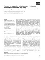
Fig. 1. Yeast two-hybrid assay for interaction between emerin and Btf. (A) Yeast two-hybrid assay for emerin truncations in pGBKT7 and Btf
truncations in pGADT7. Pairwise interactions between emerin and Btf fragments, as assayed by growth on quadruple-dropout selective medium
(minus Trp/Leu/His/Ade; right panel); left panel shows the control plate of cells grown under double-selection (minus Trp/Leu) to maintain
both plasmids. (B) Yeast two-hybrid assay for lamin A in pGBKT7 and Btf in pGADT7. Pairwise interaction of pGBKT7 and pGADT7 was
assayed by growth on quadruple-dropout medium (right); the left panel is the control, double-selective plate of cells used for the assays.
(C) Summary of Btf-interacting domains of emerin in yeast two-hybrid assay. The plus (+) mark at the right represents positive interactions.
The C-terminal fragment of emerin (residues 164–254) is sufficient to bind Btf. Interacting domains for emerin are shown at the top of the panel:
LEM and transmembrane (TM) domains are indicated. (D) Summary of emerin-interacting domains of Btf in yeast two-hybrid assay. The plus
(+) and minus (–) marks at right represent positive and negative interactions, respectively. The central fragment of Btf (residues 377–646) is
sufficient to bind emerin. Potential functional domains in Btf are indicated: RS represents the RS domain [45], boxes indicate regions with high
(green) and moderate (orange) similarity to transcription complex subunit TRAP150 [42]; the striped bar indicates the putative Bcl-2-binding
region of Btf [28].
1038 T. Haraguchi et al. (Eur. J. Biochem. 271) Ó FEBS 2004
377–761 of Btf, suggesting that this internal region of Btf is
sufficient to interact with emerin. For the studies below, we
used the KIAA0164 cDNA as full length Btf.
To determine the minimum regions of emerin and Btf
required for their interaction, we tested pairwise combina-
tions of subfragments of each protein for binding in the two-
hybrid assay (Fig. 1C,D). Full length emerin (1–254) and
two emerin fragments consisting of residues 104–254 and
164–254 were fused to the GAL4 DNA-binding domain.
These emerin fragments were tested for binding to full
length Btf (1–920) and Btf fragments 377–920, 377–761,
377–646, 377–574 and 521–761 fused to the GAL4 activator
domain (Fig. 1A). Diploid cells expressing both emerin and
Btf were tested for interaction by their growth under
quadruple selection (media lacking Leu, Trp, His and Ade).
Full length emerin (1–254) interacted with full length Btf
(1–920) and Btf fragments 377–920, 377–761 or 377–646.
Control diploids carrying only the control vectors did not
survive this selection, as expected (Fig. 1A). No interaction
was detected for full length emerin plus Btf fragment 377–
574 or 521–761, suggesting that Btf residues 377–646
comprised a minimum fragment necessary for binding to
emerin. Interestingly, this region of Btf also mediates
binding to the antideath protein, Bcl-2 (Fig. 1D, striped
bar; [28]).
We next tested emerin fragments 104–254 and 164–254
(Fig. 1C) for binding to Btf in two-hybrid assays (Fig. 1A).
These fragments represent the C-terminal half of emerin,
including its transmembrane span and small lumenal
domain. Both fragments interacted with full length Btf (1–
920) and Btf fragments 377–920, 377–761 and 377–646, but
not with fragments 377–574 or 521–761. We concluded that
the N-terminal half of emerin was not essential for binding
to Btf, and that exposed C-terminal residues 164–222 were
sufficient to bind Btf. Regions of emerin and Btf important
for their interaction in the two-hybrid assay are shown
schematically (Fig. 1C,D).
As a control, we also used the two-hybrid assay to
determine if Btf binds to lamin A (Fig. 1B). No interaction
was detected between lamin A (fused to the GAL4 DNA-
binding domain) and full length Btf (fused to the GAL4
activator domain; Fig. 1B, right), indicating that Btf does
not bind directly to lamin A in this assay.
Biochemical analysis of Btf binding to emerin
Btf residues 377–646 were sufficient to bind emerin in the
yeast two-hybrid assay. To test this result biochemically, we
synthesized four
35
S-labeled fragments of Btf in coupled
transcription/translation extracts in vitro, and assayed their
binding using a microtiter well assay (see Materials). Each
well contained a constant amount (5–10 pmole) of immo-
bilized (adsorbed), purified recombinant human emerin
(nucleoplasmic domain; residues 1–222), and BSA to block
nonspecific sites. Wells were not allowed to dry at any time
during this assay.
35
S-Labeled Btf fragments were then
added, incubated 60–90 min, washed, and bound proteins
were eluted using 5% SDS and counted. Consistent with the
two-hybrid results, the largest Btf fragment 377–920 was
positive for binding to emerin and the smallest, fragment
377–574, did not bind (Fig. 2A). Interestingly, this quanti-
tative analysis showed detectable but 50% reduced
binding of emerin to Btf fragments 377–761 and 377–646.
Similar results were found using emerin-conjugated beads
(data not shown). We concluded that Btf residues 761–920
contribute significantly to its affinity for emerin, but are not
essential. In contrast, Btf residues 574–646 are essential for
binding to emerin. The equilibrium binding affinity (K
D
)of
Btf (fragment 377–920) for emerin was 100 n
M
(range
60–280 n
M
; n ¼ 9; Fig. 2B). The stoichiometry of inter-
action was 0.8–1 mole Btf per mole emerin. These results
collectively showed that Btf has significant binding affinity
for emerin, and revealed regions within each protein that
mediate their interaction.
Mapping residues in emerin required for binding to Btf
A functional map of emerin was defined previously, with
respect to binding partners BAF and lamin A [5], and two
other binding partners [30,31]. To map the binding site for
Btfonemerin,wetested[
35
S]Btf binding to a collection of 21
purified emerin mutants, each bearing a small cluster of
site-directed alanine-substitution mutations. Half of these
Fig. 2. Biochemical assay for binding of Btf domains to emerin.
(A) Quantitation of Btf fragment binding to emerin in wells. Each
[
35
S]Btf fragment (377–920, 377–761, 377–646 or 377–574) was incu-
bated with immobilized emerin (1–222) and its binding to emerin was
determined as described in Materials and methods. Bars, S.E.M. (B)
Affinity of Btf for emerin was determined by adding increasing [
35
S]Btf
(377–920) to constant amounts of immobilized emerin (residues 1–222)
in microtiter wells. Double reciprocal plots were used to accurately
determine the affinity constant. Bars, S.E.M.
Ó FEBS 2004 Emerin binds Btf, a transcriptional repressor (Eur. J. Biochem. 271) 1039
mutations targeted amino acids conserved between emerin
and LAP2b (Table 3; [5]), whereas the remaining mutations
affected residues unique to emerin (Table 3 [30]);. Each
mutant emerin protein was expressed in bacteria, purified
by FPLC, immobilized in microtiter wells, incubated with
[
35
S]Btf (fragment 377–920), and the bound [
35
S]Btf was
counted as described above. The amounts of emerin loaded
per well were similar within a factor of 2, as determined by
SDS-elution of proteins from parallel wells and immuno-
blotting with antibodies against human emerin (data not
shown). As emerin was in five-fold excess, slight variations
in the amount of emerin per well did not affect the results.
We first considered the effects of mutations in ÔconservedÕ
residues. Strong binding to Btf was seen for wildtype emerin
(residues 1–222) and LEM-domain mutant m24 (Fig. 3A),
as expected. The binding of mutants 34, 112, 164 and 179
was slightly reduced, to 70% of wildtype. Binding was
significantly reduced, to 20–40% of wildtype, for mutants
70, 76, 196, 207 and 214 (Fig. 3A). For mutations in
Ôemerin-specificÕ residues (Fig. 3B), wildtype levels of
binding to Btf were detected for mutants 133, 151, 161
and 198, and binding was reduced slightly (to 70% of
wildtype) for mutants 122 and 145. However several emerin-
specific mutations (45E, 175, 192 and 206) showed signifi-
cantly reduced binding to Btf (25–35% of wildtype).
Background binding of [
35
S]Btf to negative control wells
containing BSA ranged from 5 to 15% of wildtype
(Fig. 3B). Collectively, this analysis implicated two regions
of emerin as important for binding to Btf: residues 45–83
and the C-terminal region (residues 175–217) (see Figs 3A,B
and 4C). Note that Ômutation cluster 76Õ consists of four
alanine substitutions spanning residues 76 through 83 in
emerin [30], extending the ÔimplicatedÕ region to residue 83.
Interestingly, disease-causing mutations S54F, D95-99 and
P183H also lie within these regions.
Disease-causing mutations S54F and D95–99 reduce
emerin binding to Btf
To determine if Btf binding was sensitive to disease-causing
mutations, we first tested [
35
S]Btf for binding to emerin
mutants S54F, D95–99 and P183H on blots (Ôblot overlayÕ
assay; Fig. 4A). Western blotting confirmed that similar
amounts of emerin protein were present in each lane
(Fig. 4A, Emr). We found positive binding of [
35
S]Btf to
wildtype emerin (residues 1–222) and mutant P183H, but no
signal for mutants S54F or D95–99 (Fig. 4A). The D95–99
mutation also disrupts binding to lamin A [5]. However, the
S54F result was remarkable, because this mutant bound
normally to all previously tested binding partners, including
BAF and lamin A.
Blot overlay assays are insensitive, as the blotted binding
partner is often at least partially denatured. To independ-
ently verify and quantify this reduced binding of Btf to
emerin mutant S54F, we measured the binding of [
35
S]Btf
to disease-associated emerins in the more sensitive and
Table 3. Mutations in emerin. Mutations 45A to 206 target Ôemerin-
specificÕ residues; mutants 24 to 214 target residues conserved between
emerin and LAP2b [5]. Mutated residues are indicated by lines.
Name of mutation Wildtype residues Mutant residues
45A 45RRR 45
AAA
45E 45RRR 45
EEE
104 104TYGEPES 104
AYGEAEA
122 122TS 122
AA
145 145EE 145AA
151 151ER 151
AA
161 161YQS 161
AAA
175 175SSL 175
AAA
192 192SSSSS 192
ASAAA
198 198SSWLTR 198
AAAAA
206 206IRPE 206
AAPA
24 24GPVV 24
AAAA
34 34YEKK 34
AAAA
S54F 54S 54
F
70 70DADMY 70
AAAMA
76 76LPKKEDAL 76
APAKADAA
112 112GPSRAVRGSVT 112
AASRAVAAAVA
133 133Q 133H
141 141SSSEEECKDR 141
AASAEECKAA
164 164ITHYRPV 164
AAHARPA
179 179LS 179
AA
183 183P 183
H
196 196SS 196
AA
207 207RP 207
AA
214 214GAGL 214
AAGA
Fig. 3. Biochemical assay for binding of Btf to emerin mutants.
Microtiter well binding assays for [
35
S]Btf binding to wildtype (WT)
emerin residues 1–222, or emerin bearing clusters of site-directed
alanine-substitution mutations in either (A) residues conserved
between emerin and LAP2b (Table 3,[5]) or (B) residues unique to
emerin (not conserved in LAP2b; Table 3). Bars, S.E.M.
1040 T. Haraguchi et al. (Eur. J. Biochem. 271) Ó FEBS 2004
quantitative microtiter well assay (Fig. 4B). Strong binding
was seen for wildtype emerin and mutant P183H, whereas
binding to mutants S54F and D95-99 was reduced by 50%.
Thus, the binding of Btf to mutant S54F was reduced
significantly in two independent assays, suggesting that
transcription factor Btf is uniquely sensitive to the S54F
mutation that causes EDMD. These results for emerin
mutants are summarized in Fig. 4C, in relation to regions
previously defined as important for binding to BAF and
lamin A [5].
Btf relocalizes from intranuclear ‘dot-like’ structures
to positions near the nuclear envelope
in Fas-antibody-treated (apoptotic) cells
To determine if Btf interacts with emerin in vivo,we
generated an antibody specific for Btf. This affinity-purified
antibody recognized a single band, in the nuclear fraction of
HeLa cells, with an apparent mass of 160 KDa in SDS/
PAGE (Fig. 5A, left panel). This band was specific, because
it was not recognized by preimmune antiserum (right panel)
and was competed for by pretreatment of antibody with the
antigen (middle panel). As this putative Btf band migrated
more slowly (160 KDa) than predicted from its ORF (106
KDa), we expressed a GFP-Btf fusion protein in HeLa cells
and compared this to endogenous Btf by Western blotting
(Fig. 5B). The affinity-purified antibody recognized the
endogenous 160 KDa band plus a second band with an
apparent mass of 180 KDa (Fig. 5B, left lane), which was
also recognized by antibodies against GFP (Fig. 5B, right
lane). We drew three conclusions: (a) our antibody speci-
fically recognized Btf; (b) HeLa cells express Btf, and
(c) endogenous Btf migrates in SDS/PAGE with an
apparent mass of 160 KDa.
We then used this specific antibody to determine if
endogenous Btf and emerin could be immunoprecipitated
from lysates of HeLa cells. However, the immunopreci-
pitations failed due to the insolubility of Btf (data not
shown). This same problem was encountered with
HA-tagged Btf in transiently transfected HeLa cells (data
not shown). We therefore used cytological methods to
localize Btf in HeLa cells. Indirect immunofluorescence
staining with the affinity-purified antibody showed that
Btf was localized inside the nucleus in dot-like structures,
distant from emerin (Fig. 5C). Thus, Btf and emerin
occupy separate nuclear domains in HeLa cells under
normal culture conditions. As muscle wasting in EDMD
is thought to involve apoptosis [34], we tested the
hypothesis that the subnuclear localization of Btf might
change in apoptosis-induced cells. Two days after trans-
fection with GFP-emerin encoding plasmids, HeLa cells
were induced to enter apoptosis by treatment with Fas
antibody plus cycoheximide [35]. In untreated cells, the
Btf and GFP-emerin signals are clearly separate (Fig. 5C).
However in Fas-antibody-treated cells, within 3 h, the
endogenous Btf relocalized to punctate positions near the
nuclear envelope, close to GFP-emerin but not spectrally
overlapping (Fig. 5D–F). This change in localization
occurred relatively early in apoptosis when the nuclei
were still relatively spherical and before chromosomes
became grossly condensed. These results suggest that the
subnuclear localization of Btf is differentially regulated
during apoptosis, and that our biochemically–character-
ized interaction between Btf and emerin may be physio-
logically relevant to regulate Btf at an early stage of cell
death. These experiments did not address whether emerin
inhibits or promotes Btf’s pro-death activity. However, as
muscles might enter apoptosis too readily when emerin is
missing, we speculate that Btf is normally inhibited by its
association with emerin and potentially other nuclear
membrane proteins.
Discussion
We found that a reportedly pro-apoptotic transcription
regulator, Btf, binds emerin with nanomolar affinity in vitro.
Importantly, Btf binding to emerin is weakened significantly
by the disease-causing S54F mutation, whereas all other
previously tested binding partners (BAF, lamin A, GCL
and splicing factor YT521-B) bind normally to S54F
(reviewed by Bengtsson and Wilson, 2004 [36]). These
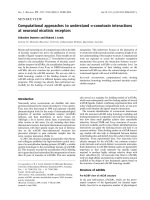
Fig. 4. Binding of Btf to disease-specific emerin mutants. (A) Blot
overlay assays for binding of [
35
S]Btf to wildtype emerin (WT) and
disease-causing emerin mutants S54F, D95–99 (D95) and P183H.
(B) Solution binding assays measuring [
35
S]Btf binding to wildtype
(WT) or mutant emerin proteins (numbered as in Fig. 3) immobilized
in microtiter wells. Bars, S.E.M. (C) Diagram mapping the proposed
Btf-binding domains in emerin, relative to reported binding domains
for BAF and lamin A [5]. No binding partner has yet been reported to
be disrupted by disease-causing mutant P183H (this report,[5]). Stars
indicate disease-causing emerin mutants.
Ó FEBS 2004 Emerin binds Btf, a transcriptional repressor (Eur. J. Biochem. 271) 1041
findings, and our discovery that Btf relocalizes near the
nuclear envelope specifically during apoptosis, suggest that
Btf is a disease-relevant binding partner for emerin. Btf is
highly expressed in skeletal muscle, although it is ubiqui-
tously expressed in other tissues tested, including heart and
brain [22,29]. Although this expression pattern for Btf does
not correlate perfectly with disease-affected tissues, some
functions of emerin are known to overlap with both LAP2b
[30,37] and MAN1 [38]. It is therefore possible that, in
patients who lack emerin, tissues that express ÔbackupÕ
LEM-domain proteins might be protected from disease. In
this regard, it will be important to determine which (if any)
other LEM-domain proteins can interact with Btf.
Emerin and its binding partners in EDMD
Emerin has many interesting binding partners in the nucleus
[36]. Btf joins a small but growing number of emerin-
binding proteins that regulate transcription (BAF [5,39],
GCL [30]) or splice site selection (YT521-B [31]), or are
proposed to regulate transcription (Lmo7; J. M. Holaska
and K. L. Wilson, unpublished results). This group of
interactors support gene expression models for emerin
function and, potentially, the disease mechanism of EDMD
and further suggest that emerin and other LEM-domain
proteins interact with a variety of overlapping binding
partners at the inner nuclear membrane.
Our mapping results showed that alanine substitutions in
two discrete regions of emerin, residues 45–83 and 175–217,
disrupt its binding to Btf. These same regions of emerin,
previously designated Ôrepressor binding domains (RBD)
1and2Õ [30] are also critical for binding to GCL [30] and
splice site regulator YT521-B [31]. Most (but not all)
mutations that disrupted emerin binding to Btf, also disrupt
its binding to GCL [30] and YT521-B [31]. Both GCL and
Btf are expressed widely in human tissues [29,30]. GCL is
Fig. 5. Btf moves near the nuclear envelope in
apoptosis-induced HeLa cells. (A) Western
blotting to verify specificity of the antibody.
Total HeLa cell extracts (total) and corres-
ponding cytoplasmic (cytosol) and nuclear
(nucleus) fractions were resolved by SDS/
PAGE, and immunoblotted with either affin-
ity-purified immune antibodies against Btf
(left panel), antigen-pretreated immune anti-
bodies (middle panel) or preimmune serum
(right panel). The immune antibody specific-
ally recognized a nuclear protein that migrated
at 160 kDa. (B) Western blots of lysates from
HeLa cells that were either nontransfected (–)
or transiently transfected to express GFP-Btf
(GFP-Btf), probed with the antibodies against
Btf (left panel) or GFP (right panel). (C)
Subnuclear localization of GFP-emerin and
endogenous Btf stained with anti-Btf Ig in
nonapoptotic HeLa cells. (D–F) Subnuclear
localization of GFP-emerin and endogenous
Btf stained with anti-Btf Ig, after 3 h of
treatment with Fas-antibody and cyclohexi-
mide to induce apoptosis in HeLa cells
(Methods).
1042 T. Haraguchi et al. (Eur. J. Biochem. 271) Ó FEBS 2004
most highly expressed in testis and is required for germ cell
formation [37,40,41], whereas Btf is highly expressed in
skeletal muscle. Our findings for Btf, coupled to previous
findings for GCL and YT521-B, strongly support the
hypothesis that two regions in the primary amino acid
sequence of emerin (RBD1 and RBD2; Fig. 4C and refs
[30,31]) form a docking site for gene regulatory proteins. We
therefore predict that Btf, GCL and YT521-B are not alone,
and that additional proteins involved in gene expression or
RNA splicing will also emerge as emerin-binding proteins.
Interestingly, the emerin-binding region of Btf is homolog-
ous to transcription complex subunit TRAP150 [42], leading
us to speculate that TRAP150 might also bind emerin. We
suggest that additional binding partners that are both
expressed in disease-affected tissues, and potentially disrup-
ted by disease-causing mutations in emerin, remain to be
discovered. An important next step is to identify genes
regulated by emerin-dependent transcription factors, to gain
specific insight into the molecular mechanisms of EDMD
disease. Our current knowledge suggests that emerin may be
anchored and stabilized at the nuclear inner membrane by
nesprin-1a and lamin A [43,44]; in turn, emerin may provide
regulated binding sites for BAF or other binding partners
(Btf, GCL and YT521-B) involved in transcription or
splicing. Btf is highly expressed in skeletal muscle, and is
therefore presumably important for muscle cell function.
A mutation that disrupts Btf binding to emerin would be
expected to affect muscles disproportionately, especially if
putative ÔbackupÕ; LEM-domain proteins are absent or
expressed at levels too low (e.g. LAP2b [30]) to compensate
for the absence of emerin.
Possible functions for Btf
The function of Btf is not fully understood. Btf was
discovered as a two-hybrid binding partner for E1B19K, a
viral protein similar to Bcl-2 [28]. When fused to the GAL4-
DNA binding domain, Btf is sufficient to repress a reporter
gene [28], showing that Btf can repress transcription in vivo.
Btf also promotes apoptosis when overexpressed in cells
[28]. Btf binds to ÔantideathÕ Bcl-2-related proteins such as
E1B19K, Bcl-2, and Bcl-XL through its C-terminal region,
and this binding is thought to promote apoptosis by
blocking their antideath activity [28]. Our two-hybrid
mapping results suggest that emerin and Bcl-2 might bind
similar regions of Btf (see Fig. 1D). We therefore hypo-
thesize that Btf has a choice, and can bind either to Bcl-2
or emerin; in cells that lack functional emerin this balance
would be lost, potentially leading to increased Btf binding to
Bcl-2 and entry into apoptosis. This hypothesis is supported
by our evidence that Btf relocalizes near emerin in
apoptosis-induced cells.
Consistent with its ability to repress transcription, Btf
localizes in HeLa cell nuclei (this report and [28]), where it is
enriched in discrete Ôdot-likeÕ structures adjacent to RNA
splicing factor SC35 (T. Haraguchi and Y. Hiraoka,
unpublished results). Btf was identified independently in
a proteomic analysis of purified interchromatin granule
clusters (IGCs), which contain > 200 proteins, including
many RNA splicing factors (N. Saitoh and D. Spector,
personal communication [Cold Spring Harbor Laborator-
ies, New York)]. The N-terminus of Btf includes Arg-Ser
repeats (a so-called ÔRS domainÕ), which are characteristic
for splicing factors and many other RNA-binding proteins
[45]. Thus, the functions of Btf are not yet understood, but
might include roles in mRNA metabolism, transcriptional
repression [28] or pro-apoptotic responses [28]. We speculate
that Btf binding to emerin in vivo is regulated at least during
cell death, and potentially also regulated by signals such as
hormones, growth/survival factors, exercise or atrophy (in
muscle). Testing these models will require further study of
Btf function both at the molecular level, and in disease-
affected tissues.
Acknowledgements
We are grateful to Drs Tsuchiya and Arahata for emerin constructs,
Dr Nagase (Kazusa DNA) for the KIAA0164 construct, Dr White
(Rutgers University) for DNA constructs of Btf
L
,Btf
S
and E1B19K,
the Riken Cell Bank for HeLa cells and Drs Saitoh and Spector (Cold
Spring Harbor Laboratories, New York) and Dr Morris (Northeast
Wales Institute, Wrexham, United Kingdom) for communicating their
results prior to publication. We also thank Ms. Kumiko Matsuno for
initial cloning of Btf in the yeast two-hybrid assay. This work was
supported by grants from the Japan Science and Technology
Corporation (CREST; to T. H. and Y. H.), Grant-in-Aid for Scientific
Research B (to T. H. and Y. H.), National Institutes of Health
Cardiology training grant (T32-HLO-7227-26; to J. M. H.), and grants
from the National Institutes of Health (GM48646) and the Scott B.
Deutschman memorial Research Award from the American Heart
Association (to K. L. W.).
References
1. Bione, S., Maestrini, E., Rivella, S., Mancini, M., Regis, S.,
Romeo, G. & Toniolo, D. (1994) Identification of a novel X-linked
gene responsible for Emery–Dreifuss muscular dystrophy. Nat.
Genet. 8, 323–327.
2. Emery, A.E.H. (1989) Emery–Dreifuss syndrome. J. Med. Genet.
26, 637–641.
3. Emery, A.E.H. (2000) Emery–Dreifuss muscular dystrophy – a 40
year retrospective. Neuromuscul. Disord. 10, 228–232.
4. Clements, L., Manilal, S., Love, D.R. & Morris, G.E. (2000)
Direct interaction between emerin and lamin A. Biochem. Biophys.
Res. Commun. 267, 709–714.
5. Lee,K.K.,Haraguchi,T.,Lee,R.S.,Koujin,T.,Hiraoka,Y.&
Wilson, K.L. (2001) Distinct functional domains in emerin bind
lamin A and DNA-bridging protein BAF. J. Cell Sci. 114, 4567–
4573.
6. Bonne, G., Di Barletta, M.R., Varnous, S., Becane, H M.,
Hammouda, E.H., Merlini, L., Muntoni, F., Greenberg, C.R.,
Gary, F., Urtizberea, J A. et al. (1999) Mutations in the gene
encoding lamin A/C cause autosomal dominant Emery–Dreifuss
muscular dystrophy. Nat. Genet. 21, 285–288.
7. De Sandre-Giovannoli, A., Chaouch, M., Kozlov, S., Vallat,
J.M., Tazir, M., Kassouri, N., Szepetowski, P., Hammadouche,
T., Vandenberghe, A., Stewart, C.L. et al. (2002) Homozygous
defects in LMNA, encoding lamin A/C nuclear-envelope pro-
teins, cause autosomal recessive axonal neuropathy in human
(Charcot–Marie–Tooth disorder type 2) and mouse. Am. J.
Hum. Genet. 70, 726–736. [Erratum appears in Am.J.Hum.Genet.
70, 1075.].
8. Novelli, G., Muchir, A., Sangiuolo, F., Helbling-Leclerc, A.,
D’Apice, M.R., Massart, C., Capon, F., Sbraccia, P., Federici, M.,
Lauro, R. et al. (2002) Mandibuloacral dysplasia is caused by a
mutation in LMNA-encoding lamin A/C. Am.J.Hum.Genet.71,
426–431.
Ó FEBS 2004 Emerin binds Btf, a transcriptional repressor (Eur. J. Biochem. 271) 1043
9. Wilson, K.L., Zastrow, M. & Lee, K.K. (2001) Nuclear lamins
and disease: insights into nuclear infrastructure. Cell 104, 647–650.
10. Burke, B. & Stewart, C.L. (2002) Life at the edge: the nuclear
envelope and human disease. Nat.Rev.Mol.CellBiol.3, 575–585.
11. Wehnert, M.S. & Bonne, G. (2002) The nuclear muscular dys-
trophies. Semin. Pediatr. Neurol. 9, 100–107.
12. Eriksson, M., Brown, W.T., Gordon, L.B., Glynn, M.W., Singer,
J.,Scott,L.,Erdos,M.R.,Robbins,C.M.,Moses,T.Y.,Berglund,
P., Dutra, A., Pak, E., Durkin, S., Csoka, A.B., Boehnke, M.,
Glover, T.W. & Collins, F.S. (2003) Recurrent de novo point
mutations in lamin A cause Hutchinson–Gilford progeria syn-
drome. Nature 423, 293–298.
13. De Sandre-Giovannoli, A., Bernard, R., Cau, P., Navarro, C.,
Amiel, J., Boccaccio, I., Lyonnet, S., Stewart, C.L., Munnich, A.,
Le Merrer, M. & Levy, N. (2003) Lamin A truncation in Hutch-
inson–Gilford progeria. Science 300, 2055.
14. Chen,L.,Lee,L.,Kudlow,B.A.,DosSantos,H.G.,Sletvold,
O., Shafeghati, Y., Botha, E.G., Garg, A., Hanson, N.B.,
Martin, G.M., Mian, I.S., Kennedy, B.K. & Oshima, J. (2003)
LMNA mutations in atypical Werner’s syndrome. Lancet 362,
440–445.
15. Wilson, K.L. (2000) The nuclear envelope, muscular dystrophy
and gene expression. Trends Cell Biol. 10, 125–129.
16. Nagano, A. & Arahata, K. (2000) Nuclear envelope proteins and
associated diseases. Curr. Opinion Neurol. 13, 533–539.
17. Cohen, M., Lee, K.K., Wilson, K.L. & Gruenbaum, Y. (2001)
Transcriptional repression, apoptosis, human disease and the
functional evolution of the nuclear lamina. Trends Bioch. Sci. 26,
41–47.
18. Manilal, S., Nguyen, T.M., Sewry, C.A. & Morris, G.E. (1996)
The Emery–Dreifuss muscular dystrophy protein, emerin, is a
nuclear membrane protein. Hum. Mol. Genet. 5, 801–808.
19. Nagano, A., Koga, R., Ogawa, M., Kurano, Y., Kawada, J.,
Okada, R., Hayashi, Y.K., Tsukahara, T. & Arahata, K. (1996)
Emerin deficiency at the nuclear membrane in patients with
Emery–Dreifuss muscular dystrophy. Nat. Genet. 12, 254–259.
20. Yorifuji, H., Tadano, Y., Tsuchiya, Y., Ogawa, M., Goto, K.,
Umetani, A., Asaka, Y. & Arahata, K. (1997) Emerin, deficiency
of which causes Emery–Dreifuss muscular dystrophy, is localized
at the inner nuclear membrane. Neurogenetics 1, 135–140.
21. Ellis, J.A., Craxton, M., Yates, J.R. & Kendrick-Jones, J. (1998)
Aberrant intracellular targeting and cell cycle-dependent phos-
phorylation of emerin contribute to the Emery–Dreifuss muscular
dystrophy phenotype. J. Cell Sci. 111, 781–792.
22. Morris, G.E. & Manilal, S. (1999) Heart to heart: from nuclear
proteins to Emery–Dreifuss muscular dystrophy. Hum. Mol.
Genet. 8, 1847–1851.
23. Fairley, E.A.L., Kendrick-Jones, J. & Ellis, J.A. (1999) The
Emery–Dreifuss muscular dystrophy phenotype arises from
aberrant targeting and binding of emerin at the inner nuclear
membrane. J. Cell Sci. 112, 2571–2582.
24. Haraguchi, T., Koujin, T., Segura-Totten, M., Lee, K.K., Mats-
uoka, Y., Yoneda, Y., Wilson, K.L. & Hiraoka, Y. (2001) BAF is
required for emerin assembly into the reforming nuclear envelope.
J. Cell Sci. 114, 4575–4585.
25. Lin, F., Blake, D.L., Callebaut, I., Skerjanc, I.S., Holmer, L.,
McBurney, M.W., Paulin-Levasseur, M. & Worman, H.J. (2000)
MAN1, an inner nuclear membrane protein that shares the LEM
domain with lamina-associated polypeptide 2 and emerin. J. Biol.
Chem. 275, 4840–4847.
26.Shumaker,D.K.,Lee,K.K.,Tanhehco,Y.C.,Craigie,R.&
Wilson, K.L. (2001) LAP2 binds to BAF-DNA complexes:
requirement for the LEM-domain and modulation by variable
regions. EMBO J. 20, 1754–1764.
27. Segura-Totten, M., Kowalski, A.K., Craigie, R. & Wilson, K.L.
(2002) Barrier-to-autointegration factor: major roles in chromatin
decondensation and nuclear assembly. J. Cell Biol. 158, 475–
485.
28. Kasof, G.M., Goyal, L. & White, E. (1999) Btf, a novel death-
promoting transcriptional repressor that interacts with Bcl-2
related proteins. Mol. Cell Biol. 19, 4390–4404.
29. Nagase, T., Seki, N., Ishikawa, K., Tanaka, A. & Nomura, N.
(1996) Prediction of the coding sequences of unidentified human
genes. V. The coding sequences of 40 new genes (KIAA0161-
KIAA0200) deduced by analysis of cDNA clones from human cell
line KG-1. DNA Res. 3, 17–24.
30. Holaska, J.M., Lee, K.K., Kowalski, A.K. & Wilson, K.L. (2003)
Transcriptional repressor germ cell-less (GCL) and BAF compete
for binding to emerin. J. Biol. Chem. 278, 6969–6975.
31. Wilkinson, F.L., Holaska, J.M., Zhang, Z., Sharma, A., Manilal,
S.,Holt,I.,Stamm,S.,Wilson,K.L.&Morris,G.E.(2003)
Emerin interacts in vitro with the splicing-associated factor,
YT521-B. Eur. J. Biochem. 270, 2459–2466.
32. Matsuoka, Y., Nishizawa, K., Yano, T., Shibata, M., Ando, S.,
Takahashi, T. & Inagaki, M. (1992) Two different protein kinases
act on a different time schedule as glial filament kinases during
mitosis. EMBO J. 11, 2895–2902.
33. Agard, D., Hiraoka, Y., Show, P. & Sedat, J.W. (1989) Fluores-
cence microscopy in three dimensions. Meth.CellBiol.30,
353–377.
34. Bonne, G., Yaou, R.B., Beroud, C., Boriani, G., Brown, S., de
Visser, M., Duboc, D., Ellis, J., Hausmanowa-Petrusewicz, I.,
Lattanzi, G., Merlini, L., Morris, G., Muntoni, F., Opolski, G.,
Pinto, Y.M., Sangiuolo, F., Toniolo, D., Trembath, R., van
Berlo, J.H., van der Kooi, A.J. & Wehnert, M. (2003) 108th
ENMC International Workshop, 3rd Workshop of the MYO-
CLUSTER project: EUROMEN, 7th International Emery–
Dreifuss Muscular Dystrophy (EDMD) Workshop, 13–15 Sep-
tember 2002, Naarden, The Netherlands. Neuromuscul. Disord. 13,
508–515.
35. Yonehara, S., Ishii, A. & Yonehara, M. (1989) A cell-killing
monoclonal antibody (anti-Fas) to a cell surface antigen co-
downregulated with the receptor of tumor necrosis factor. J. Exp.
Med. 169, 1747–1756.
36. Bengtsson, L. & Wilson, K.L. (2004) Multiple and surprising new
functions for emerin, a nuclear membrane protein. Curr. Opin.
Cell Biol. in press.
37. Nili, E., Cojocaru, G.S., Kalma, Y., Ginsberg, D., Copeland,
N.G., Gilbert, D.J., Jenkins, N.A., Berger, R., Shaklai, S.,
Amariglio, N. et al. (2001) Nuclear membrane protein
LAP2b mediates transcriptional repression alone and together
with its binding partner GCL (germ-cell-less). J. Cell Sci. 114,
3297–3307.
38. Liu, J., Lee, K.K., Segura-Totten, M., Neufeld, E., Wilson, K.L. &
Gruenbaum, Y. (2003) MAN1 and emerin have overlapping
function(s) essential for chromosome segregation and cell division
in Caenorhabditis elegans. Proc. Natl Acad. Sci. USA 100, 4598–
4603.
39. Wang, X., Xu, S., Rivolta, C., Li, L.Y., Peng, G.H., Swain, P.K.,
Sung, C.H., Swaroop, A., Berson, E.L., Dryja, T.P. et al. (2002)
Barrier to autointegration factor interacts with the cone-rod
homeobox and represses its transactivation function. J. Biol.
Chem. 277, 43288–43300.
40. Kimura,T.,Yomogida,K.,Iwai,N.,Kato,Y.&Nakano,T.
(1999) Molecular cloning and genomic organization of mouse
homologue of Drosophila germ cell-less and its expression in germ
lineage cells. Biochem. Biophys. Res. Commun. 262, 223–230.
41. Robertson, S.E., Dockendorff, T.C., Leatherman, J.L., Faulkner,
D.L. & Jongens, T.A. (1999) Germ cell-less is required only during
the establishment of the germ cell lineage of Drosophila and has
activities which are dependent and independent of its localization
to the nuclear envelope. Dev. Biol. 215, 288–297.
1044 T. Haraguchi et al. (Eur. J. Biochem. 271) Ó FEBS 2004
42. Ito, M., Yuan, C X., Malik, S., Gu, W., Fondell, J.D.,
Yamamura,S.,Fu,Z Y.,Zhang,X.,Qin,J.&Roeder,R.G.
(1999) Identity between TRAP and SMCC complexes indicates
novel pathways for the function of nuclear receptors and diverse
mammalian activators. Mol. Cell 3, 361–370.
43. Mislow, J.M., Kim, M.S., Davis, D.B. & McNally, E.M. (2002a)
Myne-1, a spectrin repeat transmembrane protein of the myocyte
inner nuclear membrane, interacts with lamin A/C. J. Cell Sci.
115, 61–70.
44. Mislow, J.M., Holaska, J.M., Kim, M.S., Lee, K.K., Segura-
Totten, M., Wilson, K.L. & McNally, E.M. (2002b) Nesprin-1a
self-associates and binds directly to emerin and lamin A in vitro.
FEBS Lett. 525, 135–140.
45. Valcarcel, J. & Green, M.R. (1996) The SR protein family:
pleiotropic functions in pre-mRNA splicing. Trends. Biochem. Sci.
21, 296–301.
Ó FEBS 2004 Emerin binds Btf, a transcriptional repressor (Eur. J. Biochem. 271) 1045