Báo cáo khoa học: S -Stereoselective piperazine-2-tert-butylcarboxamide hydrolase from Pseudomonas azotoformans IAM 1603 is a novel L-amino acid amidase doc
Bạn đang xem bản rút gọn của tài liệu. Xem và tải ngay bản đầy đủ của tài liệu tại đây (316.97 KB, 11 trang )
S
-Stereoselective piperazine-2-
tert
-butylcarboxamide hydrolase from
Pseudomonas azotoformans
IAM 1603 is a novel
L
-amino acid amidase
Hidenobu Komeda
1
, Hiroyuki Harada
1
, Shingo Washika
1
, Takeshi Sakamoto
2
, Makoto Ueda
2
and Yasuhisa Asano
1
1
Biotechnology Research Center, Toyama Prefectural University, Kosugi, Toyama, Japan;
2
Mitsubishi Chemical Group Science
and Technology Research Center, Inc., Aoba-ku, Yokohama, Kanagawa, Japan
An amidase acting on (R,S)-piperazine-2-tert-butylcarbox-
amide was purified from Pseudomonas azotoformans IAM
1603 and characterized. The enzyme acted S-stereoselec-
tively on (R,S)-piperazine-2-tert-butylcarboxamide to yield
(S)-piperazine-2-carboxylic acid. N-terminal and internal
amino acid sequences of the enzyme were determined.
ThegeneencodingtheS-stereoselective piperazine-2-tert-
butylcarboxamide amidase was cloned from the chromo-
somal DNA of the strain and sequenced. Analysis of 2.1 kb
of genomic DNA revealed the presence of two ORFs, one
of which (laaA) encodes the amidase. This enzyme, LaaA
is composed of 310 amino acid residues (molecular mass
34 514 Da), and the deduced amino acid sequence exhibits
significant similarity to hypothetical and functionally char-
acterized proline iminopeptidases from several bacteria. The
laaA gene modified in the nucleotide sequence upstream
from its start codon was overexpressed in Escherichia coli.
The activity of the recombinant LaaA enzyme in cell-free
extracts of E. coli was 13.1 unitsÆmg
)1
with
L
-prolinamide
as substrate. This enzyme was purified to electrophoretic
homogeneity by ammonium sulfate fractionation and two
column chromatography steps. On gel-filtration chroma-
tography, the enzyme appeared to be a monomer with a
molecular mass of 32 kDa. It had maximal activity at 45 °C
and pH 9.0, and was completely inactivated in the presence
of phenylhydrazine, Zn
2+
,Ag
+
,Cd
2+
or Hg
2+
. LaaA had
hydrolyzing activity toward
L
-amino acid amides such as
L
-prolinamide,
L
-proline-p-nitroanilide,
L
-alaninamide and
L
-methioninamide, but did not act on the peptide substrates
for the proline iminopeptidases despite their sequence simi-
larity to LaaA. The enzyme also acted S-stereoselectively
on (R,S)-piperidine-2-carboxamide, (R,S)-piperazine-2-car-
boxamide and (R,S)-piperazine-2-tert-butylcarboxamide.
Based on its specificity towards
L
-amino acid amides, the
enzyme was named
L
-amino acid amidase. E. coli trans-
formants overexpressing the laaA gene could be used for
the S-stereoselective hydrolysis of (R,S)-piperazine-
2-tert-butylcarboxamide.
Keywords:amidase;
L
-prolinamide; piperazine-2-tert-butyl-
carboxamide; Pseudomonas azotoformans.
Amidases (acylamide amidohydrolases, EC 3.5.1.4) cata-
lyze the hydrolysis of the carboxyl amide bonds to liberate
carboxylic acids and ammonia. Recently, various kinds of
stereoselective amidases from microbial origin have been
reported and received much attention because of their
potential use for the industrial production of optically active
compounds [1–3]. S-Enantiomer-selective amidases from
Brevibacterium sp. R312 [4], Pseudomonas chlororaphis B23
[5] and Rhodococcus rhodochrous J1 [6] were found to be
involved in nitrile metabolism with genetically linked nitrile
hydratases. S-andR-enantiomer-selective amidases, which
seemed not to be related to the nitrile metabolism, were also
found in Agrobacterium tumefaciens d3 [7] and Comamonas
acidovorans KPO-2771-4 [8], respectively. These enantio-
mer-selective amidases can be used for the production of
optically active 2-arylpropionic acids, the nonsteroid anti-
inflammatory drugs, from the corresponding racemic
amides. S-Stereoselective amino acid amidases from Pseu-
domonas putida ATCC 12633 [9], Ochrobactrum anthropi
NCIMB 40321 [10] and Mycobacterium neoaurum ATCC
25795 [11], and the R-stereoselective amino acid amidases
from O. anthropi C1-38 [12,13], O. anthropi SV3 [14],
Arthrobacter sp. NJ-26 [15] and Brevibacillus borstelensis
BCS-1 [16] were found to be useful for the production of
enantiomerically pure amino acids and their derivatives
from the corresponding racemic amino acid amides. The
genes coding for the above amidases have been isolated and
their primary structures revealed, except for the S-stereo-
selective amino acid amidases of the three microorganisms
and the R-stereoselective amino acid amidase from
Arthrobacter sp. NJ-26. While these amidases show a wide
Correspondence to Y. Asano, Biotechnology Research Center,
Toyama Prefectural University, 5180 Kurokawa, Kosugi,
Toyama 939-0398, Japan.
Fax: + 81 766 56 2498, Tel.: + 81 766 56 7500,
E-mail:
Abbreviations: LaaA,
L
-amino acid amidase; NBD-Cl, 4-chloro-
7-nitro-2,1,3-benzoxadiazole.
Enzymes: acylamide amidohydrolases (EC 3.5.1.4); proline imino-
peptidases (PIP, EC 3.4.11.5).
(Received 9 January 2004, revised 16 February 2004,
accepted 23 February 2004)
Eur. J. Biochem. 271, 1465–1475 (2004) Ó FEBS 2004 doi:10.1111/j.1432-1033.2004.04056.x
variety of substrate specificities, there is no report on the
hydrolysis of amides containing a bulky substituent at the
leaving group, such as tert-butylcarboxamide. This inability
to hydrolyze the bulky amides hindered the wide use of
amidases for the production of complex compounds.
Enantiomerically pure piperazine-2-carboxylic acid and
its tert-butylcarboxamide derivative are important chiral
building blocks for some pharmacologically active com-
pounds such as N-methyl-
D
-aspartate antagonist for glu-
tamate receptor [17], cardioprotective nucleoside transport
blocker [18] and HIV protease inhibitor [19]. (S)-Piperazine-
2-carboxylic acid has been prepared by kinetic resolution of
racemic 4-(tert-butoxycarbonyl)piperazine-2-carboxamide
with leucine aminopeptidase [18] or racemic piperazine-
2-carboxamide with Klebsiella terrigena DSM9174 cells [20].
There is no report on the kinetic resolution of (R,S)-
piperazine-2-tert-butylcarboxamide.
In this study, we screened for microorganisms that can
hydrolyze (R,S)-piperazine-2-tert-butylcarboxamide and
found the hydrolytic (amidase) activity in Pseudomonas
azotoformans IAM 1603. The amidase purified from cells of
the strain hydrolyzed S-stereoselectively (R,S)-piperazine-2-
tert-butylcarboxamide to form (S)-piperazine-2-carboxylic
acid (Fig. 1). The gene coding for the enzyme was isolated
and expressed in Escherichia coli host. The recombinant
protein was purified and characterized, and found to be a
novel
L
-stereoselective amino acid amidase, LaaA. This is
the first report revealing the primary structure of
L
-amino
acid amidase.
Materials and methods
Bacterial strains, plasmids and culture conditions
P. azotoformans IAM (Culture collection of the Institute of
Applied Microbiology) 1603 was used as the source of
enzyme and chromosomal DNA. E. coli JM109 (recA1,
endA1, gyrA96, thi, hsdR17, supE44, relA1, D(lac-proAB)/F¢
[traD36, proAB
+
, lacI
q
, lacZD M15]) was used as a host for
the recombinant plasmids. Plasmids pBluescriptII SK(–)
(Toyobo, Osaka, Japan), pUC19 (Takara Shuzo, Kyoto,
Japan) and pT7-Blue (Takara Shuzo) were used as cloning
vectors. P. azotoformans IAM 1603 was cultivated at 30 °C
on BM medium containing 10 g Bacto nutrient broth
(Difco), 10 g disodium
DL
-malate n-hydrate, 3 g K
2
HPO
4
,
1gKH
2
PO
4
,0.05gMgSO
4
•7H
2
O, 0.01 g FeSO
4
•7H
2
O,
0.01 g MnCl
2
•4H
2
O, 0.01 g CoCl
2
•6H
2
O, (NH
4
)
6
Mo
7
O
24
•
4H
2
O in 1 litre distilled water, pH 7.0. Recombinant E. coli
JM109 was cultured at 37 °C on Luria–Bertani medium [21]
containing 80 lgÆml
)1
of ampicillin. To induce the gene
under the control of the lac promoter, isopropyl-thio-b-
D
-
galactoside was added to a final concentration of 0.5 m
M
.
Purification of the amidase from
P. azotoformans
IAM 1603
P. azotoformans IAM 1603 was subcultured at 30 °Cfor
16 h in a test tube containing 5 mL BM medium. The
subculture (5 mL) was then inoculated into a 2 L Sakaguchi
flask containing 500 mL BM medium. The cultivation was
carried out at 30 °C for 8 h with reciprocal shaking. All
purification steps were performed at a temperature lower
than 5 °C. The buffer used was potassium phosphate
(pH 7.0) containing 0.1 m
M
dithiothreitol and 5 m
M
2-mercaptoethanol. The protein content of the eluates from
column chromatography was monitored by absorbance at
280 nm. Cells (125 g, wet weight) from 25 L of BM medium
were harvested by centrifugation (10 000 g at 4 °C) and
suspended in 0.1
M
buffer. The cell suspension was disrup-
ted with an ultrasonic oscillator (19 kHz insonator model
201M: Kubota, Tokyo, Japan). The sonicate was centri-
fuged at 15 000 g for 20 min at 4 °C, and the resulting
supernatant was used as the cell-free extract. The cell-free
extract was dialyzed for 12 h against three changes of
10 m
M
buffer. The dialyzed enzyme solution was then
applied to a column (5 · 20 cm) of DEAE-Toyopearl
650M (Tosoh Corp., Tokyo, Japan) previously equilibrated
with 10 m
M
buffer. After the column had been washed with
2Lof10m
M
buffer, the enzyme was eluted with a linear
gradient of NaCl (0–0.5
M
, 1.5 L each) in 10 m
M
buffer.
The active fractions were combined and then brought to
30% ammonium sulfate saturation and applied to a column
(2.5 · 20 cm) of Butyl-Toyopearl 650M (Tosoh Corp.)
previously equilibrated with 10 m
M
buffer 30% saturated
with ammonium sulfate. After the column had been washed
with 500 mL of the same buffer, the enzyme was eluted with
a linear gradient of ammonium sulfate (30–0% saturation,
500 mL each) in 10 m
M
buffer. The active fractions were
combined and dialyzed against 10 L of 10 m
M
buffer for
12 h. The dialyzed enzyme was applied to a column
(1.5 · 8 cm) of Gigapite (Seikagaku Kogyo, Tokyo, Japan)
previously equilibrated with 10 m
M
buffer. After the
columnhadbeenwashedwith50mLof10m
M
buffer,
the enzyme was eluted with a linear gradient of buffer
(0.01–1
M
, 50 mL each). The active fractions were com-
bined, concentrated with Centriprep-10 (Millipore Corp.,
MA, USA) and dialyzed against 10 L of 10 m
M
buffer for
12 h. The dialyzed enzyme was applied to a Superdex 200
HR 26/60 column (Amersham Biosciences K.K., Tokyo,
Japan) previously equilibrated with 10 m
M
buffer contain-
ing 150 m
M
NaCl and eluted with the same buffer. The
active fractions were collected and dialyzed against 10 L of
10 m
M
buffer for 12 h. The dialyzed enzyme was applied to
a MonoQ HR 5/5 column (Amersham Biosciences K.K.)
previously equilibrated with 10 m
M
buffer and then eluted
with a linear gradient of NaCl (0–0.2
M
)in10m
M
buffer.
The active fractions were combined, concentrated with
Centricon-10 (Millipore Corp.), and submitted to electro-
phoresis on a nondenaturating polyacrylamide gel, AE-6000
from Atto (Tokyo, Japan). To locate the enzymatic activity,
Fig. 1. Stereoselective hydrolysis of (R,S)-piperazine-2-tert-butyl-
carboxamide by the amidase (LaaA) from P. azotoformans IAM 1603.
1466 H. Komeda et al.(Eur. J. Biochem. 271) Ó FEBS 2004
the gel was divided into aliquots with 5 mm width and
10 m
M
buffer was added to each gel slice. The protein band
corresponding to the enzymatic activity was used for
N-terminus and internal amino acid sequencing. The
sequencing was carried out by APRO Science (Tokushima,
Japan).
Cloning of the
P. azotoformans
IAM 1603 amidase
gene (
laaA
)
For routine work with recombinant DNA, established
protocols were used [21]. Restriction endonucleases were
purchased from Takara Shuzo and alkaline phosphatase
from shrimp was purchased from Roche Diagnostics
GmbH (Mannheim, Germany). Chromosomal DNA was
prepared from P. azotoformans IAM 1603 by the method of
Misawa et al. [22]. Oligonucleotide primers were synthes-
ized on the basis of the amino acid sequences of the
N-terminal and internal peptides. The amino acid sequence
Met-Glu-Phe-Ile-Glu-Lys-Ile was used to model the oligo-
deoxynucleotide pool 5¢-ATGGAGTTCATCGAGAA
GATC-3¢ (sense strand), and Ala-Ser-Gly-His-Ala-Val-Ile
to model 5¢-GATSACSGCGTGSCCSSWSGC-3¢ (anti-
sense strand) (S ¼ CorGandW¼ AorT).PCR
amplification was performed with these primers, using
Expand
TM
high fidelity PCR system from Roche Diagnos-
tics GmbH. The reaction mixture for the PCR contained
50 lL Expand HF buffer with 1.5 m
M
MgCl
2
,eachdNTP
at a concentration of 0.2 m
M
, the sense and antisense
primers each at 1 l
M
concentration, 2.5 U Expand HF
PCR system enzyme mix and 0.5 lg of chromosomal DNA
from P. azotoformans IAM 1603 as a template. Thirty
cycles were performed, each consisting of a denaturing step
at 94 °C for 30 s (initial cycle 2 min 30 s), an annealing step
at 55 °C for 30 s and an elongation step at 72 °Cfor2min.
The PCR product (186 bp) was cloned into pT7-Blue vector
in E. coli and was used as a probe for the amidase-encoding
gene, laaA,ofP. azotoformans IAM 1603. Chromosomal
DNA of P. azotoformans IAM 1603 was completely
digested with FbaI. Southern hybridization showed an
2.1kbbandfromFbaI digestion that hybridized with the
probe. DNA fragments of 2.0–2.2 kb size range of FbaI
digestion were recovered from 0.7% (w/v) agarose gel
by use of QIAquick
TM
gel extraction kit from QIAGEN
(Tokyo, Japan) and ligated into BamHI-digested and
alkaline phosphatase-treated pBluescript II SK(–) using
Ligation Kit version 2 from Takara Shuzo. E. coli JM109
was transformed with recombinant plasmid DNA by the
method of Inoue et al. [23] and screened for the existence of
the laaA gene by colony hybridization with the probe. A
positive E. coli transformant carried a plasmid, designated
pSTB10.
DNA sequence analysis
An automatic plasmid isolation system PI-100 (Kurabo,
Osaka, Japan) was used to prepare the double-stranded
DNAs for sequencing. The plasmid pSTB10 was used as a
sequencing template. Nested unidirectional deletions were
generated with the Kilo-Sequence deletion kit (Takara
Shuzo). Nucleotide sequencing was performed using the
dideoxynucleotide chain-termination method [24] with M13
forward and reverse oligonucleotides as primers. Sequen-
cing reactions were carried out with a Thermo Sequenase
TM
cycle sequencing kit and dNTP mixture with 7-deaza-dGTP
from Amersham Biosciences K.K., and the reaction mix-
tures were run on a DNA sequencer 4000 L (Li-cor,
Lincoln, NE, USA). Both strands of DNA were sequenced.
The nucleotide sequence data reported in this paper will
appear in the DDBJ/EMBL/GenBank nucleotide sequence
databases with the accession number AB087498. Amino
acid sequences were compared with the
BLAST
program [25].
Expression of the
laaA
gene in
E. coli
A modified DNA fragment coding for the amidase was
obtained by PCR. The reaction mixture for the PCR
contained, in 50 lL, 10 m
M
Tris/HCl, pH 8.85, 25 m
M
KCl, 2 m
M
MgSO
4
,5m
M
(NH
4
)
2
SO
4
,eachdNTPata
concentration of 0.2 m
M
, a sense and an antisense primer
each at 1 l
M
concentration, 2.5 U Pwo DNA polymerase
and 200 ng plasmid pSTB10 as a template. Thirty cycles
were performed, each consisting of a denaturing step at
94 °C for 30 s (initial cycle 2 min 30 s), an anealing step at
55 °C for 30 s and an elongation step at 72 °Cfor2min.
The sense primer contained a HindIII recognition site
(underlined sequence), a ribosome-binding site (double
underlined sequence), a TAG stop codon (lowercase letters)
inframe with the lacZ gene in pUC19, and spanned
positions 676–726 in the sequence of GenBank accession
number AB087498. The antisense primer contained an XbaI
site (underlined sequence) and corresponded to the sequence
ranging from 1632 to 1654. The two primers were as
follows: sense primer, 5¢-CGATCC
AAGCTTTAAGGAGG
AAtagGAAATGGAATTCATCGAAAAAATCCG-3¢
antisense primer, 5¢-TGCATCCA
TCTAGAGCATTCA
GC-3¢. The amplified PCR product was digested with
HindIII and XbaI, separated by agarose gel electrophoresis,
and then purified with QIAquick
TM
gel extraction kit. The
amplified DNA was inserted downstream of the lac
promoter in pUC19, yielding pSTB20, and then used to
transform E. coli JM109 cells.
Purification of the amidase from
E. coli
transformant
E. coli JM109 harboring pSTB20 was subcultured at 37 °C
for 12 h in a test tube containing 5 mL Luria–Bertani
medium supplemented with ampicillin. The subculture
(5 mL) was then inoculated into a 2 L Erlenmeyer flask
containing 500 mL Luria–Bertani medium supplemented
with ampicillin and isopropyl thio-b-
D
-galactoside. After a
12 h incubation at 37 °C with rotary shaking, the cells were
harvested by centrifugation at 8000 g for 10 min at 4 °C
and washed with 0.9% (w/v) NaCl. All the purification
procedures were performed at a temperature lower than
5 °C. The buffer used throughout this purification was Tris/
HCl buffer, pH 8.0. Washed cells from 2.5 L culture were
suspended in 100 m
M
buffer and disrupted by sonication for
10 min. For the removal of intact cells and cell debris, the
sonicate was centrifuged at 15 000 g for 20 min at 4 °C.
After centrifugation, the resulting supernatant was fract-
ionated with solid ammonium sulfate. The precipitate
obtained at 50–70% saturation was collected by centrifu-
gation and dissolved in 10 m
M
buffer. The resulting enzyme
Ó FEBS 2004
L
-Amino acid amidase from P. azotoformans (Eur. J. Biochem. 271) 1467
solution was dialyzed against 10 L of the same buffer for
24 h. The dialyzed solution was applied to a column
(1.5 · 13 cm) of DEAE-Toyopearl 650M previously equil-
ibrated with 10 m
M
buffer. After the column had been
washed thoroughly with 10 m
M
buffer, the enzyme was
eluted with 100 mL 10 m
M
buffer containing 50 m
M
NaCl.
The active fractions were then brought to 30% ammonium
sulfate saturation and added to a column (1.5 · 3cm)of
Butyl-Toyopearl 650M equilibrated with 10 m
M
buffer
30% saturated with ammonium sulfate. After the column
had been washed with the same buffer, followed by 10 m
M
buffer 15% saturated with ammonium sulfate, the active
fractions were eluted with 10 m
M
buffer 10% saturated with
ammonium sulfate. The active fractions were combined and
used for characterization.
Enzyme assay
During the purification of the amidase from P. azotofor-
mans IAM 1603, the enzyme assay was carried out with
(R,S)-piperazine-2-tert-butylcarboxamide as a substrate.
The reaction mixture (0.1 mL) contained 10 lmol potas-
sium phosphate buffer (pH 7.0), 5.4 lmol (R,S)-piperazine-
2-tert-butylcarboxamide and an appropriate amount of the
enzyme. The reaction was performed at 30 °Cfor5hand
piperazine-2-carboxylic acid formed was derivatized with
4-chloro-7-nitro-2,1,3-benzoxadiazole (NBD-Cl) by the
addition of 100 lL 0.1% NBD-Cl in methanol, 100 lL
0.1
M
NaHCO
3
and 500 lLH
2
O to the reaction mixture.
After incubation at 55 °C for 1 h, the amount of derivatized
piperazine-2-carboxylic acid was determined with a Waters
600E HPLC apparatus equipped with an ODS-80Ts
column (4.6 · 150 mm) (Tosoh Corp.) at a flow rate of
0.6 mLÆmin
)1
, using the solvent system methanol/5 m
M
H
3
PO
4
(2 : 3, v/v). The eluate was detected spectrofluoro-
metrically with an excitation wavelength of 503 nm and an
emission wavelength of 541 nm. One unit of enzyme activity
was defined as the amount catalyzing the formation of
1 lmol piperazine-2-carboxylic acid per min from (R,S)-
piperazine-2-tert-butylcarboxamide under the above condi-
tions. On the other hand,
L
-prolinamide was used as a
substrate during the purification and characterization of
recombinant amidase from E. coli transformant. The
standard reaction mixture (1 mL) contained 100 lmol
Tris/HCl buffer (pH 8.0), 20 lmol
L
-prolinamide hydro-
chloride and an appropriate amount of the enzyme. The
reaction was performed at 30 °C for 5 min and stopped by
the addition of 1 mL ethanol. The amount of
L
-proline
formed in the reaction mixture was determined with the
HPLC apparatus equipped with Sumichiral OA-5000
column (4.6 · 150 mm) from Sumika Chemical Analysis
Service (Osaka, Japan) at a flow rate of 1.0 mLÆmin
)1
,using
the solvent system of 2 m
M
CuSO
4
. Absorbance of the
eluate was monitored at 254 nm. One unit of enzyme
activity was defined as the amount catalyzing the formation
of 1 lmol
L
-proline per min from
L
-prolinamide under the
above conditions. Protein was determined by the method
of Bradford [26] using BSA as standard. Enzyme activity
toward other amino acid amides and dipeptides was
determined by measuring the production of amino acids.
Amino acid amides and peptides were purchased from
Bachem (Bubendorf, Switzerland), Sigma (Tokyo, Japan)
and Tokyo Kasei Kogyo Co. Ltd (Tokyo, Japan). The
amounts of (R,S)-piperidine-2-carboxylic acid (
D
,
L
-pipe-
colic acid),
L
-alanine, (R,S)-piperazine-2-carboxylic acid,
L
-serine,
L
-arginine, glycine and
L
-lysine were quantitatively
assayed by HPLC as described for the
L
-proline. The
amounts of
L
-threonine,
L
-asparagine,
L
-glutamine,
L
-valine
and
D
-proline were assayed by HPLC using the solvent
system 2 m
M
CuSO
4
/methanol (17 : 3, v/v). The amounts
of
L
-methionine,
L
-leucine,
L
-isoleucine and
L
-aspartic acid
were assayed by HPLC using the solvent system 2 m
M
CuSO
4
/methanol (7 : 3, v/v). The amounts of
L
-histidine
and
L
-glutamic acid were assayed by HPLC using the
solvent systems 2 m
M
CuSO
4
/isopropanol 19 : 1 (v/v) and
17 : 3 (v/v), respectively. The amounts of
L
-phenylalanine,
L
-tryptophan and
L
-tyrosine were assayed by HPLC on
an ODS-80Ts column (4.6 · 150 mm) at a flow rate of
0.7 mLÆmin
)1
using the solvent system methanol/5 m
M
H
3
PO
4
(1 : 4, v/v). Absorbance of the eluate was monitored
at 254 nm. The enzyme activity toward
L
-proline-p-nitro-
anilide was assayed by the formation of p-nitroaniline. A
reaction mixture (1.0 mL) containing 5 lmol
L
-proline-
p-nitroanilide, 100 lmol Tris/HCl buffer (pH 8.0) and the
enzyme, was monitored by the change in absorbance at
405 nm with a Hitachi U-3210 spectrophotometer.
Analytical measurements
To estimate the molecular mass of the enzyme, the sample
(10 lg) was subjected to a TSK G-3000 SW column
(0.75 · 60 cm; Tosoh Corp.) on an HPLC system at a flow
rate of 0.6 mLÆmin
)1
with 0.1
M
sodium phosphate
(pH 7.0) containing 0.1
M
Na
2
SO
4
at room temperature.
Absorbance of the eluate was monitored at 280 nm. The
molecular mass of the enzyme was then calculated from the
relative mobility compared with those of the standard
proteins glutamate dehydrogenase (290 kDa), lactate dehy-
drogenase (142 kDa), enolase (67 kDa), adenylate kinase
(32 kDa) and cytochrome c (12.4 kDa) (products of Ori-
ental Yeast Co., Tokyo, Japan). SDS/PAGE analysis was
performed by the method of Laemmli [27]. Proteins were
stained with Brilliant blue G and destained in ethanol/acetic
acid/water (3 : 1 : 6, v/v/v).
Results
Purification of the amidase from
P. azotoformans
IAM 1603
An amidase activity versus (R,S)-piperazine-2-tert-butylcar-
boxamide was detected in P. azotoformans IAM 1603. Var-
ious nitrogen and carbon sources in the culture media were
tested, and the highest activity was obtained after culture in
an optimized medium (BM medium) containing Bacto
nutrient broth and
DL
-malate. HPLC analysis with Sumich-
iral OA-5000 column showed that the P. azotoformans
IAM1603cellsactedon(R,S)-piperazine-2-tert-butylcar-
boxamide to produce (S)- and (R)-piperazine-2-carboxylic
acid, with rather preferred (S)-form (Fig. 2A). To investi-
gate the stereoselectivity of the hydrolytic activity toward
the substrate, the amidase was purified from the cell free
extract of P. azotoformans IAM 1603 as described in
Materials and methods. From the DEAE-Toyopearl
1468 H. Komeda et al.(Eur. J. Biochem. 271) Ó FEBS 2004
column chromatography, two amidase fractions active on
(R,S)-piperazine-2-tert-butylcarboxamide were obtained
(data not shown). One of the fractions hydrolyzed the
substrate S-stereoselectively to produce (S)-piperazine-
2-carboxylic acid, and the other hydrolyzed it nonselec-
tively to produce (R,S)-piperazine-2-carboxylic acid. The
(S)-selective fraction was further purified with a recovery of
0.19% (Table 1). Although the final preparation from the
MonoQ column chromatography appeared to be a single
band on SDS/PAGE with a molecular mass of 34 kDa,
native polyacrylamide gel electrophoresis showed that the
sample still contained some contaminated proteins. After
the native polyacrylamide gel electrophoresis, enzymatic
activity was located by dividing the gel to assay the activity.
The corresponding protein was submitted to N-terminal
and internal amino acid sequencing, yielding the following
result: MEFIEKIREG for N-terminal and DVAASGH
AVI for internal sequences.
Cloning of the amidase gene
The oligonucleotide primers used for cloning of the amidase
gene by PCR were based on the N-terminal and internal
amino acid sequences of the purified amidase from
P. azotoformans IAM 1603. PCR with the primers and
the chromosomal DNA prepared from the strain yielded an
amplified 186 bp DNA. Nucleotide sequencing of the DNA
fragment revealed that the fragment contained the two
amino acid sequences derived from the fragments of purified
amidase. Using Southern hybridization with the 186 bp
probe, a 2.1 kb FbaI signal was obtained. From a genomic
FbaI DNA library in E. coli JM109, a clone containing a
plasmid that carried a 2.1 kb insert could be isolated. The
plasmid named pSTB10 was used to generate nested
deletion plasmids for the determination of the nucleotide
sequence. The nucleotide sequence determined was found to
be 2104 bp long and two ORFs, ORF1 and ORF2, were
present in this region. An amino acid sequence deduced
from the ORF2 contained the sequences determined by
peptide sequencing, indicating that the ORF2 codes for the
amidase. ORF2 was designated laaA. The structural gene
consists of 930 bp and codes for a protein of 310 amino
acids (molecular mass 34 514 Da). A potential ribosome-
binding site (AGGG) was located just 7 nucleotides
upstream from the start codon ATG, and there was a
palindromic sequence suggesting a termination structure
downstream from the TGA stop codon of the gene. In the
region of DNA upstream of the laaA translational start
codon, GTTACT and TATCGT sequences relating to the
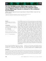
Fig. 2. Hydrolysis of (R,S)-piperazine-2-tert-butylcarboxamide by cells
of P. azotoformans IAM 1603 and stereochemical analysis of piperazine-
2-carboxylic acid produced by the purified amidase. (A) P. azotoformans
IAM 1603 was cultivated in 200 mL of BM medium for 12 h at 30 °C.
The cells were then harvested, washed with 0.9% NaCl and suspended
in 3 mL of 0.1
M
of potassium phosphate (pH 7.0). The reaction
mixture contained 10 m
M
of (R,S)-piperazine-2-tert-butylcarbox-
amide, 150 lL of the cell suspension and 0.1
M
of potassium phos-
phate (pH 7.0) in a total volume of 200 lL, and was incubated at
30 °C. The reaction was stopped at the specific time and the concen-
tration of each enantiomer of piperazine-2-carboxylic acid formed was
determined using HPLC with a Sumichiral OA-5000 column as des-
cribed in Materials and methods. Symbols: d,(S)-piperazine-2-carb-
oxylic acid; s,(R)-piperazine-2-carboxylic acid. (B) The reaction
mixture contained 10 m
M
of (R,S)-piperazine-2-tert-butylcarboxa-
mide, 10 lg of the purified amidase and 0.1
M
of potassium phosphate
(pH 7.0) in a total volume of 200 lL,andwasincubatedat30°Cfor
10 h. The stereochemistry of the piperazine-2-carboxylic acid formed
was determined using HPLC with a Sumichiral OA-5000 column as
described in Materials and methods.
Table 1. Purification of the S-stereoselective amidase from P. azoto-
formans IAM 1603. (R,S)-Piperazine-2-tert-butylcarboxamide was
used as a substrate for total activity and specific activity.
Step
Total
protein
(mg)
Total
activity
(mU)
Specific
activity
(mUÆmg
)1
)
Yield
(%)
Cell free extract 11200 59.2 5.27 · 10
)3
100
DEAE-Toyopearl 420 15.1 3.57 · 10
)2
25.4
Butyl-Toyopearl 56.2 7.24 0.128 12.2
Gigapite 7.02 1.21 0.171 2.03
Superdex HR26/60 2.10 0.85 0.405 0.14
MonoQ HR5/5 0.123 0.11 0.894 0.19
Ó FEBS 2004
L
-Amino acid amidase from P. azotoformans (Eur. J. Biochem. 271) 1469
)35 and )10 consensus promoter regions, respectively, were
identified. Alignment by the protein databases using the
BLAST
program showed that the deduced primary structure
of amidase is similar to those of putative proline iminopep-
tidases from Pseudomonas syringae (71.3% identical over
293 amino acids, TrEMBL accession number Q87WK6),
Sinorhizobium meliloti (66.2% identical over 290 amino
acids [28], TrEMBL accession number Q92M42), Xantho-
monas axonopodis (63.8% identical over 290 amino acids
[29], TrEMBL accession number Q8PIB1), Xanthomonas
campestris (63.4% identical over 290 amino acids [29],
TrEMBL accession number Q8P6Z8), Mesorhizobium loti
(58.1% identical over 291 amino acids [30], PRF accession
number 2705259DR), Salmonella typhimurium (42.6%
identical over 282 amino acids [31], TrEMBL accession
number Q8ZPP7) and Lactobacillus plantarum (35.6%
identical over 292 amino acids [32], TrEMBL accession
number Q890D8) and functionally characterized proline
iminopeptidases from Lactobacillus delbrueckii ssp. lactis
(37.1% identical over 294 amino acids [33], Swiss Prot
accession number PIP_LACDL), Lactobacillus helveticus
(35.9% identical over 295 amino acids [34], Swiss Prot
accession number PIP_LACHE) and Lactobacillus del-
brueckii ssp. bulgaricus CNRZ 397 (35.7% identical over
297 amino acids [35], PRF accession number 2105330A).
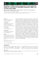
Figure 3 shows the alignment of the primary structures
of the amidase, LaaA, from P. azotoformans IAM1603,
putative proline iminopeptidase from P. syringae and
functionally characterized proline iminopeptidase from
L. delbrueckii ssp. lactis. The consensus motif (Gly-
X-Ser111-X-Gly-Gly) surrounding the catalytic serine of
the proline iminopeptidases family was conserved in LaaA
sequence. Asp251 and His278 constituting the probable
catalytic triad [36–38] with the Ser111 were also present in
the sequence. When the other ORF, ORF1, contained in
plasmid pSTB10 locating upstream of the laaA ORF, was
compared with other sequences in the databases, it was
observed that its deduced amino acid sequence showed
similarity to those of the following transcriptional regulator
proteins: hypothetical LuxR family protein from P. syrin-
gae (65.8% identical over 202 amino acids, TrEMBL
accession number Q87WK7), hypothetical protein
SMc04032 from S. meliloti (46.0% identical over 202 amino
acids [28], TrEMBL accession number Q92M41), hypo-
thetical AhyR/AsaR family protein from X. axonopodis
(46.8% identical over 201 amino acids [29], TrEMBL
accession number Q8PIB0), hypothetical AhyR/AsaR
family protein from X. campestris (45.9% identical over
205 amino acids [29], TrEMBL accession number Q8P6Z7),
hypothetical LuxR family protein from Rhodopseudomonas
palustris (31.2% identical over 189 amino acids, GenBank
accession number BX572594), VanR from Vibrio anguilla-
rum (30.1% identical over 193 amino acids [39], Swiss Prot
accession number VANR_VIBAN), BafR from Burkholde-
ria ambifaria (29.6% identical over 199 amino acids,
TrEMBL accession number Q9AER1), hypothetical pro-
tein from Bradyrhizobium japonicum (29.5% identical over
190 amino acids [40], TrEMBL accession number Q89VI3),
MupR from Pseudomonas fluorescens (26.8% identical over
194 amino acids [41], PRF accession number 2801295B) and
BviR from Burkholderia cepacia (27.8% identical over 198
amino acids [42], TrEMBL accession number Q9AHP7).
ORF1 was designated laaR. Comparison of the deduced
amino acid sequences of the P. azotoformans laaR and its
homologous genes indicated that the ORF1 lacks its
5¢ terminus part, probably coding for about 50 amino acid
residues.
Production of the LaaA in
E. coli
The direction of the laaA gene was same as that of the lac
promoter in the plasmid, pSTB10. However, the E. coli
transformant harboring pSTB10 showed no activity
towards the substrates such as (R,S)-piperazine-2-tert-
butylcarboxamide,
L
-prolinamide and
L
-proline-p-nitro-
anilide, irrespective of the addition of isopropyl
thio-b-
D
-galactoside to the culture medium. To express the
laaA gene in E. coli, we improved the sequence upstream
Fig. 3. Comparison of the amino acid sequen-
ces of the amidase (LaaA) from P. azotofor-
mans IAM 1603 and other homologous
proteins. Identical and conserved amino acids
among the sequences are marked in black and
in gray, respectively. Dashes indicate the gaps
introduced for better alignment. LaaA,
amidase from P. azotoformans IAM 1603;
Q87WK6, putative proline iminopeptidase
from Pseudomonas syringae;PIP_LACDL,
proline iminopeptidase from Lactobacillus
delbrueckii ssp. lactis. Three residues, serine,
aspartic acid and histidine that constitute
the putative catalytic triad are marked by
asterisks.
1470 H. Komeda et al.(Eur. J. Biochem. 271) Ó FEBS 2004
from the ATG start codon by PCR, with plasmid pSTB10
as a template as described in Materials and methods. The
resultant plasmid, pSTB20, in which the laaA gene was
under the control of the lac promoter of pUC19 vector, was
introduced into E. coli JM109 cells. A protein correspond-
ing to the predicted molecular mass of 34 kDa was
produced when the lac promoter was induced by isopropyl
thio-b-
D
-galactoside (data not shown). When E. coli JM109
harbouring pSTB20 was cultivated in Luria–Bertani
medium supplemented with ampicillin and isopropyl thio-
b-
D
-galactoside for 12 h at 37 °C, the level of LaaA activity
in the supernatant of the sonicated cell-free extracts of the
transformants was 0.026 and 13.2 unitsÆmg
)1
with (R,S)-
piperazine-2-tert-butylcarboxamide and
L
-prolinamide as
substrates, respectively. The cell reaction with 0.2
M
of
(R,S)-piperazine-2-tert-butylcarboxamide was carried out
by using the various concentrations of E. coli cells (0.28%,
1.41% and 2.83%, w/w) prepared from the 12 h culture
(Fig. 4).TheE. coli cells produced (S)-piperazine-
2-carboxylic acid with high optical purity (> 95% enantio-
meric excess) at all of the reaction times tested.
Purification of the LaaA from
E. coli
transformant
Recombinant LaaA was purified from the E. coli JM109
harboring pSTB20 with a recovery of 11.8% by ammonium
sulfate fractionation and DEAE-Toyopearl and Butyl-
Toyopearl column chromatographies (Table 2). The final
preparation gave a single band on SDS/PAGE with a
molecular mass of 34 kDa (Fig. 5). This value is in good
agreement with that estimated from the deduced amino acid
sequence of the LaaA. The molecular mass of the native
enzyme was about 32 kDa according to gel filtration
chromatography, indicating that the native enzyme was a
monomer. The purified enzyme catalyzed the hydrolysis of
L
-prolinamide to
L
-proline at 192 UÆmg
)1
under the stand-
ard conditions.
Stability
The purified enzyme could be stored without loss of activity
for more than six months at )20 °C in the buffer containing
50% glycerol. The stability of the enzyme was examined at
various temperatures. After the enzyme had been preincu-
bated for 5 min in 100 m
M
Tris/HCl (pH 8.0), a sample of
the enzyme solution was taken and the activity was assayed
Fig. 4. Stereoselective hydrolysis of (R,S)-piperazine-2-tert-butylcar-
boxamide by cells of E. coli JM109/pSTB20. The reaction mixture
contained 0.2
M
of (R,S)-piperazine-2-tert-butylcarboxamide, washed
E. coli cells prepared from the culture broth after a 12 h cultivation
and 0.1
M
of Tris/HCl (pH 8.0) in a total volume of 100 lL, and was
incubated at 30 °C. The reaction was stopped at the specific time and
the concentration of piperazine-2-carboxylic acid formed was deter-
mined as described in Materials and methods. Symbols: d,(S)-acid
formed with cells (0.28%,w/w); j,(S)-acidformedwithcells
(1.41%,w/w); m,(S)-acid formed with cells (2.83%,w/w); s,(R)-acid
formed with cells (0.28%,w/w); h,(R)-acid formed with cells
(1.41%,w/w); n,(R)-acid formed with cells (2.83%,w/w).
Table 2. Purification of LaaA from E. coli JM109 harboring pSTB20.
L
-Prolinamide was used as a substrate for total activity and specific
activity.
Step
Total
protein
(mg)
Total
activity
(U)
Specific
activity
(UÆmg
)1
)
Yield
(%)
Cell free extract 1020 13400 13.1 100
Ammonium sulfate 354 8720 24.6 65.1
DEAE-Toyopearl 25.5 3900 153 29.1
Butyl-Toyopearl 8.24 1580 192 11.8
Fig. 5. SDS/PAGE of LaaA. Lane 1, molecular mass standards
[phosphorylase b (94 kDa), BSA (67 kDa), ovalbumin (43 kDa),
carbonic anhydrase (30 kDa), soybean trypsin inhibitor (20.1 kDa)
and a-lactalbumin (14,4 kDa)]; lane 2, purified LaaA (5 lg).
Ó FEBS 2004
L
-Amino acid amidase from P. azotoformans (Eur. J. Biochem. 271) 1471
with
L
-prolinamide as a substrate under the standard
conditions. It exhibited the following activity: 55 °C, 0%;
50 °C, 25%; 45 °C, 81%; 40 °C, 100%; 35 °C, 100%. The
stability of the enzyme was also examined at various pH
values. The enzyme was incubated at 30 °C for 5 min in the
following buffers (final concentration 100 m
M
): acetic acid/
sodium acetate (pH 4.0–6.0), Mes/NaOH (pH 5.5–6.5),
potassium phosphate (pH 6.5–8.5), Tris/HCl (pH 7.5–9.0),
ethanolamine/HCl (pH 9.0–11.0), glycine/NaCl/NaOH
(pH 10.0–13.0). Then a sample of the enzyme solution
was taken, and the LaaA activity was assayed with
L
-prolinamide as a substrate under the standard conditions.
The enzyme was most stable in the pH range 6.0–9.5.
Effects of pH and temperature
The optimal pH for the activity of the enzyme was measured
in the buffers described above. The enzyme showed
maximum activity at pH 9.0. The enzyme reaction was
carried out at various temperatures for 5 min in 0.1
M
Tris/
HCl (pH 8.0), and enzyme activity was found to be
maximal at 45 °C. Above 45 °C, it decreased rapidly,
possibly because of instability of the enzyme at the higher
temperatures.
Effects of inhibitors and metal ions
Various compounds were investigated for their effects on
enzyme activity. We measured the enzyme activity under
standard conditions after incubation at 30 °Cfor5min
with various compounds at 1 m
M
. The enzyme was
completely inhibited by ZnSO
4
,ZnCl
2
,CdCl
2
,AgNO
3
and HgCl
2
and inhibited 73% by PbCl
2
, 70% by NiCl
2
and
52% by CoCl
2
. Other inorganic compounds such as LiBr,
H
2
BO
3
,NaCl,MgSO
4
,AlCl
3
,KCl,CaCl
2
,CrCl
3
,MnCl
2
,
FeSO
4
,Fe(NH
4
)
2
(SO
4
)
2
, CuSO
4
,RbCl,Na
2
MoO
4
(NH
4
)
6
Mo
7
O
24
, SnCl
2
,CsClandBaCl
2
did not influence
the activity. The enzyme was completely inhibited by
phenylhydrazine, however, other carbonyl reagents such
as hydroxylamine, hydrazine,
D
,
L
-penicillamine and
D
-cycloserine were not inhibitory toward the enzyme.
Chelating reagents, e.g. o-phenanthroline, 8-hydroxyquino-
line, ethylenediaminetetraacetic acid and a,a¢-dipyridyl had
no significant effect on the enzyme. The enzyme was
inhibited by thiol reagents such as p-chloromercuribenzoate
(67% inhibition), iodoacetate (40% inhibition) and
N-ethylmaleimide (24% inhibition). A serine protease
inhibitor, phenylmethanesulfonyl fluoride, a serine/cysteine
protease inhibitor, leupeptine and an aspartic protease
inhibitor, pepstatin, did not influence the activity.
Substrate specificity
To study the substrate specificity, the LaaA was used
to hydrolyze various amino acid amides and dipeptides
and the activity was assayed (Table 3). Besides
L
-prolina-
mide, the enzyme was active towards
L
-proline-p-nitro-
anilide (R,S)-piperidine-2-carboxamide,
L
-alaninamide and
L
-methioninamide (R,S)-piperazine-2-carboxamide. (R,S)-
Piperazine-2-tert-butylcarboxamide was, however, hydro-
lyzed at much lower rates than the above
L
-amino acid
amides. Dipeptides and
D
-prolinamide were not substrates
of the enzyme. The apparent K
m
value for
L
-proline-
p-nitroanilide was 0.58 m
M
,whereastheV
max
value for the
substrate was 80.9 UÆmg
)1
. Incubation of the LaaA with
L
-prolinamide and glycine did not yield a dipeptide,
L
-prolylglycine, suggesting no transpeptidase activity of
the enzyme.
Discussion
In this study, we purified an S-stereoselective amidase acting
on (R,S)-piperazine-2-tert-butylcarboxamide from P. azoto-
formans IAM 1603 and cloned the gene, laaA, coding for the
enzyme. E. coli cells overexpressing the laaA gene have been
demonstrated to be applicable to the S-stereoselective
hydrolysis of (R,S)-piperazine-2-tert-butylcarboxamide to
produce (S)-piperazine-2-carboxylic acid with high optical
purity. This is the first example that presents the stereose-
lective amidase useful for the optical resolution of a racemic
amide compound containing bulky substituents at the
leaving group.
Sequence analysis of the cloned gene, laaA, reveals
homology to proline iminopeptidases [PIP, EC 3.4.11.5],
which catalyze the removal of N-terminal proline from
peptides with high specificity, rather than to the other
amidases mentioned in the Introduction, suggesting an
evolutionary origin for LaaA from the enzymes involved in
peptide degradation. Crystal structures of proline imino-
petidases from X. campestris pv. citri [36] and Serratia
marcescens [38] have been solved. The enzyme consists of
two domains and the larger domain shows the general
topology of the a/b hydrolase fold. Ser113, Asp268 and
His296 residues (numbering of the residues are based on the
enzyme from S. marcescens) constituting the catalytic triad
are located at the interface of the two domains. Perfect
conservation of these residues in the LaaA sequence
suggests that LaaA could be categorized as a new member
of the family of proline iminopeptidases, and that the
Table 3. Substrate specificity of purified LaaA. The activity for
L
-pro-
linamide, corresponding to 192 UÆmg
)1
, was taken as 100%. The fol-
lowing compounds were not substrates for the amidase:
L
-argininamide,
L
-asparaginamide,
L
-isoasparagine,
L
-glutaminamide,
L
-isoglutamine,
glycinamide,
L
-histidinamide,
L
-lysinamide,
L
-valinamide,
D
-prolina-
mide,
L
-alanyl-
L
-alanine,
L
-alanylglycine, glycylglycine,
L
-prolyl-
L
-alanine and
L
-prolylglycine.
Substrate Relative activity (%)
L
-Prolinamide 100
L
-Proline-p-nitroanilide 40.9
(R,S)-Piperidine-2-carboxamide 32.0
L
-Alaninamide 10.6
L
-Methioninamide 4.2
(R,S)-Piperazine-2-carboxamide 3.7
L
-Phenylalaninamide 0.97
L
-Leucinamide 0.46
L
-Serinamide 0.43
L
-Tryptophanamide 0.20
(R,S)-Piperazine-2-tert-butylcarboxamide 0.20
L
-Isoleucinamide 0.17
L
-Threoninamide 0.12
L
-Tyrosinamide 0.086
1472 H. Komeda et al.(Eur. J. Biochem. 271) Ó FEBS 2004
catalytic mechanism of LaaA could be analogous to those
of the other members. However, LaaA could not act on the
peptide substrates such as
L
-prolyl-
L
-alanine,
L
-prolylgly-
cine,
L
-alanyl-
L
-alanine,
L
-alanylglycine and glycylglycine
(Table 3). Therefore, LaaA may differ from the other
members of the family with respect to its substrate
recognition. LaaA was sensitive to heavy metal salts and
thiol reagents and rather resistant to serine peptidase
inhibitors, suggesting the presence of a possible catalytic
cysteine residue. However, these features have also been
previously observed in proline iminopeptidases whose
catalytic serine residue has been identified by site-directed
mutagenesis [43] and crystal structure analysis [36,38].
LaaA was found to have hydrolyzing activity toward
L
-amino acid amides such as
L
-prolinamide,
L
-proline-
p-nitroanilide,
L
-alaninamide and
L
-methioninamide. The
enzyme also acted S-stereoselectively on (R,S)-piperidine-2-
carboxamide (R,S)-piperazine-2-carboxamide and (R,S)-
piperazine-2-tert-butylcarboxamide. Based on its substrate
specificity towards
L
-amino acid amides, LaaA should be
called
L
-amino acid amidase.
L
-Amino acid amidases were previously purified from
P. putida ATCC 12633 [9], O. anthropi NCIMB 40321 [10]
and M. neoaurum ATCC 25795 [11] and characterized.
All of the three enzymes seemed to be metalloenzymes
because their activities are inhibited by chelating reagents
such as ethylenediaminetetraacetic acid and o-phenanthro-
line and/or activated by divalent cations (Table 4). Com-
parison of the characteristics of LaaA with those of the
other
L
-amino acid amidases suggests that LaaA is unique
not only with respect to its physicochemical characteristics
but also concerning its substrate specificity. As the
primary sequences of the three amidases have never been
reported, LaaA from P. azotoformans IAM 1603 is the
first
L
-amino acid amidase whose primary sequence is
revealed.
Acknowledgements
WearegratefultoS.Iwamoto,R.KasaharaandA.Nakayama
(Toyama Prefectural University) for their technical assistance. This
work was supported by Grants-in-Aid for Scientific Research
(13760076 to H. K.) from JSPS (Japan Society for the Promotion of
Science).
References
1. Asano, Y. & Lu
¨
bbehu
¨
sen, T.L. (2000) Enzymes acting on peptides
containing
D
-amino acid. J. Biosci. Bioeng. 89, 295–306.
2. Kamphuis, J., Boesten, W.H.J., Broxterman, Q.B., Hermes,
H.F.M., van Balken, J.A.M., Meijer, E.M. & Schoemaker, H.E.
(1990) New developments in the chemoenzymatic production of
amino acids. Adv. Biochem. Eng. Biotechnol. 42, 133–186.
3. Schmid, A., Dordick, J.S., Hauer, B., Kiener, A., Wubbolts, M. &
Witholt, B. (2001) Industrial biocatalysis today and tomorrow.
Nature 409, 258–268.
4. Mayaux, J F., Cerbelaud, E., Soubrier, F., Faucher, D. & Pe
´
tre
´
,
D. (1990) Purification, cloning, and primary structure of an
enantiomer-selective amidases from Brevibacterium sp. strain
R312: structural evidence for genetic coupling with nitrile
hydratase. J. Bacteriol. 172, 6764–6773.
5. Ciskanik, L.M., Wilczek, J.M. & Fallon, R.D. (1995) Purifica-
tion and characterization of an enantioselective amidases from
Pseudomonas chlororaphis B23. Appl. Environ. Microbiol. 61,
998–1003.
Table 4. Comparison of the characteristics of LaaA from P. azot ofor mans IAM 1603 and bacterial
L
-aminoacidamidases.pCMB, p-chloro-
mercuribenzoate; DFP, diisopropylfluorophosphate; EDTA, ethylenediaminetetraacetic acid; PMSF, phenylmethylsulfonyl fluoride; DTT,
dithiothreitol.
LaaA
L
-Aminopeptidase
L
-Specific amidase
L
-Amino amidase
Origin Pseudomonas azotoformans Pseudomonas putida Ochrobactrum
anthropi
Mycobacterium
neoaurum
IAM 1603 ATCC 12633 NCIMB 40321 ATCC 25795
Molecular mass
of subunit
34 514 Da 53 000 Da 36 000 Da 40 000 Da
Number of subunits 1 8 2 3 or 4
Optimum pH 9.0 9.5 6.0–8.5 8.0–9.5
pH stability 6.0–9.5
Optimum
temperatrure
45 °C40°C70°C50°C
Heat stability 45 °C60°C55°C
Inhibitor Phenylhydrazine, pCMB,
iodoacetate,
N-ethylmaleimide,
Zn
2+
,Ag
+
,Cd
2+
,Hg
2+
pCMB, DFP, EDTA,
PMSF, o-phenanthroline,
Cu
2+
,Ca
2+
EDTA,
o-phenanthroline,
DTT, o-phenanthroline,
iodoacetamide
Activator No DTT, Mn
2+
,Mg
2+
,Co
2+
Zn
2+
,Mn
2+
,Mg
2+
Substrate specificity
L
-Prolinamide
L
-Leucinamide
L
-Prolinamide
L
-Prolinamide
L
-Proline-p-nitroanilide
(S)-Piperidine-2-carboxamide
L
-Alaninamide
L
-Methioninamide
L
-Phenylglycinamide
L
-Methioninamide
L
-Phenylalaninamide
L
-Methioninamide
L
-Phenylglycinamide
L
-Alaninamide
L
-Valinamide
L
-a-Methylvalinamide
Peptidase activity No Yes:
L
-Phe-
L
-Phe,
L
-Phe-
L
-Leu
No No
Ó FEBS 2004
L
-Amino acid amidase from P. azotoformans (Eur. J. Biochem. 271) 1473
6. Kobayashi, M., Komeda, H., Nagasawa, T., Nishiyama, M.,
Horinouchi, S., Beppu, T., Yamada, H. & Shimizu, S. (1993)
Amidase coupled with low-molecular-mass nitrile hydratase from
Rhodococcus rhodochrous J1. Sequencing and expression of the
gene and purification and characterization of the gene product.
Eur. J. Biochem. 217, 327–336.
7. Trott, S., Bauer, R., Knackmuss, H J. & Stolz, A. (2001) Genetic
and biochemical characterization of an enantioselective amidase
from Agrobacterium tumefciens strain d3. Microbiology 147, 1815–
1824.
8. Hayashi, T., Yamamoto, K., Matsuo, A., Otsubo, K., Murama-
tsu, S., Matsuda, A. & Komatsu, K. (1997) Characterization and
cloning of an enantioselective amidase from Comamonas acido-
vorans KPO-2771-4. J. Ferment. Bioeng. 83, 139–145.
9. Hermes, H.F.M., Sonke, T., Peters, P.J.H., van Balken, J.A.M.,
Kamphuis, J., Dijkhuizen, L. & Meijer, E.M. (1993) Purification
andcharacterizationofan
L
-aminopeptidase from Pseudomonas
putida ATCC 12633. Appl. Environ. Microbiol. 59, 4330–4334.
10. van den Tweel, W.J.J., van Dooren, T.J.G.M., de Jonge, P.H.,
Kaptein, B., Duchateau, A.L.L. & Kamphuis, J. (1993) Ochro-
bactrum anthropi NCIMB 40321: a new biocatalyst with broad-
spectrum
L
-specific amidases activity. Appl. Microbiol. Biotechnol.
39, 296–300.
11. Hermes, H.F.M., Tandler, R.F., Sonke, T., Dijkhuizen, L. &
Meijer, E.M. (1994) Purification and characterization of an
L
-amino amidase from Mycobacterium neoaurum ATCC 25795.
Appl. Environ. Microbiol. 60, 153–159.
12. Asano, Y., Nakazawa, A., Kato, Y. & Kondo, K. (1989) Prop-
erties of a novel
D
-stereospecific aminopeptidase from Ochrobac-
trum anthropi. J. Biol. Chem. 264, 14233–14239.
13. Asano, Y., Kato, Y., Yamada, A. & Kondo, K. (1992) Structural
similarity of
D
-aminopeptidase to carboxypeptidase DD and
b-lactamases. Biochemistry 31, 2316–2328.
14. Komeda, H. & Asano, Y. (2000) Gene cloning, nucleotide
sequencing, and purification and characterization of the
D
-stereospecific amino-acid amidase from Ochrobactrum anthropi
SV3. Eur. J. Biochem. 267, 2028–2035.
15. Ozaki, A., Kawasaki, H., Yagasaki, M. & Hashimoto, Y. (1992)
Enzymatic production of
D
-alanine from
DL
-alaninamide by novel
D
-alaninamide specific amide hydrolase. Biosci. Biotechnol. Bio-
chem. 56, 1980–1984.
16. Baek, D.H., Kwon, S J., Hong, S P., Kwak, M S., Lee, M H.,
Song, J.J., Lee, S G., Yoon, K H. & Sung, M H. (2003) Char-
acterization of a thermostable
D
-stereospecific alanine amidase
from Brevibacillus borstelensis BCS-1. Appl. Environ. Microbiol.
69, 980–986.
17. Bigge, C.F., Johnson, G., Ortwine, D.F., Drummond, J.T., Retz,
D.M.,Brahce,L.J.,Coughenour,L.L.,Marcoux,F.W.&Probert,
A.W. (1992) Exploration of N-phosphonoalkyl-, N-phosphono-
alkenyl-, and N-(phosphonoalkyl) phenyl-spaced alpha-amino
acids as competitive N-methyl-
D
-aspartic acid antagonists. J. Med.
Chem. 35, 1371–1384.
18. Bruce, M.A., Laurent, D.R.S., Poindexter, G.S., Monkovic, I.,
Huang, S. & Balasubramanian, N. (1995) Kinetic resolution of
piperazine-2-carboxamide by leucine aminopeptidase. An appli-
cation in the synthesis of the nucleoside transport blocker (-)
draflazine. Synthetic Commun. 25, 2673–2684.
19. Askin, D., Eng, K.K., Rossen, K., Purick, R.M., Wells, K.M.,
Volante, R.P. & Reider, P.J. (1994) Highly diastereoselective
reaction of a chiral, non-racemic amide enolate with (S)-glycidyl
tosylate. Synthesis of the orally active HIV-1 protease inhibitor
L-735,524. Tetrahedron Lett. 35, 673–676.
20. Eichhorn,E.,Roduit,J P.,Shaw,N.,Heinzmann,K.&Kiener,
A. (1997) Preparation of (S) piperazine-2-carboxylic acid,
(R)-piperazine-2-carboxylic acid, and (S)-piperizine-2-carboxylic
acid by kinetic resolution of the corresponding racemic
carboxamides with stereoselective amidases in whole bacterial
cells. Tetrahedron Asymmetry 8, 2533–2536.
21. Sambrook, J., Fritsch, E.F. & Maniatis, T. (1989) Molecular
Cloning: a Laboratory Manual, 2nd edn. Cold Spring Harbor
Laboratory, Cold Spring Harbor N.Y.
22. Misawa, N., Nakagawa, M., Kobayashi, K., Yamano, S., Izawa,
Y., Nakamura, K. & Harashima, K. (1990) Elucidation of the
Erwinia uredovora carotenoid biosynthetic pathway by functional
analysis of gene products expressed in Escherichia coli. J. Bacteriol.
172, 6704–6712.
23. Inoue, H., Nojima, H. & Okayama, H. (1990) High efficiency
transformation of Escherichia coli with plasmids. Gene 96, 23–28.
24. Sanger, F., Nicklen, S. & Coulson, A.R. (1977) DNA sequencing
with chain-terminating inhibitors. Proc. Natl Acad. Sci. USA 74,
5463–5467.
25. Altschul, S.F., Gish, W., Miller, W., Myers, E.W. & Lipman, D.J.
(1990) Basic local alignment search tool. J. Mol. Biol. 215,
403–410.
26. Bradford, M.M. (1976) A rapid and sensitive method for the
quantitation of microgram quantities of protein utilizing the
principle of protein-dye binding. Anal. Biochem. 72, 248–254.
27. Laemmli, U.K. (1970) Cleavage of structural proteins during the
assembly of the head of bacteriophage T4. Nature 227, 680–685.
28. Capela,D.,Barloy-Hubler,F.,Gouzy,J.,Bothe,G.,Ampe,F.,
Batut, J., Boistard, P., Becker, A., Boutry, M., Cadieu, E.,
Dreano, S., Gloux, S., Godrie, T., Goffeau, A., Kahn, D., Kiss, E.,
Lelaure, V., Masuy, D., Pohl, T., Portetelle, D., Puhler, A.,
Purnelle, B., Ramsperger, U., Renard, C., Thebault, P.,
Vandenbol, M., Weidner, S. & Galibert, F. (2001) Analysis of the
chromosome sequence of the legume symbiont Sinorhizobium
meliloti strain 1021. Proc. Natl Acad. Sci. USA 98, 9877–9882.
29. da Silva, A.C., Ferro, J.A., Reinach, F.C., Farah, C.S., Furlan,
L.R., Quaggio, R.B., Monteiro-Vitorello, C.B., Van Sluys, M.A.,
Almeida, N.F., Alves, L.M., do Amaral, A.M., Bertolini, M.C.,
Camargo, L.E., Camarotte, G., Cannavan, F., Cardozo, J.,
Chambergo, F., Ciapina, L.P., Cicarelli, R.M., Coutinho, L.L.,
Cursino-Santos, J.R., El-Dorry, H., Faria, J.B., Ferreira, A.J.,
Ferreira, R.C., Ferro, M.I., Formighieri, E.F., Franco, M.C.,
Greggio, C.C., Gruber, A., Katsuyama, A.M., Kishi, L.T., Leite,
R.P., Lemos, E.G., Lemos, M.V., Locali, E.C., Machado, M.A.,
Madeira, A.M., Martinez-Rossi, N.M., Martins, E.C., Meidanis,
J., Menck, C.F., Miyaki, C.Y., Moon, D.H., Moreira, L.M.,
Novo, M.T., Okura, V.K., Oliveira, M.C., Oliveira, V.R., Pereira,
H.A., Rossi, A., Sena, J.A., Silva, C., de Souza, R.F., Spinola,
L.A., Takita, M.A., Tamura, R.E., Teixeira, E.C., Tezza, R.I.,
Trindade dos Santos, M., Truffi, D., Tsai, S.M., White, F.F.,
Setubal, J.C. & Kitajima. J.P. (2002) Comparison of the genomes
of two Xanthomonas pathogens with differing host specificities.
Nature 417, 459–463.
30. Kaneko, T., Nakamura, Y., Sato, S., Asamizu, E., Kato, T.,
Sasamoto, S., Watanabe, A., Idesawa, K., Ishikawa, A., Kawa-
shima, K., Kimura, T., Kishida, Y., Kiyokawa, C., Kohara, M.,
Matsumoto, M., Matsuno, A., Mochizuki, Y., Nakayama, S.,
Nakazaki, N., Shimpo, S., Sugimoto, M., Takeuchi, C., Yamada,
M. & Tabata, S. (2000) Complete genome structure of the nitro-
gen-fixing symbiotic bacterium Mesorhizobium loti. DNA Res. 7,
331–338.
31. McClelland, M., Sanderson, K.E., Spieth, J., Clifton, S.W.,
Latreille, P., Courtney, L., Porwollik, S., Ali, J., Du Dante, M.F.,
Hou, S., Layman, D., Leonard, S., Nguyen, C., Scott, K., Holmes,
A., Grewal, N., Mulvaney, E., Ryan, E., Sun, H., Florea, L.,
Miller, W., Stoneking, T., Nhan, M., Waterston, R. & Wilson,
R.K. (2001) Complete genome sequence of Salmonella enterica
serovar Typhimurium LT2. Nature 413, 852–856.
32. Kleerebezem, M., Boekhorst, J., van Kranenburg, R., Molenaar,
D., Kuipers, O.P., Leer, R., Tarchini, R., Peters, S.A., Sandbrink,
1474 H. Komeda et al.(Eur. J. Biochem. 271) Ó FEBS 2004
H.M., Fiers, M.W.E.J., Stiekema, W., Klein Lankhorst, R.M.,
Bron, P.A., Hoffer, S.M., Nierop Groot, M.N., Kerkhoven, R.,
DeVries,M.,Ursing,B.,DeVos,W.M.&Siezen,R.J.(2003)
Complete genome sequence of Lactobacillus plantarum WCFS1.
Proc.NatlAcad.Sci.USA100, 1990–1995.
33. Klein, J.R., Schmidt, U. & Plapp, R. (1994) Cloning, heterologous
expression, and sequencing of a novel proline iminopeptidase
gene, pepI,fromLactobacillus delbrueckii subsp. lactis DSM 7290.
Microbiology 140, 1133–1139.
34. Varmanen, P., Rantanen, T. & Palva, A. (1996) An operon from
Lactobacillus helveticus composed of a proline iminopeptidase
gene (pepI) and two genes coding for putative members of
the ABC transporter family of proteins. Microbiology 142, 3459–
3468.
35. Atlan, D., Gilbert, C., Blanc, B. & Portalier, R. (1994) Cloning,
sequencing and characterization of the pepIP gene encoding a
proline iminopeptidase from Lactobacillus delbrueckii subsp.
bulgaricus CNRZ 397. Microbiology 140, 527–535.
36. Medrano, F.J., Alonso, J., Garcia, J.L., Romero, A., Bode, W. &
Gomis-Ruth, F.X. (1998) Structure of proline iminopeptidase
from Xanthomonas campestris pv. citri: a prototype for the prolyl
oligopeptidase family. EMBO J. 17, 1–9.
37. Morel, F., Gilbert, C., Geourjon, C., Frot-Coutaz, J., Portalier, R.
& Atlan, D. (1999) The prolyl aminopeptidase from Lactobacillus
delbrueckii subsp. bulgaricus belongs to the a/b hydrolase family.
Biochim. Biophys. Acta 1429, 501–505.
38. Yoshimoto, T., Kabashima, T., Uchikawa, K., Inoue. T., Tanaka,
N., Nakamura, K., Tsuru, M. & Ito, K. (1999) Crystal structure of
prolyl aminopeptidase from Serratia marcescens. J. Biochem. 126,
559–565.
39. Milton,D.L.,Hardman,A.,Camara,M.,Chhabra,S.R.,Bycroft,
B.W., Stewart, G.S. & Williams, P. (1997) Quorum sensing in
Vibrio anguillarum: characterization of the vanI/vanR locus and
identification of the autoinducer N-(3-oxodecanoyl)-
L
-homoserine
lactone. J. Bacteriol. 179, 3004–3012.
40. Kaneko, T., Nakamura, Y., Sato, S., Minamisawa, K., Uchiumi,
T., Sasamoto, S., Watanabe, A., Idesawa, K., Iriguchi, M.,
Kawashima, K., Kohara, M., Matsumoto, M., Shimpo, S.,
Tsuruoka,H.,Wada,T.,Yamada,M.&Tabata,S.(2002)
Complete genomic sequence of nitrogen-fixing symbiotic bacter-
ium Bradyrhizobium japonicum USDA110. DNA Res. 9, 189–197.
41. El-Sayed, A.K., Hothersall, J. & Thomas, C.M. (2001) Quorum-
sensing-dependent regulation of biosynthesis of the polyketide
antibiotic mupirocin in Pseudomonas fluorescens NCIMB 10586.
Microbiology 147, 2127–2139.
42. Lutter, E., Lewenza, S., Dennis, J.J., Visser, M.B. & Sokol, P.A.
(2001) Distribution of quorum-sensing genes in the Burkholderia
cepacia complex. Infect. Immun. 69, 4661–4666.
43. Kitazono, A., Ito, K. & Yoshimoto, T. (1994) Prolyl amino-
peptidase is not a sulfhydryl enzyme: identification of the active
serine residue by site-directed mutagenesis. J. Biochem. 116,
943–945.
Ó FEBS 2004
L
-Amino acid amidase from P. azotoformans (Eur. J. Biochem. 271) 1475