Báo cáo khoa học: NMR solution structure of Cn12, a novel peptide from the Mexican scorpion Centruroides noxius with a typical b-toxin sequence but with a-like physiological activity doc
Bạn đang xem bản rút gọn của tài liệu. Xem và tải ngay bản đầy đủ của tài liệu tại đây (743.72 KB, 13 trang )
NMR solution structure of Cn12, a novel peptide from the Mexican
scorpion
Centruroides noxius
with a typical b-toxin sequence
but with a-like physiological activity
Federico del Rı
´
o-Portilla
1
, Elizabeth Herna
´
ndez-Marı
´
n
1
, Genaro Pimienta
2
*, Fredy V. Coronas
2
,
Fernando Z. Zamudio
2
, Ricardo C. Rodrı
´
guez de la Vega
2
, Enzo Wanke
2,3
and Lourival D. Possani
2
1
Institute of Chemistry, National Autonomous University of Mexico, Mexico City, Mexico;
2
Department of Molecular Medicine and
Bioprocesses, Institute of Biotechnology, National Autonomous University of Mexico;
3
Dipartimento di Biotecnologie e Bioscienze,
Universita
´
di Milano-Bicocca, Milan, Italy
Cn12 isolated from the venom of the scorpion Centruroides
noxius has 67 amino-acid residues, closely packed with four
disulfide bridges. Its primary structure and disulfide bridges
were determined. Cn12 is not lethal to mammals and
arthropods in vivo at doses up to 100 lgperanimal.Its3D
structure was determined by proton NMR using 850 dis-
tance constraints, 36 / angles derived from 36 coupling
constants obtained by two different methods, and 22
hydrogen bonds. The overall structure has a two and half
turn a-helix (residues 24–32), three strands of antiparallel
b-sheet (residues 2–4, 37–40 and 45–48), and a type II turn
(residues 41–44). The amino-acid sequence of Cn12 resem-
bles the b scorpion toxin class, although patch-clamp
experiments showed the induction of supplementary slow
inactivation of Na
+
channels in F-11 cells (mouse neuro-
blastoma N18TG-2 · rat DRG2), which means that it
behaves more like an a scorpion toxin. This behaviour
prompted us to analyse Na
+
channel binding sites using
information from 112 Na
+
channel gene clones available in
the literature, focusing on the extracytoplasmic loops of the
S5–S6 transmembrane segments of domain I and the S3–S4
segments of domain IV, sites considered to be responsible
for binding a scorpion toxins.
Keywords: Centruroides noxius; NMR structure; patch-
clamp; scorpion toxin; sodium channel.
Scorpion toxins are relatively short peptides with a variable
length of amino acids, showing characteristic 3D folding
comprised of an a-helix and three segments of antiparallel
b-sheet structure, stabilized by several disulfide bridges
[1–5]. Their known physiological role is to block or modify
ion-channel function, causing impairment of cellular com-
munication, which leads to the depolarization of excitable
membranes and might cause death of animals stung by
scorpions [6,7].
There are several reasons why the molecular basis of
toxin specificity and molecular mechanism of action
continue to be of scientific interest: (a) to study ion channels,
the target molecules of most known scorpion toxins, in
order to understand their molecular structure and function,
thus learning more about cellular excitability; (b) to
understand the toxic effects of scorpion venoms, a pre-
requisite for the development of more effective and safer
antidotes and/or vaccines; (c) to find toxins specific for
invertebrate organisms with a view to developing biode-
gradable drugs for pest control; (d) to discover other
possible unknown target molecules for which peptides were
evolved in the venom of scorpions. The last of these is not
trivial, as the huge variability of these peptides, estimated to
be of the order of 100 000 in scorpion venom alone, and of
which only about 0.2% have been identified, leaves a wide
open field for research [4,5,8]. Several recent articles and
reviews have reported on the structural and functional
aspects of these peptides [3,8–18]. Most dealt with scorpion
toxins as ion-channels blockers or modifiers of their
function. However, the structural variability of peptides
and the different types of receptor they recognize is steadily
increasing [19], exemplified by the following novel discov-
eries: ERG-channel-specific toxins [20], analgesic peptides
[21,22], modulators of immune response [23–25], antibiotics
[26–28], antimalaria agents [29], and others.
For the ion-channel-specific peptides, two distinct groups
of toxins have been identified based on the length of the
peptide chain: short-chain peptides (23–41-amino-acids
long) which recognize and bind to various types and
subtypes of K
+
channels [13,19], ryanodine-sensitive Ca
2+
channels [11], and Cl
–
channels [30]; long-chain peptides
(59–76 amino acids), which are specific for Na
+
channels
[8,18] and T-type Ca
2+
channels [31,32].
The purpose of this paper is to describe for the first
time a Na
+
-channel-specific scorpion toxin (Na-ScTx),
isolated from the New World scorpion Centruroides
Correspondence to L. D. Possani, Department of Molecular Medicine
and Bioprocesses, Institute of Biotechnology, National Autonomous
University of Mexico, Avenida Universidad 2001, Apartado Postal
510-3, Cuernavaca 62210, Mexico. Fax: + 52 777 3172388,
Tel.: + 52 777 3171209, E-mail:
Abbreviation:Na-ScTx,scorpiontoxinspecificforNa
+
channel; TTX,
tetrodotoxin.
Note: Laboratories 1 and 2 contributed equally to this work.
*Present address: EMBL, Meyerhofstrasse 1, Heidelberg D-69117,
Germany.
(Received 4 February 2004, revised 30 March 2004,
accepted 22 April 2004)
Eur. J. Biochem. 271, 2504–2516 (2004) Ó FEBS 2004 doi:10.1111/j.1432-1033.2004.04181.x
noxius. It is structurally similar to b Na-ScTxs, but has
an a-like function. The 3D structure of this toxin was
determined by NMR. The importance of the overall
charge distribution on the surface of the toxin for its
activity is emphasized. Furthermore, considerations rela-
ted to the amino-acid sequences deduced from cloned
Na
+
-channel genes are discussed in terms of what is
known about the interacting surfaces of Na-ScTxs and
Na
+
channels.
Materials and methods
Venom source, purification procedures and lethality tests
Venom from scorpions collected in Nayarit State (Mexico)
was obtained by electrical stimulation and separated by
Sephadex G-50 gel filtration, followed by ion-exchange
chromatography on CM-cellulose columns, as previously
described [33]. For this work, fraction II-4 (v.g., fraction II
from Sephadex, and subfraction 4 from CM-cellulose) was
further separated by HPLC using previously published
conditions [34]. Lethality tests were conducted with mice,
crickets and crayfish, using the same conditions as reported
[34].
Determination of amino-acid sequence and MS analysis
The full amino-acid sequence of the toxin was obtained by
direct Edman degradation, using an automatic Beckman
sequencer (LF 3000 Protein Sequencer) and samples of
native and reduced toxin. Additional information was
generated by sequencing subpeptides obtained by HPLC
separation of toxin treated with endopeptidases lysine-C
(Boehringer, Mannheim, Germany) and Staphylococcus
aureus V8 (Boehringer, Mannheim, Germany), as previ-
ously described [8,33,34]. The last amino-acid residue was
confirmed by MS analysis, performed in a LCQ
DUO
ion-trap spectrometer from Finnigan (San Jose, CA, USA).
Determination of disulfide bridges
A sample containing 100 lg toxin was digested with
trypsin (Promega, Madison, WI, USA) in slightly acidic
conditions, followed immediately by endopeptidase V8
digestion, as previously described [8], and separated by
HPLC on a C
18
reverse-phase column with a linear
gradient from solution A [0.12% (v/v) trifluoroacetic acid
in water] to 60% solution B [0.10% (v/v) trifluoroacetic
acid in acetonitrile], run for 60 min. Several components
were fractionated (data not shown). The component
eluted at 27.78 min was identified as the disulfide bridge
between Cys11 and Cys65. Because of incomplete
digestion, the peptide eluted at 28.92 min was further
treated with chymotrypsin (Boehringer) and subsequently
separated by HPLC. The component eluted at 26.38 min
(data not shown) corresponded to the disulfide bridge
between Cys15 and Cys40. The two other disulfide
bridges were determined by NMR analysis as discussed
below. For the assignment of the first two disulfide
bridges, the peptides hydrolyzed by enzymatic digestion
were sequenced using the Beckman LF3000 Protein
Sequencer described above.
Electrophysiological data
Cell culture. Cells of the F-11 clone (mouse neuroblastoma
N18TG-2 · rat DRG) [35] were routinely cultured in
Dulbecco’s modified Eagle’s medium, containing 4.5 gÆL
)1
glucose and 10% fetal calf serum. The cells were incubated
at 37 °C in a humidified atmosphere with 5% CO
2
.
Solutions and drugs. The standard extracellular solution
contained (m
M
): NaCl 130, KCl 5, CaCl
2
2, MgCl
2
2,
Hepes/NaOH 10,
D
-glucose 5, at pH 7.40. In the high-K
+
external solution ([K
+
]
o
¼ 40 m
M
), NaCl was replaced by
an equimolar amount of KCl. The standard pipette solution
at [Ca2
+
]
i
¼ 10
–7
M
(pCa 7) contained (m
M
): K
+
-aspartate
130, NaCl 10, MgCl
2
2, CaCl
2
1.3, EGTA/KOH 10, Hepes/
KOH 10, ATP (Mg
2+
salt) 1, pH 7.30.
Patch-clamp recordings and data analysis. The currents
were recorded by means of the patch-clamp amplifier
MultiClamp 700A (Axon Instruments, Foster City, CA,
USA) at room temperature as previously described [36]
(pipette resistance 0.8–1.2 MW); cell capacitance and series
resistance errors were carefully compensated for (85–90%)
before each voltage clamp protocol run. The extracellular
solutions were delivered through a nine-hole (0.6-mm)
remote-controlled linear positioner, with an average
response time of 2–3 s, placed near the cell under study.
The Na
+
-current inactivation curves were obtained by
plotting the normalized peak current against V
m
. Final
traces were corrected with traces obtained in the presence of
20 n
M
tetrodotoxin (TTX). The activation was derived as
the normalized sodium conductance relationship (E
Na
¼
)65). To determine the amplitude of the toxin-induced slow
inactivation, we subtracted the control traces from the traces
recorded in the presence of the toxin. The amplitude of these
toxin-induced currents was analyzed by plotting the value
3 ms after the onset of the depolarizing pulse. The decaying
inactivating portion of the control traces and the currents in
the presence of toxin were fitted to one or two exponential
decaying functions, respectively, to obtain the inactiva-
tion time constants. pClamp 8 (Axon Instruments) and
Origin 4.1 (Microcal Inc, Studio City, CA, USA) software
were routinely used during data acquisition and analysis.
Preparation of the NMR sample
A sample of purified Cn12 containing 6.2 mg peptide was
dissolved in 0.8 mL H
2
O/D
2
O (9 : 1, v/v) to a final
concentration of 0.9 m
M
. The pH measured for this solution
was 3.1. After the experiments in H
2
O, the peptide was
lyophilized and redissolved in D
2
O to perform additional
experiments.
1
H-NMR spectroscopy
The experiments were performed in a Varian Unity Plus 500
spectrometer. Data were collected at 300 K. Mixing times
were 35 ms for TOCSY and 80 and 100 ms for NOESY in
H
2
OandinD
2
O. Data were processed with
NMRPIPE
[37] to
obtain 4K · 4K spectra. Spectra were analysed using
the
XEASY
program [38]. The values of the J
HN-Ha
were
estimated from TOCSY spectra with the modified
Ó FEBS 2004 NMR solution structure of Cn12 (Eur. J. Biochem. 271) 2505
J doubling in the frequency domain [39,40], when possible,
or with the strategy proposed by Wishart et al. [41].
Experimental constraints and structure calculations
Most of the distance constraints were obtained from
NOESY spectra in H
2
O; additional constraints were from
NOESY spectra in D
2
O. NOE intensities were evaluated
from the volume of the cross-peak and calibrated internally
using the
CALIBA
program [42] to generate a set of upper
limit distances. Most NOE data were obtained from
resolved signals. NOE signals for H
b
–H
b
between Cys11
and Cys65 and Cys15 and Cys40 were assigned; however,
no NOE data were observed for the other Cys pairs.
A total of 850 distance constraints were used from which
121 are sequential, 30 medium range (1 < |i–j|<4)and
92 long range (|i–j| > 4); the remainder were intraresidues.
A total of 36 / angle constraints were used based on the
J
HN-Ha
values. Using the modified J doubling method, it
was possible to evaluate 23 J
HN-Ha
values from a trace of
TOCSY spectra. It was only possible to measure 13
additional coupling constants from TOCSY spectra as
proposed by Wishart. It was found that the modified
J doubling method gives smaller values than the Wishart
method [41], and in the case of the a-helix (residues
24–32), where a value of 3.9 is expected [43], values are
closer depending on the structure obtained. Proton–
deuterium exchange of the amide groups was measured
on a sample lyophilized from H
2
O and redissolved in pure
D
2
O as described [43], in order to determine 22 hydrogen
bond constraints. Four disulfide bridge constraints were
added to the calculations. The dynamic annealing struc-
ture calculations were performed with the CNS software
suite [44].
Analysis of Na
+
-channel sequences
By searching data banks containing sequences of Na
+
channels from human, fruit fly and squid, several
representative sequences were chosen (five for humans,
three for Drosophila, and one for squid). With these
sequences, more than 200
BLAST
entries were found that
matched isoforms of Na
+
-channel sequences. An align-
ment was performed using the program
CLUSTAL X
[45],
from which 50 nonredundant sequences were selected.
Among these are sequences from several species including
mammals, insects and other invertebrates (available from
the authors on request). The proposed binding sites for
a Na-ScTxs, including regions corresponding to the S5–S6
segment from domain I and S3–S4 from domain IV of
the Na
+
channels, were re-aligned independently using
CLUSTAL X
, and some were chosen for our figure. Among
these are the ones best represented and cited in the
literature on scorpion toxins.
Results
Purification and sequencing
Figure 1A shows the results of separating 1 mg fraction II-4
(for details see Materials and methods) from the venom of
the scorpion C. noxius. For this report, about 20 HPLC
runs were performed as described, in order to obtain enough
peptide for this work. The component indicated by the star
in Fig. 1A was recovered and chromatographed again
(Fig. 1A, inset) to give the pure peptide, called Cn12. The
name comes from the abbreviation of the scorpion species
C. noxius followed by the number 12, which corresponds to
the 12th pure peptide fully characterized from this venom,
specific for Na
+
channels. This peptide corresponds to
1.3% of the total venom protein concentration.
For determination of the primary structure, it was
necessary to obtain overlapping sequences of several
peptides (Fig. 1B). The 36 most N-terminal amino-acid
residues were identified by directly sequencing the native
peptide, and the identities confirmed by sequencing a
sample of reduced and alkylated cysteine residues (vinyl
derivatives) of Cn12. The overlapping sequence spanning
Ala33 to Gly63 was determined with a peptide obtained by
Lysine-C digestion, as described in Materials and methods,
and the final segment overlapping from Gly53 to Arg66 was
obtained by sequencing a peptide digested with endo-
peptidase V8. The last residue, Ser in position 67, was
determined by MS. The molecular mass found by MS
ionization analysis was 7139.5 Da, and the theoretically
expected value was 7139.7 Da, thus confirming the full
sequence.
Disulfide bridges
As mentioned in Materials and methods, a peptide obtained
by enzymatic digestion of Cn12, and separated by HPLC
(elution time 27.78 min) provided a heterodimeric pro-
duct when directly sequenced by Edman degradation.
The sequences obtained were DGYPLASNGC and
GTVLWGDSGTXPCR, indicating unequivocally the posi-
tion of one of the disulfide bridges, which in this case was
between Cys11 and Cys65, based on the known primary
structure of Cn12 (Fig. 1B). Another core peptide from the
initial digestion (time 28.92 min) gave several amino-acid
sequences, making it impossible to assign any other disulfide
bridges. It was therefore further digested with chymotryp-
sin, which produced subpeptides, and that eluted at
26.35 min (data not shown) gave the sequences FGC and
GYC, corresponding to the disulfide bridge Cys15 and
Cys40. The peptide eluted at 17.48 min gave four sequences
corresponding to segments of the primary structure con-
taining Cys25, Cys27, Cys45 and Cys47. As we knew the
primary structure, we knew that the remaining disulfide
bridges were between Cys25 and Cys45 and Cys29 and
Cys47, or conversely between Cys25 and Cys47 and Cys29
and Cys45. The sequences of the fragments from the
chymotryptic hydrolysis showed that the other putative
disulfides, i.e. Cys25–Cys29 and Cys45–Cys47, were not
possible because the sequence would not fit the one
determined experimentally.
Modelling the 3D structure, based on the NMR data as
discussed below, showed that the only disulfide configur-
ation that would fit the results obtained is Cys25–Cys45 and
Cys29–Cys47. Furthermore, these disulfide pairs are at
exactly the same positions as those described for other Na-
ScTxs isolated from several other scorpion species [8]. Thus,
the disulfide bridges of Cn12 are assumed to be: Cys11–
Cys65, Cys15–Cys40, Cys25–Cys45 and Cys29–Cys47.
2506 F. del Rı
´
o-Portilla et al.(Eur. J. Biochem. 271) Ó FEBS 2004
Sequence comparison of Cn12 with other Na-ScTxs
A databank search with the sequence of Cn12 as the query
retrieved only scorpion venom-derived peptides classified as
Na-ScTxs. The highest identities were found with scorpion
toxins belonging to the b group (identity > 30%), including
several toxins isolated from New World scorpions. It also
shows similarities to some depressant and excitatory toxins
from Old World scorpions, as well as to the recently
characterized Birtoxin, a long-chain peptide with only three
disulfide bridges from the scorpion Parabuthus transvaalicus
[46]. The similarities to Na-ScTxs of the a group are
considerably lower (identity < 25%). On the basis of these
results, Cn12 should be classified as a member of the b
group of Na-ScTxs. To allow proper discussion in the terms
of structure–activity relationship, with regard to recognition
and affinity for Na
+
channels, Cn12 is aligned with other
Na-ScTxs in Fig. 2. For this comparison, only toxins for
which the 3D structures are known were included. The 3D
structures of three pharmacologically different classes of
Na-ScTxs are known: a Na-ScTxs (AaHII, BmKM1,
BmKM2, BmKM4, BmKM8, BmK-aTx16, BmK-aIT,
Bs-mktx, CsE-V, LqhII, LqqIII, Lqh-aIT), b Na-ScTxs
(Cn2, CsE1, CsEv1, CsEv2, CsEv3, CsEv5, Ts1/Tsc), and
an insect-specific excitatory Na-ScTx Bjxtr-IT. As expected,
the phylogenetic tree rooted with Bjxtr-IT places Cn12
closer to toxins described as b Na-ScTxs than to the a Na-
ScTxs. A recent proposal by Froy & Gurevitz [47] is that
toxins CsEv1 and CsEv3 belong to the group of so-called
a¢ Na-ScTxs. However, the lack of pharmacological data
prevents proper classification of this last group of toxins [7],
and thus they are here referred to as b Na-ScTx, on the basis
of their primary structure.
Bioassays and electrophysiological effect of Cn12
Bioassays conducted with pure Cn12 in mice, crickets and
sweet-water shrimps, using concentrations up to 100 lgper
animal produced inconclusive results. Apparently, at this
concentration, Cn12 is not toxic to any of the animals
tested. The immediate questions posed were: why is this
component, which is present at relatively high concentration
in the venom (Fig. 1A), not toxic in vivo?Whatisthe
biological function of this novel peptide? To answer these
questions, it was decided to verify the effect of Cn12 in vitro
using patch-clamp experiments.
The TTX-sensitive Na
+
current present in the tumour
cell line F-11 [35] was used to test the properties of the toxin
Cn12. Figure 3A shows representative recordings of inward
currents elicited according to the protocol shown below.
The effects of 2.8 l
M
Cn12 are illustrated in Fig. 3B. The
peak currents are higher in the presence of toxin but the
most interesting effect was a net increase in the inactivation
time constant. I
peak
vs. V plots obtained in the same cell in
Fig. 1. Final purification and amino-acid
sequence determination of Cn12. (A) A sample
of fraction II-4 from a CM-cellulose ion-
exchange column [33] containing 1 mg protein
wasappliedtoananalyticalC
18
reverse-phase
column and eluted with a linear gradient of
solution A [0.12% (v/v) trifluoroacetic acid in
water] to 60% solution B [0.10% (v/v) tri-
fluoroacetic acid in acetonitrile] run for
60 min. The component labelled with asterisk
corresponds to 33% of fraction II-4. It was
further chromatographed in the same system,
but eluted with a gradient from 10% to 40%
solution B over 40 min. Only the material
eluted under the main area of the peak, indi-
cated by the horizontal line, was collected. It
corresponded to pure toxin, whereas small
contaminants (5%) were eliminated on the
ascending and descending sections of the
chromatogram. (B) Direct Edman degrada-
tion of native peptide and reduced and
alkylated samples provided unequivocal ami-
no-acid sequence identification from Arg1 to
Asp36. Further sequencing peptides obtained
from enzymatic hydrolysis with endopeptidase
Lys-C and Staphylococcus aureus protease V8
allowed us to obtain the overlapping segments
from A33 to G63 and from Gly53 to R66,
respectively, as indicated. The last residue,
S67, was identified by MS.
Ó FEBS 2004 NMR solution structure of Cn12 (Eur. J. Biochem. 271) 2507
the control and during the application of 0.7 and 2.8 l
M
Cn12areshowninFig.3C.
In the voltage range )20 mV to +20 mV, the decaying
inactivation time course was fitted with one single expo-
nential time constant in the control, but, in the presence of
the toxin, we were forced to add another slower time
constant while maintaining unaltered the fast time constant
used in the control. The ratio of the fast to slow amplitudes
was 0.1 ± 0.02 (n ¼ 4). The complete results of four
experiments are shown in Fig. 3D where it can be seen that
the control data (open squares) and the Cn12-induced slow
inactivation component data (open circles) differ by about
one order of magnitude. Normalized voltage-dependent
activation is illustrated in Fig. 3E both in the control (open
squares) and during the action of Cn12 (open triangles);
the data show no significant difference.
Classical double-pulse inactivation protocols were used to
investigate the voltage-dependent steady-state inactivation
process. In this case also (Fig. 3E), data did not differ in the
control (open squares) and in the presence of 2.8 l
M
Cn12
(open triangles). As the toxin induced the development of a
novel slowly inactivating component (Fig. 3D), we subtrac-
ted the control traces from the toxin traces and plotted the
steady state inactivation of this toxin-induced component
(closed squares). This resulted in a right shift by about
12 mV.
Overall, these data suggest that Cn12 behaves like a weak
a Na-ScTx because it induces supplementary slow inactiva-
tion of the Na
+
channel. In other words, Cn12 interferes
with cellular communication at the level of the Na
+
channels.
NMR solution structure of Cn12
Figures 4, 5 and 6 summarize the most important data
obtained from the NMR analysis of pure Cn12. It was
possible to analyze the NMR data because of well-dispersed
signals obtained at 11.75 T. Figure 4A shows the NOE
diagram of sequential and medium range data, the chemical
shift index, and coupling constants used for the structure
calculation. Figure 4B shows a ribbon diagram of Cn12, in
which the most obvious 3D elements are shown. This was
possible because of the use of 850 distance constraints, 36 /
angles derived from 23 coupling constants measured from
TOCSY experiments using the modified J-doubling method
and 13 additional coupling constants using the Wishart
method [41], plus 22 hydrogen bonds added after the first
calculations, and the four disulfide constrains (see Materials
and methods). Over 250 structures were calculated, from
which 19 with the smallest total energy and no NOE
violations greater than 0.2 A
˚
and no angle constraints
violations greater than 5 ° were used to draw Fig. 4C.
Table 1 shows the rmsd values from different regions in the
peptide and energy mean values of calculated structures. All
three prolines were determined in trans position because of
the presence of NOE data between the previous Ha with the
proline HD
X
protons [H
a
(i–1)H
c
(i)]. For each proline, at
least one NOE was assigned. Concerning the disulfide
bridges mentioned above, if a distinct disulfide pairing
constraint is used to calculate the plausible 3D structures,
the model obtained does not fit the NMR results well,
because several NOE violations are produced and structures
with very high total energy are obtained. These data led to
the conclusion that the two disulfide bridges not yet directly
determined are indeed between Cys25 and Cys45 and Cys29
and Cys47, as mentioned above. Additional details of NMR
experimental results of Cn12 can be found in the databanks
(PDB entry 1PE4 and BRMB number 5913).
Discussion
Purification, sequence and function of known Na-ScTxs
The purification procedure and sequence determination of
Cn12 are clearly described in the Results section and require
no further discussion. However, as this paper reports the
Fig. 2. Multiple sequence alignment of Na-ScTxs. Multiple alignment of amino-acid sequences of all Na-ScTxs for which the 3D structure is known
was conducted using
CLUSTAL X
[45]. Amino-acid sequences are followed by the abbreviated name and the corresponding PDB code. The third
column displays the identity scores with respect to Cn12. In the right part of the figure a simplified phylogenetic tree is included, for which the
amino-acid sequence of Bjxtr-IT (a highly divergent insect-specific excitatory toxin) was used as the root. The tree accurately differentiates between
a and b Na-ScTxs, and the corresponding branches are labelled accordingly. Amino acids shown directly to be important in pharmacological
activity by site-directed mutagenesis are in bold, as reported in: Lqh-aIT [18], BmK M1 [55,56] and Bjxtr-IT [63]. In the upper and lower parts of the
figure, the secondary-structure elements (h, helix; b, sheet) and fully (asterisks) or partially (dots) conserved residues are indicated, respectively.
2508 F. del Rı
´
o-Portilla et al.(Eur. J. Biochem. 271) Ó FEBS 2004
structure and function of the first a-toxin specific for Na
+
channels, isolated from C. noxius, it is important to review
briefly the field, considering the data and current ideas on
the interacting surfaces of scorpion toxins and Na
+
channels.
The best studied scorpion toxins with respect to inter-
acting surfaces are the K
+
channel-specific toxins
[9,13,15,16,19]. Less information is available for those
specific for Na
+
channels. The Na-ScTxs were initially
classified as a and b toxins [48,49]. The a Na-ScTxs bind to
site 3 of the receptor from vertebrates in a voltage-
dependent manner, whereas the b Na-ScTxs bind to site 4,
producing a shift to more negative potential and the binding
is independent of the membrane potential [18,50,51].
On the basis of electrophysiological recordings and
binding and displacement experiments performed with
different animal models (mammals, insects and crusta-
ceans), several subgroups of a, a-like, and b Na-ScTxs have
been proposed [7,8,47]. Still more recently, another novel
toxin, Cn11 from C. noxius was described, the first example
of a Na
+
channel blocker peptide in crustacean prepara-
tions, in contrast with all other known Na-ScTxs that are
modifiers of channel function [34]. However, in reality a
general classification of the different types and subtypes of
Na-ScTxs is not available, which is due in part to the lack of
knowledge about their structural and functional relation-
ships. In addition, none of the different types and subtypes
of Na
+
channel have had their 3D structure determined,
unlike the K
+
channels [52].
Previous studies conducted with both a and b Na-ScTxs
showed that small differences such as single amino-acid
modifications or deletion of short stretches of sequence were
Fig. 3. Biophysical modifications produced by Cn12 on the TTX-sensitive sodium currents of the neuroblastoma cell line F-11. (A-B) Na
+
current
elicited from a holding potential of )100 mV to the test potentials from )30 to +20 according to the protocol shown below A, in the control and in
the presence of 2.8 l
M
toxin in B. (C) The I–V plot of one experiment in which Cn12 was used at concentrations of 0.7 and 2.8 l
M
in the same cell.
(D) Plot of the inactivation time constant (s
h
) as a function of test potential in the control (h) and from the traces showing the slow toxin-induced
component (s) (Materials and methods). Insets: left, control inward current at 0 mV (CON) and in the presence of 2.8 l
M
Cn12; right, the slowly
inactivating toxin-induced current obtained by subtracting the control current from the trace in the presence of toxin. (E) Plot of the voltage-
dependent activation and steady-state inactivation (n ¼ 4; Materials and methods). The Boltzmann relationships (dashed lines) were fitted with the
following parameters (mV): activation (control) V
½
–10, slope 7.1; inactivation (control) V
½
41.7, slope 5.2; inactivation (slow component) V
½
54,
slope 6.2. The amplitude of these slowly inactivating traces 3 ms after the onset was plotted (j) as a function of the conditioning potential. The left
insets show the currents elicited at 10 mV from )100, )60 and )20 mV in the control (upper) and in the presence of the toxin (lower). The central
inset shows the recordings obtained after subtraction of the control traces from the 2.8 l
M
traces. TTX-sensitive current data are plotted.
Ó FEBS 2004 NMR solution structure of Cn12 (Eur. J. Biochem. 271) 2509
enough to cause a dramatic change in toxicity to mice
[53,54]. In recent years, many publications have contri-
buted novel data and identified possible residues directly
involved in the recognition and binding to Na
+
channels
[4,5,18,33,55–65]. However, a structural and functional
characteristic unique to all ScTXs and Na
+
channels has
not been found. Rather, each novel toxin needs to be
analysed in the specific context of the structural determi-
nants of its functions and putative receptor or binding sites.
For these reasons, the report of a novel function or
structural feature of an undescribed Na-ScTx should be
taken as an important contribution to the field. We have
described here a novel peptide isolated from C. noxius
which displays a-like activity. We show that the charge
distribution on the surface of the peptide is probably one of
the significant structural features that govern its function.
Furthermore, if we comparatively rotate the 3D structure of
the known Na-ScTxs, it is evident that there are several
equivalent spatial orientations for which a clear difference in
charge distribution exists among these toxins. Here, one
such orientation, called face C, was arbitrarily chosen to
illustrate the point.
3D Structural elements of Cn12
There are three secondary-structural elements in Cn12, a
two and a half turn a-helix (residues 24–32), three strands
of antiparallel b-sheet (residues 2–4, 37–40 and 45–48), and
a type II turn (residues 41–44) (Fig. 4B). These elements
are common to all Na-ScTxs. No significant changes were
found in the secondary and tertiary structures. It can be seen
in Fig. 4C that the segment from residue 5 to 11 and that
from residue 53 to 67 is not well defined because of the lack
of long-range NOEs, probably because of the high mobility
of these regions. The a-helix is linked to the b-sheet by two
disulfide bridges, which are conserved in all long-chain
toxins [8], the CS-a/b motif [2].
Alignment of Fig. 2, using the
CLUSTAL X
program,
shows a clear cut separation of all the b Na-ScTxs from all
the a Na-ScTxs. The a Na-ScTxs have identities of the order
of 50% among themselves, the same being true for all the
b Na-ScTxs (data not shown). However, when the a and
b Na-ScTxs are compared, the identities fall below 30%. In
addition to the eight highly conserved cysteines, Tyr4 and
Gly38 are strictly conserved in all toxins, but further
Fig. 4. Structure of Cn12. (A) The most representative data for the structure determination of Cn12 (sequential and medium range NOEs,
exchangeable amide proton, chemical shift index, and coupling constants) are shown. A strong NOE is represented by a bigger rectangle. Exchange
behaviour of amide protons is indicated by black rectangles; the bigger the rectangle the slower the exchange. Arrows indicate b-strands and zig-zag
lines indicate the a-helix. (B) Ribbon diagram of Cn12 showing an a-helix at residues 24–32, three strands of antiparallel b-sheet comprising residues
2–4, 37–40 and 45–48, and a type II turn at residues 41–44. (C) The models of 19 out of 250 NMR structures calculated for Cn12 were
superimposed, showing well-defined secondary structures for segments of amino-acid residues in positions: 3–4, 24–32, 37–48, except for the
N-terminal and C-terminal regions.
2510 F. del Rı
´
o-Portilla et al.(Eur. J. Biochem. 271) Ó FEBS 2004
similarities are present in positions: R1, D2, G3, G10, Y46
and V50 (numbered according to the sequence of Cn12).
Location of charge distribution and the binding affinities
for Na
+
channels
Data from lethality tests conducted in vivo usually correlate
well with electrophysiological data obtained in vitro.Ahigh
toxicity of Na-ScTx usually means high affinity for ion
channels. This seems to be the case for Cn12. It is not toxic
in vivo at concentrations at which other toxins from the
same scorpion, such as Cn2 (a mammalian-specific toxin),
Cn5 (a crustacean-specific toxin) and Cn10 (an insect-
specific toxin) are very effective (doses of 0.4–40 lgper
individual [57,58]). As shown in Fig. 3 the affinity of Cn12
for the Na
+
channel model chosen for this study (F-11
clone, see Materials and methods) indicates that the affinity
is low (high nanomolar or even 1.0 l
M
). Thus, it seems to
fit the rule: low toxicity in vivo, low affinity in vitro.
However, this simple observation could be misleading.
There are too many variables in the toxin–channel inter-
actions. The different tissues of the experimental animals
are differently susceptible to different scorpion toxins, as
mentioned above. Peptides not toxic when intraperitoneally
injected can be highly toxic when intracranially injected
[18,22]. The types and subtypes of ion channels and other
possible receptor targets for the Na-ScTx, and their
distribution in cell membranes, are extremely variable and
may explain the differences. It is quite clear that scorpions
have evolved huge variability in peptides to capture their
prey or defend themselves from predators. A plausible
explanation for this is the presence of a coevolutionary
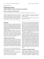
Fig. 5. Electrostatic surface potentials of selected Na-ScTxs. (A) Ribbon diagram of Cn12 (blue) superimposed with Bjxtr-IT (red), Lqh-aIT (green)
and BmK M1 (orange), taken from PDB: 1PE4, 1BCG, 1LQH and 1SN1, respectively. This figure was generated after overlapping the solved 3D
structures according to the positions occupied by similar secondary-structure elements, as indicated in Fig. 2. The orientation chosen correspondsto
the original face B described in [66]. (B) Electrostatic surface potential of the same toxins calculated with the
MOLMOL
program [71], using fully
charged residues, shown at 1.4 A
˚
van der Waals radius. Visible charged residues for each toxin are indicated by the one-letter code, in which red
means negatively charged, blue positively charged. Neutral or hydrophobic residues are in white, but not individually marked. Axes in the middle of
the figures represent the selected orientation.
Ó FEBS 2004 NMR solution structure of Cn12 (Eur. J. Biochem. 271) 2511
process. Whenever the channel changes, the toxin also
changes in order to most efficiently fit its binding site
[64,65]. However, as shown in Fig. 5A, the Cn12 scaffold
is similar to the others, and yet it is a weak modifier of
Na
+
channel function.
Figure 5B compares the distribution of the charge of
Cn12 with three toxins in which site-directed mutations
were performed, and the corresponding function studied
[18,56,57,63]. It is clear that the a-like and a anti-insect
toxins (BmK M1 and Lqh-aIT, respectively) show a quite
similar overall charge distribution, when analyzed in the
orientation originally described as face B [66]. Although
Cn12 shows an a scorpion toxin effect, it does not have the
same charge distribution, suggesting that other faces of the
3D structure of the toxins contribute to this effect. Similarly,
the insect-excitatory toxin Bjxtr-IT, defined as b scorpion
toxin, has a different charge distribution, as would be
expected. The results of site-directed mutagenesis show that
face A does not seem to be important for channel
recognition. Rather, it probably has a role in maintaining
the correct 3D folding of the molecules [55]. For example,
the residues shown to be important for the function of the
a toxins Lqh-aIT [18] and BmK M1 [55,56] are: K8, Y10,
F17, R18, W38, N44, R58, V59 and K8, W38, Y42, K62,
H64, respectively. For these two toxins, the residues in
question are mostly situated in face B, as shown in Fig. 5B.
However, this seems not to be case for the b toxin Bjxtr-IT
Fig. 6. Electrostatic surface potentials for face C of selected Na-ScTxs. Same electrostatic surfaces for toxins as in Fig. 5B, rotated from face B by
87.5 ° in the z-axis direction, 62.5 ° in the y-axis direction, and 10.0 ° in the x-axis direction, following exactly this order of rotation. This is one of
the orientations in which a pronounced difference was found. Toxin structures are labelled as in Fig. 5.
Table 1. Experimental constraints and structural statistics.
(A) Distance constraints
Intraresidue 607
Sequential 121
Medium-range 30
Long-range 92
Total 850
(B) Angle constraints 36 (u)
(C) Cartesian coordinate rmsd (A
˚
) in backbone atoms
All 1.994
Backbone
All 1.097
Residues 11–52 0.968
Helix(24–32) 0.259
b-strand(2–4) 0.421
b-strand(37–40) 0.261
b-strand(45–48) 0.173
b-sheet 0.532
b-strand and helix 0.612
(D) Energy (kcalÆmol
)1
) calculated from CNS (19 structures)
Total 97.9 (+15.6)
Bonds 5.0 (+1.1)
Angles 28.7 (+4.3)
van der Waals 35.8 (+5.5)
NOE 21.4 (+5.8)
Dihedral 1.1 (+0.5)
Impropers 6.0 (+1.5)
2512 F. del Rı
´
o-Portilla et al.(Eur. J. Biochem. 271) Ó FEBS 2004
[63], in which the important residues, although clustered in
two patches (E15, V19, N20, I22, A23, P24, H25, Y26, E30,
V34, V71, Q72, I73 and I74), are situated in different
orientations of the molecule.
On further analysis of the structures available, another
face was found, defined here as face C, which is quite
distinct with respect to charge distribution of the toxins
under analysis. Face C was obtained by rotating the four
superimposed models through 87.5 ° in the z-axis direction,
62.5 ° in the y-axis direction, and 10.0 ° in the x-axis
direction, as shown in Fig. 6. Other toxins for which the 3D
structures are known were similarly analyzed: BmK M2,
BmK M4, BmK M8, AaH II, CsE-V (a toxins), and Cn2,
CsE-v5 and Ts1 (b toxins). Using the orientation of face C
for these toxins, a distinct charge distribution is observed.
The toxins are like a Christmas tree, decorated in different
forms to interact most efficiently with their specific targets.
Comparative analysis of charge distribution in
Na
+
channels
The receptor site 3 of Na
+
channels is surmised to be
formed mainly of extracytoplasmic loops of the S3–S4
segment of domain IV and, to a lesser extent, of the segment
S5–S6 of domain I [14]. Contact with a Na-ScTx is mediated
by electrostatic and hydrophobic interactions. Nevertheless,
asshowninFig.7,Na
+
-channel isoforms constitute a
highly homogeneous family in terms of primary structure,
particularly in the S3–S4 segment of domain IV. The
variations found among the different isoforms are reduced
to point mutations, making it difficult to explain their
distinct susceptibilities to various a Na-ScTxs. For example,
it is well documented that some a Na-ScTxs display
remarkable species specificity (e.g. Na-ScTx Lqh-II binds
to rat brain synaptosomes with a 100-fold higher affinity
than to cockroach preparations; conversely Lqh-aIT binds
to neuronal preparations from cockroach with a 10 000-
fold higher affinity than to rat brain synaptosomes [51]). In
addition, some toxins are capable of discriminating between
Na
+
-channel isoforms of the same organism (e.g. the rat
brain isoform rNa
V
1.1 is 10-fold more sensitive to the action
of the a Na-ScTx Lqq5 than the cardiac isoform rNa
V
1.5
[67]). However, when the overall charge of both the toxins
and the channels are considered together, a plausible
explanation comes from analysis of the other region
involved in receptor site 3, segment S5–S6 of domain I. In
general, there is greater variability in this segment. Highly
sensitive Na
+
channels have more acidic residues than
insensitive isoforms (compare rNa
V
1.4 and rNa
V
1.5 in
Fig. 7), which would also support the observed preferred
interaction of the former with anti-mammal-specific basic
a Na-ScTxs [68–70]. In striking contrast, insect-specific a
Na-ScTxs usually present an overall neutral or, often, a
negative net charge at physiological pH. It has been
suggested that the presence of sialic acid in the S5–S6
segment from domain I of mammalian channels may
influence the binding of nonpositively charged Na-ScTxs,
disfavouring their binding because of the absence of primary
electrostatic attraction [70]. Cn12 has a slightly negative
charge at physiological pH, and the neuroblastoma cells
Fig. 7. Multiple alignment of amino-acid sequences corresponding to the receptor site 3 of Na
+
channels. A total of 50 nonredundant sequences of
Na
+
channels available in databases were aligned with
CLUSTAL X
[45]. The segments S5–S6 of domain I and S3–S4 of domain IV from selected
sequences are shown (Na
V
1.1, 1.4 and 1.5 from rat; Na
V
1.7 from human; Para from fruit fly; NachB1 from squid). Conserved residues are indicated
by asterisks, and conservative replacements by dots. Acidic amino acids in the extracytoplasmic loops are shown in bold. Critical binding residues
determined by mutagenesis are highlighted by empty circles [67,70].
Ó FEBS 2004 NMR solution structure of Cn12 (Eur. J. Biochem. 271) 2513
used in Fig. 3 (mammalian tissue) have a high content of
sialic acid. These two facts may explain the lower affinity
found for this toxin. In contrast with the charge-interacting
residues, the putative hydrophobic interactions are much
more difficult to estimate as there is less difference in the
relative abundance and no periodicity of distribution of the
hydrophobic residues.
The small number of site-directed mutants prepared with
scorpion toxins [7,18,55,56,63,68] and the study with the
Na
+
channels are insufficient to confirm or refute the
current views. This analysis indicates that different amino
acids in distinct positions of Na-ScTxs are capable of
defining their function. Similarly, the sites on the Na
+
channels known to be responsible for the binding of
Na-ScTxs are different: a vs. b effect (for those that modify
the gating mechanism) and Cn11 (a blocking toxin). We
expect that, when more data become available, the concepts
on the interaction of Na-ScTxs and Na
+
channels will be
better understood or even modified. In this communication,
we show the 3D structure of a novel scorpion toxin. We
conclude that the actual interacting surfaces depend on the
whole toxin molecule, with an important role for charge
distribution. As important must be the type or subtype of
Na
+
channel with which the toxin interacts. More work is
necessary.
Acknowledgements
This work was partially supported by grants from the Mexican Council
of Science and Technology (CONACyT) Z-005 and 40251-Q to L.D.P.
and grant number 32000N and 38616E to F.R.P. Grant IN206003
from Direccion General de Asuntos del Personal Academico (DGAPA)
of the National Autonomous University of Mexico to L.D.P. and the
scholarships given to E.H.M., G.P. and R.C.R.V. by the CONACyT
are also gratefully acknowledged.
References
1. Fontecilla-Camps, J.C., Almassy, R.J., Suddath, F.L., Watt, D.D.
& Bugg, C.E. (1980) The three-dimensional structure of a protein
from scorpion venom: a new structural class of neurotoxins. Proc.
NatlAcad.Sci.USA77, 6496–6500.
2. Tamaoki,H.,Miura,R.,Kusunoki,M.,Kyogoku,Y.,Kobaya-
shi, Y. & Moroder, L. (1998) Folding motifs induced and stabi-
lized by distinct cystine frameworks. Protein Eng. 11, 648–659.
3. Froy, O. & Gurevitz, M. (1998) Membrane potential modulators:
a thread of scarlet from plants to humans. FASEB J. 12, 1793–
1796.
4. Possani,L.D.,Merino,E.,Corona,M.,Bolivar,F.&Becerril,B.
(2000) Peptides and genes coding for scorpion toxins that affect
ion-channels. Biochimie 82, 861–868.
5. Possani, L.D., Merino, E., Corona, M. & Becerril, B. (2002)
Scorpion genes and peptides specific for potassium channels:
structure, function and evolution. In Perspectives in Molecular
Toxicology (Me
´
nez, A., ed.), pp. 201–214. John Wiley & Sons Ltd,
New York.
6. Caldero
´
n-Aranda, E.S., Dehesa-Da
´
vila, M., Cha
´
vez-Haro,A.&
Possani, L.D. (1996) Scorpion stings and their treatment in
Mexico. In Envenomings and Their Treatments (Bon, C. & Goyf-
fon, M., eds), pp. 311–320. Editions Fondation Marcel Me
´
rieux,
Lyon.
7. Gordon, D., Savarin, P., Gurevitz, M. & Zinn-Justin, S. (1998)
Functional anatomy of scorpion toxins affecting sodium channels.
J. Toxicol. Toxin Rev. 17, 131–159.
8. Possani, L.D., Becerril, B., Delepierre, M. & Tytgat, J. (1999)
Scorpion toxins specific for Na
+
-channels. Eur. J. Biochem. 264,
287–300.
9. Miller, C. (1995) The charybdotoxin family of K
+
-channel
blocking peptides. Neuron 15, 5–10.
10. Garcia,M.,Hanner,M.,Knaus,H.G.,Koch,R.,Schmalhofer,
W., Slaughter, R.S. & Kaczorowski, G.J. (1997) Pharmacology of
potassium channels. Adv. Pharmacol. 39, 425–471.
11. Valdivia, H. & Possani, L.D. (1998) Peptide toxins as probes of
Ryanodine receptor. Trends Cardiovasc. Med. 8, 111–118.
12. Possani,L.D.,Selisko,B.&Gurrola,G.(1999)Structureand
function of scorpion toxins affecting K
+
-channels. Perspect. Drug
Discov. 15/16, 15–40.
13. Tytgat, J., Chandy, K.G., Garcia, L.M., Gutman, G.A., Martin-
Eauclaire, M.F., van del Walt, J.J. & Possani, L.D. (1999) A
unified nomenclature for short chain peptides isolated from
scorpion venoms: alpha-KTx molecular subfamilies. Trends
Pharmacol. Sci. 20, 445–447.
14. Cestele, S. & Catterall, W.A. (2000) Molecular mechanisms of
neurotoxin action on voltage-gated sodium channel. Biochimie 82,
883–892.
15. Teneholz, T.C., Klenk, K.C., Matteson, D.R., Blaustein, M.P. &
Weber, D.J. (2000) Structural determinants of scorpion toxin
affinity: the charybdotoxin alpha-KTX family of K
+
-channel
blocking peptide. Rev. Physiol. Biochem. Pharmacol. 140, 135–185.
16. Garcia,M.,Gao,Y.,McManus,O.B.&Kaczorowski,G.J.(2001)
Potassium channels: from scorpion venoms to high-resolution
structure. Toxicon 39, 739–748.
17. Gurevitz,M.,Gordon,D.,Ben-Natan,S.,Turkov,M.&Froy,O.
(2001) Diversification of neurotoxin by C-terminal ÔwigglingÕ:a
scorpion recipe fopr survival. FASEB J. 15, 1201–1205.
18. Gordon, D. & Gurevitz, M. (2003) The selectivity of scorpion
alpha-toxins for sodium channel subtypes is determined by subtle
variations of the interacting surface. Toxicon 41, 125–128.
19. Rodrı
´
guez de la Vega, R.C., Merino, E., Becerril, B. & Possani,
L.D. (2003) Novel interactions between K
+
channels and scor-
pion toxins. Trends Pharmacol. Sci. 24, 222–227.
20. Corona, M., Gurrola, G.B., Merino, E., Restano-Cassulini, R.,
Valdez-Cruz, N.A., Garcı
´
a, B., Ramı
´
rez-Domı
´
nguez, M.E.,
Coronas,F.I.V.,Zamudio,F.Z.,Wanke,E.&Possani,L.D.
(2002) A large number of novel Ergtoxin-like genes and ERG
K
+
-channels blocking peptides from scorpions of the genus
Centruroides. FEBS Lett. 532, 121–126.
21. Guan, R., Wang, C.G., Wang, M. & Wang, D.C. (2001) A
depressant insect toxin with a novel analgesic effect from scorpion
Buthus martensii Karsch. Biochim. Biophys. Acta 1549, 9–18.
22. Corona, M., Coronas, F.I.V., Merino, E., Becerril, B., Gutie
´
rrez,
R., Rebolledo-Antunez, S., Garcia, D.E. & Possani, L.D. (2003) A
novel class of peptide found in scorpion venom with neuro-
depressant effects in peripheral and central nervous system of the
rat. Biochim. Biophys. Acta 1649, 58–67.
23.Chandy,K.G.,Cahalan,M.,Pennington,M.,Norton,R.S.,
Wulfh, H. & Gutman, G.A. (2000) Potassium channels in T
lymphocytes: toxins to therapeutic immunosuppressants. Toxicon
39, 1269–1276.
24. Beeton, C., Barbaria, J., Giraud, P., Devaux, J., Benoliel, A.M.,
Gola,M.,Sabatier,J.M.,Bernard,D.,Crest,M.&Beraud,E.
(2001) Selective blocking of voltage-gated K
+
channels improves
experimental autoimmune encephalomyelitis and inhibits T cell
activation. J. Immunol. 166, 936–944.
25.Beeton,C.,Wulff,H.,Barbaria,J.,Clot-Faybesse,O.,
Pennington,M.,Bernard,D.,Cahalan,M.D.,Chandy,K.G.&
Beraud, E. (2001) Selective blockade of T lymphocyte K
+
chan-
nels ameliorates experimental autoimmune encephalomyelitis: a
model for multiple sclerosis. Proc. Natl Acad. Sci. USA 98, 13942–
13947.
2514 F. del Rı
´
o-Portilla et al.(Eur. J. Biochem. 271) Ó FEBS 2004
26. Torres-Larios,A.,Gurrola,G.B.,Zamudio,F.Z.&Possani,L.D.
(2000) Hadrurin, a new antimicrobial peptide from the venom of
the scorpion Hadrurus aztecus. Eur. J. Biochem. 267, 5023–5031.
27. Corzo, G., Escoubas, P., Villegas, E., Barnham, K.J., He, W.,
Norton, R.S. & Nakajima, T. (2001) Characterization of unique
amphipathic antimicrobial peptides from venom of the scorpion
Pandinus imperator. Biochem. J. 359, 35–45.
28. Moerman,L.,Bosteels,S.,Noppe,W.,Willems,J.,Clynen,E.,
Schoofs, L., Thevissen, K., Tytgat, J., van Eldere, J., van der Walt,
J. & Verdonck, F. (2002) Antibacterial and antifungal properties
of alpha-helical, cationic peptides in the venom of scorpions from
southern Africa. Eur. J. Biochem. 269, 4799–4810.
29.Conde,R.,Zamudio,F.Z.,Rodrı
´
guez, M.H. & Possani, L.D.
(2000) Scorpine, an anti-malaria and anti-bacterial agent purified
from scorpion venom. FEBS Lett. 471, 165–168.
30. DeBin, J.A., Maggio, J.E. & Strichartz, G.R. (1993) Purification
and characterization of chlorotoxin, a chloride channel ligand
from the venom of the scorpion. Am.J.Physiol.264, C361–C369.
31. Chuang, R.S., Jaffe, H., Cribbs, L., Perez-Reyes, E. & Swartz, K.J.
(1998) Inhibition of T-type voltage-gated calcium channels by a
new scorpion toxin. Nat. Neurosci. 1, 668–674.
32. Olamendi-Portugal, T., Garcı
´
a, B.I., Lo
´
pez-Gonza
´
lez, I., van der
Walt,J.,Dyason,K.,Ulens,C.,Tytgat,J.,Felix,R.,Darszon,A.
& Possani, L.D. (2002) Two new scorpion toxins that target vol-
tage-gated Ca
2+
and Na
+
channels. Biochem. Biophys. Res.
Commun. 299, 562–568.
33. Possani, L.D. (1984) Structure of scorpion toxins. In Handbook of
Natural Toxins, Vol. 2 (Tu, A.T., ed.), pp. 513–550. Marcel
Dekker Inc., New York.
34. Ramı
´
rez-Domı
´
nguez, M.E., Olamendi-Portugal, T., Garcı
´
a, U.,
Garcı
´
a, C., Are
´
chiga, H. & Possani, L.D. (2002) Cn11, first
example of scorpion toxin that is a true channel blocker of Na
+
currents on crayfish neurons. J. Exp. Biol. 205, 869–876.
35. Platika, D., Boulos, M.H., Baizer, L. & Fishman, M.C. (1985)
Neuronal traits of clonal cell lines derived by fusion of dorsal root
ganglia neurons with neuroblastoma cells. Proc. Natl Acad. Sci.
USA 82, 3499–3503.
36. Faravelli, L., Arcangeli, A., Olivotto, M. & Wanke, E.A. (1996)
HERG-like channel in rat F-11 DRG cell line: pharmacological
identification and biophysical characterisation. J. Physiol. (Lond.)
496, 13–23.
37. Delaglio,F.,Grzesiek,S.,Vuister,G.W.,Zhu,G.,Pfeifer,J.&
Bax, A. (1995) NMRPipe: a multidimensional spectral processing
system based on UNIX pipes. J. Biomol. NMR 6, 277–293.
38. Bartels,C.,Xia,T.,Billeter,M.,Gu
¨
ntert, P. & Wu
¨
thrich, K.
(1995) The program XEASY for computer-supported NMR
spectral analysis of biological macromolecules. J. Biomol. NMR 5,
1–10.
39. del Rı
´
o-Portilla, F., Blechta, V. & Freeman, R. (1994) Measure-
ment of poorly resolved splittings by J doubling in the frequency
domain. J. Magn. Reson. 111, 132–135.
40. Garza-Garcı
´
a, A., Ponzanelli-Velazquez, G. & del Rı
´
o-Portilla, F.
(2000) Deconvolution and measurement of spin-spin splittings by
modified J doubling in the frequency domain. J. Magn. Reson.
148, 214–219.
41. Wishart, D.S., Wang, Y. & Nip, A.M. (1997) A simple method to
quantitatively measure polypeptide JHNHa1 coupling constants
from TOCSY or NOESY spectra. J. Biomol. NMR 10, 373–382.
42. Gu
¨
ntert, P., Mumenthaler, C. & Wu
¨
thrich, K. (1997) Torsion
angle dynamics for NMR structure calculation with the new
program DYANA. J. Mol. Biol. 273, 283–298.
43. Wu
¨
thrich, K. (1986) NMR of Proteins and Nucleic Acids. John
Wiley and Sons, New York.
44. Bru
¨
nger,A.T.,Adams,P.T.,Clore,G.M.,DeLano,W.L.,Gros,
P., Grosse-Kunstleve, R.W., Jiang, J.S., Kuszewski, J., Nilges, M.,
Pannu,N.S.,Read,R.J.,Rice,L.M.,Simonson,T.&Warren,
G.L. (1998) Crystallography & NMR System: a new software
suite for macromolecular structure determination. Acta Crystal-
logr. D 54, 905–921.
45. Thompson, J.D., Gibson, T.J., Plewniak, F., Jeanmougin, F. &
Higgins, D.G. (1997) The CLUSTAL_X windows interface: flex-
ible strategies for multiple sequence alignment aided by quality
analysis tools. Nucleic Acids Res. 25, 4876–4882.
46.Inceoglu,B.,Lango,J.,Wu,J.,Hawkins,P.,Southern,J.&
Hammock, B.D. (2001) Isolation and characterization of a novel
type of neurotoxic peptide from the venom of the South African
scorpion Parabuthus transvaalicus (Buthidae). Eur. J. Biochem.
268, 5407–5413.
47. Froy, O. & Gurevitz, M. (2003) New insight on scorpion diver-
gence inferred from comparative analysis of toxin structure,
pharmacology and distribution. Toxicon 42, 549–555.
48. Jover, E., Courad, F. & Rochat, H. (1980) Two types of scorpion
neurotoxins characterized by their binding to two separate
receptor sites on rat brain synaptosomes. Biochem. Biophys. Res.
Commun. 95, 1607–1614.
49. Wheeler, K.P., Watt, D.D. & Lazdunski, M. (1983) Classification
of Na
+
channel receptors specific for various scorpion toxins.
Pflu
¨
gers Arch. Eur. J. Physiol. 397, 164–165.
50. Couraud, F. & Jover, E. (1984) Mechanism of action of scorpion
toxins. In Handbook of Natural Toxins,Vol.2(Tu,A.T.,ed.),pp.
629–678. Marcel Dekker Inc., New York.
51. Gordon,D.,Martin-Eauclaire,M.F.,Cestele,S.,Kopeyan,C.,
Calier,E.,BenKhalifa,R.,Pelhate,M.&Rochat,H.(1996)
Scorpion toxins affecting sodium current inactivation bind two
distinct homologous receptor sites on brain and insect sodium
channels. J. Biol. Chem. 271, 8034–8045.
52. Jiang,Y.,Lee,A.,Chen,J.,Ruta,V.,Cadene,M.,Chait,B.T.&
MacKinnon, R. (2003) X-ray structure of a voltage-dependent
K
+
channel. Nature (London) 423, 33–41.
53. Habersetzer-Rochat, C. & Sampieri, F. (1976) Structure-function
relationships of scorpion neurotoxins. Biochemistry 152,
2254–2261.
54. Possani, L.D., Martin, B.M., Svendsen, I., Rode, G.S. & Erickson,
B.W. (1985) Scorpion toxins from Centruroides noxius and Tityus
serrulatus. Biochem. J. 229, 739–750.
55. Sun,Y.M.,Bosmans,F.,Zhu,R.H.,Goudet,C.,Xiong,Y.M.,
Tytgat, J. & Wang, D.C. (2003) Importance of the conserved
aromatic residues in the scorpion alpha-like toxin BmK M1, the
hydrophobic surface region revisited. J. Biol. Chem. 278, 24125–
24131.
56. Wang, C.G., Gilles, N., Hamon, A., Le Gall, F., Stankiewicz, M.,
Pelhate, M., Xiong, Y.M., Wang, D.C. & Chi, C.W. (2003)
Exploration of the functional site of a scorpion alpha-like toxin
by site-directed mutagenesis. Biochemistry 42, 4699–4708.
57. Selisko, B., Garcı
´
a, C., Becerril, B., Delepierre, M. & Possani,
L.D. (1996) An insect-specific toxin from Centruroides noxius
Hoffmann. cDNA, primary structure, three-dimensional model
and electrostatic surface potentials in comparison with other toxin
variants. Eur. J. Biochem. 242, 235–242.
58. Garcı
´
a, C., Becerril, B., Selisko, B., Delepierre, M. & Possani,
L.D. (1997) Isolation, characterization and comparison of a novel
crustacean toxin with a mammalian toxin from the venom of the
scorpion Centruroides noxius Hoffmann. Comp. Biochem. Physiol.
B 116, 315–322.
59. Selisko, B., Licea, A.F., Becerril, B., Zamudio, F., Possani, L.D. &
Horjales, E. (1999) Antibody BCF2 against scorpion toxin Cn2
from Centuroides noxius Hoffmann: primary structure and three-
dimensional model as free Fv fragment and complexed with its
antigen. Proteins 37, 130–143.
60. Caldero
´
n-Aranda, E.S., Selisko, B., York, E.J., Gurrola, G.B.,
Stewart, J.M. & Possani, L.D. (1999) Mapping of an epitope
recognized by a neutralizing monoclonal antibody specific to toxin
Ó FEBS 2004 NMR solution structure of Cn12 (Eur. J. Biochem. 271) 2515
Cn2 from the scorpion Centruroides noxius, using discontinuous
synthetic peptides. Eur. J. Biochem. 264, 746–755.
61.Gazarian,T.G.,Selisko,B.,Gurrola,G.B.,Herna
´
ndez, R.,
Possani, L.D. & Gazarian, K.G. (2003) Potential of peptides
selected from random phage-displayed libraries to mimic
conformational epitopes: a study on scorpion toxin Cn2 and the
neutralizing monoclonal antibody BCF2. Comb. Chem. High
Throughput Screen. 6, 119–132.
62. Polikarpov, I., Sanches, M., Maragoni, S., Toyama, M.H. &
Teplyakov, A. (1999) Crystal structure of neurotoxin Ts1 from
Tityus serrulatus provides insights into the specificity and toxicity
of scorpion toxins. J. Mol. Biol. 290, 175–184.
63. Cohen,L.,Karbat,I.,Gilles,N.,Froy,O.,Corzo,G.,Angelovici,
R., Gordon, D. & Gurevitz, M. (2004) Dissection of the functional
surface of an anti-insect excitatory toxin illuminates a putative hot
spot common to all scorpion beta-toxins affecting Na+ channels.
J. Biol. Chem. 279, 8206–8211.
64.Froy,O.,Sagiv,T.,Poreh,M.,Urbach,D.,Zilberberg,N.&
Gurevitz, M. (1999) Dynamic diversification from a putative
common ancestor of scorpion toxin affecting potassium, sodium
and chloride channels. J. Mol. Evol. 48, 187–196.
65. Zhu, S., Bosmans, F. & Tytgat, J. (2004) Adaptive evolution of
scorpion sodium channel toxins. J. Mol. Evol. 58, 145–153.
66. Fontecilla-Camps, J.C., Almassy, R.J., Ealick, S.E., Suddath,
F.L., Watt, D.D., Feldmann, R.J. & Bugg, C.E. (1981) Archi-
tecture of scorpion neurotoxins: a class of membrane-binding
proteins. Trends Biochem. Sci. 6, 291–296.
67. Rogers, J.C., Qu, Y., Tanada, T.N., Scheuer, T. & Catterall, W.A.
(1996) Molecular determinants of high affinity binding of alpha-
scorpion toxin and sea anemone toxin in the S3–S4 extracellular
loop in domain IV of the Na
+
channel alpha subunit. J. Biol.
Chem. 271, 15950–15962.
68. Zilberberg,N.,Froy,O.,Loret,E.,Cestele,S.,Arad,D.,Gordon,
D. & Gurevitz, M. (1997) Identification of structural elements of a
scorpion a-neurotoxin important for receptor site recognition.
J. Biol. Chem. 272, 14810–14816.
69. Ceste
`
le,S.,Stankiewicz,M.,Mansuelle,P.,DeWaard,M.D.,
Dargent,B.,Gilles,N.,Pelhate,M.,Rochat,H.,Martin-Eaucl-
aire, M.F. & Gordon, D. (1999) Scorpion alpha-like toxins toxic
to both mammals and insects differentially interact with receptor
site 3 on voltage-gated sodium channels in mammals and insects.
Eur. J. Neurosci. 11, 975–985.
70. Leipold, E., Lu, S., Gordon, D., Hansel, A. & Heinemann, S.H.
(2004) Combinatorial interaction of scorpion toxins Lqh-2, Lqh-3,
and LqhaIT with sodium channel receptor sites-3. Mol. Pharma-
col. 65, 685–691.
71. Koradi, R., Billeter, M. & Wu
¨
thrich, K. (1996) MOLMOL: a
program for display and analysis of macromolecular structures.
J. Mol. Graphics 14, 51–55.
2516 F. del Rı
´
o-Portilla et al.(Eur. J. Biochem. 271) Ó FEBS 2004