Báo cáo khoa học: Defining the QP-site of Escherichia coli fumarate reductase by site-directed mutagenesis, fluorescence quench titrations and EPR spectroscopy doc
Bạn đang xem bản rút gọn của tài liệu. Xem và tải ngay bản đầy đủ của tài liệu tại đây (395.79 KB, 14 trang )
Defining the Q
P
-site of Escherichia coli fumarate reductase
by site-directed mutagenesis, fluorescence quench
titrations and EPR spectroscopy
Richard A. Rothery
1
, Andrea M. Seime
1
, A M. Caroline Spiers
1
, Elena Maklashina
2,3
,
Imke Schro
¨
der
4
, Robert P. Gunsalus
4
, Gary Cecchini
2,3
and Joel H. Weiner
1
1 CIHR Membrane Protein Research Group, Department of Biochemistry, University of Alberta, Edmonton, Alberta, Canada
2 Molecular Biology Division, Veterans Affairs Medical Center, San Francisco, CA, USA
3 Department of Biochemistry and Biophysics, University of California, San Francisco, CA, USA
4 Department of Microbiology, Immunology and Molecular Genetics, University of California, Los Angeles, CA, USA
Escherichia coli, when grown anaerobically with fuma-
rate as the respiratory oxidant, develops a respiratory
chain terminated by a membrane-bound menaqui-
nol:fumarate oxidoreductase (FrdABCD
1
) [1,2]. The
enzyme comprises a catalytic dimer of the FrdA
(65.8 kDa) and FrdB (27 kDa) subunits that is
anchored to the inner surface of the cytoplasmic mem-
brane by two small hydrophobic membrane-anchor
Keywords
fumate reductase; Q-site; iron-sulfur;
menaquinol
Correspondence
R. A. Rothery, Department of Biochemistry,
474 Medical Sciences Building, University of
Alberta, Edmonton, Alberta T6G 2H7
Fax: +1 780 492 0886
Tel: +1 780 492 2229
E-mail:
(Received 13 September 2004, revised 22
October 2004, accepted 1 November 2004)
doi:10.1111/j.1742-4658.2004.4469.x
We have used fluorescence quench titrations, EPR spectroscopy and
steady-state kinetics to study the effects of site-directed mutants of FrdB,
FrdC and FrdD on the proximal menaquinol (MQH
2
) binding site (Q
P
)of
Escherichia coli fumarate reductase (FrdABCD) in cytoplasmic membrane
preparations. Fluorescence quench (FQ) titrations with the fluorophore
and MQH
2
analog 2-n-heptyl-4-hydroxyquinoline-N-oxide (HOQNO) indi-
cate that the Q
P
site is defined by residues from FrdB, FrdC and FrdD. In
FQ titrations, wild-type FrdABCD binds HOQNO with an apparent K
d
of
2.5 nm, and the following mutations significantly increase this value: FrdB-
T205H (K
d
¼ 39 nm); FrdB-V207C (K
d
¼ 20 nm); FrdC-E29L (K
d
¼
25 nm); FrdC-W86R (no detectable binding); and FrdD-H80K (K
d
¼
20 nm). In all titrations performed, data were fitted to a monophasic bind-
ing equation, indicating that no additional high-affinity HOQNO binding
sites exist in FrdABCD. In all cases where HOQNO binding is detectable
by FQ titration, it can also be observed by EPR spectroscopy. Steady-state
kinetic studies of fumarate-dependent quinol oxidation indicate that there
is a correlation between effects on HOQNO binding and effects on the
observed K
m
and k
cat
values, except in the FrdC-E29L mutant, in which
HOQNO binding is observed, but no enzyme turnover is detected. In this
case, EPR studies indicate that the lack of activity arises because the
enzyme can only remove one electron from reduced MQH
2
, resulting in it
being trapped in a form with a bound menasemiquinone radical anion.
Overall, the data support a model for FrdABCD in which there is a single
redox-active and dissociable Q-site.
Abbreviations
DmsABC, E. coli dimethylsulfoxide reductase; FQ, fluorescence quench; FrdABCD, E. coli fumarate reductase; FrdCAB, Wolinella
succinogenes fumarate reductase; HOQNO, 2-n-heptyl-4-hydroxyquinoline-N-oxide; LPC, oxidized lapachol [2-hydroxy-3-(3-methyl-2-butenyl)-
1,4-naphthoquinone]; LPCH
2
, reduced lapachol; MQ, menaquinone; MQH
2
, menaquinol; NarGHI, nitrate reductase A; SdhCAB, Bacillus
subtilis succinate dehydrogenase; SdhCDAB, E. coli and ⁄ or eukaryotic succinate dehydrogenase.
FEBS Journal 272 (2005) 313–326 ª 2004 FEBS 313
subunits, FrdC (15 kDa) and FrdD (13.1 kDa). The
crystal structure of FrdABCD has been reported at
3.3 A
˚
resolution [3,4], and has an overall architecture
similar to that of the E. coli complex II homolog
SdhCDAB (succinate:ubiquinone oxidoreductase) [5,6].
Each enzyme contains a single FAD that is covalently
bound to the catalytic subunit (FrdA ⁄ SdhA) and three
[Fe-S] clusters (a [2Fe-2S] cluster, a [4Fe-4S] cluster,
and a [3Fe-4S] cluster) coordinated by the electron-
transfer subunit (FrdB ⁄ SdhB) [1]. However, important
differences exist between the membrane-intrinsic
domains of the two enzymes [1,7]. The membrane-
intrinsic domain of SdhCDAB coordinates a single
heme b (b
556
) that is sandwiched between the SdhC
and SdhD subunits [8,9]. Quinone binding and reduc-
tion is believed to take place in the region between the
heme and the [3Fe-4S] cluster of SdhB [1,6]. In the case
of FrdABCD, the membrane-intrinsic domain does not
contain heme, but instead contains two menaquinones
at discreet sites in the crystallized form of the enzyme
[3,4]. In both enzymes, despite the available structures,
the number of functional quinone ⁄ quinol binding sites
has yet to be unequivocally determined.
The menaquinones identified in the crystal structure
of FrdABCD [3] are located at sites towards the inner
(cytoplasmic) and outer (periplasmic) sides of the mem-
brane-intrinsic domain of the enzyme (FrdCD). One
site, the Q
P
site (the proximal Q-site), is located in the
interface region between the FrdCD subunits and
the [3Fe-4S] cluster coordinating region of FrdB on the
cytoplasmic side of the membrane. The other site, the
Q
D
site (the distal Q-site) is located approximately 25 A
˚
from the Q
P
site on the opposite (periplasmic) side of
the membrane [3,10]. The relatively large distance
between the two sites may preclude direct electron-trans-
fer through the protein medium, which is believed to be
limited to a distance of approximately 14 A
˚
[11]. How-
ever, a third region of electron density has been identi-
fied recently between the Q
P
and Q
D
sites (the ‘M’ site),
and is centered approximately 13 A
˚
from each Q-site [4].
If this electron density corresponds to an additional
electron-transferring cofactor, it could provide a conduit
for electron-transfer from the Q
D
site to the Q
P
site.
However, analyses of the bioenergetics of respiratory
growth of E. coli on fumarate indicate that FrdABCD
turnover does not produce a transmembrane electro-
chemical potential [12], suggesting the presence of a sin-
gle dissociable and redox-active Q-site that is formally
located on the cytoplasmic side of the membrane.
Menaquinol (MQH
2
) oxidation by FrdABCD has
been studied using a combination of site-directed muta-
genesis, enzymology, EPR spectroscopy and X-ray crys-
tallography. Initial mutagenesis studies suggested that
there may be two Q-sites present – a polar Q
B
site
(equivalent to the Q
P
site), and an apolar Q
A
site (equiv-
alent to the Q
D
site) [13–15]. Investigation of the steady-
state kinetics of quinol-dependent fumarate reduction
by FrdABCD suggests that MQH
2
binding and oxida-
tion occur at a single site [16]. Kinetic studies carried
out in the presence of HOQNO or alkylated dinitro-
phenol derivatives also support the presence of a single
MQH
2
oxidation site [17]. By exploiting the fluorescent
properties of HOQNO in fluorescence quench (FQ)
titrations, we determined that this inhibitor binds at a
single high-affinity site within FrdABCD [18,19]. EPR
studies indicate that this high-affinity site is conforma-
tionally linked to the [3Fe-4S] cluster of FrdB [18]. The
emerging hypothesis that there is a single site for MQH
2
or HOQNO binding has been complicated recently by
the observation in crystallographic studies that the Q
D
site is unoccupied when HOQNO or a dinitrophenol
derivative is bound at the Q
P
site [4]. Given the available
structural information on FrdABCD, it would therefore
be of interest to examine the effects of a range of site-
directed mutants on the HOQNO binding properties
and enzymology of the enzyme.
In this paper, we evaluate the effects of mutation of
amino acid residues located in the vicinity of the Q
P
site on HOQNO binding to FrdABCD. We have deter-
mined the effect of each mutation on HOQNO binding
detected by FQ titration and EPR spectroscopy. We
have also investigated the effects the mutants have on
the steady-state kinetics of fumarate-dependent quinol
oxidation.
Results
Selection of mutants of FrdB, FrdC and FrdD
The following residues are located within approximately
5A
˚
of the menaquinone (MQ) observed at the Q
P
-site
in the structure of FrdABCD: T205, F206, Q225 and
K228 from FrdB; R28, E29, W86, L89 and A93 from
FrdC; and W14, F17, G18, H80, R81 and H84 from
FrdD [3,4,10]. Site-directed mutants of some of these res-
idues have been generated and partially characterized,
including the following: FrdC-E29L [14,20], FrdC-
W86R, FrdD-H80K and FrdD-H84K [14]. In the con-
text of this study, mutants of the following residues
located at a slightly greater distance from the Q
P
site are
also potentially of interest: FrdB-V207 (% 8A
˚
from Q
P
,
a FrdB-V207C mutant) [21], and FrdC-A32 (% 9A
˚
from Q
P
, a FrdC-A32V mutant) [14]. At an even greater
distance away from the Q
P
site is FrdC-F38 (% 18 A
˚
), a
mutation at this position (FrdC-F38M [14]), would be
expected to have little effect on MQH
2
binding and
Quinol binding to E. coli fumarate reductase R. A. Rothery et al.
314 FEBS Journal 272 (2005) 313–326 ª 2004 FEBS
oxidation. Finally, we generated a mutant of FrdB-T205
(FrdB-T205H) to assess the role of the [3Fe-4S] cluster
binding domain of FrdB in defining the Q
P
-site. This
residue is sandwiched between the [3Fe-4S] cluster and
the Q
P
site. All eight mutant enzymes were studied to
assess the effects of the mutations on MQH
2
binding
using FQ titrations, EPR spectroscopy and steady-state
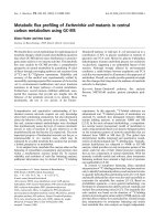
kinetic studies. The locations of all the mutated residues
located within % 10 A
˚
of the Q
P
site are illustrated in
Fig. 1. HOQNO has a very similar structure to that of
MQ, and as a result appears to bind to the Q
P
site in an
almost identical way (compare Fig. 1A and B with C
and D). This similarity in both structure and binding
renders HOQNO an excellent inhibitor with which to
characterize the Q
P
site of FrdABCD.
FQ titrations of HOQNO binding to mutant
FrdABCD
HOQNO is a close structural analog of MQH
2
⁄ MQ
and is a very potent inhibitor of FrdABCD [16,18].
When excited at 341 nm, free HOQNO in aqueous
solution fluoresces with an emission wavelength of
479 nm. Its fluorescence is completely quenched when
bound to FrdABCD and certain other E. coli respirat-
ory chain enzymes (including dimethylsulfoxide reduc-
tase and nitrate reductase A [18,19,22–24]). This
enables its binding to a Q-site to be analyzed by FQ
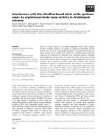
titration. Figure 2 shows representative titrations of
membranes containing the wild-type and mutant
enzymes studied herein. Data for all of the mutants is
presented in Table 1. DW35 membranes lacking
FrdABCD (Fig. 2A) do not exhibit high-affinity
HOQNO binding. The following FrdABCD mutants
bind HOQNO with K
d
values equivalent to that of the
wild-type enzyme (K
d
¼ 2.5 nm; Fig. 2B): FrdC-A32V
(2.5 nm; not shown), FrdC-F38M (2.5 nm; not shown)
and FrdD-H84K (3.0 nm, not shown). At the opposite
extreme, it is clear that the FrdC-W86R mutant does
not exhibit high-affinity HOQNO binding (Fig. 2E).
This mutant appears to have a similar phenotype to
that of the previously reported FrdC-H82R mutant
[18,25]. Intermediate effects are observed with the fol-
lowing mutants: FrdB-T205H (K
d
¼ 39 nm; Fig. 2C),
FrdB-V207C (20 nm; not shown), FrdC-E29L (25 nm;
Fig. 2D) and FrdD-H80K (20 nm; Fig. 2F). Based on
Fig. 1. Positions of the mutated residues
close to the Q
P
site studied herein. A and B
show views of the MQ-bound form of FrdA-
BCD (1L0V), whereas C and D show views
of the HOQNO-bound form (1KF6). A and C
represent views from an identical perspec-
tive, as do panels B and D (Experimental
procedures). (A) Looking along the axis defi-
ned by the two keto-oxygens of the prox-
imal menaquinone (MQ) naphthoquinone
bicycle. (B) Looking along the axis of the
MQ towards the isoprenoid chain. (C) The
same perspective as A, but with HOQNO
bound. (D) The same perspective as B, but
with HOQNO bound. In all panels, FrdB and
FrdA are above the MQ ⁄ HOQNO plane, and
FrdC and FrdD are substantially below the
MQ ⁄ HOQNO plane. Residues from FrdB,
FrdC and FrdD have labels starting with ‘B-’,
‘C-’, and ‘D-’, respectively.
R. A. Rothery et al. Quinol binding to E. coli fumarate reductase
FEBS Journal 272 (2005) 313–326 ª 2004 FEBS 315
these observations and the FrdABCD structure [3,4], it
is clear that residues from FrdB, FrdC and FrdD play
important roles in defining the Q
P
site. In every case
where binding is detected, the data can be fitted to an
equation (Eqn 1) describing noncooperative binding at
a single site within FrdABCD.
Table 1 shows the calculated specific concentration
of HOQNO binding sites for each mutant in which
binding is detected by FQ titration. It also shows the
concentration of FrdABCD calculated by EPR spin
quantitation of both the [2Fe-2S] and [3Fe-4S] clusters.
In each case, the estimated number of Q-sites per
enzyme is very close to unity, indicating that HOQNO
binding occurs at a single site within FrdABCD. Based
on enzymes that bind HOQNO, 1.02 ± 0.12 sites were
observed per [3Fe-4S] cluster and 1.05 ± 0.09 sites
were observed per [2Fe-2S] cluster.
Detection of HOQNO binding by EPR
spectroscopy
Figure 3 shows the effect of HOQNO on the EPR
spectrum around g ¼ 2.0 of ferricyanide-oxidized
HB101 membrane samples containing wild-type and
mutant FrdABCD. EPR spectra of membranes lacking
overexpressed FrdABCD exhibit low-intensity features
around g ¼ 2.0 upon which HOQNO has little effect
(Fig. 3A). Spectra of membranes containing over-
expressed wild-type FrdABCD exhibit the EPR spec-
trum of its oxidized [3Fe-4S] cluster (Fig. 3B). This
spectrum is nearly isotropic with a peak at g ¼ 2.02
(g
z
) and a broad trough immediately up-field. As has
been reported previously [18,20], addition of HOQNO
elicits the observation of an additional peak-trough at
approximately g ¼ 1.98 (g
xy
).
Both of the FrdB mutants studied herein (FrdB-
T205H and FrdB-V207C) have significant effects on
the EPR properties of FrdABCD. In the case of the
FrdB-T205H mutant, the [3Fe-4S] cluster line-shape is
narrower than that of the wild-type (note the position
of the trough in the spectrum without HOQNO;
Fig. 3C). As is the case for the wild-type enzyme, addi-
tion of HOQNO results in the resolution of a peak-
trough on the high-field side of the g ¼ 2.02 peak. This
peak-trough is centered at a g-value reflecting the
narrower spectrum of the [3Fe-4S] cluster in the
Fig. 2. Representative fluorescence quench titrations of HOQNO binding to wild-type and mutant FrdABCD in DW35 membranes. Titrations
were carried out using membranes from E. coli DW35 transformed with plasmids encoding wild-type and mutant FrdABCD at total mem-
brane protein concentrations of 0.2 (e), 0.3 (h), 0.4 (n), and 0.5 mgÆmL
)1
(s). Data were fitted to the following specific enzyme concentra-
tions (nmolÆ mg protein
)1
)andK
d
values (nM): (A) background, 0.36, > 500; (B) wild-type, 3.54, 2.5; (C) FrdB-T205H, 3.13, 39; (D) FrdC-E29L,
3.26, 25; (E) FrdC-W86R, negligible binding; (F) FrdD-H80K, 3.61, 20. Note that in the cases of the background and FrdC-W86R mutant
membranes, the data presented represent insignificant binding.
Quinol binding to E. coli fumarate reductase R. A. Rothery et al.
316 FEBS Journal 272 (2005) 313–326 ª 2004 FEBS
FrdB-T205H mutant in the absence of inhibitor
(g
xy
¼ 2.0 in the presence of inhibitor rather than at
1.98). Figure 3D shows the spectrum of oxidized mem-
branes containing overexpressed FrdB-V207C mutant
enzyme. In agreement with Manadori et al. [21], little
or no [3Fe-4S] cluster is assembled into this mutant
enzyme (compare Fig. 3A and D), and therefore
HOQNO binding cannot be detected by its perturba-
tion of the EPR spectrum of the oxidized enzyme (see
below).
In contrast to the results of Ha
¨
gerha
¨
ll et al. [20], the
EPR experiments reported herein indicate that HO-
QNO elicits an effect on the EPR line-shape of the
[3Fe-4S] cluster of the FrdC-E29L mutant enzyme
(Fig. 3E). This result is consistent with the observation
of HOQNO binding by FQ titration (Fig. 2D and
Table 1). For the other mutations located within the
membrane anchor subunits (FrdC and FrdD), there is
a strong correlation between the observation of an
HOQNO-induced line-shape change and the observa-
tion of inhibitor binding in FQ titrations (compare
Figs 2 and 3, Table 1). Thus, no EPR line-shape
change is elicited on the FrdC-W86R mutant [3Fe-4S]
cluster spectrum (Fig. 3G).
HOQNO binding to reduced wild-type and
FrdC-V207C mutant enzyme
The EPR properties of reduced wild-type FrdABCD
are complicated by spin–spin interactions between the
paramagnetic [Fe-S] clusters present (viz. between the
S ¼ ½ [2Fe-2S] and [4Fe-4S] clusters and the S ¼ 2
reduced [3Fe-4S] cluster) [26]. The clusters have mid-
point potentials (E
m
values) of % )79 mV ([2Fe-2S]
c1uster [27]), )320 mV ([4Fe-4S] c1uster [26]), and
)70 mV ([3Fe-4S] c1uster [18,21,26]). Because of the
pairing of the [3Fe-4S] cluster with the [4Fe-4S] cluster
in a 7Fe ferredoxin-type motif, we examined the possi-
bility that HOQNO binding to the Q
P
site may affect
the EPR properties of the fully reduced enzyme.
Figure 4A shows that HOQNO has no effect on the
spectrum of dithionite-reduced HB101 membranes
lacking overexpressed FrdABCD. No differences are
observed between the spectrum recorded in the absence
of HOQNO (Fig. 4Ai) and that recorded in its pres-
ence (Fig. 4Aii). The spectrum of reduced membranes
containing overexpressed wild-type FrdABCD recor-
ded in the absence of HOQNO has an intense peak at
g ¼ 2.02 (g
z
) and a peak-trough at g ¼ 1.93 (g
xy
)
(Fig. 4Bi). These comprise the EPR spectrum of the
[2Fe-2S] cluster of FrdB [27]. The EPR spectrum of
the [4Fe-4S] cluster manifests itself as a very broad,
rapidly relaxing signal underlying that of the [2Fe-4S]
cluster [21,26] with peaks at g ¼ 2.18 and troughs at
g ¼ 1.82 and g ¼ 1.66. No significant effect is elicited
on this spectrum by HOQNO (compare Fig. 4Bi and
Bii).
Figure 4C shows similar spectra recorded of mem-
branes containing the overexpressed FrdB-V207C
mutant that contains a [4Fe-4S] cluster in place of the
[3Fe-4S] cluster of the wild-type enzyme [21]. In this
case, the broad underlying spectrum arises from the
Table 1. Effect of the FrdABCD mutations on HOQNO binding determined by FQ titrations and EPR spectroscopy in E. coli strain DW35.
The concentration of the dithionite-reduced [2Fe-2S] cluster was estimated by double integration of EPR spectra recorded at 40 K under
nonsaturating conditions using a CuEDTA concentration standard [47]. The concentration of the ferricyanide-oxidized [3Fe-4S] cluster was
estimated by double integration of EPR spectra recorded at 9 K under nonsaturating conditions using a Cu-EDTA concentration standard
[47]. The effect of HOQNO on the [3Fe-4S] cluster EPR line-shape was determined using E. coli HB101 membranes. Samples and EPR con-
ditions were as described for Figs 3 and 4. ND, not detected.
Membrane
preparation
HOQNO
K
d
(nM)
[Q-sites] (nmolÆmg
)1
)
by FQ
[2Fe-2S] (nmolÆmg
)1
)
by EPR
[3Fe-4S] (nmolÆmg
)1
)
by EPR
Q-sites per
[2Fe-2S]
Q-sites per
[3Fe-4S]
EPR
effect
Background ND ND ND
a
ND
a
ND ND No
FrdABCD 2.5 3.54 3.48 3.60 1.02 0.98 Yes
FrdB-T205H 39.0 3.13 3.21 2.49 0.98 1.26 Yes
FrdB-V207C 20.0 1.44 1.39 0.16 1.04 ND
b
Yes
c
FrdC-E29L 25.0 3.26 2.74 3.11 1.19 1.05 Yes
FrdC-A32V 2.5 2.97 2.96 3.16 1.00 0.94 Yes
FrdC-F38M 2.5 3.26 3.14 3.44 1.04 0.95 Yes
FrdC-W86R ND ND 2.37 2.34 ND ND No
FrdD-H80K 20.0 3.61 3.15 3.47 1.15 1.04 Yes
FrdD-H84K 3.0 3.68 3.09 3.47 1.19 1.06 Yes
a
Features clearly attributable to either a [2Fe-2S] cluster or a [3Fe-4S] are not detected in spectra of reduced and oxidized membrane sam-
ples from E. coli strain DW35.
b
The FrdB-V207C mutant contains a [4Fe-4S] cluster in place of the [3Fe-4S] cluster of the wild-type enzyme.
c
In this case, the effect of HOQNO was determined by analyses of spectra of dithionite-reduced samples recorded as described in the
legend to Fig. 4.
R. A. Rothery et al. Quinol binding to E. coli fumarate reductase
FEBS Journal 272 (2005) 313–326 ª 2004 FEBS 317
spin-coupled pair of [4Fe-4S] clusters and comprises a
peak at g ¼ 2.29, and troughs at g ¼ 1.87 and 1.67.
Addition of HOQNO causes the appearance of a peak
at g ¼ 1.98 (compare Figure 4Ci and ii). Overall, these
data are consistent with there being a perturbation of
the engineered [4Fe-4S] cluster in the FrdB-V207C
mutant by HOQNO, and with there being no pertur-
bation of the [4Fe-4S] cluster of the wild-type enzyme.
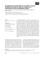
Fig. 4. Effect of HOQNO on the engineered [4Fe-4S] cluster EPR
spectrum of FrdB-V207C FrdABCD in HB101 membranes. Mem-
branes were incubated in the absence of (i) or presence of (ii)
0.5 m
M HOQNO for 5 min, then reduced with 5 mM dithionite
under argon for 5 min prior to being frozen in liquid nitrogen. Spec-
tra are presented of membranes containing no overexpressed
enzyme (A), and membranes containing overexpressed wild-type
(B), and FrdB-V207C (C). EPR spectra were recorded as described
for Fig. 3.
Fig. 3. Effect of HOQNO on the [3Fe-4S] cluster EPR spectrum of
wild-type and mutant FrdABCD in HB101 membranes. Membranes
were incubated with 0.5 m
M HOQNO (thick lines) or an equivalent
volume of ethanol for 5 min (thin lines), then oxidized with 0.2 m
M
ferricyanide for two minutes prior to being frozen in liquid nitrogen.
Spectra are shown of membranes containing no overexpressed
enzyme (A), and membranes containing overexpressed wild-type
(B), FrdB-T205H (C), FrdB-V207C (D), FrdC-E29L (E), FrdC-A32V (F),
FrdC-W86R (G), and FrdD-H80K (H). EPR spectra were recorded
under the following conditions: temperature, 12 K; microwave
power, 20 mW at 9.47 GHz; modulation amplitude, 10 G
pp
at 100
KHz. Spectra were normalized to a nominal protein concentration
of 30 mgÆmL
)1
. In addition, the absolute intensity of the g ¼ 2.02
peaks were normalized for each pair of spectra.
Quinol binding to E. coli fumarate reductase R. A. Rothery et al.
318 FEBS Journal 272 (2005) 313–326 ª 2004 FEBS
Effect of the mutations on the quinol:fumarate
oxidoreductase activity of FrdABCD
In order to gain a broader understanding of the effects
of the mutants on the physiological quinol oxidation
reaction catalyzed by FrdABCD, we studied their
effects on the steady-state kinetics of the quinol:
fumarate oxidoreductase reaction using the MQH
2
analog lapachol [2-hydroxy-3-(3-methyl-2-butenyl)-1,4-
naphthoquinone; LPC]. When reduced, this substrate
(LPCH
2
) has significant structural similarity to MQH
2
,
and in its oxidized form has a convenient absorbance
peak in the visible region at 481 nm in aqueous solu-
tion [16]. Figure 5 shows representative Eadie–Hofstee
plots describing the steady-state kinetic behavior of
wild-type and a subset of the mutants of FrdABCD in
DW35 membranes. The wild-type enzyme has a K
m
for LPCH
2
of approximately 225 lm and a k
cat
of
approximately 71 s
)1
. The FrdB-T205H and FrdD-
H80K mutants have increased K
m
values (of 355 lm
and 670 lm, respectively), but have similar k
cat
values
to that of the wild-type (68 s
)1
and 67 s
)1
, respect-
ively). The FrdC-A32V mutant exhibits quite different
behavior, with a decrease observed in both the K
m
and
the k
cat
values (to 115 lm and 31 s
)1
, respectively).
Likewise, the FrdB-V207C mutant also displayed a
decrease in both K
m
and k
cat
(Table 2). Despite the
HOQNO binding observed both by EPR and FQ titra-
tion, the FrdC-E29L mutant exhibited no quinol:fuma-
rate oxidoreductase activity. Kinetic data for all of the
mutants are summarized in Table 2.
Detection of a menasemiquinone radical anion
in the FrdC-E29L mutant
The FrdC-E29L mutant is unusual because it retains
high-affinity HOQNO binding (Table 1 and Fig. 2),
but demonstrates no fumarate-dependent LPCH
2
oxi-
dation. It has been demonstrated previously by redox
potentiometry to stabilize a menasemiquinone radical
Fig. 5. Determination of steady-state kinetic parameters for wild-
type and mutant FrdABCD. e, wild-type, K
m
¼ 225 lM, k
cat
¼
71 s
)1
. h, FrdAB
T205H
CD; K
m
¼ 355 lM, k
cat
¼ 68 s
)1
. s,
FrdABCD
H80K
, K
m
¼ 670 lM, k
cat
¼ 67 s
)1
. n, FrdABC
A32V
C, K
m
¼
115 l
M, k
cat
¼ 31 s
)1
. Assays at a range of LPCH
2
concentra-
tions were carried out as described in the Experimental proce-
dures.
Table 2. Effect of the FrdABCD mutants on the kinetic parameters for lapachol oxidation in E. coli strain DW35. Growth, ability of the
DW35 based strains used herein to support anaerobic growth using glycerol as carbon source and fumarate as respiratory oxidant. HOQNO
binding is as judged by the data presented in Table 1. Group, classification of mutant phenotypes: 0, no quinol oxidation, no high-affinity
HOQNO binding, does not support growth; 1, normal or modulated K
m
and normal k
cat
for quinol oxidation, high-affinity HOQNO binding,
supports growth; 2 ) normal or modulated K
m
with decreased k
cat
, high-affinity HOQNO binding, supports growth; 3, no quinol oxidation,
high-affinity HOQNO binding, does not support growth. NA, not applicable. Membranes from the background strain, E. coli DW35, do not
contain FrdABCD. ND, not detected.
Membrane preparation K
m
a
(lM) k
cat
a
(s
)1
) Growth on GF HOQNO Binding Group
Background ND ND No No NA
FrdABCD 225 ± 25 71 ± 3 Yes Yes 1
FrdB-T205H 355 ± 34 68 ± 4 Yes Yes 1
FrdB-V207C 203 ± 17 31 ± 1 Yes Yes 2
FrdC-E29L ND ND No Yes 3
FrdC-A32V 115 ± 6 31 ± 1 Yes Yes 2
FrdC-F38M 385 ± 40 82 ± 5 Yes Yes 1
FrdC-W86R ND ND No No 0
FrdD-H80K 670 ± 47 67 ± 3 Yes Yes 1
FrdD-H84K 451 ± 34 75 ± 3 Yes Yes 1
a
Kinetic parameters were determined from Eadie–Hofstee plots such as those presented in Fig. 5.
R. A. Rothery et al. Quinol binding to E. coli fumarate reductase
FEBS Journal 272 (2005) 313–326 ª 2004 FEBS 319
anion [20]. Thus, a plausible explanation for the lack
of quinol:fumarate oxidoreductase activity is that this
mutant becomes trapped in a state in which a mena-
semiquinone radical anion is bound to the Q
P
site. We
tested this hypothesis by attempting to observe turn-
over-induced radical species in the wild-type and
FrdC-E29L mutant enzymes. Figure 6 shows EPR
spectra recorded at 150K of variously treated mem-
brane preparations. No g ¼ 2.00 radical signal is
detected in oxidized and dithionite-reduced mem-
branes containing overexpressed wild-type enzyme
(Fig. 6A,B). Addition of fumarate to dithionite-
reduced membranes containing wild-type enzyme elicits
the observation of a small g ¼ 2.006 signal consistent
with the appearance of a menasemiquinone radical
intermediate under turnover conditions. As is the case
for the wild-type enzyme, dithionite-reduced mem-
branes containing the FrdC-E29L mutant enzyme exhi-
bit no radical signal. A significant signal is observed
in oxidized membranes containing mutant enzyme. An
intense g ¼ 2.006 signal is observed when the FrdC-
E29L mutant enzyme is reduced with dithionite and
then oxidized with fumarate, consistent with this
mutant becoming trapped in a menasemiquinone
bound form when enzyme turnover is attempted
(Fig. 6G,H).
Discussion
We have investigated the effects of a number of point
mutations on the affinity of FrdABCD for HOQNO.
In each case where HOQNO binding is detected, there
is a striking correlation between the concentration of
binding sites and the concentration of enzyme deter-
mined by EPR spin quantitation of the [2Fe-2S] and
[3Fe-4S] clusters (Table 1). Where modulation of the
K
d
for HOQNO is detected, the FQ data can be fitted
to a binding equation describing noncooperative bind-
ing at a single site within FrdABCD. These observa-
tions are consistent with the presence of a single
redox-active dissociable Q-site in FrdABCD, and indi-
cate that this site coincides with the Q
P
site observed
in the crystal structures of Iverson et al. [3,4]. The
HOQNO binding data agree with the structure of
FrdABCD incubated in the presence of HOQNO, in
which the inhibitor is bound exclusively at the Q
P
site.
We previously reported the effect of HOQNO on
the EPR line-shape of the [3Fe-4S] cluster of FrdB,
and showed that a point mutation in FrdC, FrdC-
H82R, eliminated both this effect and HOQNO bind-
ing detected by FQ titration [18]. However, the posi-
tion of FrdC-H82 within the hydrophobic core of
FrdC (> 5 A
˚
away from Q
P
), along with the relatively
severe Arg substitution, warranted re-examination of
HOQNO binding to FrdABCD using a range of avail-
able mutations. It is quite possible that the FrdC-
H82R mutation causes relatively gross conformational
changes that could affect both the Q
P
and Q
D
sites.
While some of the mutations studied herein may fall
into the same category as the FrdC-H82R mutant (i.e.
Fig. 6. Demonstration that turnover of the FrdC-E29L mutant is
stalled with a menasemiquinone radical-bound form in E. coli
DW35 membranes. EPR spectra were recorded of DW35 mem-
branes containing wild-type enzyme (A–D) and FrdC-E29L mutant
enzyme (E–H). (A, E), membranes reduced with 5 m
M dithionite for
2 min; (B) and (F), oxidized membranes. (C) and (G), membranes
reduced with dithionite for 2 min, then treated with 25 m
M fuma-
rate for 30 s. (D) and (H), as for (C) and (G), but with the incubation
with fumarate for 1 min. EPR spectra were recorded at 150 K using
a microwave power of 20 mW at 9.44 GHz and a modulation ampli-
tude of 1.2 G
pp
. Spectra were normalized to a protein concentration
of 30 mgÆmL
)1
.
Quinol binding to E. coli fumarate reductase R. A. Rothery et al.
320 FEBS Journal 272 (2005) 313–326 ª 2004 FEBS
the FrdC-W86R mutant), we were able to study a
range of mutations that are more likely to have local
effects within the protein. Overall, there is a good cor-
relation between the location of the mutated residues
and the severity of the observed effects on HOQNO
binding (compare Figure 1 and Table 1).
An effect on the EPR spectrum of the [3Fe-4S] clus-
ter is clearly observed in each case where HOQNO
binding is detected by FQ titration. In addition, we
were able to observe that this effect is not propagated
beyond the location of the [3Fe-4S] cluster (Fig. 4).
The FrdB-V207C mutant contains a [4Fe-4S] cluster in
place of the [3Fe-4S] cluster of the wild-type enzyme,
so that the mutant enzyme contains two [4Fe-4S] clus-
ters coordinated by a motif similar to those found in
the bacterial 8Fe ferredoxins [21]. In this mutant, the
converted cluster is paramagnetic in its reduced state,
but its spectroscopic analysis is complicated by spin–
spin interactions with the other two reduced clusters of
the enzyme (Fig. 4). Despite this, we were able to dem-
onstrate that HOQNO elicits a line-shape change on
the EPR spectrum of the fully reduced FrdB-V207C
mutant. Overall, the combination of FQ and EPR data
confirm that the Q
P
site is defined by residues from
FrdB, FrdC and FrdD.
Our observation that the Q
P
site is closely coupled
to the [3Fe-4S] cluster of FrdB bears interesting com-
parison with data reported for the membrane-bound
E. coli dimethylsulfoxide reductase (DmsABC). This
enzyme is a complex iron–sulfur molybdoenzyme that,
like FrdABCD, contains no heme within its mem-
brane anchor domain (DmsC) [28]. The electron
transfer subunit of DmsABC (DmsB) contains four
[4Fe-4S] clusters, and one of these can be changed to
a [3Fe-4S] cluster by site-directed mutagenesis (in a
DmsB-C102S mutant) [29]. Treatment of this
mutant with HOQNO results in a perturbation of the
[3Fe-4S] cluster EPR spectrum that is similar to that
reported for the [3Fe-4S] cluster of FrdABCD [18,30].
It is therefore likely that the dissociable Q-site of
DmsABC is located in the interface region between
the membrane-anchor (DmsC) and the electron-trans-
fer subunit (DmsB).
Comparison of the FQ titration, EPR and steady-
state kinetic data on the FrdABCD mutants reported
herein supports their assignments to the following
groups:
0 – no enzyme activity, no high-affinity HOQNO
binding, unable to support growth. Members: the
FrdC-W86R mutant and the FrdC-H82R mutant pre-
viously reported by us [18,25].
1 – normal or modulated K
m
, normal k
cat
, high-affinity
HQONO binding, able to support growth. Members:
the wild-type enzyme, the FrdB-T205H, FrdC-F38M,
FrdD-H80K and FrdD-H84K mutants.
2 – normal or modulated K
m
, decreased k
cat
, high-
affinity HOQNO binding, able to support growth.
Members: the FrdB-V207C and FrdC-A32V mutants.
3 – no quinol oxidation, high-affinity HOQNO bind-
ing, unable to support growth. Member: the FrdC-
E29L mutant.
Overall, the kinetic data presented herein are consis-
tent with the occurrence of simple Michaelis–Menten
kinetics, with LCPH
2
binding and oxidation occurring
at a single Q-site (Fig. 5). However, it is notable that
mutants that appear to have little effect on HOQNO
binding can modulate the observed steady-state kinet-
ics of the enzyme. For example, the FrdC-A32V
mutant significantly decreases the observed k
cat
.A
possible explanation for this is that the increased bulk
of the hydrophobic sidechain is able to stabilize qui-
nol ⁄ quinone species at the Q
P
site, decreasing the rate
of substrate entry and product egress. The other
mutant with a significantly decreased k
cat
, the FrdB-
V207C mutant contains a low potential [4Fe-4S] clus-
ter (E
m
of % )370 mV [21] in place of the native
[3Fe-4S] cluster with an E
m
of % )70 mV). In this case,
it is likely that the relative inefficiency of the low-
potential [4Fe-4S] cluster in accepting electrons from
reduced quinol explains the decreased k
cat
.
The two FrdD mutants studied herein produced
somewhat unexpected results: both are HisfiLys resi-
due changes (FrdD-H80 and FrdD-H84), yet only the
FrdD-H80K mutant has a significant effect on both
the K
d
for HOQNO and the K
m
for LPCH
2
. Careful
examination of the structure of FrdABCD (PDB file
L0V [4], Fig. 1) reveals a possible explanation for this.
Whilst the sidechain of FrdD-H84 is marginally closer
to the MQ at the Q
P
site than that of FrdD-H80, the
axis of the His-84 imidazole points slightly away from
the MQ naphthoquinone bicycle, whereas that of the
His-80 imidazole appears to be pointing at least parti-
ally towards it. Thus, it is more likely that the side-
chain of the Lys substitution of FrdD-H80 elicits an
effect on HOQNO binding and LPCH
2
oxidation than
the Lys substitution of FrdD-H84. Although this
explanation appears plausible, it should be noted that
it is based on structural data of fairly low resolution
(3.3 A
˚
) [3,4].
The FrdB-T205H mutant is of interest in establish-
ing the role of FrdB in defining the Q
P
site. As men-
tioned previously (Results), this mutation was chosen
because of the location of FrdB-T205H with respect to
the Q
P
site, the [3Fe-4S] cluster and the interface
between FrdB and the membrane anchor subunits.
With the exception of the FrdC-W86R mutant, the
R. A. Rothery et al. Quinol binding to E. coli fumarate reductase
FEBS Journal 272 (2005) 313–326 ª 2004 FEBS 321
FrdB-T205H mutant has the largest effect on the K
d
for HOQNO, raising it from % 2.5 nm to 39 nm (Fig. 2
and Table 1). In addition to its effect on HOQNO
binding, this mutant is also of interest for the follow-
ing reasons: (a) it has a subtle effect on the [3Fe-4S]
cluster EPR line-shape of both the untreated and HO-
QNO treated enzyme (the linewidth is significantly nar-
rowed, compare Fig. 3B and C) and (b) it changes the
sequence of the [3Fe-4S] cluster-coordinating Cys
group so that it contains the critical His residue that is
present after the first Cys in the carboxin-sensitive
complex II enzymes [31]. We are currently investi-
gating the effect of this mutation on the carboxin-sen-
sitivity of FrdABCD (E Maklashina, RA Rothery, JH
Weiner and G Cecchini, unpublished data).
Of the mutants classified above, the single member of
the Class 3 subgroup is particularly interesting. The
FrdC-E29L mutant has no quinol:fumarate oxidoreduc-
tase activity, yet it retains HOQNO binding measured
by both the FQ and EPR methods (Figs 2 and 3).
Ha
¨
gerha
¨
ll and coworkers [20] demonstrated by potenti-
ometric titration and EPR spectroscopy that a mena-
semiquinone radical anion is stabilized in this mutant.
Examination of FrdABCD structure reveals that the
position of FrdC-E29 is suitable for it to act as a proton
acceptor during enzyme turnover [3,4]. Furthermore, it
is widely believed that HOQNO represents a good ana-
log of the menasemiquinone radical intermediate
[32,33]. Our observation of a radical when enzyme turn-
over is attempted indicates that the mutant is only able
to accept a single electron from MQH
2
, resulting in a
bound and stabilized menasemiquinone intermediate,
thus explaining the observed binding of HOQNO and
the lack of quinol:fumarate oxidoreductase activity.
In addition to the E. coli complex II homologs
(FrdABCD and SdhCDAB), a high-resolution struc-
ture is available for one additional bacterial complex
II homolog. This is the Wolinella succinogenes fuma-
rate reductase (FrdCAB) [34,35] which belongs to a
distinct class of complex II homologs that includes the
Bacillus subtilis succinate dehydrogenase (SdhCAB)
[33]. These enzymes have a single membrane anchor
subunit (FrdC and SdhC, respectively) that contains
two hemes. The structure of the W. succinogenes Frd-
CAB [35] reveals that one heme is proximal to the
membrane-extrinsic dimer (heme b
P
), whilst the other
is distal to it (heme b
D
). It has been demonstrated that
a point mutation (FrdC-E66Q) that eliminates MQH
2
oxidation by FrdCAB is located at a site (a Q
D
site) in
close proximity to heme b
D
towards the periplasmic
side of FrdC [34]. In B. subtilis SdhCAB, the heme b
D
is essential for electron-transfer to MQ [36], and this
heme is the only one that appears to be affected by
HOQNO [32]. Thus, in contrast to the case in E. coli
FrdABCD, in W. succinogenes FrdCAB and B. subtilis
SdhCAB, available evidence points towards a model
for quinone ⁄ quinol binding in which the redox-active
dissociable Q-site is located towards the periplasmic
side of the membrane anchor domain (at a Q
D
site),
and that electron-transfer across the membrane
to ⁄ from the catalytic dimer is mediated by the two
hemes in a manner similar to that observed in E. coli
nitrate reductase A (NarGHI) [24,37–40] and suggested
for formate dehydrogenase N [41].
The role of the Q
D
site in FrdABCD remains unre-
solved. The data presented herein suggest a model for
the enzyme in which quinol binding and oxidation
occur exclusively at the Q
P
site. This is supported by
theoretical models of through-protein electron transfer
which indicate that the 25 A
˚
distance between the Q
P
and Q
D
menaquinones identified in the protein struc-
ture is too far to allow for physiologically relevant
electron transfer between these sites [11]. Our prelimin-
ary investigations of mutants (such as FrdD-F57V and
FrdC-V35A) surrounding the MQ
D
observed in the
protein structure indicate that these have no effect on
the HOQNO binding detected by FQ titration and by
EPR; and have little effect on quinol:fumarate oxidore-
ductase activities. A full description of these mutants
will appear in a later communication (E Maklashina,
RA Rothery, JH Weiner and G Cecchini, unpublished
data). Thus, it is likely that the Q
D
site plays no direct
role in menaquinol oxidation.
Overall, by using a range of FrdB, FrdC, and FrdD
mutants, we have demonstrated that in every case
where HOQNO binding is detected, it occurs at a sin-
gle site within FrdABCD. In agreement with the struc-
tural data of Iverson and coworkers [3,4], we provide
biochemical and biophysical evidence for the location
of the dissociable and redox-active Q site of FrdABCD
being in the interface region between the FrdCD mem-
brane-intrinsic domain and the FrdB electron-transfer
subunit. These studies provide important information
on the mechanism of MQH
2
oxidation by FrdABCD.
Experimental procedures
Bacterial strains and plasmids
E. coli DW35 (zjd::Tn10D(frdABCD)18 sdhC::Kan araD139
D(argF-lac)U169 rpsL150 relA1 flbB5301 deoC1 pfsF25 rbsR
[14] does not express FrdABCD or SdhCDAB. E. coli
HB101 (supE44 hsdS20 (r
B
–
m
B
–
) recA13 ara-14 proA2 lacY1
galK2 rpsL20 xyl-5 mtl-1) is a wild-type strain that expres-
ses plasmid-encoded FrdABCD to very high levels and
generates more consistent EPR data than that obtained
Quinol binding to E. coli fumarate reductase R. A. Rothery et al.
322 FEBS Journal 272 (2005) 313–326 ª 2004 FEBS
from DW35. Wild-type FrdABCD was expressed from
plasmid pH3 [42]. Mutant derivatives of pH3 were obtained
using standard molecular genetic procedures [43] as follows:
FrdC and FrdD mutants: FrdC-E29L, FrdC-A32V, FrdC-
F38M, FrdC-W86R, FrdD-H80K, FrdD-H84K: these
mutants were originally generated and partially character-
ized using plasmid pDW100 (frdC
+
D
+
) in combination
with a second plasmid, pFRD23 (frdA
+
B
+
) [14]. In order
to express high levels of FrdABCD, it was necessary to sub-
clone the mutated frdCD genes of pDW100 as a DraIII-
XhoI fragment into appropriately cut pH3. FrdB mutants:
FrdAB
V207C
CD was encoded by pH3-V207C [21]. A plas-
mid encoding FrdAB
T205H
CD (pH3-T205H) was generated
by site-directed mutagenesis using the methodology des-
cribed by Cecchini et al. [44].
Cell growth
DW35 strains
E. coli DW35 and its transformants were grown overnight
in 5 L batches in a B. Braun Biostat B fermenter (B. Braun
Biotech International, Melsungen, Germany) at 37 °Cin
the presence of 100 lgÆmL
)1
streptomycin (Amresco, Solon,
OH) and 50 lgÆmL
)1
kanamycin (Fisher Biotech, Fair
Lawn, NJ). Transformants were grown in a medium that
also contained 100 lgÆmL
)1
ampicillin (Amresco). The
growth medium contained 12 gÆL
)1
tryptone, 24 g ÆL
)1
yeast
extract, 5 gÆL
)1
NaCl and 4 mLÆL
)1
glycerol.
HB101 strains
E. coli HB101 and its transformants were grown overnight
in 2 L batches at 37 °C on Terrific Broth [43] in the pres-
ence of 100 lgÆmL
)1
streptomycin. The growth medium
used to culture plasmid-transformed HB101 also contained
100 lgÆmL
)1
ampicillin. In all cases, cells were harvested by
centrifugation at 10 000 g for 15 min at 4 °C, washed in a
buffer containing 100 mm Mops ⁄ KOH and 5 mm EDTA
(pH 7.0), and were flash frozen in liquid nitrogen prior to
being stored at )70 °C.
Isolation of cytoplasmic membranes
Crude membranes were prepared by French pressure cell
lysis and differential centrifugation at 150 000 g for 1.5 h at
4 °C in 100 mm Mops ⁄ KOH and 5 mm EDTA (pH 7.0)
which contained the protease inhibitor phenylmethanesulfo-
nyl fluoride (0.2 mm) [29]. Cytoplasmic membranes were
isolated from resuspended crude membranes by layering
them on top of a 55% (w ⁄ v) sucrose step (made up in buf-
fer) in an ultracentrifuge tube. Following centrifugation at
40 000 r.p.m. for 1.5 h in a Beckman 50.2Ti rotor
(150 000 g at 4 °C), the floating band enriched in the cyto-
plasmic membrane fraction was removed, diluted in buffer,
and subjected to a further centrifugation. Finally, to ensure
complete removal of residual sucrose, the pellet was resus-
pended in buffer and recentrifuged [24]. Membranes were
then resuspended in buffer to a protein concentration of
approximately 30 mgÆmL
)1
, flash frozen in liquid nitrogen,
and stored at )70 °C until use.
FQ titrations with HOQNO
The affinity of FrdABCD for HOQNO (Sigma-Aldrich,
Oakville, Ontario, Canada) was determined using FQ titra-
tions performed as described previously using a Perkin
Elmer (Norwalk, CT) LS-50B luminescence spectrometer
[18,23,45]. Fluorescence intensities were measured using an
excitation wavelength of 341 nm and an emission wave-
length of 479 nm. All experiments were carried out at room
temperature (23 °C) and pH 7.0 in 100 mm Mops ⁄ KOH
and 5 mm EDTA. HOQNO was added to the fluorescence
cuvette from a 0.25-mm stock ethanolic solution. A range
of protein concentrations was used as indicated in the indi-
vidual figure legends. The observed fluorescence (F
obs
) was
fitted to an equation (Eqn 1) describing ligand binding to a
single site as described previously [45,46]:
F
obs
¼ðf
bound
À f
free
ÞÂ Q À
ffiffiffiffiffiffiffiffiffiffiffiffiffiffiffiffiffiffiffiffiffiffiffiffiffiffiffiffiffiffiffiffiffiffiffiffiffiffiffiffiffiffiffiffi
ðQ
2
À n
s
½E
tot
½I
tot
Þ
q
þ f
free
½I
tot
ð1Þ
with
Q ¼
1
2
Âð½I
tot
þK
d
þ n
s
½E
tot
Þ ð2Þ
and
½I
tot
¼½I
bound
þ½I
free
ð3Þ
These equations are from reference [45]. The specific fluo-
rescences of the bound and free inhibitor are f
bound
and
f
free
, respectively. [I
tot
], [I
bound
] and [I
free
] are the concentra-
tions of total, bound and free inhibitor, respectively. [E
tot
]
is the total concentration of enzyme, and n
s
is the number
of binding sites. In the analyses presented herein, [E
tot
]is
deemed to be proportional to protein concentration. The
fluorescence of bound HOQNO is assumed to be zero
(f
bound
¼ 0) [18,19,22–24].
Preparation of EPR samples
In all cases, samples were prepared in 3 mm internal diam-
eter quartz EPR tubes. Following appropriate treatment(s),
samples were frozen rapidly in liquid nitrogen-chilled eth-
anol and were stored under liquid nitrogen prior to EPR
characterization. To investigate the effect of HOQNO on
the EPR line-shape of the [3Fe-4S] cluster, 500 lL mem-
brane samples at % 30 mgÆmL
)1
were incubated in the pres-
R. A. Rothery et al. Quinol binding to E. coli fumarate reductase
FEBS Journal 272 (2005) 313–326 ª 2004 FEBS 323
ence of 0.5 mm HOQNO for 5 min before being oxidized
with 0.2 mm ferricyanide for % 2 min. Samples were trans-
ferred to 3 mm internal diameter quartz EPR tubes prior to
being frozen and stored as described above. To investigate
the effect of HOQNO on the EPR line-shape of fully
reduced FrdABCD and FrdAB
V207C
CD, 150 lL membrane
samples at 30 mgÆmL
)1
were incubated with 0.5 m m HO-
QNO, then reduced with 5 mm dithionite for 5 min under
an argon atmosphere before being frozen and stored as des-
cribed above. For EPR spin quantitations, reduced samples
were prepared by incubation with 5 mm dithionite for
5 min, and oxidized samples were prepared by incubation
with 0.2 mm ferricyanide for 2 min. To investigate the
appearance of menasemiquinone radical species, samples
were reduced with 5 mm dithionite for 2 min and then trea-
ted with 25 mm fumarate for 30 s or 1 min before being
frozen as described above.
EPR spectroscopy
EPR spectra were recorded using a Bruker ESP 300 spec-
trometer (Bruker Biospin, Rheinstetten, Germany)
equipped with an Oxford Instruments (Abingdon, Oxon,
UK) ESR-900 flowing helium cryostat and a Hewlett Pack-
ard 5350B microwave frequency counter (Hewlett Packard,
Santa Clara, CA). For investigations of menasemiquinone
radical species, a Bruker liquid nitrogen evaporating cryo-
stat was used (a Bruker ER4111 VT Variable Temperature
Unit). Spin quantitations were carried out as previously
described [47] using a 1 mm Cu-EDTA standard and a Bru-
ker Elexsys E500 spectrometer equipped with an Oxford
Instruments ESR-900 flowing helium cryostat.
Protein assays
Protein concentrations were determined by the Lowry
method, modified by the inclusion of 1% (w ⁄ v) sodium
dodecyl sulfate in the incubation mixture to solubilize mem-
brane proteins [48].
Enzyme assays
Quinol:fumarate oxidoreductase assays were carried out at
room temperature (23 °C) in N
2
-saturated 100 mm Mops ⁄
KOH and 5 mm EDTA (pH 7.0) using 20 mm potassium
fumarate and 60–600 lm reduced lapachol [2-hydroxy-3-
(3-methyl-2-butenyl)-1,4-naphthoquinol, LPCH
2
; Sigma-
Aldrich] as described previously [16,18]. The appearance of
oxidized lapachol (LPC) in the assay mixture was followed
at 481 nm using a standard laboratory spectrophotometer.
For each membrane preparation, K
m
and k
cat
were deter-
mined by generating Eadie–Hofstee plots (v vs. v ⁄ s), and the
protein concentration was between 0.016 mgÆmL
)1
and
0.056 mgÆmL
)1
.
Structural alignment and molecular graphics
Protein structures of MQ-bound and HOQNO-bound
FrdABCD (PDB files 1L0V and 1KF6, respectively [4])
were manipulated using the program pymol (version 0.97,
Delano Scientific LLC, ). Prior
to generating the views presented in Fig. 1, the structures
(all subunits) were aligned with a root mean square devi-
ation of 0.17 A
˚
for the superposition of 1021 C-a atoms
between the two forms.
Acknowledgements
The authors wish to thank: Delilah Mroczko for her
assistance with the B. Braun Biostat B fermentation
system, and Monica Palak for the preparation of mem-
brane samples.
This work was funded by the Canadian Institutes of
Health Research and the Canada Foundation for
Innovation. A M.C.S. and A.M.S. were supported by
Alberta Heritage Foundation for Medical Research
Summer Studentships. J.H.W. holds a Canada
Research Chair in Membrane Biochemistry. Further
funding was provided by National Institutes of Health
grants to G.C. (GM61606) and R.P.G (GM49649 and
AI21678). G.C. also received support from the Depart-
ment of Veteran Affairs.
References
1 Cecchini G, Schro
¨
der I, Gunsalus RP & Maklashina E
(2002) Succinate dehydrogenase and fumarate reductase
from Escherichia coli. Biochim Biophys Acta 1553,
140–157.
2 Cole ST, Condon C, Lemire BD & Weiner JH (1985)
Molecular biology, biochemistry, and bioenergetics of
fumarate reductase, a complex membrane-bound iron-
sulfur flavoenzyme of Escherichia coli. Biochim Biophys
Acta 811, 381–403.
3 Iverson TM, Luna-Chavez C, Cecchini G & Rees DC
(1999) Structure of the Escherichia coli fumarate
reductase respiratory complex. Science 284, 1961–
1966.
4 Iverson TM, Luna-Chavez C, Croal LR, Cecchini G &
Rees DC (2002) Crystallographic studies of the Escheri-
chia coli quinol-fumarate reductase with inhibitors
bound to the quinol-binding site. J Biol Chem 277,
16124–16130.
5To
¨
rnroth S, Yankovskaya V, Cecchini G & Iwata S
(2002) Purification, crystallisation and preliminary crys-
tallographic studies of succinate: ubiquinone oxido-
reductase from Escherichia coli. Biochim Biophys Acta
1553, 171–176.
Quinol binding to E. coli fumarate reductase R. A. Rothery et al.
324 FEBS Journal 272 (2005) 313–326 ª 2004 FEBS
6 Yankovskaya V, Horsefield R, To
¨
rnroth S, Luna-
Chavez C, Miyoshi H, Le
´
ger C, Byrne B, Cecchini G &
Iwata S (2003) Architecture of succinate dehydrogenase
and reactive oxygen species generation. Science 299,
700–704.
7 Cecchini G, Maklashina E, Yankovskaya V, Iverson
TM & Iwata S (2003) Variation in proton donor ⁄ accep-
tor pathways in succinate: quinone oxidoreductases.
FEBS Lett 545, 31–38.
8 Vibat CR, Cecchini G, Nakamura K, Kita K & Gennis
RB (1998) Localization of histidine residues responsible
for heme axial ligation in cytochrome b
556
of complex II
(succinate: ubiquinone oxidoreductase) in Escherichia
coli. Biochemistry 37, 4148–4159.
9 Maklashina E, Rothery RA, Weiner JH & Cecchini G
(2001) Retention of heme in axial ligand mutants of suc-
cinate-ubiquinone oxidoreductase (complex II) from
Escherichia coli. J Biol Chem 276, 18968–18976.
10 Iverson TM, Luna-Chavez C, Schro
¨
der I, Cecchini G &
Rees DC (2000) Analyzing your complexes: structure of
the quinol-fumarate reductase respiratory complex. Curr
Opin Struct Biol 10, 448–455.
11 Page CC, Moser CC, Chen X & Dutton PL (1999) Nat-
ural engineering principles of electron tunneling in bio-
logical oxidation-reduction. Nature 402, 47–52.
12 Tran QH, Bongaerts J, Vlad D & Unden G (1997)
Requirement for the proton pumping NADH dehydro-
genase I of Escherichia coli in respiration from NADH
to fumarate and its bioenergetic implications. Eur J Bio-
chem 244, 155–160.
13 Westenberg DJ, Gunsalus RP, Ackrell BAC &
Cecchini G (1990) Electron transfer from menaquinol
to fumarate. Fumarate reductase anchor polypeptide
mutants of Escherichia coli. J Biol Chem 265, 19560–
19567.
14 Westenberg DJ, Gunsalus RP, Ackrell BAC, Sices H &
Cecchini G (1993) Escherichia coli fumarate reductase
frdC and frdD mutants. Identification of amino acid
residues involved in catalytic activity with quinones.
J Biol Chem 268, 815–822.
15 Yankovskaya V, Sablin SO, Ramsay RR, Singer TP,
Ackrell BAC, Cecchini G & Miyoshi H (1996) Inhibitor
probes of the quinone binding sites of mammalian com-
plex II and Escherichia coli fumarate reductase. J Biol
Chem 271, 21020–21024.
16 Rothery RA, Chatterjee I, Kiema G, McDermott MT
& Weiner JH (1998) Hydroxylated naphthoquinones as
substrates for Escherichia coli anaerobic reductases. Bio-
chem J 332, 35–41.
17 Maklashina E & Cecchini G (1999) Comparison of cata-
lytic activity and inhibitors of quinone reactions of suc-
cinate dehydrogenase (Succinate-ubiquinone
oxidoreductase) and fumarate reductase (Menaquinol-
fumarate oxidoreductase) from Escherichia coli. Arch
Biochem Biophys 369, 223–232.
18 Rothery RA & Weiner JH (1998) Interaction of a
menaquinol binding site with the [3Fe-4S] cluster of
Escherichia coli fumarate reductase. Eur J Biochem 254,
588–595.
19 Zhao Z, Rothery RA & Weiner JH (1999) Stopped-flow
studies of the binding of 2-n-heptyl-4-hydroxyquinoline-
N-oxide to fumarate reductase of Escherichia coli. Eur J
Biochem 260, 50–56.
20 Ha
¨
gerha
¨
ll C, Magnitsky S, Sled VD, Schro
¨
der I, Gunsa-
lus RP, Cecchini G & Ohnishi T (1999) An Escherichia
coli mutant quinol: fumarate reductase contains an
EPR-detectable semiquinone stabilized at the proximal
quinone-binding site. J Biol Chem 274, 26157–26164.
21 Manadori A, Cecchini G, Shro
¨
der I, Gunsalus RP, Werth
MT & Johnson MK (1992) [3Fe-4S] to [4Fe-4S] cluster
conversion in Escherichia coli fumarate reductase by site-
directed mutagenesis. Biochemistry 31, 2703–2712.
22 Zhao Z & Weiner JH (1998) Interaction of HOQNO
with dimethyl sulfoxide reductase of Escherichia coli.
J Biol Chem 273, 20758–20763.
23 Rothery RA, Blasco F, Magalon A, Asso M & Weiner
JH (1999) The hemes of Escherichia coli nitrate reduc-
tase A (NarGHI): potentiometric effects of inhibitor
binding to NarI. Biochemistry 38, 12747–12757.
24 Rothery RA, Blasco F & Weiner JH (2001) Electron
transfer from heme b
L
to the [3Fe-4S] cluster of Escheri-
chia coli nitrate reductase A (NarGHI). Biochemistry 40,
5260–5268.
25 Weiner JH, Cammack R, Cole ST, Condon C, Honore
´
N, Lemire BD & Shaw G (1986) A mutant of Escheri-
chia coli fumarate reductase decoupled from electron
transport. Proc Natl Acad Sci USA 83, 2056–2060.
26 Cammack R, Patil DS & Weiner JH (1986) Evidence
that centre 2 in Escherichia coli fumarate reductase is a
[4Fe-4S] cluster. Biochim Biophys Acta 870, 545–551.
27 Werth MT, Cecchini G, Manodori A, Ackrell BAC,
Schro
¨
der I, Gunsalus RP & Johnson MK (1990) Site-
directed mutagenesis of conserved cysteine residues in
Escherichia coli fumarate reductase: modification of the
spectroscopic and electrochemical properties of the
[2Fe-2S] cluster. Proc Natl Acad Sci USA 87, 8965–8969.
28 Weiner JH, Rothery RA, Sambasivarao D & Trieber
CA (1992) Molecular analysis of dimethylsulfoxide
reductase: a complex iron-sulfur molybdoenzyme of
Escherichia coli. Biochim Biophys Acta 1102, 1–18.
29 Rothery RA & Weiner JH (1991) Alteration of the iron-
sulfur composition of Escherichia coli dimethyl sulfoxide
reductase by site-directed mutagenesis. Biochemistry 30,
8296–8305.
30 Rothery RA & Weiner JH (1996) Interaction of an engi-
neered [3Fe-4S] cluster with a menaquinol binding site
of Escherichia coli DMSO reductase. Biochemistry 35,
3247–3257.
31 Matsson M & Hederstedt L (2001) The carboxin-bind-
ing site on Paracoccus denitrificans succinate: quinone
R. A. Rothery et al. Quinol binding to E. coli fumarate reductase
FEBS Journal 272 (2005) 313–326 ª 2004 FEBS 325
reductase identified by mutations. J Bioenerg Biomembr
33, 99–105.
32 Smirnova IA, Ha
¨
gerha
¨
ll C, Konstantinov AA & Heder-
stedt L (1995) HOQNO interaction with cytochrome b
in succinate: menaquinone oxidoreductase from Bacillus
subtilis. FEBS Lett 359, 23–26.
33 Hederstedt L (2002) Succinate: quinone oxidoreductase
in the bacteria Paracoccus denitrificans and Bacillus sub-
tilis. Biochim Biophys Acta 1553, 74–83.
34 Lancaster CRD, Groß R, Haas A, Ritter M, Ma
¨
ntele
W & Simon J (2000) Essential role of Glu-66 for mena-
quinol oxidation indicates transmembrane electrochemi-
cal potential generation by Wolinella succinogenes
fumarate reductase. Proc Natl Acad Sci USA 97, 13051–
13056.
35 Lancaster CRD, Kro
¨
ger A, Auer M & Michel H (1999)
Structure of fumarate reductase from Wolinella succino-
genes at 2.2A
˚
resolution. Nature 402, 377–385.
36 Matsson M, Tolstoy D, Aasa R & Hederstedt L (2000)
The distal heme center in Bacillus subtilis succinate:
quinone reductase is crucial for electron transfer to
menaquinone. Biochemistry 39, 8617–8624.
37 Rothery RA, Blasco F, Magalon A & Weiner JH (2001)
The diheme cytochrome b subunit (NarI) of Escherichia
coli nitrate reductase A (NarGHI): structure, function,
and interaction with quinols. J Mol Microbiol Biotech-
nol 3, 273–283.
38 Zhao Z, Rothery RA & Weiner JH (2003) transient
kinetic studies of heme reduction in Escherichia coli
nitrate reductase A (NarGHI) by menaquinol. Biochem-
istry 42, 5403–5413.
39 Zhao Z, Rothery RA & Weiner JH (2003) Effects of
site-directed mutations on heme reduction in Escherichia
coli nitrate reductase A by menaquinol: a stopped-flow
study. Biochemistry 42, 14225–14233.
40 Bertero MG, Rothery RA, Palak M, Hou C, Lim D,
Blasco F, Weiner JH & Strynadka NC (2003) Insights
into the respiratory electron transfer pathway from the
structure of nitrate reductase A. Nat Struct Biol 10,
681–687.
41 Jormakka M, Tornroth S, Byrne B & Iwata S (2002)
Molecular basis of proton motive force generation:
structure of formate dehydrogenase-N. Science 295 ,
1863–1868.
42 Blaut M, Whittaker K, Valdovinos A, Ackrell BAC,
Gunsalus RP & Cecchini G (1989) Fumarate reductase
mutants of Escherichia coli that lack covalently bound
flavin. J Biol Chem 264, 13599–13604.
43 Sambrook J, Fritsch EF & Maniatis T (1989) Molecular
Cloning: a Laboratory Manual, 2nd edn. Cold Spring
Harbor Laboratory Press, Cold Spring Harbor, NY.
44 Cecchini G, Sices H, Schro
¨
der I & Gunsalus RP (1995)
Aerobic inactivation of fumarate reductase from Escher-
ichia coli by mutation of the [3Fe-4S]-quinone binding
domain. J Bacteriol 177, 4587–4592.
45 Okun JG, Lu
¨
mmen P & Brandt U (1999) Three classes
of inhibitors share a common binding domain in mito-
chondrial complex I (NADH:ubiquinone oxidoreduc-
tase). J Biol Chem 274, 2626–2630.
46 Brandt U & von Jagow G (1991) Analysis of inhibitor
binding to the mitochondrial cytochrome c reductase by
fluorescence quench titration. Evidence for a ‘catalytic
switch’ at the Q
o
center. Eur J Biochem 195, 163–170.
47 Paulsen KE, Stankovich MT & Orville AM (1993) Elec-
tron paramagnetic resonance spectroelectrochemical
titration. Methods Enzymol 227 , 396–411.
48 Markwell MAD, Haas SM, Bieber LL & Tolbert NE
(1978) A modification of the Lowry procedure to sim-
plify protein determination in membrane and lipopro-
tein samples. Anal Biochem 87, 206–210.
Quinol binding to E. coli fumarate reductase R. A. Rothery et al.
326 FEBS Journal 272 (2005) 313–326 ª 2004 FEBS