Preparation of hundred-micron carbon spheres using solvent extraction in a simple microchannel device
Bạn đang xem bản rút gọn của tài liệu. Xem và tải ngay bản đầy đủ của tài liệu tại đây (5.88 MB, 7 trang )
Microporous and Mesoporous Materials 343 (2022) 112186
Contents lists available at ScienceDirect
Microporous and Mesoporous Materials
journal homepage: www.elsevier.com/locate/micromeso
Preparation of hundred-micron carbon spheres using solvent extraction in a
simple microchannel device
Jie Li a, Zhenheng Xu a, Liang Yu b, *, Lixiong Zhang a, **
a
b
State Key Laboratory of Materials-Oriented Chemical Engineering, College of Chemistry and Chemical Engineering, Nanjing Tech University, Nanjing, 211816, China
Chemical Technology, Luleå University of Technology, SE-971 87, Luleå, Sweden
A R T I C L E I N F O
A B S T R A C T
Keywords:
Hundred-micron carbon spheres
Porous carbon microspheres
Solvent extraction
Microfluidics
Phenol phenolic resin
Carbon microspheres with a uniform size of about 170 μm were prepared in a simple co-flow microfluidic device
using solvent extraction method. An ethanol solution of colloidal silica and phenol formaldehyde (PF) resol was
used as the dispersion phase, and a mixture of hexane and diisopropylamine was used as the continuous phase.
The droplets of PF resol resin/silica were generated in the continuous phase. Colloidal silica assisted the for
mation of the spherical structure and worked as a pore generator. The continuous phase was also used as
extractant and catalyst for PF resin/silica microspheres formation. Curing, drying, carbonization and leaching
were used for the post-treatment of the PF resin/silica microspheres to obtain porous carbon microspheres. The
carbon microspheres displayed a narrow size distribution and a high surface area of 679 m2/g coupled with
adjustable mesopores and large mesopore volume. Carbon microspheres prepared from the dispersion phase with
different PF/silica ratios (denoted as carbon/silica (C/Si) ratios) were studied and the formation mechanism of
the PF/silica microspheres was deeply explored.
1. Introduction
Porous microspheres with size ranging from 100 to 1000 μm are
widely used in various practical applications due to their suitable size for
transportation and recovery [1]. Because of the low density, high
chemical stability and thermal conductivity, hundred-micron porous
carbon spheres have been used in air and water purification, blood pu
rification, CO2 adsorption, and electronic and energy storage devices
[2–11].
So far, polymerization is the most popular method for the prepara
tion of hundred-micron carbon microspheres [11] and carbon spheres
synthesized from polymers displayed higher mechanical strength [8].
Divinylbenzene-based spherical activated carbon with size between 200
and 1200 μm were used for water purification, i.e. removal of organic
pollutants from water [6]. The spherical activated carbon showed two
times higher methylene blue adsorption capacity compared to the
commercially available spherical activated carbon. However, due to the
low mesopore volume, the highest observed methylene blue adsorption
capacity was only 32 mg/g. Yang et al. synthesized phenolic resin-based
activated carbon spheres with different pore size distributions by using
polyethylene glycol and polyvinyl butyral as pore-forming agents [7].
The size of the mesopores was between 3 and 5 nm in the carbon
spheres. Therefore, they showed excellent adsorption capacity for large
molecules e.g., creatinine and VB12. Activated carbon spheres with the
diameter of 200–950 μm were prepared by carbonization of commer
cially available polystyrene-based ion-exchange resin spheres [8]. The
polystyrene-based activated carbon spheres were 3 times harder than
the pitch-based activated carbons spheres. Due to the large pore volume
of 1.35 cm3/g, the spheres displayed high adsorption capacity of 153
mg/g for dibenzothiophene. Polystyrene-based microporous activated
carbon spheres with a narrower size distribution between 500 and 800
μm have been prepared by suspension polymerization of styrene
monomer and followed by some post-treatment processes including
sulfonation, oxidation, carbonization, and activation. The obtained
carbon spheres displayed a high surface area (1526 m2/g) and a large
pore volume (0.73 cm3/g). The adsorption capacity for CO2 was also
high about 4.21 mmol/g at 25 ◦ C and ambient pressure [9]. A so-called
inverse-microemulsion-polymerization-phase-separation method has
been developed for the preparation of carbon spheres using phenolic
resin [10]. The carbon spheres with the size of about 100 μm were
* Corresponding author.
** Corresponding author.
E-mail addresses: (L. Yu), (L. Zhang).
/>Received 4 April 2022; Received in revised form 29 July 2022; Accepted 12 August 2022
Available online 20 August 2022
1387-1811/© 2022 The Authors. Published by Elsevier Inc. This is an open access article under the CC BY-NC-ND license ( />
J. Li et al.
Microporous and Mesoporous Materials 343 (2022) 112186
composed of carbon nanoparticles with the size smaller than 100 nm.
Therefore, the carbon spheres displayed a hierarchical structure and
high total pore volume of 2.78 cm3/g. Due to the unique structure, the
carbon spheres showed a rapid transport of electrolyte ions, and there
fore displayed high potential in superior rate performance carbon-based
supercapacitors. Polymerization method is easy to scale up. However,
the wide size distribution of the spheres products is always a drawback
of the method.
Phenolic resin is a decent carbon source for the synthesis of highquality carbon microspheres due to the high carbon content. However,
the polymerization of phenol and formaldehyde is a relatively slow
process due to the high viscosity of the mixture of phenol and formal
dehyde [12]. Normally, a reaction time of 5 h at 95 ◦ C is needed for the
polymerization process [12]. Singh et al., prepared macrosize phenolic
beads (100–200 μm) by a suspension polymerization method with the
reaction temperature of 96 ◦ C and reaction time of 4 h [13]. The
phenolic beads can be used as precursors for the preparation of carbon
sphere. However, the beads showed a broad size distribution and poor
structural properties, e.g., low porosity. In some cases, additives are
added to improve the formation process and reduce the polymerization
time as well as improve the porosity of the obtained microspheres
[14–16]. However, the issue of the broad size distribution of micro
spheres prepared from the conventional polymerization remains.
Therefore, a new method is necessary.
As it is well known, microchannel technique could reduce the reac
tion time and temperature for time consuming and high temperature
demanding systems [17]. In addition, microchannel technique could
prepare microspheres with more uniform size compared to the con
ventional polymerization method [18–20]. Steinbacher et al., summa
rized the application of microchannel technique for the preparation of
even size microdroplets and microspheres [17]. Our group also has
developed several microchannel devices for the preparation of nano
particles, microspheres, and microcapsule materials in the last 15 years
[18,21–23]. Silica microspheres with the uniform size of 100 μm and
various structures including solid, hollow, hollow with a hole and
filbert-like solid structures were prepared in a simple T-type micro
channel device [21]. Monodisperse carbon hollow spheres were also
prepared in such device [18]. To improve the textural properties of the
obtained carbon spheres, colloidal silica is often used as additive for the
preparation of porous carbon. Our previous report showed that the pore
size of carbon spheres increased from 0.6 to 6.2 nm after removal of
silica nanoparticles from poly (furfuryl alcohol)-silica composite mi
crospheres [22]. Therefore, the adsorption capacity for dye increased
significantly. Silica nanoparticles also have been employed to tailor
ăber method [24]. The
porosity in carbon spheres using a modified Sto
obtained carbon spheres showed both microporous and mesoporous
structure with surface areas between 326 and 1500 m2/g and total pore
volumes between 0.26 and 1.22 cm3/g. Meanwhile, a high adsorption
capacity of 7.8 mmol/g for CO2 were observed at 0 ◦ C and 1 bar [24]. So
far, large size (hundred-micron meters) mesoporous carbon spheres with
an even size distribution have rarely been reported.
In the present work, hundred-micron carbon spheres with control
lable porous structure were prepared using a simple microchannel de
vice in combination with a solvent extraction method. PF resin/silica
microspheres were first prepared in the simple microchannel device.
The microspheres were carbonized to obtained carbon/silica spheres.
Subsequently, porous carbon spheres were prepared after removal of
silica. The synthesized time was much shorter and the obtained carbon
spheres were more even compared to the conventional polymerization
method. The conditions for the formation of PF resin/silica micro
spheres in the microchannel device were optimized and the effect of
carbon/silica ratio on the textural properties of the porous carbon
spheres was investigated. In addition, the mechanism of the formation of
the microspheres in the microchannel was discussed.
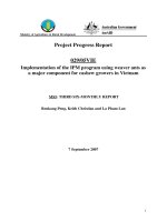
Fig. 1. Experimental device diagram.
2. Experimental
2.1. Preparation of colloidal silica
Colloidal silica with the concentration of about 10 wt% was prepared
by mixing 2.6 g TEOS with 3.0 g ethanol and followed by slowly adding
1.35 g HCl (4 × 10− 3 M). The mixture was stirred for 2 h at room
temperature.
2.2. Preparation of phenol formaldehyde resol
Phenol formaldehyde resol with a solids content of about 80 wt%
was prepared using phenol and formaldehyde (37 wt%) as precursors
and 20 wt% NaOH aqueous solution as catalyst as described in previous
work [25]. A mixture was first prepared by melting 12.2 g phenol at
42 ◦ C and subsequently adding 2.6 g 20 wt% NaOH under stirring. Af
terwards, formaldehyde solution (21 g) was added to the mixture. The
mixture was heated to 75 ◦ C and maintained for 60 min until the mixture
became red. The mixture was then cooled down to room temperature
and neutralized to pH of 7 using 7 × 10− 2 M HCl. The water in the
mixture was removed by drying at 45 ◦ C for about 2 days. The obtained
product was dissolved into ethanol to remove the NaCl precipitation.
Finally, the phenol formaldehyde resol resin was obtained after drying
at 45 ◦ C to remove ethanol.
2.3. Preparation of carbon microspheres
Fig. 1 shows the microchannel device that used in the present work
for the preparation of carbon microspheres precursors, i.e. phenol
formaldehyde resin microspheres. The device has been described in our
previous work [26].
Briefly, two PTFE tubes (red pipes in Fig. 1a) with the inner diameter
of 0.56 mm were fixed in a T-type microchannel for continuous phase.
The length of the PTFE tube for the outlet of the T-type microchannel
was 100 mm. A needle with the inner diameter of 0.26 mm was inserted
into the other inlet of the T-type microchannel. The head of the needle
should be placed at 2 mm after the joint point of T as shown in Fig. 1a.
The needle inlet will be used for dispersed phase.
The dispersed phase was a mixture of the colloidal silica, phenol
formaldehyde resol, and ethanol. The ethanol was used to control the
solid concentration in the dispersed phase. Continuous phase, i.e., the oil
phase was a mixture of n-hexane, liquid paraffin, and diisopropylamine.
The dispersed phase and continuous phase were fed into the T-type
microchannel at flowrates of 0.2 and 6.0 ml/h, respectively. Liquid
microdroplets were formed at the outlet of the needle due to the
shearing force of the continuous phase. A condenser column with the
length of 750 mm and inner diameter of 20 mm connected to a flask was
placed under the outlet of the T-type microchannel to collect the
generated microspheres (Fig. 1b). The composition of the liquid mixture
in the column was the same as the continuous phase. The microspheres
were maintained in the oil phase for extraction. PF resin/silica
2
J. Li et al.
Microporous and Mesoporous Materials 343 (2022) 112186
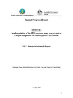
Fig. 2. Photographs of the obtained PF/silica microspheres with different m
(C/Si), (a) ∞; (b) 17; (c) 15.
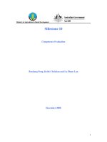
Fig. 3. SEM images of (a) the overall morphology and (b) the surface of PF/
silica microspheres with m (C/Si) = 15.
microspheres were obtained in the flask after ethanol and water were
extracted. The microspheres were cured at 100 ◦ C for 12 h and washed
by n-hexane for three times to obtain the solid PF resin/silica micro
spheres. For comparison, solid phenol formaldehyde resin was prepared
by mixing 1.0 g 80 wt% phenol formaldehyde resol with 0.1 g diiso
propylamine, and subsequently curing at 100 ◦ C oven for 12 h. The
obtained solid phenol formaldehyde resin was washed by n-hexane for
three times.
A tubular furnace was used to carbonize the PF resin/silica micro
spheres under nitrogen atmosphere. The temperature was first increased
to 350 ◦ C and maintained for 3 h, followed by increasing to 700 ◦ C and
maintained for 7 h. Porous carbon microspheres were eventually ob
tained by immersing the carbon/silica spheres in 10 wt% HF for 2 h to
remove silica nanoparticles. Solid phenol formaldehyde resin was
carbonized at the same conditions to prepare carbon for comparison.
2.4. Characterizations
An optical microscope (Olympus CX31) and SEM instruments (Phi
lips Quanta 200 and Hitachi− S4800) were used to observe the
morphology of the obtained products. N2 adsorption –desorption iso
therms were determined at liquid nitrogen temperature using BEL
SORPII instrument. Prior the measurement, the samples were degassed
at 120 ◦ C for 6 h to remove any impurity. The total pore volume was
calculated at p/p0 of 0.99 and the BET method was used to estimate the
surface area. Pore size distributions were analyzed using the non-local
density functional theory (NLDFT). A miniature double-frequency nu
merical control ultrasonic cleaning machine (KQ-100VDV) was used to
evaluate the mechanical strength of the carbon microspheres. The mi
crospheres were dispersed in water and treated in the ultrasonic clean
ing machine under a frequency of 45 kHz for 30 min. The particles size
distribution of the synthesized colloidal silica was measured using a
Malvern Zetasizer (Nano-ZS) instrument.
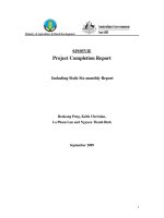
Fig. 4. Optical micrograph of the process of extraction of microdroplets pre
pared from m (C/Si) of 15 (a), 13 (b), 11 (c), 8.5 (d), and 6.5 (e). The size of the
scale is 400 μm.
silica. Spherical product could be obtained when m (C/Si) was 17, but
the microspheres agglomerated slightly, see Fig. 2b. This resulted from
the low colloidal silica concentrations. Monodispersing microspheres
were obtained when m (C/Si) decreased to 15, i.e., when the concen
tration of colloidal silica increased. The well dispersing microspheres
formed in the column provided promising microspheres precursors for
the late high temperature curing and carbonization.
Fig. 3 shows the SEM images of the PF/silica microspheres prepared
from a mixture with m (C/Si) of 15. The microspheres have a uniform
size of about 150 μm, which is much smaller than the liquid micro
spheres generated from the microchannel due to the contraction during
extraction. To investigate the formation process of the PF/silica micro
spheres, more experiments were carried out using dispersed phase with
different m (C/Si). Different amounts of ethanol were used to maintain a
constant solid concentration in the dispersed phase. The size of the
microdroplets at the outlet of the needle was 400 μm. Fig. 4 shows the
variations of the microdroplets in the extract as a function of extracting
time, which prepared from the m (C/Si) of 15, 13, 11, 8.5, and 6.5,
respectively.
Fig. 4 shows that the freshly generated microdroplets are trans
parent, no matter with the ratio of m (C/Si), and the microdroplets
became dark with the increase of the extracting time due to the gelation
of colloidal silica and the polymerization of phenol formaldehyde resol.
The color of the microdroplets changed evenly when m (C/Si) ≥11.
However, with the decrease of m (C/Si), i.e., increased the colloidal
silica content, a transparent shell and a dark core were observed, which
3. Results and discussion
3.1. Effect of silica contents
Fig. 2 shows the photographs of the microspheres prepared using
dispersed phases with different ratios of colloidal silica, phenol form
aldehyde resol resin, and ethanol. The mass ratios of carbon to silicon (m
(C/Si)) in the dispersed phases were ∞, 17 and 15. However, the total
content of solid in the dispersed phase was maintained constant at 35 wt
%. The continuous phase was comprised of n-hexane, liquid paraffin,
and diisopropylamine with the volume ratio of 2:2:1.
Fig. 2 shows that a large liquid droplet was generated from the small
droplets prepared without colloidal silica, i.e., the droplets agglomer
ated when the m (C/Si) was infinity. This was due to the condensation
polymerization of phenol formaldehyde resol is a slow process and an
alkaline catalyst is needed, otherwise, the short residence time in the
condensation column was not sufficient for the formation of stable
phenol formaldehyde resin microspheres. Therefore, a large liquid
droplet was obtained in the flask. However, stable PF/silica micro
spheres could be formed in the column due to the gelation of colloidal
3
J. Li et al.
Microporous and Mesoporous Materials 343 (2022) 112186
Meanwhile, the particle size of the obtained PF/silica microspheres
decreased with the increase of the m (C/Si) as shown in Fig. 5, which
showed that the content of colloidal silica has a significant effect on the
microsphere size. It is important to note that the total solids content in
the dispersed phase was maintained constant at 35 wt% as mentioned
above when m (C/Si) was changed. More information about shell and
core is explored as below.
Fig. 6 shows the PF/silica microspheres prepared from different ra
tios of m (C/Si). Silica gel, i.e., a hydrogel with three-dimension struc
ture will generate in the microdroplets when the content of colloidal
silica was high. Therefore, a more porous structure was obtained in the
microspheres, see Fig. 6b. Microspheres with more compact structure
was observed when the content of colloidal silica decreased, i.e., the
content of phenol formaldehyde resol increased. Since diisopropylamine
in the extract catalysed the polymerization of phenol formaldehyde
resol, the compact structures, mainly the compact shells were generated
in the microsphere. Therefore, the PF/silica microspheres with shells
thickness of about 4 μm, 15 μm and 18 μm were observed, when m (C/Si)
increased from 6.5 to 15, see Fig. 6. We, therefore, can conclude that the
core and the shell of PF/silica microspheres were mainly silica and
phenol formaldehyde resin, respectively.
Fig. 5. Size of PF/silica microspheres as a function of m (C/Si).
probably resulted from the phase separation during the gelation of
colloidal silica (water phase) and the polymerization of phenol formal
dehyde resol (oil phase).
3.2. Effect of diisopropylamine concentration
Diisopropylamine was selected as catalyst for the polymerization of
Fig. 6. SEM images of the whole, surface, cross-section and edge of the cross-section of the PF/silica microspheres prepared at m (C/Si) = 6.5 (˜
ad), m (C/Si) = 13 (e
~ h), and m (C/Si) = 15 (i ~ l).
Fig. 7. SEM images of cross-section of PF/silica microsphere prepared at m (C/Si) = 15 and different diisopropylamine concentrations, (a) 10 wt%; (b) 40 wt%; (c)
60 wt%, and the cross-section edge of (d) 10 wt%, (e) 40 wt%, (f) 60 wt%.
4
J. Li et al.
Microporous and Mesoporous Materials 343 (2022) 112186
separation. In addition, silicate could be generated when the pH is high
enough, i.e., at high diisopropylamine concentration, which could be
easily rinsed away. Therefore, the carbon content will be high and silica
content will be much lower at the surface. These explain the formation
of shell on the surface of the microspheres (see Fig. 7c, f) and the lower
silicon in the shell when the concentration of diisopropylamine
increased to 60 wt%. Table 1 summarizes the elements distribution in
the microspheres.
The effect of diisopropylamine concentration on the formation pro
cess of the microspheres were further explored by observing the change
of the microdroplets in the extract with different compositions. Fig. 8
shows the change of the colour of the microdroplets in the extract with
low diisopropylamine concentration. The microspheres became dark
evenly. However, the results in Fig. 9, where the diisopropylamine
concentration was higher, show a shell on the microsphere due to the
fast polymerization of phenol formaldehyde on the surface and gelation
of colloidal silica in the centre. These images show clear phase
separation.
Table 1
Effect of diisopropylamine concentration on the distribution of elements in PF/
silica microspheres.
Diisopropylamine
concentration (wt.%)
10
40
60
Distribution of elements in core/shell of the
microsphere (wt.%)
C
63.10/
59.43
65.28/
57.24
51.62/
61.55
Si
6.65/
2.82
8.68/
4.61
14.05/
3.19
N
0.06/
2.41
1.49/
3.88
0.09/
4.03
O
30.20/
35.34
24.55/
34.26
34.24/
31.23
3.3. Textural properties of the microspheres
Fig. 10 shows the SEM images of the microspheres after curing,
carbonization and removal of silica. Moreover, the microspheres were
prepared from a dispersed phase with m (C/Si) = 11 and solid content of
35 wt%.
As it has been mentioned above, the microdroplets generated at the
outlet of the needle were about 400 μm. However, the size of the mi
crospheres was contracted to about 260 μm after polymerization and
extraction. The size of the microsphere further decreased to about 170
μm after carbonization, and the sizes before and after removal of silica
were almost the same.
Fig. 11 shows the N2 adsorption-desorption isotherms of micro
spheres prepared at different conditions. The adsorption solely occurred
at the initial pressure range for carbon/silica and carbon microspheres,
which indicates only micropores generated in the microspheres. The
micropores similar with the activated carbon. More micropores and
mesopores were generated after removal of silica. Fig. 11 shows the pore
size distributions of the obtained microsphere at different conditions.
The mesopores sizes of the carbon microspheres synthesized from C/Si
= 6.5 was in the range of 5.7–8.3 nm. Larger mesopores up to 12.3 nm
were observed in the carbon microspheres prepared from C/Si = 11.
Only micropores could be observed in C(PF) particles. The micropo
rosity in the carbon microspheres also increased after silica removal, and
the size of the micropores is much smaller than the size of the silica
particles in the colloidal silica (Fig. 11d). This indicated that even
smaller silica particles were generated in the carbon/silica micro
spheres. The smaller silica particles were probably developed from the
dissolution of the TEOS-generated silica during the experiment [27,28].
Table 2 summarizes the textural properties of the prepared micro
spheres at different conditions. The surface area of pure carbon was only
126 m2/g, which was much smaller than the carbon/silica microspheres.
The surface area of carbon/silica microspheres increased significantly
after removal of silica. For instance, the surface area of carbon/silica
microspheres prepared from a dispersed phase with m (C/Si) = 6.5 was
329 m2/g, and it increased to 679 m2/g after removal of silica, mean
while, the total pore volume increased from 0.15 to 0.50 cm3/g. These
results indicated that colloidal silica was used not only as the assistant
for the formation of the spherical structure, but also for the generation of
porous structure, especially for the mesoporous structure, which makes
the microsphere a wide application at macromolecules adsorption, drug
delivery, supercapacitors, and catalysis. It is important to note that the
microspheres well preserved the spherical morphology (Fig. 10) after
the treatment in an ultrasonic bath (45 kHz) for 30 min. The results
indicated a good mechanical strength of the carbon microspheres.
Porous carbon materials have been extensively studied in the last
decades. Macropore-rich activated carbon microspheres with size of
Fig. 8. Optical micrographs of the extraction process of PF/silica microdroplets
in the extraction phase. The PF/silica were prepared with C/Si = 15 and solid
content of 35 wt%. The composition of extraction phase was liquid paraffin: nhexane: diisopropylamine = 8:9:3. The size of the scale is 400 μm.
Fig. 9. Optical micrographs of the extraction process of PF/silica microdroplets
in the extraction phase. The PF/silica were prepared with C/Si = 15 and solid
content of 35 wt%. The composition of extraction phase was liquid paraffin: nhexane: diisopropylamine = 8:3:9. The size of the scale is 300 μm.
phenol formaldehyde resol. Meanwhile, it could enhance the gelation of
colloidal silica. The advantages of diisopropylamine catalyst are the
lower reaction temperature, and high solubility in n-hexane and liquid
paraffin.
Since the pH of the colloidal silica used in the present work was
about 5, silica gel was formed when the microspheres interacted with
diisopropylamine. The concentration of diisopropylamine will affect the
gelation rate of colloidal silica and the polymerization rate of phenol
formaldehyde. At low diisopropylamine concentration, the gelation and
polymerization rate were low. Diisopropylamine could diffuse into the
microspheres before polymerization completed, therefore the elements
distribution was quite even. However, when the diisopropylamine
concentration was high, phenol formaldehyde will be polymerized at the
surface of the microspheres quickly, and more phenol formaldehyde
resol will diffuse to the surface for polymerization. Meanwhile, some
diisopropylamine will diffuse into the microsphere and therefore silica
gel formed inside the microspheres. This process is similar to phase
5
J. Li et al.
Microporous and Mesoporous Materials 343 (2022) 112186
Fig. 10. SEM images of microspheres after (a) aging; (b) carbonization; (c) removal of SiO2 by hydrofluoric acid. The dispersed phase that used for the preparation of
the microspheres had a m (C/Si) = 11 and solid content of 35 wt%. SEM images with different magnifications (d) and (e) of the microspheres after treatment in an
ultrasonic bath (45 kHz) for 30 min.
Fig. 11. (a) N2 adsorption-desorption isotherms; (b) and (c) NLDFT pore size distributions of carbon/silica microspheres and carbon microspheres after removal of
SiO2; (d) particle size distribution of the colloidal silica prepared in the present work.
about 100 μm have been prepared using inverse-microemulsionpolymerization-phase-separation coupling method. The polymerization
was performed at 120 ◦ C for 12 h. Phenolic resin was used as carbon
source, ethylene glycol was used as pore generator and hexamethy
lenetetramine was used as the hardener for polymerization. The carbon
microspheres displayed a surface area as high as 1622 m2/g, but almost
no mesoporous was observed [10]. In addition, the microemulsion
polymerization method is batch with complicated operation procedures,
i.e., mixing and temperature control, and the prepared microsphere
normally showed a wide particle size distribution. The method using
microchannel technology can produce microspheres continuously with
narrow size distributions, and the temperature in microchannel is even
due to the enhanced heat transfer. In addition, the microspheres are
formed in a much shorter time about 15 min in the microchannel at
room temperature. Colloidal silica is more economical than the other
organic pore generators, and the microchannel method is easy to scale
up, which shows high potential for large applications.
4. Conclusions
Carbon/silica microspheres and carbon microspheres with a size of
hundred-micron have been successfully prepared using microchannel
technique. The process was quite economical due to the gentle reaction
conditions, i.e., room temperature and short reaction time. Colloidal
silica can significantly reduce the time for the formation of microsphere
due to the gelation. The use of colloidal silica can also increase surface
area and pore volume, both microporous and mesoporous, significantly.
The textural properties of the microspheres can be adjusted easily by
colloidal silica content, which enables the microsphere material a great
potential as macromolecules adsorbent, drug delivery material,
6
J. Li et al.
Microporous and Mesoporous Materials 343 (2022) 112186
[3] C. Liu, X. Liang, X. Liu, Q. Wang, L. Zhan, R. Zhang, W. Qiao, L. Ling, Surface
modification of pitch-based spherical activated carbon by CVD of NH3 to improve
its adsorption to uric acid, Appl. Surf. Sci. 254 (2008) 6701–6705.
[4] K.T. Nagesh, Porous carbon spheres: recent developments and applications, AIMS
Mater. Sci. 5 (2018) 1016–1052.
[5] X. Zhao, H. Chen, F. Kong, Y. Zhang, S. Wang, S. Liu, L.A. Lucia, P. Fatehi, H. Pang,
Fabrication, characteristics and applications of carbon materials with different
morphologies and porous structures produced from wood liquefaction: a review,
Chem. Eng. J. 364 (2019) 226–243.
[6] S. Yenisoy-Karakas¸, A. Aygün, M. Günes¸, E. Tahtasakal, Physical and chemical
characteristics of polymer-based spherical activated carbon and its ability to adsorb
organics, Carbon 42 (2004) 477–484.
[7] J.-B. Yang, L.-C. Ling, L. Liu, F.-Y. Kang, Z.-H. Huang, H. Wu, Preparation and
properties of phenolic resin-based activated carbon spheres with controlled pore
size distribution, Carbon 40 (2002) 911–916.
[8] Q. Wang, X. Liang, W. Qiao, C. Liu, X. Liu, L. Zhan, L. Ling, Preparation of
polystyrene-based activated carbon spheres with high surface area and their
adsorption to dibenzothiophene, Fuel Process. Technol. 90 (2009) 381–387.
[9] Y. Sun, J. Zhao, J. Wang, N. Tang, R. Zhao, D. Zhang, T. Guan, K. Li, Sulfur-doped
millimeter-sized microporous activated carbon spheres derived from sulfonated
poly(styrene–divinylbenzene) for CO2 capture, J. Phys. Chem. C 121 (2017)
10000–10009.
[10] D. Zhang, J. Zhao, C. Feng, R. Zhao, Y. Sun, T. Guan, B. Han, N. Tang, J. Wang,
K. Li, J. Qiao, J. Zhang, Scalable synthesis of hierarchical macropore-rich activated
carbon microspheres assembled by carbon nanoparticles for high rate performance
supercapacitors, J. Power Sources 342 (2017) 363–370.
[11] S. Li, A. Pasc, V. Fierro, A. Celzard, Hollow carbon spheres, synthesis and
applications – a review, J. Mater. Chem. 4 (2016) 12686–12713.
[12] G. He, B. Riedl, Phenol-urea-formaldehyde cocondensed resol resins: their
synthesis, curing kinetics, and network properties, J. Polym. Sci. B Polym. Phys. 41
(2003) 1929–1938.
[13] A. Singh, R.K. Yadav, A. Srivastava, Synthesis of resole-type phenolic beads from
phenol and formaldehyde by suspension polymerization technique, J. Appl. Polym.
Sci. 112 (2009) 1005–1011.
[14] K. Nakanishi, N. Soga, Phase separation in silica sol-gel system containing
polyacrylic acid. IV. Effect of chemical additives, J. Non-Cryst. Solids 142 (1992)
45–54.
[15] L. Zhang, Y. Zhang, Y. Xue, H. Duan, Y. Cui, Enzymatic synthesis of soluble phenol
polymer in water using anionic surfactant as additive, Polym. Int. 62 (2013)
1277–1282.
[16] H. Jiang, J. Wang, Z. Duan, F. Li, Study on the microstructure evolution of phenolformaldehyde resin modified by ceramic additive, Front. Mater. Sci. China 1
(2007) 35–39.
[17] J.L. Steinbacher, D.T. McQuade, Polymer chemistry in flow: new polymers, beads,
capsules, and fibers, J. Polym. Sci. Polym. Chem. 44 (2006) 6505–6533.
[18] Y. Pan, M. Ju, C. Wang, L. Zhang, N. Xu, Versatile preparation of monodisperse
poly(furfuryl alcohol) and carbon hollow spheres in a simple microfluidic device,
Chem. Commun. 46 (2010) 3732–3734.
[19] C.-H. Choi, J. Kim, J.-O. Nam, S.-M. Kang, S.-G. Jeong, C.-S. Lee, Microfluidic
design of complex emulsions, Chem. Phys. Chem. 15 (2014) 21–29.
[20] R.J. Krupadam, B.A. Korde, M. Ashokkumar, S.D. Kolev, Novel molecularly
imprinted polymeric microspheres for preconcentration and preservation of
polycyclic aromatic hydrocarbons from environmental samples, Anal. Bioanal.
Chem. 406 (2014) 5313–5321.
[21] M. Ju, X. Ji, C. Wang, R. Shen, L. Zhang, Preparation of solid, hollow, hole-shell
and asymmetric silica microspheres by microfluidic-assisted solvent extraction
process, Chem. Eng. J. 250 (2014) 112–118.
[22] Y. Liu, M. Ju, C. Wang, L. Zhang, X. Liu, Preparation of monodisperse mesoporous
carbon microspheres from poly(furfuryl alcohol)–silica composite microspheres
produced in a microfluidic device, J. Mater. Chem. 21 (2011) 15049–15056.
[23] M. Ju, C. Zeng, C. Wang, L. Zhang, Preparation of ultrafine carbon spheres by
controlled polymerization of furfuryl alcohol in microdroplets, Ind. Eng. Chem.
Res. 53 (2014) 3084–3090.
[24] J. Marszewska, M. Jaroniec, Tailoring porosity in carbon spheres for fast carbon
dioxide adsorption, J. Colloid Interface Sci. 487 (2017) 162–174.
[25] J. Cai, L. Jiang, H. Wei, C. Wang, L. Yu, L. Zhang, Preparation of carbon/cobalt
composite from phenolic resin and ZIF-67 for efficient tannic acid adsorption,
Microporous Mesoporous Mater. 287 (2019) 9–17.
[26] M. Ju, Y. Li, L. Yu, C. Wang, L. Zhang, Two-phase diffusion technique for the
preparation of ultramacroporous/mesoporous silica microspheres via interface
hydrolysis, diffusion, and gelation of TEOS, Langmuir 34 (2018) 2046–2056.
[27] J. Liu, X. Wang, J. Gao, Y. Zhang, Q. Lu, M. Liu, Hollow porous carbon spheres with
hierarchical nanoarchitecture for application of the high performance
supercapacitors, Electrochim. Acta 211 (2016) 183–192.
[28] J. G´
orka, M. Jaroniec, Hierarchically porous phenolic resin-based carbons obtained
by block copolymer-colloidal silica templating and post-synthesis activation with
carbon dioxide and water vapor, Carbon 49 (2011) 154–160.
[29] J.B. Joo, P. Kim, W. Kim, J. Kim, N.D. Kim, J. Yi, Simple preparation of hollow
carbon sphere via templating method, Curr. Appl. Phys. 8 (2008) 814–817.
[30] H.-J. Liu, W.-J. Cui, L.-H. Jin, C.-X. Wang, Y.-Y. Xia, Preparation of threedimensional ordered mesoporous carbon sphere arrays by a two-step templating
route and their application for supercapacitors, J. Mater. Chem. 19 (2009)
3661–3667.
Table 2
Summary of the textural properties of different mesoporous carbon
microspheres.
Microspheres
Particle
size (μm)
Surface
area (m2/
g)
Total pore
volume
(cm3/g)
dp
(nm)
Ref.
C(PF) particles
n/a
126
0.07
1.57
Carbon/silica
microspheresa
Carbon
microspheresa
Carbon/silica
microspheresb
Carbon
microspheresb
Activated carbon
microspheres
Macroporous
carbon spheres
Ordered
mesoporous
carbon sphere
Phenolic resinbased carbon
spheres
170
295
0.14
1.3
170
625
0.38
11.3
174
329
0.15
1.4
174
679
0.50
6.2
~100
1622
2.78
>10
This
work
This
work
This
work
This
work
This
work
[10]
0.4
451
0.28
n/a
[29]
0.3
601
1.70
10.4
[30]
n/a
797
0.35
2
[7]
a
and b microspheres prepared from dispersed phases with m(C/Si) = 11 and 6.5,
respectively.
stationary phase of chromatography, supercapacitor, and catalyst
carrier.
Notes
The authors declare no confliction of interest.
CRediT authorship contribution statement
Jie Li: Writing – original draft, Investigation, Formal analysis.
Zhenheng Xu: Investigation, Formal analysis. Liang Yu: Writing – re
view & editing, Supervision, Formal analysis, Data curation. Lixiong
Zhang: Writing – review & editing, Funding acquisition,
Conceptualization.
Declaration of competing interest
The authors declare that they have no known competing financial
interests or personal relationships that could have appeared to influence
the work reported in this paper.
Data availability
Data will be made available on request.
Acknowledgment
The authors acknowledge the financial support from the Priority
Academic Program Development of Jiangsu Higher Education
Institutions.
References
[1] J. Zang, P. Tian, G. Yang, S. Jia, S. Zhou, H. Xu, Y. Wang, A facile preparation of
pomegranate-like porous carbon by carbonization and activation of phenolic resin
prepared via hydrothermal synthesis in KOH solution for high performance
supercapacitor electrodes, Adv. Powder Technol. 30 (2019) 2900–2907.
[2] N.P. Wickramaratne, M. Jaroniec, Activated carbon spheres for CO2 adsorption,
ACS Appl. Mater. Interfaces 5 (2013) 1849–1855.
7