Single grain infrared photoluminescence (IRPL) measurements of feldspars for dating
Bạn đang xem bản rút gọn của tài liệu. Xem và tải ngay bản đầy đủ của tài liệu tại đây (1.28 MB, 8 trang )
Radiation Measurements 133 (2020) 106313
Contents lists available at ScienceDirect
Radiation Measurements
journal homepage: />
Single grain infrared photoluminescence (IRPL) measurements of feldspars
for dating
G.A.T. Duller a, *, M. Gunn b, H.M. Roberts a
a
b
Department of Geography and Earth Sciences, Aberystwyth University, UK
Department of Physics, Aberystwyth University, UK
A R T I C L E I N F O
A B S T R A C T
Keywords:
Luminescence
Imaging
EM-CCD
Feldspar
Dose determination
Existing infrared photoluminescence (IRPL) systems have used pulsed infrared stimulation (~830 nm) and
measured IRPL emission (at 880 or 955 nm) using time resolved data collection with photomultipliers. Break
through of the infrared stimulation light overwhelms the IRPL, but the delayed emission during the laser-off
period has been used instead.
This paper describes a system for measurement of the IRPL signal from single sand-sized grains of feldspar. The
attachment uses an electron-multiplying charge-coupled device (EMCCD) imaging system, and has two in
novations that make it possible to use such a detector to obtain IRPL data. First, the optical detection system has
been designed to minimise stray light and maximise the efficiency with which filters reject the stimulation light.
This acts to reduce, but not eliminate, the breakthrough. Second, by placing the sample to be measured in a
clearly defined sample grid, the spatial resolution provided by the EMCCD has been used to differentiate between
regions of the image where IRPL is emitted and adjacent regions where only breakthrough is expected. This
allows quantification of the breakthrough and effective subtraction to isolate the IRPL signal from the grains of
interest.
The attachment has been used to measure IRPL from single sand-sized grains of feldspar from an aeolian dune
from New Zealand. A 1W UV LED (365 nm) is also added to the system and this is effective at resetting the IRPL
signal, permitting a single aliquot regenerative dose (SAR) protocol to be used to measure equivalent dose (De).
Measurement of a known laboratory dose (104 Gy) demonstrates the reproducibility of the attachment, with no
overdispersion observed in the resulting single grain De values. The recovered dose is within 10% of the given
dose. The natural IRPL signal yields De values from single grains with low overdispersion (22%) and giving a
weighted mean value (103 � 5.8 Gy) that is consistent with that obtained using post-IR IRSL measurements (105
� 3.8 Gy). The attachment described here provides IRPL measurements on single grains suitable for exploring the
potential of this novel and exciting signal for dating geological sediments.
1. Introduction
The discovery of a luminescence emission at 955 nm in feldspars
when stimulated in the near infrared (Prasad et al., 2017) opens up
exciting new opportunities for archaeological and geological dating
applications. This infrared photoluminescence signal (IRPL) signal, and
a second emission at 880 nm (Kumar et al., 2018), are reported to have a
number of properties that make them attractive for dating. First, the
signal does not deplete during measurement, making it possible to
improve the precision of individual measurements by extending the
period of stimulation. Secondly, the signal is thought not to suffer from
anomalous fading, a major challenge for other methods based on anal
ysis of stimulated luminescence from feldspars. However, an additional
observation in initial measurements (Prasad et al., 2017) is that the
signal does not bleach rapidly with exposure to daylight, and this could
potentially be an impediment to the use of this signal for dating sedi
ments. In previous research using other luminescence signals, where the
exposure of sediments to daylight may vary from one mineral grain to
another, single grain measurements have proven valuable for dating
(Duller, 2008). This paper describes the development of instrumentation
for measuring the 955 nm IRPL emission from single sand-sized (~200
μm diameter) feldspar grains. The performance of this instrumentation
* Corresponding author.
E-mail address: (G.A.T. Duller).
/>Received 26 August 2019; Received in revised form 12 January 2020; Accepted 12 March 2020
Available online 19 March 2020
1350-4487/© 2020 The Authors. Published by Elsevier Ltd. This is an open access article under the CC BY license ( />
G.A.T. Duller et al.
Radiation Measurements 133 (2020) 106313
is assessed using potassium-rich feldspar from a sample of aeolian sand
from New Zealand.
Kook et al. (2018) describe three approaches for measuring IRPL
using an 830 nm laser for stimulation, and then a combination of long
pass filters at 925 nm (LP925) and a band pass filter at 950 nm (BP950)
to reject the stimulation light, and allow transmission of IRPL at 880 or
955 nm. Although the LP925 and BP950 filters have very low trans
mission at 830 nm when light strikes them normally, their performance
is degraded significantly when light passes through obliquely (Fig. 1(a)
and (b)), and breakthrough of the 830 nm stimulation light into the
detector is a major problem. Two of the three systems that Kook et al.
(2018) describe pulse the 830 nm IR laser (typically 5 μs on pulse, 95 μs
off period) and use time resolved measurements with IR sensitive pho
tomultiplier tubes so that they can ignore the signal during the on-period
of the 830 nm stimulation and just use the IRPL emission in the
off-period of the 830 nm laser (typically the period 3–92μs after each IR
pulse). In order to make measurements of single grains of feldspar, they
based one system around a focussed laser system (Duller et al., 2003).
Kook et al. (2018) also describe a system based around use of an electron
multiplying charge coupled device (EMCCD) camera. The EMCCD de
tector described by Kook et al. (2015) has been shown to be a sensitive
detector capable of resolving luminescence emissions from single grains
of quartz (Thomsen et al., 2015) at ~340 nm. However, unlike typical
photomultipliers used for luminescence dating (e.g. ET EMD-9107 or
EMI 9635) the EMCCD also has high sensitivity at wavelengths in the
yellow (e.g. 580 nm, see Duller et al., 2015) and into the infrared,
including up to 955 nm (Kook et al., 2018). Kook et al. (2018) showed an
image of IRPL collected using the EMCCD, but no analytical data are
shown, possibly because of the high breakthrough of the IR stimulation
into the detector. Due to the reset and readout clock speeds, EMCCD
images cannot be collected at a rate suitable for time-resolved
measurements.
The aim of this work is to design an attachment for the Risø TL/OSL
reader to measure IRPL from single mineral grains using an EMCCD.
Two approaches are described that make this possible. Firstly, an optical
design is described that optimises the performance of the detection fil
ters in rejecting the stimulation light. Secondly, the spatially resolved
nature of the data is exploited to provide an assessment of the break
through during every data collection, allowing this breakthrough signal
to be effectively subtracted, yielding the IRPL signal. The performance of
the attachment is demonstrated in a series of measurements of labora
tory and natural doses.
2. Instrument description
Following the work of Kook et al. (2018) the Evolve EMCCD camera
(Photometrics) was mounted on a Risø TL/OSL reader, but this was done
using a bespoke head and detection optics designed and manufactured at
Aberystwyth University to reduce the breakthrough of the IR stimulation
(Fig. 2). The head has ports at 45� for optical stimulation of the sample
using a 200 mW 850 nm IR laser diode mounted in a Thorlabs TEC
temperature-controlled mount. The diode is driven from a Thorlabs
bench top laser diode current controller operated in constant power
mode with feedback provided by the laser diodes inbuilt photodiode.
The laser diode output is cleaned up with an Edmund Optics BP850 � 10
nm OD4 filter to remove the low level but broad tails in its emission. The
laser was scattered by a ground glass diffuser to provide uniform illu
mination with an irradiance of 20 mW cmÀ 2 at the sample. On the
detection side, the 850 nm stimulation was rejected using three Edmund
Optics LP925 OD4 filters, and a BP950 x 50 OD4 filter was used to isolate
the 955 nm emission (Fig. 1). This is the same filter combination
described by Kook et al. (2018). The filters are interference filters
designed to operate with light arriving perpendicular to the filter sur
face, and their transmission properties shift progressively to shorter
wavelength as the angle of incidence of light on the filters increases
(Fig. 1(a) and (b)). An examination of the Risø DASH head (see Fig. 2 in
Lapp et al., 2015.) indicates that light can reach the filters at large angles
of incidence with a small number of scattering events from the inside of
the head and lens tube. The reflectance of the black anodise on the head
was measured to be in excess of 20% at 850 nm, and so the likelihood of
this happening is large. A critical part of the design of the IRPL head
discussed in the present paper has been to control stray light to prevent
the stimulation light reaching the filters at large angles of incidence.
This has been achieved in two ways. Firstly, long focal length imaging
optics along with a baffle located near the sample are used to restrict the
angles of rays entering the optical system as shown in Fig. 2. The im
aging optics consist of 100 mm and 80 mm Anti-Reflection coated IR
achromatic doublets to achieve a magnification of 0.8 with the filters
located between the lenses. Secondly, stray light has been minimised by
coating all surfaces in the optical assembly with a matt black paint with
a reflectance of less than 4% at 850 nm. The combination of these two
approaches has meant that it has been possible to reduce the break
through to a level where IRPL can be measured using the EMCCD, and
this is described in the remainder of this paper.
The head designed for IRPL measurements has eight ports where
stimulation or detection units may be placed (Fig. 2) and one of these
was fitted with a 300 mW 880 nm LED delivering ~40 mW cmÀ 2 at the
sample. Although an Electron Tubes EMD-9107 PMT was also mounted
on the head, the collection efficiency was very poor due to the viewing
geometry and limited aperture of the collecting optics, meaning that it
was not possible to make IRSL measurements. Where IRSL measure
ments were needed, a DASH head Lapp et al (2015) equipped with 870
nm IR LEDs (180 mW cmÀ 2) was used, fitted with a BG3 and BG-39 filter.
Resetting the IRPL signal has been reported as difficult (Prasad et al.,
2017) so we mounted a 1W 365 nm Thorlabs LED and collimating lens
on the reader (Fig. 2) to provide a computer controlled bleaching unit
with an irradiance of 0.6 W cmÀ 2. The impact of this UV LED on the IRPL
Fig. 1. (a) Transmission characteristics of LP925 when measured at angles
from zero to 40� from perpendicular. (b) Similar data as shown in (a) but for the
BP950 filter.
2
G.A.T. Duller et al.
Radiation Measurements 133 (2020) 106313
Fig. 2. Left: Photograph of the IRPL unit mounted on
a Risø reader, with the EM-CCD mounted at the top.
Also visible in the foreground (with the red housing)
is the 365 nm LED used for bleaching the IRPL signal.
Right: Schematic of the IRPL unit showing: (a) 850
nm stimulation laser; (b) laser clean-up filter
(Edmund Optics™ 850 � 10nm); (c) ground glass
diffuser; (d) the sample; (e) baffle; (f) 100 mm NIR
achromatic doublet objective lens (Thorlabs™
AC254-100-B); (g) detection filters (Edmund Optics™
– two LP925 and one BP950 � 50nm); (h) 80 mm NIR
achromatic doublet imaging lens (Thorlabs™ AC25480-B); (i) Image plane – Photometrics™ EMCCD; (j)
Focus mechanism; (k) Additional ports for LEDs,
PMTs etc. Eight ports in total; (l) all internal surfaces
painted matt black. (For interpretation of the refer
ences to colour in this figure legend, the reader is
referred to the Web version of this article.)
and IRSL signal is discussed later in this paper.
The additional stimulation and bleaching units described above were
connected to the External Light Source signals that are built into the
DASH head (Lapp et al., 2015), and this allowed full automatic control of
the new attachment with the standard software used for measuring se
quences on Risø instruments.
2.1. Spatial discrimination of IRPL signal and breakthrough
Potassium-rich feldspar separated from an aeolian dune in North
Island New Zealand (GDNZ16; Duller, 1996) was used for characteri
sation of this instrument. The sample had been sieved at 180–212 μm
and undergone density separation at 2.62 and 2.58 g cmÀ 3 to isolate a
potassium-rich fraction. Grains of this material were mounted on single
grain discs consisting of an array of 10 by 10 holes, each 300 μm deep
and 300 μm in diameter. Optical stimulation at 850 nm was for 1.25 s,
and an image was collected every 0.25 s or every 0.1 s (to aid compar
ison, values in the text are all expressed as counts per 0.1s). The IRPL
image of the single grain disc shown in Fig. 3 was after a dose of ~100
Gy. Prior to this measurement the grains had been preheated at 260 � C
for 60 s.
The single grain holder has a defined grid of 100 holes containing
grains, and the three locating holes around the margins of the disc allow
a coordinate system to be defined that marks where each grain hole is
within the image (Duller et al., 1999; Kook et al., 2015). The defined
geometry of the sample on the disc makes it possible to discriminate
spatially between those regions where one would only expect to observe
breakthrough, and those where one would expect to see IRPL (on top of
any breakthrough). This spatial discrimination provides an alternative
to the time resolved discrimination between signal and breakthrough
described by Kook et al. (2018) for photomultipliers.
To measure breakthrough of the 850 nm stimulation into the de
tector and subtract it from the IRPL signal, two sets of regions of interest
were defined for the EMCCD images collected. The first set of regions of
interest are centred on each of the holes where the 100 grains are
mounted (shown in red in Fig. 4(a)). The second set of regions of interest
are centred between the grain holes in an 11 by 11 grid (shown in green
in Fig. 4(a)). Thus each grain hole has 4 regions of interest around it
where there should be no IRPL, and the average of these four values can
be used to define the background due to breakthrough for that hole.
Fig. 3. Image of IRPL emission from a single grain disc with grains of
potassium-rich feldspar from GDZN16. The image is 512 by 512 pixels and the
field of view is approximately 10 mm across. This frame of the image was
collected for a period of 0.1 s. The different colours in the image show the
intensity of the IRPL signal per pixel. (For interpretation of the references to
colour in this figure legend, the reader is referred to the Web version of
this article.)
Multiplying mode of the EMCCD and one in non-EM mode. In non-EM
mode the maximum intensity of the observed signal is ~4000 counts
per pixel. Kook et al. (2015) recommended defining a region of interest
450 μm in diameter over each single grain hole in order to optimise the
signal from each grain while minimising cross-talk from grains in
adjacent holes. Summing the signal from this 0.16 mm2 area, the in
tensity is up to 240,000 counts per 0.25 s, but this includes breakthrough
from the 850 nm stimulation. Averaging the signal from regions of
3. Initial characterisation of the instrument
Two sets of measurements were made, one using the Electron
3
G.A.T. Duller et al.
Radiation Measurements 133 (2020) 106313
Fig. 4. (a) Regions of interest defined both to estimate the IRPL signal (shown as red circles) and to define the background due to breakthrough (shown as green
circles). (b) Net IRPL signal for the holes with sample (red circles in part a). Each run involved 2.5 s of stimulation at 850 nm, but data are expressed as signal per
0.1s. (For interpretation of the references to colour in this figure legend, the reader is referred to the Web version of this article.)
interest (each 0.16 mm2 in area) located away from the single grain
holes gives a mean signal of 125657 � 5441 counts or 279 � 12 counts
per pixel per 0.25s. In non-EM mode, CCD readout noise (noise intro
duced during the process of measuring the charge accumulated in each
pixel) is significant, and is 11.1 counts per pixel. The remainder (~268
counts per pixel per 0.25s) of this signal arises from breakthrough of the
850 nm stimulation wavelength through the detection filters. In electron
multiplying mode the dynamic range of the EMCCD is diminished, but
the read noise is negligible. Analysis of the same sample in EM-mode
gave a read noise of 0.003 counts per pixel and a signal of 228 � 11
counts per pixel per 0.25s (equivalent to 91 counts per pixel per 0.1s).
To assess the reproducibility of the IRPL measurements, and to see
whether the signal is depleted with repeated measurement, the sequence
shown in Table 1 was used. In this analysis the signals from all 100 re
gions of interest centred on the grains have been summed, and the signal
from the 121 ROIs used for the background scaled by a factor of 1.21
before subtracting it from the 100 ROIs where grains are located. The
signal from breakthrough is ~100 counts per pixel per 0.1s, similar to
the value of 91 counts per pixel per 0.1s obtained previously. After
subtracting this background, the net signal averaged across all 100 grain
holes is ~30 counts per pixel, equating to a total signal of ~1.1 million
counts per 0.1s (Fig. 4(b)). A small decline in IRPL intensity is observed
(0.18% per 2.5 s measurement), consistent with previous reports (Prasad
et al., 2017). There is variability of 0.97% in the IRPL signal about a
linear fit to the data (Fig. 4(b)), and it is likely that this results from
variations in the power output from the 850 nm laser diode.
The intensity of IRPL measurements will depend linearly upon the
power of the IR stimulation source, so having a stable power output from
the 850 nm laser diode is important. The breakthrough of the 850 nm
laser into the EMCCD detector as measured by the ROIs in between the
100 sample holes provides a direct measure of the intensity of the 850
nm stimulation, and so variations in this breakthrough from one mea
surement to another might be used to correct for minor variations in
stimulation power.
To assess whether this method could correct for variations in stim
ulation power, the average breakthrough (as measured in the array of
121 ROIs shown as green circles in Fig. 4(a)) was divided by the average
breakthrough measured over all 20 measurements, to calculate the
relative power for each measurement cycle. This value of relative power
was then used to normalise the net IRPL signal and calculate the powercorrected values shown in Fig. 4(b). This power-corrected data set shows
the same trend as the uncorrected data, implying that for this data set
there is no evidence for systematic changes in IR power. The powercorrected data shows slightly lower variability than the data after sim
ple subtraction (0.62% compared with 0.97%), but the pattern of vari
ability is the same as the original, implying that the correction has not
been entirely successful. Since the uncorrected data showed very limited
variability (0.97%), subsequent analysis has not used this form of power
correction.
4. Impact of 365 nm illumination upon IRPL, IRSL and post-IR
IRSL signals
To undertake a single aliquot regenerative dose (SAR) protocol, the
luminescence signal needs to be reset after each measurement. The IRPL
signal does not decrease rapidly with measurement time, so some other
method of resetting the signal is needed. Prasad et al. (2017) used
stimulation at 470 nm while holding the sample at 300 � C, but this was
not possible using the instrument described here. Instead, a 1W 365 nm
LED has been mounted on the reader. The sequence shown in Table 2(a)
was used to measure the impact of exposure to this UV LED upon the
IRPL signal. Since measurement causes negligible depletion of the IRPL
signal, a single beta dose was given and preheated, prior to an alter
nating sequence of IRPL measurement and UV exposure, eventually
giving a cumulative length of UV exposure of 20,000 s (Fig. 5). The same
sample was then measured using the sequence in Table 2(b) to measure
the impact of the UV exposure upon the IRSL and post-IR IRSL225 signals
and provide a point of comparison with previously published bleaching
data for these signals (e.g. Colarossi et al., 2015; Buylaert et al., 2012).
For these measurements a DASH head equipped with 870Δ45 nm LEDs
was used along with the EMCCD and BG3 and BG39 filters to observe the
Table 1
Experimental procedure to assess the reproducibility of IRPL measurements and
depletion of the signal with measurement time.
Step
1
2
3
4
5
ß irradiation 665 Gy
TL to 260 � C at 5 � C.sÀ 1 and hold for 60s
IR (880 nm) at 50 � C for 200s
IRPL (850 nm) at 50 � C for 2.5s (5s measurement time, including time before
and after IR stimulation with no optical stimulation)
Repeat step 4 a total of 20 times
4
G.A.T. Duller et al.
Radiation Measurements 133 (2020) 106313
Table 2(a)
Impact of UV LED on the IRPL signal.
Table 2(b)
Impact of UV LED on the IRSL and pIR IRSL225 signal.
Step
1
2
3
4
5
6
Step
ß irradiation 665 Gy
TL to 260 � C at 5 � C.sÀ 1 and hold for 60s
IR (880 nm) at 50 � C for 200s
IRPL (850 nm) at 50 � C for 2.5s (5s including off time before and after IR
stimulation)
UV exposure for 0, 0, 2, 3, 5, 10, 30 etc seconds
Repeat steps 4 and 5 to a total cumulative UV exposure time of 20,000 s
1
2
3
4
5
6
7
8
9
10
11
Signal
ß dose 62 Gy
TL to 260 � C at 5 � C.sÀ 1 and hold for 60s
UV exposure for 0, 1, 2, 5, 10, 20..50ks
IRSL at 50 � C for 200s
IRSL at 225 � C for 100s
UV exposure for 600s
ß dose 52 Gy
TL to 260 � C at 5 � C.sÀ 1 and hold for 60s
IRSL at 50 � C for 200s
IRSL at 225 � C for 100s
UV exposure for 600s
Repeat steps 1 to 11
IRSL50
pIR IRSL225
IRSL50
pIR IRSL225
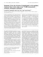
A similar experiment as described above was undertaken on the same
sample, to provide direct comparison of the behaviour of the IRSL signal
at different measurement temperatures. The procedure followed is
identical to that used for the IRPL measurement except that following
the preheat to 320 � C the IRSL signal is measured at the different tem
peratures for 200 s in order to deplete the IRSL signal. The IRSL signal
increases in intensity by a factor of over 30 times in this temperature
range. This increase is due to greater efficiency with which charge is
excited from the ground state and subsequently transported via bandtail states to recombination centres (Jain and Ankjaergaard, 2011).
Fig. 5. Impact of 1W 365 nm UV LED upon different luminescence signals from
feldspar. The procedure described in Table 2(a) was used for measurements of
the IRPL signal and Table 2(b) for the IRSL50 and pIR IRSL225 signal.
6. Single aliquot regenerative dose measurements
6.1. Measurement of a laboratory radiation dose
~400 nm IRSL emission. As has been observed many times previously,
the IRSL50 signal is reset more rapidly than the post-IR IRSL225 signal
(Fig. 5). In spite of the slower rate of bleaching for the post-IR IRSL225
signal it does appear to be well reset in many natural depositional en
vironments and has proved to be a very valuable chronometer for dating
sediments. The IRPL signal appears to bleach at a very similar rate as the
post-IR IRSL225 signal, and thus there is also potential for the IRPL signal
in sediment dating applications. The solar spectrum in nature is very
different to that from the 365 nm LED, and hence measurement of
samples bleached in nature will be important in future work. The ability
to look at grain to grain variations in the degree of resetting is clearly
advantageous. This is explored in section 6, but before that, the tem
perature dependence of the IRPL and IRSL signals are compared.
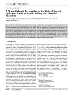
A single aliquot regenerative dose (SAR) protocol using the IRPL
signal is shown in Table 3. Grains were exposed to the UV LED for 600 s
after measurement of Lx and Tx in order to reduce the IRPL signal. To
assess the effectiveness of the procedure the set of grains that had pre
viously been measured to assess the impact of UV exposure upon the
IRPL and IRSL signals were used. Grains were given a beta dose of 104
Gy, and then the sequence in Table 3 was followed to build up a dose
response curve. The aim of this experiment was to see whether this SAR
sequence was able to recycle a dose, and reset the IRPL signal. Fig. 7(a)
shows the IRPL signal from a single grain in response to regeneration
doses ranging from zero to 166 Gy. The background subtracted from this
signal varies depending upon the signal measured in the four adjacent
background ROIs, but is typically ~40,000 counts per 0.1s channel per
ROI. The IRPL signal does not decline during the 2 s of stimulation at
5. Temperature dependence of IRPL and IRSL signals
The temperature dependence of the IRPL signal was investigated in
order to decide the optimum temperature at which to make IRPL mea
surements using the attachment described here. Kumar et al. (2018)
demonstrated that over the temperature range from 7 K to 295 K, the
955 nm IRPL emission remains constant from 7 K to ~200 K, and then
drops from 200 K to 295 K, but does not reach zero. An aliquot of po
tassium rich feldspar from GDNZ16 was used to extend the temperature
range for such measurements. The aliquot was given a beta dose of 166
Gy, preheated at 320 � C for 60 s and then had the IRSL signal measured
using the 880 nm LED while holding the sample at 50 � C. Following this,
the IRPL signal was measured at temperatures from 50 to 300 � C
(323–573 K). After each measurement, the aliquot was bleached using
the UV LED for 1200 s to remove the IRPL signal, and the aliquot was
then irradiated again and the cycle repeated. The IRPL signal measured
in the range 50–300 � C drops monotonically (Fig. 6), continuing the
trend seen by Kumar et al. (2018) at lower temperatures. This drop has
previously been interpreted as thermal quenching of the IRPL emission
process (Kumar et al., 2018), and is in stark contrast to the increase in
the IRSL signal that has been reported many times, as first seen by Duller
and Wintle (1991).
Fig. 6. Change in IRPL and IRSL signals with measurement temperature. Note
the different y-axes used for the IRPL and IRSL data. The temperature is shown
both in centigrade and kelvin to allow comparison with previously pub
lished data.
5
G.A.T. Duller et al.
Radiation Measurements 133 (2020) 106313
Table 3
Single aliquot regenerative dose (SAR) sequence used for IRPL measurements.
Step
1
2
3
4
5
6
7
8
9
10
11
Signal
Natural or Laboratory Irradiation
Preheat to 260 � C at 5 � C.sÀ 1 and then hold for 60s
IR (880 nm) for 200s, sample at 50 � C
IRPL (850 nm) for 2.5s (0.25s/image) at room temp
UV (365 nm) for 600 s at room temp
Test Dose (52 Gy)
Preheat to 260 � C at 5 � C.sÀ 1 and then hold for 60s
IR (880 nm) for 200s, sample at 50 � C
IRPL (850 nm) for 2.5s (0.25s/image) at room temp
UV (365 nm) for 600 s at room temp
Return to step 1
Lx
Tx
850 nm (as expected from Fig. 4(b)) and the small variation in IRPL
intensity during each measurement is thought to relate to fluctuations in
the intensity of the 850 nm laser diode (cf. Fig. 4(b)). To reduce the
impact of these small fluctuations, the IRPL signal is summed for the
period from 2 to 4s. The signals obtained are then used to construct a
dose response curve for each grain (e.g. Fig. 7(b)). Signals have been
screened to accept only those grains where recycling is within 10% of
unity, the uncertainty on the test dose is less than 10%, and signals grow
monotonically. Of the 100 grains on the sample disc, 23 gave data
suitable for constructing a dose response curve, and a radial plot of the
De values is shown in Fig. 7(c). As would be expected where the grains
have previously been conditioned in prior experiments, the De values do
not show any overdispersion. The mean De value is 96.4 � 1.5 Gy, 93%
of the given dose of 104 Gy. The results demonstrate that the instrument
is able to generate dose response curves from individual grains of feld
spar, and that the SAR procedure in Table 3 is able to recover a given
laboratory radiation dose.
6.2. Measurement of a natural radiation dose
A set of grains from GDNZ16 that had not previously been measured
were placed in a single grain holder, and the sequence shown in Table 3
used to measure the natural IRPL signal and De from the grains. Of the
100 grains measured, 22 gave data that passed the rejection criteria
outlined above. The IRPL dose response curve was measured to a
maximum regenerative dose of 1325 Gy (e.g. Fig. 8(a)) and dose
response curves have an average D0 value of 261 Gy, but this value
varied from 122 to 432 for individual grains. For the 22 grains for which
a De value was obtained, the average background was 59291 � 1717
counts per 0.25s (sum of ~450 pixels), and the average natural signal
(including the background) from the 22 grains was 94201 � 59291
counts per 0.25 s. Thus the average signal-to-noise ratio is only 0.59. In
spite of this low signal-to-noise ratio, dose response curves such as that
shown in Fig. 8(a) could be produced, and the radial plot shows that
consistent De values were obtained (Fig. 8(b)). The overdispersion of the
22 IRPL De values is 22%, near the lower end of the range observed in
the few examples of single grain IRSL data sets from potassium rich
feldspars (e.g. Reimann et al., 2012; Neudorf et al., 2012; Smedley et al.,
2016; Riedesel et al., 2018). The weighted mean IRPL De value obtained
is 103 � 5.8 Gy, 18% larger than the value of 87.0 � 3.7 Gy obtained by
Duller (1996). However, this earlier value may not be reliable since it
used a single aliquot additive dose method that is no longer used, and it
measured the IRSL emission at 50 � C but did not make any assessment of
anomalous fading. To provide a more appropriate comparison, the De
value for this sample was measured using a SAR protocol with a post-IR
IRSL signal.
The IRPL head was replaced with the DASH head to allow IRSL
measurements to be made. A second set of single grains of feldspar from
GDNZ16 were mounted on single grain discs and a post-IR IRSL225 SAR
sequence applied. The SAR sequence is almost identical to that described
in Table 3 except that the IRSL measurements were for 100 s while the
Fig. 7. IRPL instrument reproducibility assessment using single grains of
GDNZ16. The grains were given a beta dose of 104 Gy before starting the SAR
sequence. (a) An example of the net IRPL signal measured for a single grain
after different regeneration doses varying from zero to 166 Gy. (b) The SAR
dose response curve for the grain whose data are shown in (a). (c) Radial plot of
De values obtained for 23 grains. The overdispersion is zero demonstrating the
reproducibility of the attachment, and the recovered dose is 96.4 � 1.5 Gy, 93%
of the given dose of 104 Gy.
sample was held at a temperature of 50 � C, and the IRPL measurement
was replaced by a post-IR IRSL measurement for 200 s while holding the
sample at 225 � C. For the post-IR IRSL225 signal, a total of 76 grains of
the 100 grains measured passed the rejection criteria and gave a
weighted mean De of 105 � 3.8 Gy (Fig. 8), indistinguishable from the
IRPL value. The overdispersion of the post-IR IRSL225 De data is 25%,
similar to the IRPL value.
7. Conclusions
The use of an optical system designed to maximise the effectiveness
6
G.A.T. Duller et al.
Radiation Measurements 133 (2020) 106313
the advantages of working with an imaging detector and a sample that is
clearly defined within the field of view is that the background can be
measured by analysis of areas away from the sample and subtracted.
Reducing the breakthrough from the 850 nm stimulation will be
important in future developments, but subtraction of the background in
this way provides a system capable of measuring the IRPL signal from
single grains suitable for dating.
Declaration of competing interest
The authors declare that they have no known competing financial
interests or personal relationships that could have appeared to influence
the work reported in this paper.
Acknowledgements
This work was supported by the UK Space Agency CREST3 program
under grant ST/P001998/1. Research in Next Generation Luminescence
methods in Aberystwyth is supported by NERC grant CC003, and by
HEFCW infrastructure funding for SPARCL. Colleagues at DTU NuTech
(especially MyungHo Kook and Per Sørensen) are gratefully thanked for
their advice about interfacing to the DA-20 TL/OSL instrument. The two
anonymous referees are thanked for their comments which helped to
improve the clarity of the manuscript.
References
Buylaert, J.-P., Jain, M., Murray, A.S., Thomsen, K.J., Thiel, C., Sohbati, R., 2012.
A robust feldspar luminescence dating method for Middle and Late Pleistocene
sediments. Boreas 41, 435–451.
Colarossi, D., Duller, G.A.T., Roberts, H.M., Tooth, S., Lyons, R., 2015. Comparison of
paired quartz OSL and feldspar post-IR IRSL dose distributions in poorly bleached
fluvial sediments from South Africa. Quat. Geochronol. 30, 233–238.
Duller, G.A.T., 1996. The age of the Koputaroa dunes, southwest North Island, New
Zealand. Palaeogeogr. Palaeoclimatol. Palaeoecol. 121, 105–114.
Duller, G.A.T., 2008. Single grain optical dating of Quaternary sediments: why aliquot
size matters in luminescence dating. Boreas 37, 589–612.
Duller, G.A.T., Bøtter-Jensen, L., Kohsiek, P., Murray, A.S., 1999. A high-sensitivity
optically stimulated luminescence scanning system for measurement of single sandsized grains. Radiat. Protect. Dosim. 84, 325–330.
Duller, G.A.T., Bøtter-Jensen, L., Murray, A.S., 2003. Combining infrared- and greenlaser stimulation sources in single-grain luminescence measurements of feldspar and
quartz. Radiat. Meas. 37, 543–550.
Duller, G.A.T., Kook, M., Stirling, R.J., Roberts, H.M., Murray, A.S., 2015. Spatiallyresolved thermoluminescence from snail opercula using an EMCCD. Radiat. Meas.
81, 157–162.
Duller, G.A.T., Wintle, A.G., 1991. On infrared stimulated luminescence at elevated
temperatures. Nucl. Tracks Radiat. Meas. 18, 379–384.
Jain, M., Ankjaergaard, C., 2011. Towards a non-fading signal in feldspar: insight into
charge transport and tunnelling from time-resolved optically stimulated
luminescence. Radiat. Meas. 46, 292–309.
Kook, M., Lapp, T., Murray, A.S., Thomsen, K.J., Jain, M., 2015. A luminescence imaging
system for the routine measurement of single-grain OSL dose distributions. Radiat.
Meas. 81, 171–177.
Kook, M., Kumar, R., Murray, A.S., Thomsen, K.J., Jain, M., 2018. Instrumentation for
the non-destructive optical measurement of trapped electrons in feldspar. Radiat.
Meas. 120, 247–252.
Kumar, R., Kook, M., Murray, A.S., Jain, M., 2018. Towards direct measurement of
electrons in metastable states in K-feldspar: do infrared-photoluminescence and
radioluminescence probe the same trap? Radiat. Meas. 120, 7–13.
Lapp, T., Kook, M., Murray, A.S., Thomsen, K.J., Buylaert, J.P., Jain, M., 2015. A new
luminescence detection and stimulation head for the Risø TL/OSL reader. Radiat.
Meas. 81, 178–184.
Neudorf, C.M., Roberts, R.G., Jacobs, Z., 2012. Sources of overdispersion in a K-rich
feldspar sample from north-central India: insights from De, K content and IRSL age
distributions for individual grains. Radiat. Meas. 47, 696–702.
Prasad, A.K., Poolton, N.R.J., Kook, M., Jain, M., 2017. Optical dating in a new light: a
direct, non-destructive probe of trapped electrons. Sci. Rep. 7, 12097.
Reimann, T., Thomsen, K.J., Jain, M., Murray, A.S., Frechen, M., 2012. Single-grain
dating of young sediments using the pIRIR signal from feldspar. Quat. Geochronol.
11, 28–41.
Riedesel, S., Brill, D., Roberts, H.M., Duller, G.A.T., Garrett, E., Zander, A.M., King, G.E.,
Tamura, T., Burow, C., Cunningham, A., Seeliger, M., DeBatist, M., Heyvaert, V.M.
A., Fujiwara, O., Brückner, H., 2018. Single-grain feldspar luminescence chronology
Fig. 8. (a) IRPL dose response curve for a single grain of GDNZ16. (b) Radial
plot of De values for grains of GDNZ16. De data are shown from IRPL mea
surements (filled circles) and from post-IR IRSL225 measurements
(open triangles).
of the LP925 and BP950 rejection filters, and using the well-defined
spatial definition of the sample within the image has made possible an
IRPL system for single grains based on an EM-CCD detector. Although
the signal-to-noise ratio is still low in comparison with the performance
typically achieved for IRSL measurements, this IRPL system is able to
generate reproducible data, with a typical variability of less than 1%
between replicate measurements of the same sample (Fig. 4(b)). The
955 nm IRPL emission is strongly affected by thermal quenching, with
the signal dropping monotonically from 50 � C to 300 � C (Fig. 6), and
thus subsequent measurements of IRPL have been made at room tem
perature to maximise the signal. A single aliquot regenerative dose
(SAR) method has been applied, using a UV LED to reset the IRPL signal
between regeneration cycles. A dose recovery experiment demonstrates
the reproducibility of the IRPL measurements, that a dose can be
recovered within 10% of the given dose, and the suitability of the UV
LED for resetting the signal. The natural De for single grains of an aeolian
dune sand (GDNZ16) from New Zealand measured using IRPL (103 �
5.8 Gy) is consistent with the value derived using a post-IR IRSL225
signal (105 � 3.8 Gy). The age control for GDNZ16 is poorly constrained
(Duller, 1996) so it is not possible to assess the accuracy of this De
determination, but future work using IRPL will focus on analysis of
samples with robust independent age control in order to assess the ac
curacy of ages that can be calculated using this new signal.
Whilst the breakthrough from the 850 nm stimulation is high, one of
7
G.A.T. Duller et al.
Radiation Measurements 133 (2020) 106313
of historical extreme wave event deposits recorded in a coastal lowland, Pacific coast
of central Japan. Quat. Geochronol. 45, 37–49.
Smedley, R.K., Glasser, N.F., Duller, G.A.T., 2016. Luminescence dating of glacial
advances at lago Buenos Aires (~46 � S), Patagonia. Quat. Sci. Rev. 134, 59–73.
Thomsen, K.J., Kook, M., Murray, A.S., Jain, M., Lapp, T., 2015. Single-grain results from
an EMCCD-based imaging system. Radiat. Meas. 81, 185–191.
8