Annealing properties of the PVA-GTA-I gel dosimeter
Bạn đang xem bản rút gọn của tài liệu. Xem và tải ngay bản đầy đủ của tài liệu tại đây (3.39 MB, 6 trang )
Radiation Measurements 149 (2021) 106674
Contents lists available at ScienceDirect
Radiation Measurements
journal homepage: www.elsevier.com/locate/radmeas
Annealing properties of the PVA-GTA-I gel dosimeter
˜ o a, b, c, Chryzel Angelica B. Gonzales a, Akito Saito d, Takuya Wada e,
Jolan E. Tan
Yasushi Nagata f, Hiroshi Yasuda a, *
a
Department of Radiation Biophysics, Research Institute for Radiation Biology and Medicine (RIRBM), Hiroshima University, Kasumi 1-2-3, Minami-ku, Hiroshima,
734-8553, Japan
b
Graduate School of Biomedical and Health Sciences, Hiroshima University, Kasumi 1-2-3 Minami-ku, Hiroshima, 734-8553, Japan
c
Phoenix Leader Education Program (Hiroshima Initiative) for Renaissance from Radiation Disaster, Hiroshima University, Kasumi 1-2-3 Minami-ku, Hiroshima, 7348553, Japan
d
Department of Radiation Oncology, Hiroshima University Hospital, Kasumi 1-2-3, Minami-ku, Hiroshima, 734-8551, Japan
e
Section of Radiation Therapy, Department of Clinical Support, Hiroshima University Hospital, Kasumi 1-2-3, Minami-ku, Hiroshima, 734-8551, Japan
f
Department of Radiation Oncology, Institute of Biomedical and Health Sciences, Hiroshima University Hospital, Kasumi 1-2-3, Minami-ku, Hiroshima, 734-8551, Japan
A R T I C L E I N F O
A B S T R A C T
Keywords:
Radiochromic gel dosimeter
PVA-GTA-I
Annealing
LINAC X-rays
γ-rays
Radiation therapy
Reusable
The feature of reusability of a gel dosimeter is of particular interest for application to three-dimensional
dosimetry in clinical settings. As one of the reusable materials, the radiochromic gel formula composed of
polyvinyl alcohol (PVA), glutaraldehyde (GTA), and iodide (I), abbreviated as “PVA-GTA-I gel dosimeter”, is
investigated in the present study. The annealing properties of the PVA-GTA-I gel were examined for energetic Xrays from a medical linear accelerator, while its reusability was confirmed for 137Cs-source γ-rays. The radiationinduced colorings of the PVA-GTA-I gel irradiated with few Gy were erased within several hours, while the
reactions induced by higher dose (~20 Gy) were more persistent and required longer time up to 24 h to be
completely reversed. The linear dose response of the PVA-GTA-I dosimeter was well reproduced after repetition
of annealing.
1. Introduction
Gel dosimeters are chemically-based materials that allow 3D dose
distribution analysis due to their unique characteristic of recording
spatial dose information within its medium (Baldock et al., 2010;
Romanyukha et al., 2011; Schreiner, 2015). This attribute is of great
importance in the dosimetry and quality assurance of modern radiation
therapy plans, where current and emerging techniques require more
accurate methods to quantify ionizing radiation fields in three di
mensions (3D). In recent studies, there have been considerable interests
in the development and improvement of radiochromic gel dosimeters
that rely on the radiation-induced color transformation which is pro
portional to the absorbed dose (Solc and Spvek, 2009; Vandecasteele
et al., 2011; Nasr et al., 2015; Oldham, 2015; Colnot et al., 2018; Kouvati
et al., 2019). Using these materials couple with optical techniques such
as spectrophotometry or optical computed tomography (optical CT), the
3D radiation dose distribution in a human tissue or organ could be
approximately measured.
˜ o et al., 2019, 2020), we investigated the
In our earlier studies (Tan
rudimentary dose-response characteristics of an experimental radio
chromic gel formula for 137Cs-source γ-rays. The formula is composed of
mostly water, polyvinyl alcohol (PVA) crosslinked with glutaraldehyde
(GTA), potassium iodide (KI), fructose, and glucono-δ-lactone (GDL),
which was abbreviated as the ‘PVA-GTA-I gel dosimeter’. The
PVA-GTA-I gel dosimeter was developed through modification of the
polyvinyl alcohol-iodide (PVA-I) based gel (Miyoshi et al., 2016; Suna
gawa et al., 2017; Hayashi et al., 2019, 2020) in conjunction with
glutaraldehyde (GTA) as a cross-linking agent which was applied to
Fricke-gels (d’Errico et al., 2017; Marini et al., 2017). This PVA-GTA-I
gel dosimeter converts from transparent to color red after irradiation.
It is relatively easy to fabricate and has high transparency and sensi
tivity. Moreover, this gel formula could be reinitialized through
˜ o et al., 2019).
annealing, as qualitatively confirmed by the authors (Tan
This feature is comparable to the PRESAGE-RU radiochromic plastic
formulation which was also reported to have potential reusability
through the rapid decay of its coloration in dark storage and room
temperature conditions (Juang et al., 2015). In the PVA-GTA-I gel, the
main cause of color loss is through heating which results in the
* Corresponding author.
E-mail address: (H. Yasuda).
/>Received 24 June 2021; Received in revised form 6 September 2021; Accepted 20 October 2021
Available online 25 October 2021
1350-4487/© 2021 The Authors. Published by Elsevier Ltd. This is an open access article under the CC BY license ( />
J.E. Ta˜
no et al.
Radiation Measurements 149 (2021) 106674
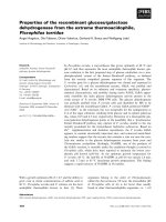
Fig. 1. Irradiation (a) set-up and (b) geometric dimensions of the PVA-GTA-I gel samples using X-rays from the 6-MV medical linear accelerator (LINAC).
separation or dissociation of the triiodide ions (I3− ) and the PVA-GTA
complex. Additionally, the presence of the sugar (i.e., fructose) further
reduces the I3− to mono iodide ions (3I− ) which finally leads to the
reinitialized or colorless state of the gel. Since it is only in more recent
years that the PVA-GTA-I gel is found to have such advantageous
characteristics, further research is needed in terms of the quantitative
analyses of the annealing properties.
Thus, in the present study, the authors focus on further investigating
the annealing properties of the PVA-GTA-I gel dosimeter, expecting that
a practical method to reinitialize the dosimeter between the fractions of
dose delivery to radiotherapy patients will be established. More
concretely, we analyze the absorbance changes with the investigation of
the effect of natural oxidation on the annealed gel samples, trying to
provide possible implications towards its application for radiation
therapy.
irradiation set-up and the measurement duration, three separate solu
tions of the same PVA-GTA-I gel formula were fabricated. The annealing
schedule was divided into three batches: the first batch for 0.25, 0.5,
0.75 and 1.0 h annealing; the second batch for 4, 8, and 10 h annealing;
and the third batch for 12, 16, 20, and 24 h annealing. Each annealing
time has a corresponding set of samples which comprises 27 cuvette
samples (i.e., three cuvette samples for each dose level). A total of 297
cuvette samples of the PVA-GTA-I gel dosimeter were prepared for these
experiments. The dose-responses of the three sample batches were also
analyzed to check the fabrication reproducibility.
2.2. Medical linear accelerator irradiation
The irradiations of the PVA-GTA-I gel dosimeters were conducted
using a 6-MV high-energy photon beam produced by TrueBeam™
medical linear accelerator (LINAC) (Varian Medical Systems, CA, USA)
located at Hiroshima University Hospital (Hiroshima, Japan). As illus
trated in Fig. 1, the gel dosimeters were positioned perpendicular to the
beam axis and in between solid water-equivalent phantoms (PH-40
Tough Water Phantom®, Kyoto Kagaku Co., Ltd., Kyoto, Japan). The
source-to-skin distance (SSD) and source-to-axis distance (SAD) were
98.4 cm and 100 cm, respectively. The irradiation field size was 15 × 15
cm2, and a fixed dose rate at 600 MU⋅min− 1 was employed. The gel
dosimeters were irradiated with photon beams at 8 dose levels of 1, 2, 3,
4, 7, 10, 15, and 20 Gy with one set of samples left unirradiated (con
trol). Shortly after irradiation, all the samples were stored inside the
refrigerator at 15 ◦ C for about 24 h. Following this, the samples were
then stabilized at room temperature for 2 h before performing the initial
measurements.
2. Materials and methods
The methods used in this study comprised of preparation of the PVAGTA-I gel dosimeter samples, irradiation of the samples using high en
ergy X-rays and annealing the irradiated samples at a selected temper
ature wherein the absorbance decays were then quantitatively
measured. While, the same-composition material was irradiated with
137
Cs-source γ-rays for confirmation of its reusability.
2.1. Gel sample preparation
The synthesis of the gel samples was performed by first dissolving 10
wt % of partially saponified polyvinyl alcohol (PVA) (86–90 mol%
saponification, polymerization degree = 1000) in Milli-Q™ ultrapure
water (resistivity = 18.2 M Ω cm). The potassium iodide (KI), fructose,
glucono-δ-lactone (GDL), and 25% aq. glutaraldehyde (GTA) with con
centrations of 1.42, 1.54, 1.53, and 0.36 wt %, respectively, were then
added into the PVA solution at room temperature (20 ◦ C) until a ho
mogeneous mixture was achieved. All the reagents used were of
analytical grade from FUJIFILM Wako Pure Chemical Corporation
(Osaka, Japan). The resulting liquid solution was transferred into the
uncovered PMMA cuvettes (10 × 10 × 45 mm3) and placed inside a
vacuum chamber (− 0.08 MPa) for 2 h to inhibit the formation of air
pockets within the gel samples. After removing from the vacuum, the
cuvette samples were covered with lids and parafilm to minimize water
loss during the subsequent heating process (i.e., for gel solidification).
The samples were heated at 50 ◦ C for 12 h in a sterilizing oven (Sanyo
MOV-112S, Sanyo Electric Biomedical Co., Ltd., Osaka, Japan) and then
stabilized at room temperature for 2 h before irradiation.
For preparation of a large number of samples and limitations in the
2.3. Absorbance measurement and annealing process
The measurements of the optical absorbances were conducted using
a NanoDrop OneC™ UV–Vis spectrophotometer (Thermo Fisher Scien
tific Inc., MA, USA). The absorbance value at the 486 nm peak was used
as the reference point for all the results in the present study. Following
initial measurements, the PVA-GTA-I gel dosimeter samples were
immediately annealed in a pre-heated oven at 50 ◦ C with time intervals
of 0.25, 0.5, 0.75, 1, 4, 8, 10, 12, 16, 20, and 24 h. The gel dosimeters
were always stabilized at room temperature for 2 h before the UV–Vis
measurements. The change of absorbance (ΔAbs) was obtained from the
difference between the measured absorbance of the irradiated sample
(Absi ) and unirradiated (control) sample (Absc ), as defined in Eq. (1):
ΔAbs = Absi –Absc
2
(1)
J.E. Ta˜
no et al.
Radiation Measurements 149 (2021) 106674
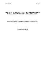
Fig. 2. Irradiation set-up of the PVA-GTA-I gel samples using the
source γ-rays.
137
Fig. 4. Dose vs. ΔAbs plots of the three batches of gel samples fabricated with
the same PVA-GTA-I gel formula. Linear fitting was applied to each batch; the
error bar represents 1 s.d.
Cs-
Canada). The experimental set-up is shown in Fig. 2. The gel dosimeters
were positioned at the center of the sample container (φ 260 × 100 mm)
perpendicular to the sources. Three sets of irradiations were performed
to the PVA-GTA-I gel dosimeters for six dose levels: 1, 2, 3, 5, 7, and 10
Gy with the dose rate of 0.806 Gy min− 1. Soon after each succeeding
irradiations and measurements, the samples were annealed repeatedly
for 12 h at 50 ◦ C for the reinitialization process. All samples were stored
inside the refrigerator at 5 ◦ C in between multiple irradiations and
measurements. It should be noted that high-energy X-rays were not used
for the reusability test due to the limited access in the facility caused by
the COVID-19 pandemic, hence the 137Cs γ-ray source were used as an
alternative radiation source which was more accessible.
3. Results and discussion
3.1. Fabrication reproducibility
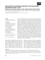
The photographic images of the PVA-GTA-I gels irradiated with
0 (control), 1, 2, 3, 4, 7, 10, 15 and 20 Gy of LINAC X-rays and annealed
for 0 (before starting), 1, 12 and 24 h are shown in Fig. 3. It was visually
confirmed that the radiation-induced colorings of the PVA-GTA-I do
simeters were reversed to the initial state after 24 h at longest.
Fig. 4 shows the dose- ΔAbs plots of the three batches of the PVAGTA-I gel dosimeter. The dose-response was linear for all batches with
average Re = 0.99 and the average sensitivity defined by the slope (the m
value indicated in the figure) was around 0.035 Gy-1. The small varia
tions in sensitivity could be attributable to several factors such as at
mospheric temperature and humidity during the storage and
stabilization stages. Table 1 shows the summary of the one-way ANOVA
test which was conducted to examine difference between the ΔAbs
values among the different PVA-GTA-I gel batches. A p-value of less than
0.05 was required to reject the null hypothesis H0. The result of the
ANOVA (F(2,24) = 0.0085, P = 0.99), which did not reject the H0,
confirmed the good reproducibility in fabrication of the different
batches of PVA-GTA-I gels.
Fig. 3. Images of the PVA-GTA-I gels irradiated with 0 (control), 1, 2, 3, 4, 7,
10, 15 and 20 Gy of 6-MV LINAC X-rays at 0h, 1h, 12h and 24 h of annealing
duration. All the samples were stored in a refrigerator after irradiation until the
annealing was started.
2.4. Reusability test with
137
Cs-source γ-rays
3.2. Behavior of the annealing decay curve
Reusability of the PVA-GTA-I gels having the same composition was
examined using 137Cs source γ-rays from the Gammacell®40 Exactor
Low Dose Rate Research Irradiator (Best Theratronics Ltd., Ottawa,
Fig. 5 presents the ΔAbs with respect to annealing time of the PVAGTA-I gel dosimeters irradiated with different doses. Each data point
3
J.E. Ta˜
no et al.
Radiation Measurements 149 (2021) 106674
Table 1
Summary of one-way ANOVA test for the fabrication reproducibility.
SUMMARY
Groups
Count
Sum
Average
Variance
Batch 1
Batch 2
Batch 3
9
9
9
2.051889
2.182519
2.146694
0.227988
0.242502
0.238522
0.056427
0.062168
0.059264
Source of Variation
SS
df
MS
F
P-value
F crit
Between Groups
Within Groups
Total
0.001012
1.422867
1.42388
2
24
26
0.000506
0.059286
0.008538
0.991501
3.402826
ANOVA
Table 2
Fitting parameters (see equation (2)) and goodness-of-fit values for each dose
level of the PVA-GTA-I gel dosimeters annealed at 50 ◦ C for up to 24 h.
Fitting parameters
+ A2 e(−
λ2 *t)
λ1 (h− 1)
A2 (arb.
unit)
λ2 (h− 1)
A1/
A2
ratio
R2
χ2
1
0.0442
3.33
0
–
–
0.946
2
0.0846
3.33
0.0057
0.006
14.8
0.997
3
0.1112
3.33
0.0259
0.468
4.30
0.993
4
0.1225
3.33
0.0648
0.478
1.89
0.997
7
0.1927
3.33
0.1508
0.335
1.28
0.997
10
0.2035
3.33
0.2924
0.245
0.70
0.998
15
0.2815
3.33
0.4718
0.156
0.60
0.996
20
0.2146
3.33
0.7749
0.118
0.28
0.996
1.65
×
10− 4
4.04
×
10− 5
2.70
×
10− 4
2.40
×
10− 4
8.89
×
10− 4
1.35
×
10− 3
5.26
×
10− 3
1.05
×
10− 2
(χ2) values were also calculated. The values of those parameters are
summarized in Table 2. Additionally, the absorbance half-life was
determined from the decay coefficient by Eq. (3):
is an average of three samples with three measurement series obtained at
the 486 nm peak. The error bars shown in the plot represent one stan
dard deviation (1 s.d.) that were mostly located within the data markers.
It was found that the time changes of ΔAbs values be fitted well with
a two-phase exponential decay function, as described by Eq. (2):
λ1 *t)
A1 (arb.
unit)
Note: The parameter values in this table correspond to Eq. (2) in the text and the
fitting curves in Fig. 5; A1 & λ1 correspond to the fast component and A2 & λ2 the
slow component; R2 and χ2 are the coefficient of determination and chi-square
value, respectively.
Fig. 5. Annealing time vs. ΔAbs plots of the PVA-GTA-I gel dosimeters irradi
ated with doses from 1 to 20 Gy. The inset graph shows the magnified view of
the lower dose region (1–4 Gy). Each dose level is fitted with the two-phase
decay function and is represented by the line. Note that the ΔAbs values were
normalized from 0 to 1 for illustration purposes.
A(t) = A1 e(−
Goodness-of-fit
Dose
(Gy)
Tn =
ln 2
λn
(3)
where Tn and λn are the half-life and decay coefficient at a given phase
order n (n = 1 or 2), respectively.
From the results shown in Fig. 5 and Table 2, it was found that the
annealing decay curves of the ΔAbs of the PVA-GTA-I gel dosimeters can
be approximated well with the two-phase decay model. It was seen that
the contribution ratio of the fast decay component to slow decay
component (i.e, A1/A2) became smaller with increasing dose level; it
was considered that the contribution of the slow decay component was
negligible for 2 Gy or less. The average half-life (T2 ) of the slow decay
component calculated by Eq. (3) for 3 Gy or higher dose ranged from 1.5
h for 3 Gy to 5.9 h for 20 Gy, while T1 was constant as 12 min. Further
(2)
where A(t) is the absorbance at a given time t; A1 and A2 are the am
plitudes of each decay phase; and λ1 and λ2 are the fast and slow decay
constants, respectively. Through the careful regression analyses, it was
found that the value of λ1 could be given as a constant (= 3.33); the other
three parameters (A1 , A2 and λ2 ) were judged to be variable quantities
and then determined by the least-squares method. Additionally, to verify
the goodness-of-fit, the coefficient of determination (R2) and chi-square
4
J.E. Ta˜
no et al.
Radiation Measurements 149 (2021) 106674
Fig. 6. Annealing time vs. ΔAbs subplots of the PVA-GTA-I gel samples measured soon after annealing (black marker) and 7d after annealing (red marker) for
different dose levels. Each point is the average ΔAbs from three samples and the error bar represents 1 s.d. (For interpretation of the references to color in this figure
legend, the reader is referred to the Web version of this article.)
analysis of the two-phase decay model reveals the estimated annealing
time for the effective reinitialization (i.e., normalized ΔAbs ≤ 0.003) of
the irradiated gel samples: 1 Gy ≈ 1h, 2 Gy ≈ 1.25h, 3 Gy ≈ 4.75h, 4 Gy
≈ 6.5h, 7 Gy ≈ 11.75h, 10 Gy ≈ 18.75h, 15 Gy ≈ 32.5h, 20 Gy ≈ 47.25h.
These results suggest that the radiation-induced coloring in the PVAGTA-I gel dosimeter is erased quickly at the lower dose range (~few
Gy) by annealing, whereas the reactions induced by highly dense ioni
zation (~20 Gy) are more persistent and require a longer time to be
completely reversed.
3.3. Auto-oxidation 7 days post-annealing
The natural oxidation in the PVA-GTA-I gel dosimeter was measured
after 7 days had elapsed post-annealing. The results are shown in Fig. 6
in which subplots of the ΔAbs values from the gel dosimeter samples
measured soon (~2h) after annealing (black marker) and 7d after
annealing (red marker) for different dose levels. The error bars represent
1 s.d. The purpose of this subplots was to confirm if the gel dosimeter is
effectively reinitialized since previous observations from unpublished
experiments showed that the PVA-GTA-I gel dosimeter (especially those
irradiated >10Gy) tend to have residual absorbance after annealing. To
further elaborate, a freshly annealed PVA-GTA-I gel dosimeter is color
less in visual inspection and displayed an absorbance value comparable
to an unirradiated dosimeter after the spectrophotometry measurement.
However, the gel coloration “relapsed” within several hours or days,
which indicated that the gel is not sufficiently annealed and that some
slow decay components are still present. In the context of the data
presented in the study, the contrast of the ΔAbs values between the 2h
and 7d post-annealing measurements provides further evidence in the
effective reinitialization time of the PVA-GTA-I gel dosimeter for each
dose level. For instance, in the 10 Gy sample measured soon after
annealing, the ΔAbs values from 12 to 24h of annealing are all ≤0.003.
However, measuring the sample again 7d after annealing reveals that
the ΔAbs at the 12h annealing point significantly increased to 0.024,
while those annealed from 16h to 24h maintained their values within
≤0.003. This observation implies that 12h of annealing at 50 ◦ C is not
enough to effectively reinitialize the PVA-GTA-I gel. In general, the PVA-
Fig. 7. Results of the reusability test of the PVA-GTA-I gel dosimeter. The same
samples were irradiated repeatedly with 137Cs-source γ-rays (1, 2, 3, 5, 7, 10
Gy) after annealing. All the samples were stored in the refrigerator for 5 ◦ C
between irradiations and measurements.
GTA-I gel dosimeters irradiated with few Gy were reinitialized in several
hours, while those with higher doses (~20 Gy) needed a longer time up
to 24 h. Thus, it is commonly recommended to apply the annealing
condition of 24 h at 50 ◦ C to erase the colorings of the PVA-GTA-I gel
dosimeters irradiated with a fractionated delivering dose in radiation
therapy.
5
J.E. Ta˜
no et al.
Radiation Measurements 149 (2021) 106674
3.4. Confirmation of reusability
17K09072 and 18KK0417).
Fig. 7 shows the dose responses of the PVA-GTA-I gels that were
irradiated with 137Cs source γ-rays repeatedly after annealing. Each data
point is an average and the error bar is one standard deviation of the
data obtained from three samples; most of the error bars were located
within the data markers. As expected from the preceding study using a
˜ o et al., 2019), the linear dose
similar-composition PVA-GTA-I gel (Tan
response up to 10 Gy was reproduced well after repetition of annealing;
the sensitivities were 3.9 × 10− 2, 3.9 × 10− 2, and 4.0 × 10− 2 for the
first, second, and third irradiation, respectively. Accordingly, it is
considered that the PVA-GTA-I gels developed in the present study could
be used repeatedly at least three times by application of an appropriate
annealing process.
References
Baldock, C., De Deene, Y., Doran, S., Ibbott, G., Jirasek, A., Lepage, M., McAuley, K.B.,
Oldham, M., Schreiner, L.J., 2010. Polymer gel dosimetry. Phys. Med. Biol. 55
/>Colnot, J., Huet, C., Gschwind, R., Clairand, I., 2018. Characterisation of two new
radiochromic gel dosimeters TruViewTM and ClearViewTM in combination with the
vistaTM optical CT scanner: a feasibility study. Phys. Med. 52, 154–164. https://doi.
org/10.1016/j.ejmp.2018.07.002.
d’Errico, F., Lazzeri, L., Dondi, D., Mariani, M., Marrale, M., Souza, S.O., Gambarini, G.,
2017. Novel GTA-PVA Fricke gels for three-dimensional dose mapping in
radiotherapy. Radiat. Meas. 106, 612–617. />radmeas.2017.07.003.
Hayashi, S., Ono, K., Fujino, K., Fujimoto, S., 2019. Influence of the components of a
radiochromic PVA – iodide gel dosimeter on the optical dose response. J. Phys. Conf.
Ser. 1305, 012031 />Hayashi, S., Ono, K., Fujino, K., Ikeda, S., Tanaka, K., 2020. Novel radiochromic gel
dosimeter based on a polyvinyl alcohol – iodide complex. Radiat. Meas. 131,
106226. />Juang, T., Adamovics, J., Oldham, M., 2015. Characterization of a reusable PRESAGE®
3D dosimeter. J. Phys. Conf. Ser. 573, 6–11. />573/1/012039.
Kouvati, K., Jaszczak, M., Papagiannis, P., Kadlubowski, S., Wach, R., Maras, P.,
Dudek, M., Kozicki, M., 2019. Leuco crystal violet-Pluronic F-127 3D radiochromic
gel dosimeter. Phys. Med. Biol. 64, 175017. />ab2f5d.
Marini, A., Lazzeri, L., Cascone, M.G., Ciolini, R., Tana, L., D’Errico, F., 2017. Fricke gel
dosimeters with low-diffusion and high-sensitivity based on a chemically crosslinked PVA matrix. Radiat. Meas. 106, 618–621. />radmeas.2017.02.012.
Miyoshi, H., Masahiko, Y., Maeda, S., Yamada, K., Matsumura, J., 2016. Reversible
radiochromic plate based on polyvinyl alcohol-iodide complex containing silica
nanoparticles. J. Radioanal. Nucl. Chem. 308, 469–475. />s10967-015-4465-y.
Nasr, A.T., Alexander, K.M., Olding, T., Schreiner, L.J., McAuley, K.B., 2015. Leucocrystal-violet micelle gel dosimeters: II. Recipe optimization and testing. Phys. Med.
Biol. 60, 4685–4704. />Oldham, M., 2015. Radiochromic 3D detectors. J. Phys. Conf. Ser. 573 />10.1088/1742-6596/573/1/012006.
Romanyukha, A., Trompier, F., Reyes, R.A., Melanson, M.A., 2011. EPR measurements of
fi ngernails in Q-band. Radiat. Meas. 46, 888–892. />radmeas.2011.04.004.
Schreiner, L.J., 2015. True 3D chemical dosimetry (gels, plastics): development and
clinical role. J. Phys. Conf. Ser. 573 />012003.
Solc, J., Spvek, V., 2009. New 3D radiochromic gel dosimeters with inhibited diffusion.
J. Phys. Conf. Ser. 164, 1–7. />Sunagawa, T., Harvel, G., Aoki, Y., Umeda, M., Hayami, J., Sakakibara, K., Goto, H.,
Ebina, T., Taguchi, M., Nagasawa, N., Yoshihashi, S., Hatashita, M., Kume, K.,
Sakura, T., 2017. Development of the gel indicator using PVA and KI. Mem. Fukui
Univ. Technol. 47, 105–110 (in Japanese).
Ta˜
no, J., Hayashi, S., Hirota, S., Gonzales, C.A., Yasuda, H., 2019. Development of a
reusable PVA-GTA-I gel dosimeter for 3D radiation dose assessments. J. Phys. Conf.
Ser. 1305 />Ta˜
no, J.E., Hayashi, S. i, Hirota, S., Gonzales, C.A.B., Yasuda, H., 2020. Effect of the
glucono-δ-lactone concentration on the sensitivity and stability of PVA-GTA-I
radiochromic gel dosimeter. Radiat. Meas. 134 />radmeas.2020.106311.
Vandecasteele, J., Ghysel, S., Baete, S.H., Deene, Y. De, 2011. Radio-physical properties
of micelle leucodye 3D integrating gel dosimeters. Phys. Med. Biol. 56, 627–651.
/>
4. Conclusions
In the present study, the annealing properties of the PVA-GTA-I gel
dosimeter were quantitatively examined for the first time. As a result, it
was found that the annealing responses of PVA-GTA-I gel dosimeters
irradiated with X-rays of up to 20 Gy from a medical linear accelerator
could be well approximated with two-phase exponential decay func
tions. The radiation-induced colorings at few Gy were easily erased in
several hours, whereas a longer annealing time of up to 24 h was
required for reinitializing the samples irradiated with a higher dose. The
identical, linear dose response was well maintained after repeated
annealing processes for γ-ray irradiation. These findings will be an
important basis for using the PVA-GTA-I gel dosimeter as a tool for
dosimetry in radiation therapy. The feature of reusability of this novel
dosimeter is favorable as it opens the prospect of routine use in clinical
settings where multiple, detailed dose verifications need to be per
formed. For establishing a practical use of the PVA-GTA-I gel dosimeter
as part of the general protocol of radiation therapy, it is highly recom
mended to conduct further studies to clarify the possible extent of its
repeated use and the best storage conditions that can minimize the
natural oxidation as well as the fundamental physico-chemical mecha
nisms behind the annealing effects observed. Research in the quest for
resolutions on these subjects is currently underway.
Declaration of competing interest
The authors declare that they have no known competing financial
interests or personal relationships that could have appeared to influence
the work reported in this paper.
Acknowledgments
The authors would like to thank the staff of the Department of Ra
diation Oncology of Hiroshima University Hospital for their technical
support. This work was supported by the JSPS KAKENHI (Grant Number
6