Analytical characterization of DNA and RNA oligonucleotides by hydrophilic interaction liquid chromatography-tandem mass spectrometry
Bạn đang xem bản rút gọn của tài liệu. Xem và tải ngay bản đầy đủ của tài liệu tại đây (2.66 MB, 11 trang )
Journal of Chromatography A 1648 (2021) 462184
Contents lists available at ScienceDirect
Journal of Chromatography A
journal homepage: www.elsevier.com/locate/chroma
Analytical characterization of DNA and RNA oligonucleotides by
hydrophilic interaction liquid chromatography-tandem mass
spectrometry
Ming Huang, Xiaobin Xu∗, Haibo Qiu∗, Ning Li
Regeneron Pharmaceuticals Inc., Tarrytown, NY 10591, USA
a r t i c l e
i n f o
Article history:
Received 7 January 2021
Revised 28 March 2021
Accepted 18 April 2021
Available online 27 April 2021
Keywords:
Oligonucleotides
siRNAs
Hydrophilic interaction liquid
chromatography
Tandem mass spectrometry
Phosphorothioate
Synthetic metabolites
a b s t r a c t
Liquid chromatography-mass spectrometry has been widely implemented as a powerful tool for providing in-depth characterization of nucleic acid therapeutic modalities, such as anti-sense oligonucleotides
and small interfering RNAs (siRNAs). In this study, we developed a generic hydrophilic interaction liquid chromatography (HILIC) hyphenated with tandem mass spectrometry method in the absence of ionpairing reagents and demonstrated its capability as an attractive and robust alternative for oligonucleotide and siRNA analysis. HILIC separation of mixtures of unmodified and fully phosphorothioatemodified DNA oligonucleotides and their synthetic 3’ exonuclease-digested metabolites were also assessed. High-resolution mass spectrometric (HRMS) analysis was used to determine the deconvoluted
masses of oligonucleotide and siRNA standards and their impurities. To enable unbiased sequence characterization with tandem mass spectrometry (MS/MS), we also optimized higher-energy C-trap dissociation (HCD) on improving the sequence coverage of DNA and RNA oligonucleotides. Lastly, we evaluated
on-column sensitivity for a phosphorothioate oligonucleotide by performing targeted analysis with either
targeted selected ion monitoring (tSIM) or parallel reaction monitoring (PRM). Higher on-column sensitivity of 13 ng, equivalent to 2.0 pmol, of a phosphorothioate oligonucleotide was achieved by tSIM analysis
as compared to PRM analysis.
© 2021 The Authors. Published by Elsevier B.V.
This is an open access article under the CC BY-NC-ND license
( />
1. Introduction
The continued optimism for synthetic nucleic acid drugs has fueled an increasing interest in antisense oligonucleotide therapy and
RNA interference (RNAi) therapy. Due to their mechanisms of gene
silencing and modulation of gene expression, oligonucleotides and
small interfering RNAs (siRNAs) represent a distinct class of therapeutic molecules. Recently, an increasing number of these drug
candidates have been approved or have progressed to later-stage
clinical trials [1,2]. However, there are different regulatory strategies for the approval pathway of nucleic acid therapeutics. These
drug candidates are categorized by the Food and Drug Administration (FDA) as small molecule drug candidates while they are categorized as biologics by European Medicines Agency (EMA) [3]. Both
oligonucleotides and siRNAs are large, highly negatively charged
∗
Corresponding authors.
E-mail addresses: (X. Xu),
(H. Qiu).
molecules and show fundamentally different chemical and physiological properties relative to small molecule and antibody drugs.
Driven by drug development needs and regulatory requirements,
numerous analytical strategies have been explored to provide indepth characterization of various drug facets, such as synthetic impurities, chemical modifications, and metabolites.
In 1990 Andrew Alpert coined the term “hydrophilic interaction chromatography” (HILIC) to describe a liquid chromatographic
technique for separating polar or ionized analytes. The HILIC retention mechanisms involved the partition of solutes between the
bulk mobile phase and an enriched water layer partially immobilized on the stationary phase surface, as well as secondary electrostatic and hydrogen bonding interactions [4]. Since then, HILIC coupled to mass spectrometry (HILIC−MS) has been broadly adopted
in the characterization of different classes of biomolecules, including proteins, glycopeptides, and small molecules, such as glycans
and saccharides [5,6]. Because polar ribosyl residues and ionizable phosphate groups in nucleic acid structures are hydrophilic,
early applications of HILIC include bioanalysis of nucleosides and
/>0021-9673/© 2021 The Authors. Published by Elsevier B.V. This is an open access article under the CC BY-NC-ND license ( />
M. Huang, X. Xu, H. Qiu et al.
Journal of Chromatography A 1648 (2021) 462184
LC−MS grade) were purchased from Sigma-Aldrich (St. Louis,
MO, USA). HPLC-grade acetonitrile was purchased from Honeywell (Charlotte, NC, USA). Deionized water was provided by a
Milli-Q integral water purification system installed with a MilliPak Express 20 filter (Milli-Q system, Millipore, El Passo, TX, USA).
All DNA and RNA oligonucleotides used in this study were purchased from Integrated DNA Technologies Inc. (IDT; Coralville, IA,
USA), and their sequences are listed in Supplementary Table 1. All
synthetic oligonucleotides contained 3 - and 5 -terminal hydroxyl
groups. The luciferase-targeted siRNA (siLuc) and its corresponding sense and anti-sense strands were purchased from IDT (Supplementary Table 1). MISSION® siRNA Universal Negative Control
#1 (SIC001, 13317 g/mol) and MISSION® siRNA Universal Negative
Control #2 (SIC002, 13302 g/mol) were purchased from MilliporeSigma (Burlington, MA, USA), and their sequences are proprietary.
All samples were dissolved in sterile nuclease-free OmniPur® Water (MilliporeSigma, Burlington, MA, USA) and stored as 200 μM
stock solutions at –20°C. Aliquots were prepared to avoid repeated
freeze-thaw cycles.
HILIC mobile phases were prepared by pre-mixing 500 mM
aqueous ammonium acetate (AA) or ammonium formate (AF), water and acetonitrile (ACN) to yield appropriate salt concentrations.
Mobile phase A (MPA) was composed of 70% ACN buffered with
AA or AF, and mobile phase B (MPB) was composed of 30% ACN
buffered with AA or AF. For HILIC analysis, sample diluent with
identical buffer composition with MPA (weak solvent) was used for
all analyzed oligonucleotides, and the on-column injection amount
was 20 pmol (2 μL) if not otherwise specified. The (n – x) oligonucleotide mixture was prepared by reconstitution of lyophilized
equimolar mixtures of each oligonucleotide with sample diluent at
a nominal concentration of 10 μM.
nucleotides in different types of sample matrices [7–9]. Recently,
HILIC has gained more attention as a promising alternative to
the widely used ion-pairing reversed-phase (IP-RP) LC methods
for oligonucleotide analysis in an effort to mitigate ion-pairing
(IP) reagent disadvantages, including reduced MS signal intensity
caused by ionization suppression and potential contamination introduced to multipurpose instruments [10]. In addition, HILIC exhibits high MS compatibility because it uses volatile mobile phase
additives (e.g. ammonium acetate), whereas another commonly
used chromatographic method, anion-exchange chromatography
(AEX), requires high concentrations of non-volatile salts which impair the coupling to MS detection [10].
Easter et al first demonstrated the applicability of HILIC to
oligonucleotide analysis [11,12]. HILIC in the presence of ionpairing reagents (IP-HILIC) was also pursued to render selective
and orthogonal analysis of oligonucleotides [11,13]. Although these
studies provided proof-of-concept evidence that HILIC can be utilized for the separation of oligonucleotides, these methods still suffer from poor column stability, long equilibration time between
runs, and limited resolution for oligonucleotides of similar chain
lengths. Till recently, oligonucleotide LC−MS analyses facilitated by
HILIC in the absence of ion-pairing reagents has presented promise
for characterizing oligonucleotide therapeutics [14–17]. From the
development of different HILIC stationary phases, increased applications of HILIC in oligonucleotide characterization emerged. To
date, different types of column chemistries have been successfully
applied to oligonucleotide analysis to promote selectivity, including
weak ion-exchange and neutral (silica-, diol-, amide- and cyanofunctionalized) stationary phases. Easter et al first presented the
use of a diol-bonded HILIC column that couples crossed-linked
diol functional groups with the neutral, silica stationary phase [11].
Loube et al also explored HILIC separation of oligonucleotides using a diol-derivatized polymer-based column and showed moderate separation efficiency for separating (n – x) shortmers of unmodified or phosphorothioate (PS) oligonucleotides from the fulllength products (FLPs) [14]. Demelenne et al’s more recent study
further demonstrated the excellent performance of a BEH amide
column functionalized with nonionic carbamoyl groups in rendering high peak capacities for unmodified and PS oligonucleotides
[16]. Besides, with good orthogonality, HILIC has been coupled to
IP-RPLC or AEX in a two-dimensional (2D)-LC fashion to comprehensively characterize synthetic impurities of therapeutic oligonucleotides [10]. In this study, we developed a generic HILIC hyphenated with high-resolution mass spectrometric (HILIC−HRMS)
method to provide rapid, robust, and in-depth analytical characterization of DNA/RNA oligonucleotides. This approach could be further extended to the characterization of duplexed siRNAs. Furthermore, HILIC separations of structurally similar synthetic metabolites of unmodified and PS-modified DNA oligonucleotides were
assessed, and the coupling of HILIC to HRMS in profiling impurities and degradants was explored. To facilitate unbiased sequence characterization, we also optimized higher-energy C-trap
dissociation (HCD) fragmentation conditions in sequencing DNA
and RNA oligonucleotides by MS/MS. The study presented herein
demonstrated the analytical robustness of HILIC as an alternative chromatographic approach for in-depth characterization of
oligonucleotides and siRNAs, when completely liberated from any
IP reagents.
2.2. LC–MS/MS analysis
The LC–MS/MS platform was composed of a Waters Acquity
I-class ultra-performance liquid chromatography (UPLC) system
(Waters, Milford, MA, USA) interfaced to a Q Exactive Hybrid
quadrupole Orbitrap mass spectrometer (Thermo Fisher Scientific,
Waltham, MA, USA) by a heated electrospray ionization (H-ESI)
source. Samples were analyzed on a BEH Amide UHPLC column
(Waters, Milford, MA, USA; 2.1 mm × 150 mm, 1.7 μm particle size, 130 A˚ pore size). For regular analyses, DNA and RNA
oligonucleotides were eluted with a linear gradient that was increased from 20% to 70% MPB over 10 min at a flow rate of 0.25
mL/min with column temperature set as 30°C. siRNAs were eluted
with a 10-min linear gradient that was increased from 60% to 70%
MPB. For oligonucleotide impurity characterization, analytes were
eluted with a 15-min linear gradient that was increased from 20%
to 65% MPB. The gradient was then ramped to 80% MPB over 1
min and held for 2 min, before dropping to 20% MPA and then
getting re-equilibrated at 20% MPA for 10 min before the next
run. The eluents were monitored at a wavelength of 260 nm using either a photodiode array (PDA) or tunable ultraviolet (TUV)
detector and then electro-sprayed into MS. The following parameters were used for MS analysis: negative polarity (static spray,
3.0 kV), ion funnel radiofrequency (RF) level at 60%, sheath gas
(40 a.u.), auxiliary gas (15 a.u.), sweep gas (0 a.u.), ion transfer
tube temperature of 325 °C, vaporizer temperature of 350 °C, mass
range 40 0–20 0 0 m/z, mass resolution 70,0 0 0 full-width half maximum (FWHM), automatic gain control (AGC) 5e5, injection time
(IT) 100 ms, intensity threshold 1e3, charge state selection 1–4,
and each full scan spectrum consisted of accumulation of 1 microscan. Data dependent MS/MS (top 7) were acquired by employing higher-energy C-trap dissociation (HCD) with a 2.0 m/z
quadrupole isolation window, AGC of 1e5, IT 200 ms, and each
MS/MS spectrum consisted of accumulation of 1 microscan. For
2. Experimental
2.1. Materials and reagents
For mobile phase preparation, glacial acetic acid, ammonium
hydroxide (25% for LC−MS LiChropurTM ), ammonium formate
(LiChropurTM LC−MS grade) and ammonium acetate (LiChropurTM
2
M. Huang, X. Xu, H. Qiu et al.
Journal of Chromatography A 1648 (2021) 462184
HCD fragmentation, normalized collision energy (NCE) was set at
15% and first mass m/z was set as 70 at a mass resolution of 30,0 0 0
FWHM. For on-column sensitivity assessment, targeted selected
ion monitoring (tSIM) and parallel reaction monitoring (PRM) acquisition were performed at a mass resolution of 70,0 0 0 FWHM
and a mass isolation window of 2.0 m/z, AGC of 1e6, and IT of
100 ms. In PRM mode, the data were acquired according to a predetermined inclusion list containing the accurate mass and normalized collisional energies (NCE) of analytes. Additionally, other
MS parameters were set as the AGC of 2e5, IT of 100 ms and isolation window of 2.0 m/z.
2.3. Data processing
High-resolution mass spectra were deconvoluted by the PMIIntact MassTM software (Protein Metrics, San Carlos, CA, USA) to
obtain both deconvoluted monoisotopic mass and average mass.
Automated MS/MS fragment annotation was achieved by using BioPharma FinderTM 4.0 (Thermo Fisher Scientific, Waltham, MA, USA)
as described in the text. Other DDA or tSIM raw files were analyzed by Xcalibur Qual Browser software version 2.2 (Thermo
Fisher Scientific, Waltham, MA, USA). To generate response curves
for sensitivity evaluation, extracted ion chromatograms (EICs) were
generated for each standard injection from the most abundant
charge state with an m/z extraction window of 10 ppm. The peak
areas of the EICs were plotted versus concentration to generate linear regression results (slope and R2 ). The limit of detection (LOD)
of each method was determined based on a target signal-to-noise
ratio (S/N) above 3, as calculated in the Qual Browser software. For
impurity profiling, the 5’ and 3’ shortmer sequences were calculated by Mongo Oligo Mass Calculator v2.08 (University at Albany,
SUNY). PRM data was analyzed by Skyline version 4.2.0 (University
of Washington).
3. Results and discussion
3.1. BEH amide column evaluation in oligonucleotide analysis
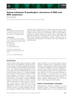
Fig. 1. HILIC retention of siRNAs, RNA oligonucleotides, DNA oligonucleotides and
phosphorothioate DNA oligonucleotides (grouped by color shaded areas) on a BEH
amide column in mobile phases with (A) 25 mM ammonium formate or ammonium
acetate; (B) ammonium acetate (pH 6.8) of various concentrations (2.5 mM, 7.5 mM,
15 mM, and 25 mM); (C) 15 mM of ammonium acetate of different pH (pH 5.5, pH
6.8, pH 9.0).
Many efforts have been made to understand the HILIC retention mechanisms of oligonucleotides by exploring the use of different HILIC stationary phases. Among them, the superior separation efficiency obtained using BEH amide column independently
reported by several research groups encouraged us to further evaluate the performance of this column for oligonucleotide analysis [10,15,16]. Mechanistically, the polar amide functional groups
may be more effective at interacting with the aqueous portion of
the mobile phase and forming the stagnant water layer required
for HILIC. The carbamoyl groups within the amide phase can participate in hydrogen-bonding as hydrogen-bond donors and further interact with hydroxyl groups in the analytes [18]. Additionally, with smaller diameters of packing materials, improved chromatographic performance was achieved due to more effective mass
transfer (Van Deemter equation). As a result, we selected the BEH
amide column to develop a generic HILIC method suitable for routine oligonucleotide analysis. To validate the method robustness,
we analyzed both single-stranded DNA and RNA oligonucleotides
as well as double-stranded siRNAs in five replicates with a 10-min
gradient and a column re-equilibration time of 10 min between
each injection. High repeatability of retention times and peak areas
were obtained with <10% coefficient of variance (CV) (Table S2). It
is also worth noting that our optimized HILIC method with 20-min
run time is 3 times faster than most HILIC applications for oligonucleotides that employ a rather long gradient and re-equilibration
time (60-min run time) [14, 16].
3.2. Impacts of mobile phase additives (salt types, concentrations and
pH) on oligonucleotide/siRNA analysis
Next, we compared the influence of commonly used HILIC mobile phase additives on HILIC separations of oligonucleotides. We
first evaluated two commonly used additives ammonium formate
(AF) and ammonium acetate (AA) at concentrations of 25 mM, and
we observed comparable peak capacities between the two groups.
As illustrated by Fig. 1A, regardless of salt types and concentrations, a general HILIC retention order was observed: duplexed RNA
> RNA oligonucleotide > DNA oligonucleotide > PS-modified DNA
oligonucleotide. Besides, the elution order of DNA oligonucleotides
was dependent on their chain length, which could be attributed
to the net negative charges carried. This is consistent with previous studies that used poly-deoxy(thymidylic) acids (dT) ladders for
benchmarking LC separations [14–16]. Meanwhile, DNA oligonucleotides carrying backbone PS modifications showed diminished
HILIC retention due to the higher hydrophobicity comparing to
phosphate backbone, with the displacement of oxygen with the
3
M. Huang, X. Xu, H. Qiu et al.
Journal of Chromatography A 1648 (2021) 462184
further increasing the charge, in proportion to the T(U) + G content [21]. Interestingly, we also observed slightly more noticeable
peak tailing at pH 5.5 while a more symmetrical peak profile at
pH 9.0, which was also reported by Goyon et al [10]. Alterations of
mobile phase pH, however, did not have obvious effects on modulating LC peak resolution.
less electronegative sulfur [13]. Moreover, AF-containing mobile
phases displayed slightly greater HILIC elution strength and diminished retention on the BEH amide column, as reflected by a decrease of the retention factor or k values (Fig. 1A). Such a phenomenon was, however, opposed to the previous findings made on
a TSK-gel Amide-80 column that AA poses greater elution strength
compared to AF [11,17]. Weaker elution strength of AF in comparison to AA was also indicated on a modified diol column, as the
analyte retention is more attributed to the pH rather than ionic
strength of the mobile phase [14]. The differential elution strength
between AF- and AA-containing mobile phases on the BEH amide
column may be attributed to stronger ion exchange interactions of
the formate ions.
Adjustments of ionic strengths in HILIC mobile phases can alter method selectivity, column retention, and separation efficiency
[19]. Lobue et al previously investigated buffer salt concentrations
ranging from 2.5 mM to 15 mM and determined 15 mM AA (pH
5.5) as the optimal concentration for good chromatographic peak
shapes and MS response on a polymer-based diol-bonded column
[14]. The authors found that lower AA concentrations generally
gave rise to broadened LC peaks and worse peak shape. Similar
mobile phase conditions, with 15 mM AA (pH 5.5) being chosen
as an optimal mobile phase additive, were reported in an independent study from Demelenne et al on a BEH amide column [16].
Moreover, in another two studies that used BEH amide column,
MacNeill et al employed 10 mM AF (pH 9.0) for quantification of
a PS oligonucleotide, while Goyon et al chose 25 mM AA with no
pH adjustment [10,15]. Taken together, we reckoned that a systematic screening of salt concentrations and pH could benefit HILIC
method development for oligonucleotide using the BEH amide column. In the present investigation, four different salt concentrations of AA were tested: 2.5 mM, 7.5 mM, 15 mM and 25 mM.
As shown in Fig. 1B, the observed k values or HILIC retention increased with elevated salt concentrations, consistent with previous
findings made on different HILIC stationary phases [14,17,20]. Such
a phenomenon may suggest that solvation instead of ion exchange
remains the dominating retention mechanisms [19]. For BEH amide
column, the underlying mechanisms accountable for this observation are likely to be the expansion of the aqueous layer adsorbed
on the stationary phase surface and as such, more solvated salt
ions accumulate in this layer and consequently contribute to a
thickening of the water layer and increased retention via hydrogen
bonding [17,19,20].
Packed with hybrid silica particles, the BEH amide column exhibits greater tolerance to high pH (>8) than pure silica particles, allowing more flexible method development. By testing mobile phases composed of 15 mM AA at different pH (pH 5.5, pH
6.8, and pH 9.0), we observed that elevated mobile phase pH with
addition of ammonium hydroxide (pH 9.0) led to lower HILIC retention of most oligonucleotides, whereas addition of acetic acid
(pH 5.5) generally gave rise to higher HILIC retention (Fig. 1C). Our
findings were in line with a recent study by Kilanowska et al, in
which a minor increase of k was observed with decrease of pH values independent of salt types [17]. However, opposing results indicating increasing of HILIC retention of a PS oligonucleotide with increase of pH were also seen, which was justified by higher charge
carried by oligonucleotides at higher pH [10]. In theory, nucleotide
subunits contain both basic nitrogen atoms within the nucleobase
and acidic phosphate groups and, as such, mobile phase pH determines the ionization and charge state. This can in turn modulate the polarity or hydrophilicity of the analyte and likewise the
HILIC retention [19]. At pH < 8, each nucleotide contributes to one
negative charge, so each additional nucleotide increases the overall charge on the molecule and consequently increases HILIC retention. As the eluent pH further increases to > 8, the tautomeric
oxygen on each G and T (U for RNA) becomes an oxyanion, thereby
3.3. Assessing effects of column temperature on HILIC analysis of
oligonucleotides and siRNAs
In HILIC mode, column temperatures can be optimized to enhance selectivity, lower solvent viscosity, and increase mass transfer rates. Most HILIC applications in oligonucleotides use relatively low temperature (40°C) for separation [14,16]. In a study by
Goyon et al, it was shown that higher column temperature of a
BEH amide column led to increased peak capacity but lower signal response when coupled to UV detection [10]. In the present
investigation, a panel of different column temperatures ranging
from 30°C to 80°C (with 10°C intervals) were tested. As suggested by the HILIC–UV traces in Figure S1, increased column
retention and peak capacity yet lowered UV response were observed with elevated temperature, which is in keeping with the
trend observed by Goyon et al. The mechanisms accounting for
higher peak resolution at elevated column temperature could be
attributed to minimized non-specific interactions that may arise
from internal hydrogen bonding [22]. However, analyte adsorption
may occur at the surface of HILIC stationary phase through electrostatic interactions or hydrogen bonding, and increasing temperature may result in greater exposure of polar groups present in
analytes [10,18]. Besides, higher column temperature could result
in reduction of the adsorbed water-rich layer on the stationary
phase, thereby modulating the partition of the analytes between
the bulk mobile phase and adsorbed water-rich layer and consequently retention behavior [23]. Consequently, 30°C was chosen as
the column temperature for the HILIC separation of single-stranded
oligonucleotides.
In its native conformation, the sense (S) and anti-sense (AS)
strands of duplex siRNA are primarily associated through noncovalent interactions such as hydrogen bond interaction and base
stacking. Increased column temperature is therefore expected to
impact the peak shapes of duplexed siRNA compounds by disrupting the double-stranded structures. For further investigation, column temperatures ranging from 30°C to 80°C were applied and the
duplex, S and AS strands of an siRNA that targets luciferase (siLuc)
were subject to HILIC–UV analysis. As shown in Figure S2, we observed complete or partial melting of the duplex strands with elevated column heating at 70°C. The results demonstrated that elevating column temperature to as low as 70°C led to peak broadening due to duplex melting, yet the conformation of S/AS strands
remained largely unchanged (Figure S2). The denaturation or melting of the duplexed structures was further confirmed by MS analysis. As such, it is generally advisable to employ a column temperature below the melting point (Tm ) of the double-stranded nucleic acids, under which the duplex structures remain largely intact. That said, on-column Tm depression can occur in the presence
of an organic solvent (i.e., ACN) in the mobile phase, and optimal
column temperature should, therefore, be carefully evaluated [24].
Furthermore, it is generally recognized that siRNAs appear to be
more hydrophilic compared to their corresponding S/AS strands, as
their hydrophobic bases are shielded from solvents in duplex conformation [25]. Indeed, we observed substantially elevated HILIC
retention of the duplexed structures in contrast to the corresponding S and AS strands (Figure S2), inferring that our current HILIC
conditions could preserve non-covalent duplexes of nucleic acids
under a lower column temperature (30°C).
4
M. Huang, X. Xu, H. Qiu et al.
Journal of Chromatography A 1648 (2021) 462184
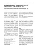
Fig. 2. Full-scan and deconvoluted mass spectra for six DNA oligonucleotides (CMV-F, M13-R, SP6, TRC-F, T3, and T7) analyzed by HILIC−MS.
3.4. Impacts of mobile phase additives on full-scan MS analysis of
oligonucleotides and siRNAs
matographic performance and reasonable MS signal intensities, 15
mM AA was chosen as the final mobile phase salt concentration.
Moreover, oligonucleotide precursor ions with longer chain lengths
were populated in higher charge states (Figure S4A), and distinct
differences in charge envelope profiles were noticed for three mobile phase pH tested: AA (pH 5.5), AA (pH 9.0) and AF (pH 5)
(Figure S4B). The impacts of mobile phase composition on charge
states were not solely dependent on buffer pH, but also can be related to salt types. Collectively, to ensure that less water content
was needed for analyte elution and thereby higher ESI desolvation
efficiency, we tend to use 15 mM AA (pH 9.0) as mobile phase additives optimized for the BEH amide column, which provided an
equivalent chromatographic performance to the one obtained at
pH 5.5.
Furthermore, we also showed that the acquired full-scan mass
spectra of DNA/RNA oligonucleotides and siRNA duplexes acquired
could be successfully deconvoluted using the PMI-Intact MassTM
software (Protein Metrics, San Carlos, CA, USA). The accurate mass
for each of the DNA oligonucleotides (Fig. 2) could be readily determined, with additional peaks of sodium (Na), potassium (K) and
acetate (Ac) adducts observed. Moreover, full-scan mass spectra
The advantages of coupling HILIC to mass spectrometric (MS)
analysis include more efficient sample desolvation and ionization
facilitated by the high organic content in the eluents, as such contributing to superior electrospray ionization (ESI)-MS sensitivity.
Because nucleic acid drugs are a class of molecules that exhibit
dominant negative charges, their charge state distribution could
be impacted by the generic pKa of the molecules, conformational
changes, and formation of cationic adducts. We first compared the
MS response obtained with the use of mobile phases containing AF
(25 mM) and different concentrations of AA: 2.5 mM, 7.5 mM, 15
mM and 25 mM. The highest MS response for most DNA oligonucleotides was achieved with a salt concentration of 15 mM AA,
except for a longer 25-mer TRC-F (Figure S3). The lowest MS response was observed for 25 mM AF (Figure S3). In addition, with
decrease of salt concentration, we observed a systematic shift of
charge state distributions of oligonucleotide polyanions to lower
m/z values, which align with the results shown by Guo et al via
direct infusion experiments [26]. Together, to balance good chro-
5
M. Huang, X. Xu, H. Qiu et al.
Journal of Chromatography A 1648 (2021) 462184
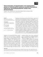
Fig. 3. Full-scan and deconvoluted mass spectra for intact siRNA duplexes (NT siRNA#1, NT siRNA#2 and siLuc) analyzed by HILIC−MS.
Fig. 4. (A) HILIC–UV chromatograms showing the separation of the mixture of synthetic 3’ (n – x) truncated sequences of a DNA oligonucleotide TRC-F. (B) HILIC–UV
chromatograms showing the separation of the 4-oligo (upper panel) and 9-oligo (lower panel) mixture of synthetic 3’ (n – x) truncated sequences of a PS oligonucleotide
TRC-FPS.
of siRNA S/AS strands that were individually analyzed or partially
denatured during ESI process could also be deconvoluted (Figure
S5). While the analysis of duplexed nucleic acids by HILIC–MS appears to be absent from the current body of literature, it was
demonstrated by others that effective IP-RPLC–MS analysis of intact duplex RNA could be achieved by using suitable mobile phases
under non-denaturing conditions [27–29]. Nevertheless, some IP
reagents used in IP-RPLC applications can result in duplex dissociation in both chromatographic and MS detection, presumably due
to the disruption of hydrogen bonds with higher ionic strength
[29,30]. We showed in the previous section that the siRNA duplexes could be retained under HILIC conditions. More importantly,
full-scan and deconvoluted mass spectra for native siRNA duplexes
corroborated the preservation of gas-phase duplex conformation by
MS analysis (Fig. 3). Taken together, the results have evidenced
that HILIC–MS could provide an attractive alternative to analyzing oligonucleotides and siRNAs, and this is the first demonstra-
tion of HILIC–MS application in providing native analyses of siRNA
duplexes.
Besides, for both siRNA duplexes and single-stranded oligonucleotides, the metal adduction peaks observed in HILIC−MS were
of relatively low abundance (Figs. 2, 3 and S5). In line with our
results, it was previously reported that the addition of ammonium
acetate during ESI process can aid in the removal of alkali metal
adducts with volatile ammonium ions (NH4 + ) bound to the negatively charged backbone of oligonucleotides [31]. Together, these
observations support another advantage of HILIC in mitigating the
adduct formation issues that are commonly encountered in IPRPLC applications [32].
3.5. HILIC separation of synthetic 3’ (n − x) truncated sequences of
oligonucleotides
In-depth characterization of nucleic acid drug products necessitates unambiguous profiling of structural variants of oligonu6
M. Huang, X. Xu, H. Qiu et al.
Journal of Chromatography A 1648 (2021) 462184
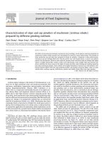
Fig. 5. (A) Full view and 60× zoom-in view illustrating the HILIC–UV profiles of the DNA oligonucleotide CMV-F. The dashed square highlights the peak region that was
further resolved by MS analysis. Extracted ion chromatograms depicting a panel of (B) 3’ and (C) 5’ (n – x) shortmer impurities present in CMV-F using the most abundant
[M – 4H]4– precursor. (D) Summary of the identified impurity sequences of CMV-F and their relative abundance present in the sample.
cleotides or siRNA arising from synthetic impurities, degradants,
and metabolites generated in vitro or in vivo [33]. Oligonucleotide and siRNA therapeutics are degraded mainly by cleavage of the phosphodiester linkages by nucleases. In vitro experiments showed that cleavage at the 3 -terminus resulting from 3’exonucleolytic activities is the primary form of metabolites, followed by 5 -exonuclease and endonuclease cleavage products [34].
Chemical modifications made to the ribose or phosphate backbone
are, hence, routinely incorporated into oligonucleotide and siRNA
molecule design in an effort to mitigate exo- and endo-nuclease
activity in order to ensure sustained drug efficacy [35]. In addition
to metabolites, process-related impurities can arise from chemical
synthesis and often include families of shortmer (n – x) and longmer (n + x) sequences. For example, an n – 1 family of impurities
would consist of multiple n – base (B), where B can be any base in
the sequence [36].
Structurally similar synthetic (n – x) sequences of oligonucleotides have been largely employed to examine HILIC separations
of shortmer impurity or metabolite mimics [14,16]. Therefore, we
first evaluated separations of an equimolar mixture of up to (n
– 8) 3’ truncated synthetic sequences of a 25-mer DNA oligonucleotide TRC-F. By extending to a 20-minute linear HILIC gradient,
we successfully achieved good separation of all (n – x) shortmer
sequences and the FLP (Fig. 4A). However, when it comes to a fully
PS-modified version of TRC-F, i.e. TRC-FPS, it was particularly difficult to resolve the 3’ (n – 1) sequence and the FLP sequences.
Specifically, the 3’ (n – 3) and 3’ (n – 2) sequences could be well
resolved from the FLP, with the other 3’ (n – x) sequences eluted
closer together (Fig. 4B). The challenge of separating PS oligonucleotides arises from the compromised LC peak resolution caused
by the inherent stereochemical configuration of the PS linkages
(“Sp” or “Rp”). One single PS linkage can introduce a chiral center at phosphorus in addition to the D-ribose chiral centers, giving rise to 2N diastereoisomers (number of PS bonds = N) and
chromatographic peak splitting or broadening. In IP-RPLC, negative charges on the oligonucleotide phosphate backbone are neutralized by positively charged alkylammonium ions in the mobile
phase. Hydrophobic interactions between the oligonucleotide bases
and the reversed-phase column play a major role in the separation, and the different hydrophobicity of the individual bases
contributes to differences in retention. In comparison, HILIC chromatography depends on hydrophilicity, which is not highly variable
between oligonucleotide units largely due to the presence of phosphate groups. Therefore, HILIC separation of oligonucleotides is not
as robust as it is with IP-RPLC approaches. Further modifications
with mobile phase modifiers or derivatization are needed to improve selectivity.
3.6. Oligonucleotide impurity analysis by HILIC–MS
Although extensive purification process removes most of impurities from oligonucleotide therapeutics, low levels of remaining
impurities or degraded products could significantly impact both
drug safety and efficacy. The commonly identified impurities of
oligonucleotide therapeutics usually include chain shortened (n –
x) products [37]. Many hybridization-based oligonucleotide assays
do not accurately distinguish large impurities or degradants from
the full-length oligonucleotide of interest, despite their ultra-high
sensitivity in quantifying impurities or metabolites [38]. In recent
years, MS has gradually become the method of choice in that it
can provide unambiguous and comprehensive impurity identification/quantitation and metabolite profiling for both oligonucleotide
and siRNA modalities [39].
As illustrated by the HILIC–UV traces in Figure S6, various small
impurity peaks could be readily separated with an extended linear
gradient (20% to 65% MPB in 15 min). These impurities could result from degradation through freeze-thaw cycles and the presence
of trace nuclease in the samples. Interestingly, for the PS oligonu7
M. Huang, X. Xu, H. Qiu et al.
Journal of Chromatography A 1648 (2021) 462184
Fig. 6. (A) Annotated HCD-MS/MS fragmentation for the [M – 5H]5– precursor (m/z 1309.2167) of a DNA oligonucleotide CMV-F (NCE 15%). (B) MS/MS fragment coverage
map for the results shown in (A), generated from Thermo Biopharma Finder 4.0.
cleotide T7-FPS, there seemed to be a lesser amount of impurities compared to the amount of impurities for unmodified DNA
or RNA oligonucleotides, possibly due to the increased resistance
of PS bonds against nucleolytic activities. Taking advantage of the
downstream HRMS analysis, we attempted to further characterize
the identities of some of these impurity peaks. In the elution window highlighted by the dashed box in the HILIC–UV profile of the
20-mer DNA oligonucleotide CMV-F (Fig. 5A), we performed targeted MS extraction using the m/z value of the most abundant precursor ions ([M – 4H]4– ) of each (n – x) sequence from either 3’ or
5’ terminus to calculate signal intensities. Based on the peak intensities extracted for the corresponding 3’ and 5’ (n – x) shortmer sequences (Fig. 5B, C), our results evidenced that the generation of 3’ truncated degradants are more prevalent than those
generated from the 5’ end for CMV-F (Fig. 5D). For other impurity
peaks with earlier elution times, we speculated that there could be
both exo- and endo-nuclease activities present that led to oligonucleotide impurities with shorter chain length. Overall, we demonstrated the feasibility of coupling the HILIC method to UV/MS detection for impurity profiling of oligonucleotides.
3.7. Oligonucleotide sequence characterization by HILIC–MS/MS
Sequencing elucidation of DNA or RNA oligonucleotides composed of up to 70 residues by MS/MS fragmentation of multiply
charged oligonucleotide precursor ions in negative ESI mode has
been intensively studied in the past few decades. McLuckey et al
indicated that the major fragmentation pathways by collisionalinduced dissociation (CID) of oligonucleotides can proceed with
multiple pathways [40]. Among the backbone cleavage products, a–
B and w-type ions are the most dominant species for DNA oligonucleotides, while y- and c-type ions remain more favored for RNA
oligonucleotides [41]. The Q-Exactive mass spectrometer features
beam-type CID, sometimes referred to as higher-energy C-trap dissociation (HCD), which relies on an octupole collision cell that operates at higher collisional energies than CID [42]. Furthermore, it
is known that precursor charge states can affect the efficiency of
fragmentation, as described in previous studies on peptides and
other biomolecules [43]. As such, we attempted to evaluate how
the commonly observed precursor charge states in the HILIC–MS
mode and the normalized collision energy (NCE) applied for HCD8
M. Huang, X. Xu, H. Qiu et al.
Journal of Chromatography A 1648 (2021) 462184
Fig. 7. Assessment of on-column sensitivity and linearity range of the HILIC–MS method by (A) tSIM scan and (B) PRM scan.
MS/MS fragmentation could impact signature fragment ions and
sequence coverage for both DNA and RNA oligonucleotides.
The complexity of a typical oligonucleotide mapping experiment resulting from a great number of structurally diverse fragmentation ions presents a significant throughput hurdle. Fortunately, this challenge has been mitigated via recent developments
and advancements in software tools that facilitate automated and
unbiased data analysis. In this study, a newly developed data analysis module incorporated into Biopharma FinderTM 4.0 (Thermo
Fisher Scientific) was utilized to achieve automated data processing and sequence annotation for HCD-MS/MS data acquired from
oligonucleotides. Benefited from this tool, the overall % sequence
coverage was systematically examined for oligonucleotides with
various base composition (deoxyribonucleic acid, ribonucleic acid),
chain length (17-mer to 25-mer), backbone modification (unmodified, partially or fully PS-modified), and sequence composition.
The most abundant precursors ([M – 4H]4– , [M – 5H]5– and [M
– 6H]6– ) observed in the HILIC–MS mode were chosen for analysis, and a panel of % normalized collisional energy (%NCE) values ranging from 13% to 23% were examined. In general, precursor
ions of higher charge states require higher NCE to achieve good
sequence coverage, and an opposite trend was observed for lowercharge-state precursors (Figure S7). For most DNA or RNA oligonucleotides selected in this study, the optimal NCE was determined
to be 15% for their most abundant charge states. For higher charge
states such as [M – 6H]6– , only partial fragmentation of parent ions
was observed with low %NCE applied, resulting in low sequence
coverage. As depicted in Fig. 6A, Biopharma FinderTM 4.0 enabled
rapid and accurate fragment annotations (predominantly a–B and
w-type ions) for the DNA oligonucleotide CMV-F. By employing 15%
NCE for the [M – 5H]5– precursor (m/z 1309.2167), 90% sequence
coverage was achieved, with the intensities of each fragment highlighted in the sequence coverage map (Fig. 6B).
and HILIC–MS in the field. Loube et al showed enhanced MS signal
response by HILIC–MS for oligonucleotides of shorter chain-lengths
and lower hydrophobicity, as compared to IP-RPLC–MS [14]. Nevertheless, Kilanowska et al also pointed out that ACN in the mobile phase may actually cause ionization suppression of oligonucleotides due to its aprotic properties [17]. In the work by Easter
et al, the LOD values were observed at the picomolar level with the
coupling of inductively coupled plasma (ICP)–MS to monitor the
signature 31 P16 O+ ion [11]. In another study, analyses of oligonucleotides were performed by HILIC–MS analysis with an LOD value
of 2.5 pmol loaded on-column for a 20-mer oligonucleotide [12]. A
similar LOD value of 2.0 pmol on-column was also reported for a
22-mer oligonucleotide using 2D-LC–MS analysis [45].
We first determined the optimal HCD energy for chemically
modified oligonucleotides including the most prevalent therapeutic
oligonucleotide design that involve full PS backbone modifications
on T7 (T7-FPS) and TRC-F (TRC-FPS). Notably, CID- or HCD-MS/MS
fragmentation of oligonucleotides with PS modifications gave rise
to a diagnostic fragment ion PSO2 – (m/z 94.93), which was broadly
used for targeted MS analysis such as multiple-reaction monitoring
(MRM) [17,20]. Our PRM results shown in Figure S8 indicated that
higher %NCE may favor generation of the signature PSO2 – fragment
ions for quantification purpose (sensitivity), whereas lower %NCE
could benefit more comprehensive sequence annotation (specificity). Next, we sought to examine the on-column sensitivity for
T7-FPS by performing tSIM or PRM analysis. As displayed in Fig. 7,
tSIM analysis displayed higher S/N ratios or sensitivity in comparison to ratios or sensitivity of PRM analysis in all injections
analyzed. Even with the lowest injection of 13 ng (equivalent to
2.0 pmol), a clear S/N profile was achieved for the top two most
abundant charge states. Furthermore, we believe that PRM analysis
could warrant higher selectivity for the analysis of structurally similar variants in complex mixtures by incorporating signature fragment ions.
3.8. Quantitative analysis of oligonucleotides
4. Conclusions
Quantitative analysis of therapeutic oligonucleotides often represents a fundamental part in determining their pharmacokinetic
and pharmacodynamic (PK/PD) properties during drug development. LC–MS based bioanalytical method can achieve the sensitivity of 1 ng/mL when coupled to upstream sample preparation techniques such as solid-phase extraction (SPE) [44]. Thus, we reckon
that it is essential to evaluate the sensitivity of our HILIC–MS
method to better suit bioanalysis requirements. There have been
controversies on the sensitivity comparison between IP-RPLC–MS
Until now, IP-RPLC remains the most frequently used technique
in providing analytical characterization of oligonucleotides. There
is, however, a drastically increasing trend of using HILIC applications to render complementary and more in-depth analysis of
oligonucleotides and other nucleic acid therapeutics. In this study,
we established a universal HILIC–MS/MS method that could provide a comprehensive solution for the analysis of oligonucleotides
and siRNAs, including separation, mass determination, sequence
9
M. Huang, X. Xu, H. Qiu et al.
Journal of Chromatography A 1648 (2021) 462184
characterization, impurity profiling, as well as the potential in
quantitative analysis of oligonucleotides and siRNAs to support
drug development.
We have evaluated the impacts of mobile phases additives
on both chromatographic separation and MS response of the
HILIC–MS method. Accurate intact mass measurement for oligonucleotides was successfully achieved by HRMS analysis. More importantly, for the first time, we presented the utility of this HILIC–
MS method that preserves the gas-phase duplexed conformation
in rendering native analysis of siRNA duplexes. Furthermore, the
HILIC–MS described herein could facilitate straightforward impurity analysis of oligonucleotides. Another highlight of the established method was the hyphenation of HILIC with MS/MS for unbiased sequence annotation, which could be further applied to
oligonucleotides with backbone or ribose chemical modifications
for structural characterization. We showed that employing low
NCE (15%) on an orbitrap instrument could facilitate comprehensive HCD-MS/MS sequence coverages for 17- to 25-mer oligonucleotides, and that automated MS/MS annotation could be achieved
with improved ease. Additionally, we have demonstrated that the
use of HILIC–MS based on tSIM mode, as compared to PRM mode,
improved the overall on-column sensitivity. However, for complex mixtures of structurally similar oligonucleotide impurities and
metabolites, selectivity could be further enhanced with PRM analysis by selecting structure-related signature fragment ions.
In-depth characterization and identification of synthetic impurities present in drug products and metabolites generated in vitro
or in vivo remain critical tasks in risk mitigation and clinical studies. Extensive work that relies on LC–MS approach has been done
previously to identify trace impurities [46,47], in vivo metabolites [48] and in vitro metabolites [49] for better risk mitigation
of manufacturing and clinical studies. Importantly, the established
HILIC–MS/MS assay can be readily employed in combination with
SPE to fully support bioanalysis of clinical samples. We envisioned
that the developed HILIC–MS/MS approach could efficiently support synthetic oligonucleotide and siRNA chemistry with a wide array of modifications and provide rapid and flexible in-depth characterization of oligonucleotide or siRNA drug products in the absence of IP reagents.
References
[1] X. Shen, D.R. Corey, Chemistry, mechanism and clinical status of antisense oligonucleotides and duplex RNAs, Nucl. Acids Res. 46 (2018) 1584–
1600.
[2] C.A. Stein, D. Castanotto, FDA-approved oligonucleotide therapies in 2017, Mol.
Ther. 25 (2017) 1069–1075.
[3] C. Iglesias-Lopez, A. Agusti, M. Obach, A. Vallano, Regulatory framework for
advanced therapy medicinal products in Europe and United States, Front. Pharmacol. 10 (2019) 921.
[4] A.J. Alpert, Hydrophilic-interaction chromatography for the separation of peptides, nucleic acids and other polar compounds, J. Chromatogr. 499 (1990)
177–196.
[5] Q. Zhang, F.Q. Yang, L. Ge, Y.J. Hu, Z.N. Xia, Recent applications of hydrophilic
interaction liquid chromatography in pharmaceutical analysis, J. Sep. Sci. 40
(2017) 49–80.
[6] T. Ikegami, Hydrophilic interaction chromatography for the analysis of biopharmaceutical drugs and therapeutic peptides: a review based on the separation
characteristics of the hydrophilic interaction chromatography phases, J. Sep.
Sci. 42 (2019) 130–213.
[7] G. Marrubini, B.E. Mendoza, G. Massolini, Separation of purine and pyrimidine
bases and nucleosides by hydrophilic interaction chromatography, J. Sep. Sci.
33 (2010) 803–816.
[8] H.Q. Zhao, X. Wang, H.M. Li, B. Yang, H.J. Yang, L. Huang, Characterization of
nucleosides and nucleobases in natural Cordyceps by HILIC-ESI/TOF/MS and
HILIC-ESI/MS, Molecules 18 (2013) 9755–9769.
[9] K. Inoue, R. Obara, T. Hino, H. Oka, Development and application of an
HILIC-MS/MS method for the quantitation of nucleotides in infant formula, J.
Agric. Food Chem. 58 (2010) 9918–9924.
[10] A. Goyon, K. Zhang, Characterization of antisense oligonucleotide impurities
by ion-pairing reversed-phase and anion exchange chromatography coupled to
hydrophilic interaction liquid chromatography/mass spectrometry using a versatile two-dimensional liquid chromatography setup, Anal. Chem. 92 (2020)
5944–5951.
[11] R.N. Easter, K.K. Kroning, J.A. Caruso, P.A. Limbach, Separation and identification of oligonucleotides by hydrophilic interaction liquid chromatography
(HILIC)-inductively coupled plasma mass spectrometry (ICPMS), Analyst 135
(2010) 2560–2565.
[12] L. Gong, J.S. McCullagh, Analysis of oligonucleotides by hydrophilic interaction
liquid chromatography coupled to negative ion electrospray ionization mass
spectrometry, J. Chromatogr. A 1218 (2011) 5480–5486.
[13] L. Gong, Analysis of oligonucleotides by ion-pairing hydrophilic interaction liquid chromatography/electrospray ionization mass spectrometry, Rapid Commun. Mass Spectrom. 31 (2017) 2125–2134.
[14] P.A. Lobue, M. Jora, B. Addepalli, P.A. Limbach, Oligonucleotide analysis by hydrophilic interaction liquid chromatography-mass spectrometry in the absence
of ion-pair reagents, J. Chromatogr. A 1595 (2019) 39–48.
[15] R. MacNeill, T. Hutchinson, V. Acharya, R. Stromeyer, S. Ohorodnik, An oligonucleotide bioanalytical LC-SRM methodology entirely liberated from ion-pairing,
Bioanalysis 11 (2019) 1157–1169.
[16] A. Demelenne, M.-J. Gou, G. Nys, C. Parulski, J. Crommen, A.-C. Servais, M. Fillet, Evaluation of hydrophilic interaction liquid chromatography, capillary zone
electrophoresis and drift tube ion-mobility quadrupole time of flight mass
spectrometry for the characterization of phosphodiester and phosphorothioate
oligonucleotides, J. Chromatogr. A 1614 (2020) 460716.
´
[17] A. Kilanowska, B. Buszewski, S. Studzinska
, Application of hydrophilic interaction liquid chromatography coupled with tandem mass spectrometry for the
retention and sensitivity studies of antisense oligonucleotides, J. Chromatogr.
A 1622 (2020) 461100.
[18] R.-I. Chirita, C. West, A.-L. Finaru, C. Elfakir, Approach to hydrophilic interaction
chromatography column selection: application to neurotransmitters analysis, J.
Chromatogr. A 1217 (2010) 3091–3104.
[19] B. Buszewski, S. Noga, Hydrophilic interaction liquid chromatography (HILIC)–a
powerful separation technique, Anal. Bioanal. Chem. 402 (2012) 231–
247.
[20] S. Studzinska, F. Lobodzinski, B. Buszewski, Application of hydrophilic interaction liquid chromatography coupled with mass spectrometry in the analysis
of phosphorothioate oligonucleotides in serum, J. Chromatogr. B Anal. Technol.
Biomed. Life Sci. 1040 (2017) 282–288.
[21] J.R. Thayer, V. Barreto, S. Rao, C. Pohl, Control of oligonucleotide retention
on a pH-stabilized strong anion exchange column, Anal. Biochem. 338 (2005)
39–47.
[22] Z. Hao, B. Xiao, N. Weng, Impact of column temperature and mobile phase
components on selectivity of hydrophilic interaction chromatography (HILIC),
J. Sep. Sci. 31 (2008) 1449–1464.
[23] Y. Guo, N. Bhalodia, B. Fattal, I. Serris, Evaluating the adsorbed water layer
on polar stationary phases for hydrophilic interaction chromatography (HILIC),
Separations 6 (2019).
[24] L. Li, J.P. Foley, R. Helmy, Simultaneous separation of small interfering RNA and
lipids using ion-pair reversed-phase liquid chromatography, J. Chromatogr. A
1601 (2019) 145–154.
[25] S.T. Crooke, J.L. Witztum, C.F. Bennett, B.F. Baker, RNA-targeted therapeutics,
Cell Metab. 29 (2019) 501.
[26] X. Guo, M.F. Bruist, D.L. Davis, C.M. Bentzley, Secondary structural characterization of oligonucleotide strands using electrospray ionization mass spectrometry, Nucl. Acids Res. 33 (2005) 3659–3666.
Authors’ contribution
Ming Huang: Methodology, Data curation, Investigation, Writing
- original draft, Writing - review & editing. Xiaobin Xu: Conceptualization, Project administration, Methodology, Writing - review
& editing. Haibo Qiu: Conceptualization, Supervision, Writing - review & editing. Ning Li: Supervision, Writing - review & editing.
Declaration of Competing Interest
All authors were employees of Regeneron Pharmaceuticals, Inc
while engaged in the study and may hold stock and/or stock options in the company. All authors have patent applications that
were based on the research in the paper.
Acknowledgement
This study was sponsored by Regeneron Pharmaceuticals, Inc.
The authors would like to thank Ashley Roberts from Scientific Writing Group for assistance in drafting and polishing this
manuscript.
Supplementary materials
Supplementary material associated with this article can be
found, in the online version, at doi:10.1016/j.chroma.2021.462184.
10
M. Huang, X. Xu, H. Qiu et al.
Journal of Chromatography A 1648 (2021) 462184
[27] M. Beverly, K. Hartsough, L. Machemer, Liquid chromatography/electrospray
mass spectrometric analysis of metabolites from an inhibitory RNA duplex,
Rapid Commun. Mass Spectrom. 19 (2005) 1675–1682.
[28] M. Beverly, K. Hartsough, L. Machemer, P. Pavco, J. Lockridge, Liquid chromatography electrospray ionization mass spectrometry analysis of the ocular
metabolites from a short interfering RNA duplex, J. Chromatogr. B Anal. Technol. Biomed. Life Sci. 835 (2006) 62–70.
[29] S.M. McCarthy, M. Gilar, J. Gebler, Reversed-phase ion-pair liquid chromatography analysis and purification of small interfering RNA, Anal. Biochem. 390
(2009) 181–188.
[30] B. Noll, S. Seiffert, H.-P. Vornlocher, I. Roehl, Characterization of small interfering RNA by non-denaturing ion-pair reversed-phase liquid chromatography, J.
Chromatogr. A 1218 (2011) 5609–5617.
[31] S. Shah, S.H. Friedman, An ESI-MS method for characterization of native and
modified oligonucleotides used for RNA interference and other biological applications, Nat. Protoc. 3 (2008) 351–356.
[32] R.E. Birdsall, M. Gilar, H. Shion, Y.Q. Yu, W. Chen, Reduction of metal adducts in
oligonucleotide mass spectra in ion-pair reversed-phase chromatography/mass
spectrometry analysis, Rapid Commun. Mass Spectrom. 30 (2016) 1667–1679.
[33] N.M. Elzahar, N. Magdy, A.M. El-Kosasy, M.G. Bartlett, Degradation product
characterization of therapeutic oligonucleotides using liquid chromatography
mass spectrometry, Anal. Bioanal. Chem. 410 (2018) 3375–3384.
[34] R.M. Crooke, M.J. Graham, M.J. Martin, K.M. Lemonidis, T. Wyrzykiewiecz,
L.L. Cummins, Metabolism of antisense oligonucleotides in rat liver homogenates, J. Pharmacol. Exp. Ther. 292 (20 0 0) 140–149.
[35] R.S. Geary, T.A. Watanabe, L. Truong, S. Freier, E.A. Lesnik, N.B. Sioufi,
H. Sasmor, M. Manoharan, A.A. Levin, Pharmacokinetic properties of
2 -O-(2-methoxyethyl)-modified oligonucleotide analogs in rats, J. Pharmacol.
Exp. Ther. 296 (2001) 890–897.
[36] D. Capaldi, A. Teasdale, S. Henry, N. Akhtar, C. den Besten, S. Gao-Sheridan, M. Kretschmer, N. Sharpe, B. Andrews, B. Burm, J. Foy, Impurities in
oligonucleotide drug substances and drug products, Nucl. Acid Ther. 27 (2017)
309–322.
[37] S.G. Roussis, A novel and intuitive method of displaying and interacting with
mass difference information: application to oligonucleotide drug impurities, J.
Am. Soc. Mass Spectrom. 26 (2015) 1150–1164.
[38] G. Zhang, J. Lin, K. Srinivasan, O. Kavetskaia, J.N. Duncan, Strategies for bioanalysis of an oligonucleotide class macromolecule from rat plasma using
liquid chromatography-tandem mass spectrometry, Anal. Chem. 79 (2007)
3416–3424.
[39] S. Pourshahian, T, herapeutic oligonucleotides, impurities, degradants, and
their characterization by mass spectrometry, 2019 n/a.
[40] S.A. McLuckey, G.J. Van Berkel, G.L. Glish, Tandem mass spectrometry of small,
multiply charged oligonucleotides, J. Am. Soc. Mass Spectrom. 3 (1992) 60–70.
[41] T.Y. Huang, A. Kharlamova, J. Liu, S.A. McLuckey, Ion trap collision-induced dissociation of multiply deprotonated RNA: c/y-ions versus (a-B)/w-ions, J. Am.
Soc. Mass Spectrom. 19 (2008) 1832–1840.
[42] J.V. Olsen, B. Macek, O. Lange, A. Makarov, S. Horning, M. Mann, Higher-energy
C-trap dissociation for peptide modification analysis, Nat. Methods 4 (2007)
709–712.
[43] G. Sonsmann, A. Römer, D. Schomburg, Investigation of the influence of charge
derivatization on the fragmentation of multiply protonated peptides, J. Am.
Soc. Mass Spectrom. 13 (2002) 47–58.
[44] J.M. Sutton, J. Kim, N.M. El Zahar, M.G. Bartlett, Bioanalysis and biotransformation of oligonucleotide therapeutics by liquid chromatography-mass spectrometry, Mass Spectrom. Rev. (2020) , n/a.
[45] F. Li, X. Su, S. Bäurer, M. Lämmerhofer, Multiple heart-cutting mixed-mode
chromatography-reversed-phase 2D-liquid chromatography method for separation and mass spectrometric characterization of synthetic oligonucleotides, J.
Chromatogr. A 1625 (2020) 461338.
[46] S.G. Roussis, I. Cedillo, C. Rentel, Semi-quantitative determination of co-eluting
impurities in oligonucleotide drugs using ion-pair reversed-phase liquid chromatography mass spectrometry, J. Chromatogr. A 1584 (2019) 106–114.
[47] S.G. Roussis, I. Cedillo, C. Rentel, Automated determination of early eluting
oligonucleotide impurities using ion-pair reversed-phase liquid chromatography high resolution-mass spectrometry, Anal. Biochem. 595 (2020) 113623.
[48] J. Liu, J. Li, C. Tran, K. Aluri, X. Zhang, V. Clausen, I. Zlatev, L. Guan, S. Chong,
K. Charisse, J.T. Wu, D. Najarian, Y. Xu, Oligonucleotide quantification and
metabolite profiling by high-resolution and accurate mass spectrometry, Bioanalysis 11 (2019) 1967–1980.
[49] J. Kim, N.M. El Zahar, M.G. Bartlett, In vitro metabolism of 2 -ribose unmodified and modified phosphorothioate oligonucleotide therapeutics using liquid
chromatography mass spectrometry, Biomed. Chromatogr. (2020) e4839 n/a.
11