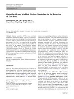
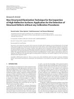
Novel technique for rapid detection of a-globin gene mutations and deletions pot
Bạn đang xem bản rút gọn của tài liệu. Xem và tải ngay bản đầy đủ của tài liệu tại đây (875.15 KB, 8 trang )
Novel technique for rapid detection of a-globin gene
mutations and deletions
JINGZHONG LIU, XINGYUAN JIA, NING TANG, XU ZHANG, XIAOYI WU, REN CAI,
LIRONG WANG, QUANZHANG LIU, BAI XIAO, JIM ZHU, and QINGTAO WANG
BEIJING AND GUANGXI, CHINA
Populations in Southeast Asiaand South China have high frequencies of a-thalassemia
caused by a-globin gene mutations and/or deletions. This study was designed to find
an efficient and simple diagnostic test for the mutations and deletions. A duplex poly-
merase chain reaction (PCR)/denaturing high-pressure liquid chromatography
(DHPLC) was used to detect the mutations and deletions. A blinded study of 110
samples, which included 92 a-thalassemia samples with various genotypes and 18
normal DNA samples, was carried out by the methods. The duplex PCR products of
the sample with known Constand spring mutation (CS)/aa, Quonsze mutation (QS)/
aa, and Weastmead mutation (WS)/aa DNA showed significantly different profiles,
which suggests that DHPLC analysis at 63.8
C can detect potential mutations directly.
The DHPLC at 50
C analysis can distinguish the SEA and nondeletional alleles. The
new assay is 100% concordant with the original genotype. In conclusion, the tech-
nique including the duplex PCR assay followed by DHPLC analysis can be used to di-
agnose a-thalassemia; this methodology is simple, rapid, accurate, semiautomatic,
and high output, and thus, it is suitable for large-scale screening. (Translational Re-
search 2010;155:148–155)
Abbreviations: CS ¼ Constand spring mutation; DC ¼ dissociation curve analysis; DHPLC ¼
denaturing high-performance liquid chromatography; Duplex PCR ¼ duplex polymerase chain
reaction; GC ¼ guanine-cytosine; QS ¼ Quonsze mutation; RDB ¼ reverse dot-blot; TEAA ¼
triethylammonium acetate; WS ¼ Weastmead mutation
a-Thalassemia is the most common recessively inherited
hemoglobin disorder.
1
Unlike a-thalassemia, in which
nondeletional mutations predominate, most recognized
a-thalassemia involve deletion of 1 or both a-globin
genes. The a-globin gene cluster is located on chromo-
some 16. a2 and a1 are highly homologous, with se-
quence identity greater than 96% between the Z1 and
Z2 boxes and high guanine-cytosine (GC) content.
2
In
Southeast Asia and southern China, most a-thalassemia
is caused by the deletion of 1 (-a4.2/, -a3.7/; termed a-
thalassemia-2) or 2 ( SEA, THAI; termed a-thalasse-
mia-1) of the 2 functional a-globin genes
3,4
(Fig 1).
The SEA is the most common type of a-thalassemia-
1. Even though carriers of the a-thalassemia-1 with
SEA type do not manifest any clinical symptoms, cou-
ples who are both carriers have a 25% chance of
From the Basic Medical Research Center, Beijing Chaoyang Hospital,
Affiliate of The Capital Medical University, Beijing, China; Institute of
Basic Medical Sciences, Chinese Academy of Medical Sciences &
Peking Union Medical College, Beijing, China; Liuzhou Women and
Children’s Hospital, Liuzhou City, Guangxi, China; Transgenomic
Ltd, Beijing, China; Beijing Deyi Clinical Diagnostic Laboratory,
Beijing, China.
Supported by Grant JS96004 from the Natural Science Foundation of
Beijing, China
Submitted for publication August 4, 2009, revision submitted October
14, 2009; accepted for publication October 16, 2009.
Reprint requests: Jingzhong Liu, PhD, Basic Medical Research Center,
Beijing Chaoyang Hospital, Affiliate of Capital Medical University,
8 Gongtinanlu, Chaoyang District, Beijing, China 100020.; e-mail:
; (J.L.).
1931-5244/$ – see front matter
Ó 2010 Mosby, Inc. All rights reserved.
doi:10.1016/j.trsl.2009.10.003
148
conceiving a homozygous fetus, which manifests as
Bart’s hydrops fetalis, the most severe thalassemic syn-
drome. All these fetuses die in utero or soon after birth.
In addition, approximately 75% of mothers who carry fe-
tuses with homozygous for the thalassemia-1 SEA type
will develop toxemia of pregnancy. An investigation of
the thalassemia-1 SEA type is therefore essential for
carrier couples and for prenatal diagnosis of conception
by couples who are both carriers of this type of gene de-
letion. Diagnostic assays of the a-thalassemia-2 are also
important for genetic counseling, because when
occurring in compound geterozygosity with SEA, the
2a4.2/ or 2a3.7/ will cause HbH disease with a thalas-
semia intermedia phenotype.
Nondeletional a-thalassemia, a more severe expres-
sion, may also be induced by point mutation of the a2
gene (Constand spring mutation [CS], Quonsze mutation
[QS], and Weastmead mutation [WS])
1,5,6
(see the lower
part of Fig 1). In an investigation of 59 cases of HbH
diseases from Guangxi, China, 27 cases (45.8%) were
confirmed to be nondeletional.
7
To date, techniques for
the detection of a-globin gene deletion have included
Southern blot analysis,
8
multiplex gap-polymerase chain
reaction (mPCR),
3,4,9
and real-time PCR with SYBR
Green1 combined with dissociation curve (DC)
analysis.
10
The techniques for the detection of a-globin gene
mutations include PCR product sequencing, reverse
dot-blot (RDB) analysis,
11
and others, but these methods
are not ideal, because they are time consuming, labor
intensive, and expensive. Denaturing high-pressure
liquid chromatography (DHPLC) is a recently developed
technique that uses parti ally denaturing conditions to
detect gene mutations, small insertions, and deletions
based on heteroduplex formation by PCR products
amplified from wild-type alleles and mutant alleles.
The DHPLC has been used successfully to detect muta-
tions in various pathogenic genes, such as the b-globin
gene.
12
Hung et al
13,14
have reported molecular assays
for a-thalassemia-2 deletions based on DHPLC detec-
tion, but no studies have reported the detection of a-glo-
bin gene mutations. This lack of research may be largely
because of the high homology of the a2 and a1 genes
and difficulties in designing specific primers and PCR
amplifications based on high GC content. Rapid and
accurate testing methods are needed to address the diag-
nostic challenges of identifying both deletions and muta-
tions using the same assay. This study reports on 1 such
method. We have developed a duplex PCR followed by
the DHPLC for detecting the SEA and the nondele-
tional alleles under 50
C condition, and for detecting
the 3 known point mutations under 63.8
C condition.
METHODS
Samples. In all, 34 DNA samples with known geno-
types including aCSa/aa (4 cases), aQSa/aa (2 cases),
aWSa/aa (2 cases), /aa (5 cases), - -/ (2 cases), 3.7/
aa (10 cases), 3.7/ (2 cases), 4.2/aa (5 cases), and
4.2/ (2 cases) were used to establish the methodology.
In all, 110 blood samples were collected from Liuzhou
city in southern China from January 2007 to May 2008,
and they were submitted to the laboratory for the blinded
study. Informed consent was obtained from the subjects.
The study conformed to the Chinese ethical guidelines
for human and animal research, and it was approved by
the Beijing Chaoyang Hospital Ethics Committee.
Primer design and duplex PCR assay. Given the minor
sequence differences of IVS-II and the 3’untranslated re-
gion between the 2 functional a-globin genes a2 and a1,
a 206-bp fragment, including the third exon and both
flanking sequences, was designed for amplification
(Fig 1). The upstream primer P1 u sed the difference in
7 bp between the a2 and 1 sequences; thus, 3 bases in
the 3’ end of P1 differed from those in the a 1 sequence.
The downstream primer P2 sequence had more bases
that differed between the a2 and the a1. Both the P1
and the P2 ensure a specific amplification of a 206-bp
segment from the a2. The amplified product was
designed as short as 206 bp that will facilitate mutation
detection using the DHPLC. The most frequently found
mutations (C S, QS, and WS) were all in this 206-bp
AT A GLANCE COMMENTARY
Liu J, et al.
Background
Populations in Southeast Asia and southern China
have high frequencies of a-thalassemia caused by
a-globin gene mutations and deletions. This study
was designed to find an efficient and simple diag-
nostic test for the mutations and deletions.
Translational Significance
A duplex polymerase chain reaction (PCR)/ dena-
turing high-performance liquid chromatography
(DHPLC) was used to detect the mutations and de-
letions. A blinded study of 110 samples including
92 a-thalassemia with various genotypes was con-
ducted. The new assay is 100% concordant with the
original genotypes. The new technique can be used
to diagnose a-thalassemia because it is simple,
rapid, accurate, semiautomatic, and high-output;
thus, it is suitable for large-scale screening.
Translational Research
Volume 155, Number 3 Liu et al 149
fragment, which allowed rapid detection of the 3 poten-
tial mutation types using DHPLC under partially dena-
tured conditions. The sequences the P1 and P2 are
shown in blue in the lower part of Fig 1. Given the dif-
ficulties in the above primer design, it was easier to de-
sign the 2 primers that represented the SEA deletion
according to the principles of gap-PCR. Primer se-
quences that satisfied the following conditions were
qualified: good specificity for amplification, length
shorter than 206 bp, ability to resolve the peak associ-
ated with this fragment in the DHPLC profile at 50
C,
and noninterference with recognition of the specific mu-
tation profile at 63.8
C. The P3 and P4 primers that were
designed in this study satisfied the above conditions and
formed a successful duplex PCR assay with the P1 and
P2 primers. SEA/ and aa /oraTa/ were detected by
the duplex PCR. The 25-mL PCR mixture contained
2.5 mL103 buffer, 2.0 mL deoxyribonucleotide triphos-
phate, 2.5 mL MgCl
2
, 5.0 mL betaine, 0.5 mL SYBR
Green1, 0.36 mg primers 1–3, 0.26 mg primer 4, 1 mg
Gold-Taq DNA polymerase, and 3 mL/0.6 mg of DNA.
The negative and positive controls were included. PCR
assays were performed using an ABI Prism 5700 ther-
macycler (Applied Biosystems, Foster City, Calif).
PCR conditions were 94
C for 10 min followed by 38
cycles of 94
C for 20 s, 65
C for 30 s, and 72
C for
40 s. The annealing temperature was decreased by 1
C
each cycle until it reached 61
C (34 cycles), and the final
extension was conducted at 72
C for 6 min. Samples
were then placed on ice for DHPLC analysis.
Two gap-PCRs. 2a3.7 and 2a4.2 were detected by
2 gap-PCR using primer pairs (p5 and p6) and (p7 and
p8), respec tively. The sequences of the 4 primers were
shown in previously published paper.
4
The conditions
were the same as the duplex PCR, except that extension
time was 2 min.
DHPLC analysis. DHPLC analysis was conducted on
the WAVE nucleic acid fragment analysis system (Trans-
genomic, Omaha, Nebr) as previously described.
12
The
duplex PCR products were analyzed at 50
C in a linear
acetonitrile gradient with triethylammonium acetate
(TEAA) as the mobi le phase, using buffer A (0.1 mol/L
Fig 1. Designing the primers P1 and P2 as well as P3 and P4. The upper part shows locations of the 4 primers in the
a-gene cluster and the 3 deletions. The lower part shows sequences around the amplicon from the P1 and P2. The
bases of the HBA1, which are different from the HBA2, are in red. The sequences in blue are primer P1 and the
complementary sequence of primer P2. The 3 HBA2 bases in bold blue are the potentially mutable bases. Compared
with nt1839-1845 of HBA1, HBA2 deletes 7 bp. (Color version of the figure is available online.)
Translational Research
150 Liu et al March 2010
TEAA) and buffer B (0.1 mol/L TEAA with 25% aceto-
nitrile) (Transgenomic). The initial buffer concentrations
were 49.8% B, with a gradient over the 12.5-min run time
to 65%, and the flow rate was 0.9 mL/min. The duplex
PCR products were also analyzed for mutations at
63.8
C using the same method. The initial buffer concen-
trations were 51.1% B, with a gradient over the 4.5-min
run time to 60.1%, and the flow rate was 0.9 mL/min.
For deletion detection of the 2 gap-PCR products, the ini-
tial buffer concentrations were 65.7% B with a linear gra-
dient over the 4.5-min run time to 74.7%, and the flow
rate was 0.9 mL/min. The eluted DNA fragments were
detected at a wavelength of 260 nm.
The blinded study. A cohort of 110 blinded samples in-
cluding patients and normal individuals were detected
and analyzed with the methods described in this article
(Fig 2). The results were compared with their original
genotype with the multiplex PCR/agarose gel electro-
phoresis, RDB, and sequencing by different inves tiga-
tors from Liuzhou Women and Children’s Hospital.
A concordance was calculated.
RESULTS
Single-tube duplex PCR detection for SEA/ and
nondeletional a-globin alleles (aa/oraTa/).
Duplex
PCR product results are shown in Fig 3. An absorption
peak appeared at 5.1 6 0.1 min in all samples with aa,
aCSa,oraQSa alleles (Fig 3, 2–7), which indicates
that they were positive for the 206-bp product. An ab-
sorption peak appeared at 1.0 6 0.1 min in all samples
with SEA deletion alleles (Fig 3, 1–3), which indicates
that they were positive in the 78-bp product. A second
peak appeared simultaneously in samples 2 and 3, which
were in accordance with the known genotype ( /aa and
/aCSa). The peak shapes of other samples were also in
accordance with their known genotypes.
Rapid detection of CS, QS, and WS point
mutations.
With DHPLC, potential mutations in the du-
plex PCR product can be detected rapidly according to
profile differences. Figure 4 shows the analysis profile s
for the duplex PCR products of 6 known genotypes at
63.8
C. aa/aa had no mutated samples; thus, only
1 peak appeared at 4.8 min (samples 5 and 6). Homodu-
plex double peaks were found at 4.7–4.8 min in the
CS/aa sample, and heteroduplex double peaks appeared
simultaneously at 4.1–4.2 min (Fig 4, 1 and 2). A single
peak appeared at 4.8 6 0.1 min in the QS/aa samples
(Fig 4, 3 and 4), and a heteroduplex double peak
appeared simultaneously at 4.5–4.6 min. The 3 continu-
ous peaks that appeared at 4.25–4.45 min in the WS/aa
samples (Fig 5, 1 and 2) had completely different profiles
than those of the carriers with the 2 above mutations.
Fig 2. A diagram explaining how the duplex PCR and the 2 gap-PCRs
followed by the DHPLC were used to carry out a complete diagnosis of
3 deletions and 3 mutations, which causes a-thalassemia in southern
China and Southeast Asia.
Fig 3. DHPLC profiles for the duplex PCR products of 7 DNA samples
of known genotypes at 50
C. 1 5 SEA/ SEA; 2 5 SEA/aa;35
SEA/aCSa;45 aa/aa;5523.7/aa;6524.2/aa;75 aQSaa/
aa. The peak appeared at 5.1 6 0.1 min, which indicates aa, aCSa,
or aQSa alleles, whereas a peak appeared at 1.0 6 0.1 min, which
indicates the positive SEA allele. (Color version of figure is available
online.)
Fig 4. DHPLC profiles for the duplex PCR products of 6 samples at
63.8
C. Four had mutations and 2 did not. 1 and 2 5 aCSa/aa;
3 and 4 5 aQSa/aa; 5 and 6 5 aa/aa. (Color version of figure is
available online.)
Translational Research
Volume 155, Number 3 Liu et al 151
Table I summarized the criteria for the diagnosis of the 3
mutations according to DHPLC profiles at 63.8
C from
the 36 known samples with well-known different geno-
types.
Detection of 2a3.7 and 2a4.2 with DHPLC. Figure 6
shows the DHPLC profile for the gap-PCR product
(1.6 kb) that was used to detect 2a3.7 at 50
C. A
peak can be found at 2.5 6 0.1 min for all samples
with 2a3.7 alleles (Fig 6, 1–5); no corresponding
peak was found in the samples without -a3.7 alleles
(Fig 6, 6 and 7). Figure 7 shows the DHPLC profile
for a second gap-PCR product (1.5 kb) that was used
to detect 2a4.2 at 50
C. A peak can be found at
2.3 6 0.1 min (Fig 7, 1–5). The detected 2a3.7 and
24.2 genotypes by DHPLC were fully concordant
with by the DC analysis (Figs 8 and 9), and the previ-
ously published results.
10
DC analysis of duplex PCR and gap-PCR products. The
products from the 3 PCR assays were also subjected to
DC analysis to obtain the genotype diagnosis rapidly for
various types of deletion mutations within a-thalassemia.
Figure 8 shows the DC profiles for the duplex PCR prod-
ucts of 4 different genotype samples. The peak at
85 6 0.2
C represents the 78-bp product (ie, the SEA
positive identifying with the absorption peak appeared at
1.0 6 0.1 min in DHPLC) (Fig 3). The peak at
89 6 0.2
C represents the 206-bp product (ie, aa or
aTa positive identifying with the absorption peak
appeared at 5.1 6 0.1 min in DHPLC) (Fig 3). Figure 9
shows the DC profiles for the gap-PCR products to detect
2a3.7 or 2a4.2 deletions. A broad peak appeared at 84–
86
C in all samples with the 2a3.7 allele (Fig 9, A),
whereas a peak appeared at 82 6 0.2
C in all samples
with the 2a4.2 allele (Fig 9, B). These results were in
accordance with those by the DHPLC method.
The blinded study results of 110 DNA samples. A total of
110 DNA samples with various a-thalassemia–causing
deletions and mutations identified by 4 gap-PCRs as
well as the DC analysis were compared to test the speci-
ficity and sensitivity of the new assay by blind analysis.
Table II shows the results for 110 DNA samples
analyzed by using the duplex PCR and the 2 gap-PCRs
combined with DHPLC. The data indicate that the
3 PCR and DHPLC methods in this investigation can
be used to carry out accurate and rapid diagnosis of all
deletion-type a-thalassemia. Normal allele and mutant
allele (aTa) were distinguished by DHPLC at 63.8
C.
According to the profiles in Figs 4 and 5, this investiga-
tion detected 9 cases of nondeletional HbH and 12 cases
Fig 5. DHPLC profiles for the duplex PCR products of DNA from 4
known mutation carriers and 1 negative control at 63.8
C. 1 5 aWSa/
aa;25 aCSa/aa;35 aQSa/aa;45 aa/aa;55 negative control.
(Color version of figure is available online.)
Fig 6. DHPLC profiles for the gap-PCR products to detect 2a3.7 for 7
DNA samples of known genotypes at 50
C. 1 and 2 52a3.7/aa;
3 52a3.7/ 4 52a3.7/-a3.7; 5 52a3.7/-a4.2; 6 5 aa/aa;
7 52a4.2/aa. All samples carrying 2a3.7 allele showed a peak at
2.5 6 0.1 min. (Color version of figure is available online.)
Fig 7. DHPLC profiles for the gap-PCR products to detect -a4.2 in 7
DNA samples of known genotypes at 50
C. 1 and 2 5 -a4.2/aa;35 -
a4.2/ 4 5 -a4.2/-a4.2; 5 5 -a3.7/-a4.2; 6 5 aa/aa;75 -a3.7/aa.
All samples carrying -a4.2 allele showed a peak at 2.3 6 0.1 min.
(Color version of figure is available online.)
Translational Research
152 Liu et al March 2010
of mutation carrier samples, as follows: 5 cases of aCSa/
aa, 4 cases of aQSa/aa, and 3 cases of aWSa/aa
(Table I). These results were in accordance with those
from the mPCR, RDB, and sequencing methods. In
a word, the blinded study results of the 110 DNA samples
using the new assay is 100% concordant with the
genotypes detected by the current standard methods.
DISCUSSION
This investigation described a novel technology that
could be adopted into the clinical laboratory because it
has severa l features. The technology includes 1 duplex
PCR and 2 gap-PCRs followed by DHPLC that can
detect 3 deletions and 3 mutations at same time. The
206-bp product replaced the 1.9-kb amplicon in the pre-
vious methods,
4,10
and it can be used to identify whether
nondeletion alleles (aa or aTa) exist. The homozygous
and heterozygous deletions can thus be distinguished,
and a complete diagnosis can be made for the genotypes
of all deletion-type a-thalassemia (Table II). Further-
more, the 3 kinds of potential point mutations that cause
nondeletional a-thalassemia can be detected by DHPLC
at 63.8
C(Table I), which takes only about 10 min.
More than 100 samples can be processed overnight auto-
matically. Thus, this methodology was more convenient,
cost effective, and less expensive than the original
methods.
10,15
This throughput enables this technique to
be adapted for large-scale population screening. The
new assay is 100% concordant with the original geno-
type. Moreover, only 1 SEA deletion was found for
2 HbH disease patients by using the duplex PCR and
the 2 gap-PCRs followed by an analysis of DHPLC at
50
C. But both the 2 gap-PCRs gave negative results
for the 2a3.7 or 2a4.2 deletion. We predicted that there
must be a mutation in the 2 DNAs. Each product of the
duplex PCR was mixed with an equal amount of the
product of the PCR from a well-known normal DNA
as template. The mixtures were heat denatured at 94
C
for 4 min and then renatured by decreasing the tempera-
ture to room temperature. The 2 samples were analyzed
by DHPLC at 63.8
C; the same profiles as the CS muta-
tion carrier were obtained as expected.
These results indicate that this methodology has predic-
tive values. The optimization of reaction conditions is im-
portant to achieve excellent detection results. Particularly
importantfactors include primer design,the concentration
ratio between the 2 pairs of primers, the proper amount of
DMSO, the amount of betaine (which will damage the
DHPLC analytical column if too much is used), and the
application of a touchdown heat cycle. The DNA extrac-
tion method and the quality of the extract significantly
influenced the effectiveness of PCR amplification and
the results of DHPLC detection. In all, 11 samples had
low peaks. After purification of the DNA samples, the
low peak problem was resolved. The cutoff values of
the peaks for a positive aa and positive SEA were 0.4
and 0.5, respectively. Therefore, it is likely that the low
peak was caused by low DNA quality or concentration.
An integrative diagnosis can be carried out on deletional
a-thalassemia by observing comprehensively the loca-
tions of the 4 potential peaks in the DHPLC or DC anal-
yses for the 3 PCR products. Both DHPLC and DC can
be used to obtain accurate diagnostic results rapidly and
may be used according to a given laboratory’s available
equipment. If the laboratory is equipped for both instru-
ments, then we recommend using the real-time PCR
and DC to diagnose deletion types, followed by a DHPLC
analysis of the duplex PCR products at 63.8
C for sam-
ples that require mutation detection. If a confident diagno-
sis cannot be carried out with DHPLC or DC profiles, this
indicates poor quality of the DNA sample, and purifica-
tion procedures or reextraction should be carried out be-
fore the method is repeated.
CONCLUSION
Use of the duplex PCR and the 2 gap-PCR assays
designed in this investigation in combination with
DHPLC and/or DC analysis allows the complete
Fig 8. DC profiles for the duplex PCR products of 4 DNA samples of
known genotypes: aa/aa, / , /aa, /aCSa. The peak at
85 6 0.2
C represents the SEA allele; the peak at 89 6 0.2
C repre-
sents the aa or aTa alleles. (Color version of figure is available
online.)
Table I. Criteria for the diagnosis of mutated
a-thalassemia according to DHPLC profiles at 63.8
C
4.05–
4.20min
4.2–
4.4min
4.55–
4.62min
4.75–
4.85min
Gene
mutant
———1peak aa/aa
2 peaks — — 2 peaks CS/aa
— — 2 peaks 1 peak QS/aa
— 3 peaks — — WS/aa
Translational Research
Volume 155, Number 3 Liu et al 153
diagnosis of the 3 kinds of mutations and 3 kinds of de-
letions for a-thalassemia that are common in China and
Southeast Asia. The new assay is 100% concord ant with
the origi nal genotype. The method has predictive values
and is simple, rapid, accurate, semiautomatic, and cost
effective.
Fig 9. A, DC profiles for the gap-PCR products to detect 2a3.7 in 4 DNA samples of known genotypes: 2a3.7/
aa,2a3.7/ ,aa/aa, /aa. B, DC profiles for the gap-PCR products to detect 2a4.2 in 4 DNA samples of 4
known genotypes: 2a4.2/aa,2a4.2/ , aa/aa, /aa. (Color version of figure is available online.)
Table II. Diagnosis results of deletional a-globin genotypes for 110 DNA samples by DHPLC at 50
C followed by
DHPLC at 63.8
C for the duplex PCR products
Duplex PCR Gap PCR
Diagnosis
206bp 78bp 1.6kb 1.5kb
29 11—— /aa (including /CS 4 cases, /QS 3
cases, and /WS 2 cases)
24 1 — 1 — 2a
3.7
/aa
10 1 –——2a
4.2
/aa
2— — 112a
3.7
/-a
4.2
6— 11 — 2a
3.7
/
4— 1 — 12a
4.2
/
2— — 1 — 2a
3.7
/-a
3.7
1— — — 12a
4.2
/-a
4.2
2— 1 — — /
30 1 ———aa/aa (including CS/aa 5 cases, QS/aa 4
cases, and WS/aa 3 cases)
Translational Research
154 Liu et al March 2010
We thank Dr. Zhang Ze-Yun and Miss Yang Hai-Yun for their
assistances in DHPLC analysis.
REFERENCES
1. Higgs DR, Vickers MA, Wilkie AO, Pretorius IM, Jarman AP,
Weatherall DJ. A review of the molecular genetics of the human
alpha-globin gene cluster. Blood 1989;73:1081–104.
2. Lacerra G, Fiorito M, Musollin G, et al. Sequence variations of the
alpha-globin genes: scanning of high GC content genes with
DHPLC and DG-DGGE. Hum Mutat 2004;24:338–49.
3. Chong SS, Boehm CD, Cutting GR, Higgs DR. Simplified
multiplex-PCR diagnosis of common Southeast Asian deletional
determinants of alpha-thalassemia. Clin Chem 2000;46:1692–5.
4. Liu JZ, Ou CY, Wang LR, Xiao B, Huang LJ, Chen LC. Detection
of three common deletional alpha-thalassemia determinants in
southern China by a single-tube multiplex polymerase chain
reaction method. Hemoglobin 2004;28:39–44.
5. Huisman THJ, Carver MFH, Efremov GD. A syllabus of human
hemoglobin variants. Augusta, Ga: The Sickle Cell Anemia
Foundation, 1996. 111–2.
6. Wen XJ, Liang X, Jin Q, Lin WX. The nondeletional types of HbH
disease in Guangxi. Hemoglobin 1992;16:45–50.
7. Cai R, Liu JZ, Wang L, et al. Study on molecular epidemiology of
the alpha-thalassemia in Liuzhou City, Guangxi Autonomous
Region, China. Hemoglobin 2004;28:325–33.
8. Olivieri NF, Chang LS, Doon AO, Michelson AM, Orkin SH. An
alpha-gene initiation codon mutation in a black family with HbH
disease. Blood 1987;70:729–32.
9. Chong SS, Boehm CD, Higgs DR, Cutting GR. Single tube
multiplex PCR screen for common deletional determinants of
alpha-thalassemia. Blood 2000;95:360–2.
10. Liu JZ, Yan M, Wang ZY, Wang L, Zhou Y, Xiao B. Molecular
diagnosis of alpha-thalassemia by combining real-time PCR with
SYBR Green 1 and dissociation curve analysis. Trans Res 2006;
148:6–12.
11. Maggio A, Giombona A, Cai SP, Wall J, Kan YW, Chehab FF.
Rapid and simultaneous typing of hemoglobin S, hemoglobin C,
and seven Mediterranean beta-thalassemia mutations by covalent
reverse dot-blot analysis: application to prenatal diagnosis in Si-
cily. Blood 1993;81:239–42.
12. Xiao W, Oefer PJ. Denaturing high-performance liquid chroma-
tography: a review. Hum Mutat 2001;17:439–74.
13. Hung CC, Lee CN, Chen CP, et al. Molecular assay of -alpha (3.7)
and –alpha (4.2) deletions causing alpha-thalassemia by denatur-
ing high-performance liquid chromotography. Clin Biochem
2007;l40:817–21.
14. Ou-Yang H, Hua L, Mo QH, Xu XM. Rapid, accurate genotyping
of the common–alpha 4.2 thalassemia deletion based on the use of
denaturing HPLC. J Clin Pathol 2004;57:159–63.
15. Duan S, Li HY, Chen Z, et al. Study on gene mutations of alpha-
thalassemia in the South of China. Zhongguo Shiyan Xueye Xue
Za Zhi 2003;11:54–60.
Translational Research
Volume 155, Number 3 Liu et al 155