Báo cáo Y học: Atlantic salmon possess three mitogen activated protein kinase kinase 6 paralogs responding differently to stress pot
Bạn đang xem bản rút gọn của tài liệu. Xem và tải ngay bản đầy đủ của tài liệu tại đây (623.81 KB, 16 trang )
Atlantic salmon possess three mitogen activated protein
kinase kinase 6 paralogs responding differently to stress
Tom E. Hansen
1,2
,Pa
˚
l Puntervoll
3
, Ole Morten Seternes
4
and Jorunn B. Jørgensen
1,2
1 Department of Marine Biotechnology, Norwegian College of Fishery Science, University of Tromsø, Norway
2 The Norwegian Structural Biology Centre (NorStruct), University of Tromsø, Norway
3 Computational Biology Unit, Bergen Centre for Computational Science, Norway
4 Department of Pharmacology, Institute of Medical Biology, University of Tromsø, Norway
The p38 group of mitogen-activated protein kinases
(MAPKs) is activated by pro-inflammatory cytokines
and environmental stress [1]. We have recently cloned
three different Atlantic salmon (Salmo salar) p38
cDNA variants (As-p38a, b1 and b2) [2]. All three
variants were phosphorylated after treatment of cells
with the stressors sodium arsenite and sorbitol. Addi-
tionally, the pro-inflammatory stimulants bacterial
lipopolysaccharide (LPS), CpG oligonucleotides and
the cytokine interleukin (IL)-1 were shown to activate
the p38 signalling pathway in salmon macrophages.
The inhibition of tumor necrosis factor (TNF)-2 and
IL-1b expression in LPS stimulated salmon macro-
phages by the p38 specific inhibitor SB203580, high-
lights the importance of p38 in the regulation of
cytokine expression also in fish.
The activation of the MAP kinase pathway is
achieved through a three component protein kinase
Keywords
MAP kinase; MKK3; MKK6; p38; salmon
Correspondence
J. B. Jørgensen, Department of Marine
Biotechnology, Norwegian College of
Fishery Science, University of Tromsø,
N-9037 Tromsø, Norway
Fax: +47 77 64 60 20
Tel: +47 77 64 67 16
E-mail:
(Received 8 May 2008, revised 28 July
2008, accepted 4 August 2008)
doi:10.1111/j.1742-4658.2008.06628.x
Mitogen activated protein kinase kinase (MKK) 3 and 6 are the main p38
mitogen-activated protein kinase activators in mammals. In the present
study, three Atlantic salmon MKK6 orthologs were identified. The deduced
amino acid sequences of the salmon MKK6 proteins were highly similar to
mammalian MKK6 sequences, and they were ubiquitously expressed. All
three were shown to be upstream activators of salmon p38. In cells exposed
to sorbitol, sodium arsenite and UV radiation, the different salmon
MKK6s were shown to be selectively activated. Thus, our results suggest a
specific function of the three salmon MKK6s depending on which stress
stimuli the cells are exposed to. Phylogenetic analysis of MKK6 and
MKK3 sequences from different species indicate that salmon is unique in
having three MKK6 gene copies, whereas other fish species possess one or
two MKK6 genes. Interestingly, in contrast to mammals, fish do not have
an MKK3 gene. We propose that two major duplication events have
occurred for the ancestral MKK3 ⁄ 6 gene: one in tetrapods yielding MKK3
and MKK6, and another one in fish yielding two MKK6 paralogs. The
third MKK6 copy found in salmon is probably the result of the salmonid-
specific tetraploidization event. In conclusion, we report for the first time
in any species the existence of three MKK6 genes displaying distinct expres-
sion and activation patterns. Furthermore, MKK3 is dispensable in some
vertebrates because it is absent from fish genomes despite being present in
chicken and all mammals sequenced so far.
Abbreviations
As, Atlantic salmon; CHSE, Chinook salmon embryo; eEF2, eukaryotic elongation factor 2; EST, expressed sequence tag; GFP, green
fluorescent protein; GST, glutathione S-transferase; IL, interleukin; LPS, lipopolysaccharide; MAP3K, MAPK kinase kinase; MAPK, mitogen-
activated protein kinase; MKK, MAP kinase kinase; TNF, tumor necrosis factor.
FEBS Journal 275 (2008) 4887–4902 ª 2008 The Authors Journal compilation ª 2008 FEBS 4887
cascade consisting of the MAPK kinase kinase
(MAP3K), the MAPK kinase (MKK or MAP2K) and
the MAPK. The MAPKs are activated upon phosphor-
ylation of Thr and Tyr in the activation loop by specific
MKKs [1,3,4], whereas the MKKs are activated by
phosphorylation of their Ser and Thr residues in the
activation loop by MAP3Ks [5]. Once activated, the
MAPKs may translocate into the nucleus and phos-
phorylate specific target molecules on Ser or Thr resi-
dues. Activated MAPKs phosphorylate a wide array of
targets localized both in the cytoplasm and the nucleus,
including transcription factors and other kinases that
facilitate the transcription of MAPK regulated genes [6].
The principal MKKs for p38 in mammalian cells are
MKK3 and MKK6 and, in some cases, MKK4 [7].
MKK substrate specificity is mediated by the interac-
tion motif located in the N-terminal part of the kinase
[8–12]. Four p38 isoforms, a, b, c and d, are found in
mammalian species and a selective activation of each
of the isoforms by MKK3 [13,14] and MKK6 has been
reported [15–18]. MKK6 and MKK3b activate all four
p38 members, whereas MKK3 activates all except
p38b [13–16,18–23]. MKK4, which primarily activates
c-Jun N-terminal kinase, is shown to participate in the
activation of p38 under certain type of stress [7]. Alto-
gether, this selective recognition by different MKKs
and MAPKs highlights some of the complexity of the
mammalian MAPK cascade.
Mouse knockout experiments have been useful in
defining the physiological roles for MKK3 and
MKK6. Although mice lacking either MKK3 or
MKK6 are viable, the disruption of both will result in
death during early development [7,24–26]. Single dis-
ruption of MKK3 has revealed an essential role for
this kinase in the regulation of TNF-a induced cyto-
kine expression in embryonic fibroblasts and IL-12
production in LPS-stimulated macrophages [24,25].
Targeted deletion of MKK6 in mice shows impaired
deletion of double positive thymocytes [26]. Analysis
of fibroblast from mice lacking both MKK3 and
MKK6 demonstrates redundant but also essential roles
for MKK3 and MKK6 in mediating TNF-a stimulated
p38 activation. By contrast, MKK3 and MKK6 are
not essential for UV-induced p38 activation [7].
The MAPK signaling pathway is highly conserved
through evolution and MKK homologues have been
identified in both vertebrates [21,23,27–31] and inverte-
brate species [32], and also in yeast [33]. In fish, two
MKKs from the MKK3 ⁄ 6 family have been cloned:
one from carp (Cyprinus carpio) and one from
zebrafish (Danio rerio) [27,29]. In the present study, we
have identified and characterized three Atlantic
salmon MKK cDNAs: Atlantic salmon MKK6a (As-
MKK6a), As-MKK6b and As-MKK6c.
Results
Cloning of As-MKK6a, As-MKK6b and As-MKK6c
With degenerated primers and RACE-PCR, we were
able to amplify a cDNA encoding 336 amino acids
showing the strongest amino acid similarity to human
MKK6 (85% identity). The sequence was given the
name As-MKK6a (GenBank accession number
AY641477). A blast analysis in the NCBI database
with the As-MKK6a sequence revealed the presence of
two rainbow trout expressed sequence tag (EST) clones
with high similarity to As-MKK6a. Based on the
sequences of the two ESTs, we were able to clone two
other MKK cDNAs that encoded a 357 amino acid
protein (As-MKK6b; GenBank accession number
EU234532) and a 359 amino acid protein (As-MKK6c;
GenBank accession number EU234533). The
As-MKK6b and As-MKK6c showed 93% nucleotide
sequence identity, respectively. The nonmatching
nucleotides were spread throughout the whole ORF,
which suggests that As-MKK6b and As-MKK6c rep-
resent two MKK6 isoforms. The predicted sizes of the
As-MKK6 proteins were 38 kDa (As-MKK6a) and
40 kDa (As-MKK6b and As-MKK6c). An alignment
of the As-MKK6 sequences and selected MKK6
sequences from other species is shown in Fig. 1A. Note
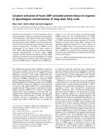
Fig. 1. Alignment of MKK6 sequences and the phylogenetic tree of MKK3 and MKK6 sequences. (A) The multiple sequence alignment of
selected MKK6 sequences is shown, emphasizing any differences. Identical amino acid residues are denoted by dots, different residues are
shown in lowercase, and gaps are shown as hyphens. The conserved phosphorylation site residues Ser and Thr lying within subdomain VIII
are marked by arrows, and the conserved N-terminal p38 docking motif [(R ⁄ K)
2
-(X)
2-6
-L ⁄ I-X-L ⁄ I] is framed by a grey box. (B) The phylogenetic
tree was built from an extended alignment that included additional MKK6 sequences and selected MKK3 sequences. Clade credibility values
are indicated, and the insect MKK3 ⁄ 6 sequences were used as outgroup. For clarity, non-salmon fish sequences occurring in the same clade
as As-MKK6a were named MKK6a and those clustering with As-MKK6b were named MKK6b. The accession numbers for the sequences
used are: MKK3 ⁄ 6: drosophila, Q9U983; mosquito, Q7PRZ7; ciona, Q4H382. MKK3: chicken, Q5ZL06; cow, A4IFH7; mouse, O09110;
human, P46734. MKK6a: tetraodon, Q4SGJ8; fugu, SINFRUP00000137389; stickleback, ENSGACP00000014471; medaka, ENS-
ORLP00000009468. MKK6b: tetraodon, Q4S8I5; fugu, SINFRUP00000128869; medaka, ENSORLP00000017352; carp, Q9I959; zebrafish,
Q6IQW6. Sequences were retrieved from UniProt (six character long accession numbers) or
ENSEMBL.
Atlantic salmon MKK6 orthologs T. E. Hansen et al.
4888 FEBS Journal 275 (2008) 4887–4902 ª 2008 The Authors Journal compilation ª 2008 FEBS
A
B
T. E. Hansen et al. Atlantic salmon MKK6 orthologs
FEBS Journal 275 (2008) 4887–4902 ª 2008 The Authors Journal compilation ª 2008 FEBS 4889
that the salmon MKK6 sequences contain both the
N-terminal p38 MAPK docking motif and the phos-
phorylation sites lying within subdomain VIII.
Phylogenetic analysis of MKK3 and MKK6
sequences
The initial blast searches performed with the
As-MKK sequences suggested the need for a thorough
phylogenetic analysis of the two closely related MKK3
and MKK6 sequence families from mammals and fish
for two reasons. First, although the As-MKK6
sequences are more similar to human MKK6 (80–85%
identity) than MKK3 (73–74% identity), the most clo-
sely related zebrafish sequence (80–87% identity) was
first named MKK3 [27]. Second, the initial blast
searches indicated that, in contrast to salmon, zebra-
fish and carp only appear to have one copy of the
MKK3 ⁄ 6 gene.
A phylogenetic tree was constructed for the
MKK3 ⁄ 6 sequences as described in the Experimental
procedures (Fig. 1B). Two equally striking observa-
tions can be made from the tree. First, MKK3 does
not appear to be present in fish. Second, the MKK6
gene appears to have undergone duplication in fish: all
six fish species analysed have at least one MKK6 gene;
green pufferfish, fugu and medaka have two copies;
and salmon is the only species with three copies.
Hence, a second duplication appears to have occurred
in salmon, resulting in As-MKK6c and As-MKK6b.
Tissue distribution of As-MKK6a, As-MKK6b and
As-MKK6
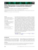
By northern blotting, a 4.0 kb transcript representing
As-MKK6a was detected in all the tissues tested with
the highest expression in the ovary. Two smaller tran-
scripts of 1.7 kb and 1.4 kb were also found in the
ovary. The 1.7 kb transcript was detected as a faint
band in the other organs tested (Fig. 2A). By using a
probe encompassing the entire coding region of
As-MKK6b, we revealed only a single transcript of
approximately 1.7 kb showing equal expression levels
in all tissues examined (Fig. 2B). Due to the high
sequence similarity between MKK6b and c, it is likely
that the MKK6b probe detects both these transcripts
on the northern blot. To distinguish between them,
RT-PCR with primers specific for MKK6a, b and c
were designed and used for expression analysis. The
RT-PCR was performed with mRNA from the same
tissues and mRNA from macrophages (Fig. 2C) and,
consistent with the northern analysis, all three
As-MKK genes were shown to be expressed in these
tissues. The expression of MKK6a was predominant in
the liver compared to MKK6 b and c. Analysis of
MKK6 expression in head kidney macrophages
revealed that only As-MKK6a and c were detected in
these cells.
As-MKK antibody specificity
The specificity of the As-MKK6a, b and c antibodies
was tested by western blot analysis of immunoprecipi-
tated myc-tagged MKK6a, b and c constructs
expressed in Chinook salmon embryo (CHSE)-214
cells. The purified antiserum raised against the
PPPHQSKGEMSQPKG peptide showed specificity to
As-MKK6b ⁄ c, but not to As-MKK6a (Fig. 3A), and
A
B
C
Fig. 2. Tissue expression analyses of salmon MKK6a, MKK6b and
MKK6c. (A) A blot containing poly(A)+ RNA isolated from various
salmon tissues was hybridized with probes specific for either
As-MKK6a (upper panel) or (B) As-MKK6 b ⁄ c (upper panel). b-actin
expression was used as a loading control in (A) and (B) (lower pan-
els). (C) Expression of the As-MKK6a, b and c in various tissues
and in head kidney macrophages examined by RT-PCR, using
MKK6a, b and c specific primers.
Atlantic salmon MKK6 orthologs T. E. Hansen et al.
4890 FEBS Journal 275 (2008) 4887–4902 ª 2008 The Authors Journal compilation ª 2008 FEBS
was named anti-MKK6b ⁄ c. By contrast, the purified
antiserum raised against the SQPKGGKRKPGLKLS
peptide recognized all three As-MKKs and was named
anti-pan-MKK6. These antisera were tested on lysate
from primary nonstimulated macrophages (Fig. 3B,C).
A band at the predicted size of As-MKK6b ⁄ c (40 kDa)
was detected using the As-MKK6b ⁄ c antibody in these
cells (Fig. 3B). This band most likely represents MKK6c
because RT-PCR analysis revealed that MKK6b was
not expressed in macrophages (Fig.2C). In addition, a
faint band at the same size as As-MKK6a was apparent
in the macrophages. Whether this band represents
another salmon MKK6 variant or is due to unspecific
binding in not known. The pan-MKK6 antibody recog-
nized two proteins with the predicted sizes of
As-MKK6b ⁄ c and 6a (Fig. 3C). These results show that
the peptide antibodies raised against the As-MKK6s,
despite some cross-reactivity, can be used to detect
salmon MKKs in tissues and cells.
Phosphorylation of As-MKKs by UV irradiation
The MKKs are phosphorylated and activated by
MAP3Ks [5]. The C-terminal part of MKKs contains
a stretch of approximately 20 amino acids immediately
on the C-terminal side of the MKK catalytic domain
reported to be important in the docking of the
MAP3Ks to the MKKs [34]. Similar C-terminal dock-
ing sites sequences are present in the three As-MKKs.
To investigate whether the As-MKKs are phosphor-
ylated at Ser and Thr residues within subdomain VIII
of the activation loop, a specific antibody to phosphor-
ylated Ser189 of human MKK3 and Ser207 of human
MKK6 was used. CHSE-214 cells were transfected
with myc-tagged MKK wild-type constructs and UV
radiated for 30 min, followed by immunoprecipitation
of the tagged MKKs. UV irradiation is a cellular stres-
sor known to engage multiple signalling pathways end-
ing in p38 activation [35]. As shown in Fig. 4, all three
As-MKKs were phosphorylated in UV radiated
CHSE-214 cells. The amount of phosphorylated
As-MKK6b was considerably lower (Fig. 4B) than the
amount of As-MKK6c (Fig. 4C) and 6a (Fig. 4A).
Phosphorylation of As-MKK6b was not detected in
the total lysate, whereas phosphorylated As-MKK6c
A
C
B
Fig. 3. Antibody specificity against As-MKKs. (A) CHSE-214 cells
were transfected with expression vectors encoding either
As-MKK6a wid-type (wt), 6b wt or 6c wt containing an N-terminal
myc epitope tag. After 48 h, the cells were lysed and the myc-
tagged proteins were immunoprecipitated using myc antibody. The
immunoblots of precipitated wt myc-MKKs were examined using
anti-pan-MKK6 (recognizing As-MKK6a, b and c, upper panel), anti-
MKK6b ⁄ c (recognizing As-MKK6b and c, middle panel) and anti-
myc sera (lower panel). Primary salmon macrophages were
harvested and As-MKK6a, 6b and 6c were visualized by western
blotting of whole cell extract using anti-As-MKK6b ⁄ c (B) and anti-
pan-MKK6 sera (C). Similar results were obtained in two separate
experiments.
A
B
C
Fig. 4. As-MKK6a, b and c are phosphorylated upon stress treat-
ment. CHSE-214 cells were transfected with myc-MKK6a wild-type
(wt) (A), myc-MKK6b wt (B) and myc-MKK6c wt (C). At 48 h post
transfection, the cells were treated with 120 mJÆcm
)2
of UV radia-
tion (30 min) or left untreated. Cell lysates were harvested and wt
myc-MKKs were immunoprecipitated (IP) from the cell lysates. The
immunoblot of the whole cell extracts (WCE) and IP myc-MKKs wt
were analysed by anti-p-MKK3 ⁄ 6 (p-MKK; upper and middle panel)
and anti-myc (lower panel) sera.
T. E. Hansen et al. Atlantic salmon MKK6 orthologs
FEBS Journal 275 (2008) 4887–4902 ª 2008 The Authors Journal compilation ª 2008 FEBS 4891
and 6a were found in the same lysate, and the latter
two were even detected in the nonstimulated cells.
These results indicate that As-MKK6b are poorly
phosphorylated in UV radiated CHSE-214 cells com-
pared to As-MKK6c and 6a.
Ectopically expressed As-MKK6a, b and c are
differently activated by diverse types of stress
The p38 signalling cascade is activated by diverse clas-
ses of stress [1,28,36]. To explore the activation of
salmon MKKs by different stressors, CHSE-214 cells
over-expressing myc-tagged MKK6a, b or c were trea-
ted with sodium arsenite or sorbitol for 30 min.
Sodium arsenite is a oxidative stress inducer that acti-
vates p38 [37,38], whereas sorbitol induces osmotic
stress [39]. A time course over 60 min with the same
type of treatments and also including UV radiation for
30 min was performed for As-MKK6a transfected
cells. The kinase activity of immunoprecipitated myc-
tagged proteins was assayed using recombinant His-
p38a as MKK substrate. High MKK6a activity was
detected for all three stress treatments (Fig. 5A). The
time course of sodium arsenite treatment revealed no
activity before 15 min post treatment and the activity
remained at the same level to 60 min post treatment.
In sorbitol treated cells, As-MKK6a activity was
detected at 5 min post treatment and the activity
remained from 15–60 min post treatment. Sorbitol
treatment gave also strong As-MKK6b activation,
whereas a modest change in the As-MKK6b activity
was detected upon sodium arsenite treatment (Fig. 5B)
Interestingly, the results for As-MKK6c were opposite,
whereas the addition of sodium arsenite to the cells
induced activation was sorbitol ineffective (Fig. 5C).
The latter suggests that sodium arsenite is a poor
MKK6c activator. UV radiation was capable of acti-
vating both As-MKK6b and c (results not shown).
The differences cannot be attributed to variability in
protein expression because western blot analysis of cell
extracts detected equal amounts of total myc-tagged
As-MKK6s. The observed difference in As-MKK6a, b
and c activation by different stimuli suggests that these
salmon MKK6s are differentially regulated.
Sorbitol induced activation of p38 in salmon TO
cells does not require MKK6a, b or c
The phosphorylation of endogenous MKKs was exam-
ined in TO cells using the commercial MKK3 ⁄ 6 phos-
pho-specific antibody. By sodium arsenite stimulation,
one band was detected with a regular substrate,
whereas three different bands were detected with a
ultrasensitive substrate, varying in size in the range
38–44 kDa (Fig. 6A). Band 1 in Fig. 6A, with an
approximately size of 38 kDa, may correspond to
MKK6a. An increased phosphorylation of the band
was detected already after 5 min and the phosphoryla-
tion was more or less constant over the whole time
course. Another band of approximately 40 kDa was
detected after 15 min (Fig. 6A, band 2). This band
corresponded in size to MKK6b or 6c. RT-PCR
results obtained with primers specific for As-MKK6b
and 6c showed that neither MKK6b nor MKK6c was
expressed in TO cells (results not shown), which
excludes the possibility that band 2 represents these
MKK6 variants. The phospho-MKK3 ⁄ 6 antibody is
known to weakly cross react with phosphorylated
MKK4 [7]. A third band (Fig. 6A, band 3) was
detected in the experiment, which corresponds to the
size of MKK4 (approximately 44 kDa) and may repre-
sent a salmon MKK4 ortholog. In sorbitol stimulated
A
B
C
Fig. 5. Activation of As-MKKs by diverse stress. (A) CHSE-214 cells
were transfected with myc-tagged As-MKK6a wild-type (wt)
expression vector and treated with sodium arsenite (SA; 250 l
M),
sorbitol (0.3
M), UV radiation (120 mJÆcm
)2
) for indicated time
points, or left untreated. Cells were lysed and myc-tagged proteins
were immunoprecipitated from the whole cell extracts (WCE) fol-
lowed by an in vitro kinase assay (KA) using salmon His-As-p38a as
a substrate. As-MKKs activities were analyzed by western blot anal-
ysis detecting phosphorylated His-As-p38a. (B) CHSE-214 cells
were transfected with myc-tagged As-MKK6b wt expression vector
and treated with sodium arsenite (SA; 250 l
M), sorbitol (0.3 M), or
otherwise as in (A). (C) CHSE-214 cells transfected with myc-
tagged As-MKK6c wt expression vector, or otherwise as in (A).
Phosphorylated His-As-p38a was detected by immunoblotting using
anti-phospho-p38 serum (p-p38; upper panel) and exogenously
expressed myc-MKKs was detected in cell extracts using anti-myc
serum (lower panel). Experiments were performed twice with
reproducible results.
Atlantic salmon MKK6 orthologs T. E. Hansen et al.
4892 FEBS Journal 275 (2008) 4887–4902 ª 2008 The Authors Journal compilation ª 2008 FEBS
TO cells, only the 38 kDa band was detected at levels
comparable with the control, indicating that this
stimulant did not induce MKK6 phosphorylation
(Fig. 6A). Interestingly, although we were unable to
detect any MKK6 activation upon sorbitol
treatment, it was shown to phosphorylate salmon p38
(Fig. 6A).
Sodium arsenite activation of endogenous MKK6 in
TO cells was further demonstrated by measuring the
ability of MKKs to phosphorylate p38 in vitro. The
MKKs were immunoprecipitated by the pan-MKK6
antibody before measuring their ability to phosphory-
late p38. Figure 6B shows that higher p38 phosphory-
lation by salmon MKK6a was observed at 50 min post
stimulation compared to the activity at 20 min of stim-
ulation. A kinase assay of sodium arsenite and sorbitol
stimulated TO cells over-expressing As-MKK6a
revealed that only sodium arsenite activated
As-MKK6a in TO cells (Fig. 6C). These results are in
agreement with the results shown in Fig. 6A, where no
endogenous MKK phosphorylation was detected in
sorbitol stimulated TO cells.
Activation of p38 independently of MKK3 ⁄ 6, but
dependent on p38 autophosphorylation, has been
A
B
D
C
Fig. 6. Sorbitol induced activation of p38 MAPK in TO cells does not involve any of the MKK6 paralogs. (A) TO cells were treated with
sodium arsenite (250 l
M), sorbitol (0.3 M) at indicated time points, or left untreated. Cells were harvested and phosphorylated As-MKK6a, b,
c (p-MKK, first and second panel) and were visualized by western blotting using regular substrate (West Pico) or ultrasensitive substrate
(West Femto) respectively. Protein loading was verified in the whole cell extracts using the anti-eEF2 serum (lower panel). Phosphorylated
As-p38a was detected by immunoblotting using anti-phospho-p38 serum (p-p38; third panel). (B) TO cells were either treated with 250 l
M
sodium arsenite or left untreated. Cells were harvested at indicated time points and endogenous As-MKK6a, b and c were immunoprecipitated
with anti-pan-MKK6 from the whole cell extracts (WCE). Activities were detected by kinase assay (KA) using His-As-p38 as substrate. Phos-
phorylated His-As-p38a was detected by immunoblotting using anti-phospho-p38 serum (p-p38; upper panel) and eEF2 was detected from
whole cell extracts (lower panel). (C) TO cells were transfected with myc-tagged As-MKK6a wild-type expression vector. After 48 h, the cells
were treated with 250 l
M sodium arsenite, 0.3 M sorbitol for 30 min, or left untreated. Cells were lysed and myc-tagged proteins were im-
munoprecipitated from the lysate followed by an in vitro kinase assay. Phosphorylated His-As-p38a was detected by immunoblotting using
anti-phospho-p38 serum (p-p38; upper panel). To confirm exogenous protein expression, the whole cell extract were blotted and probed with
anti-myc serum (lower panel). (D) TO cells were transfected with GFP tagged As-p38a. After 48 h, the cells were treated with 10 l
M
SB203580 for 1 h or left untreated, followed by stimulation with 250 lM sodium arsenite (SA) or 0.3 M sorbitol for 30 min. Cells were lysed
and phosphorylated GFP-p38a were detected by immunoblotting using anti-phospho-p38 serum (p-p38; first panel). The expression of GFP-
p38a was verified with anti-GFP (second panel). Phosphorylation of endogenous MK2 was detected with anti-phospho-MK2 (third panel) and
anti-eEF2 (fourth panel) was used as a loading control. Experiments were performed twice with reproducible results.
T. E. Hansen et al. Atlantic salmon MKK6 orthologs
FEBS Journal 275 (2008) 4887–4902 ª 2008 The Authors Journal compilation ª 2008 FEBS 4893
reported previously [40,41]. Because the p38 specific
inhibitor SB203580 blocks the ability of p38 to be
autophosphorylated [42], we further examined
whether this inhibitor would prevent p38 phosphory-
lation in stress-activated TO cells. As shown in
Fig. 6D, p38 phosphorylation was not affected by
the SB inhibitor, suggesting that p38 activation in
sorbitol stimulated TO cells is not due to p38 auto-
phosphorylation.
Salmon MKK6a, As-MKK6b and As-MKK6c
activate As-p38a, As-p38b1 and As-p38b2 in
CHSE-214 cells
The p38 MAP kinases are known substrates for
MKK3 and MKK6 in mammalian cells [13,15,16,19–
21,23]. We have recently described three p38a
variants in Atlantic salmon, which all possess the
putative dual phosphorylation motif Thr-Glu-Tyr in
the activation loop as well as the docking motifs
reported to be important for docking to activators,
substrate and regulators. Moreover, all three As-p38a
variants were shown to be phosphorylated in CHSE-
214 cells stressed with sodium arsenite [2]. To
explore the ability of As-MKK6a, As-MKK6b and
As-MKK6c to activate the different As-p38 variants,
constitutively active As-MKKs were constructed.
Mammalian constitutive active MKK3 ⁄ 6 is generated
by replacing the phosphorylation sites Ser and Thr
with the phospho-mimicking glutamic acid (EE) [43].
Constitutive active As-MKKs were generated based
on the same principle. Furthermore, catalytic inactive
As-MKKs mutants were constructed by replacing the
aspartic acid in the conserved DFG motif, known to
be essential for catalytic activity [44], with alanine
(DA). CHSE-214 cells were co-transfected with gluta-
thione S-transferase (GST)-MKK EE or DA and
myc-tagged p38 variants, followed by immunoprecipi-
tation and p38 kinase assay using recombinant ATF-
2 as substrate. All three As-p38 variants were
activated by constitutive active As-MKK6a,
As-MKK6b and As-MKK6c (Fig. 7). As-MKK6b
caused the strongest As-p38a activation among the
three MKKs (Fig. 7A, upper panel), whereas
As-MKK6c was the dominant activator of As-p38b1
(Fig. 7B, upper panel). In the case of As-p38b2, all
three MKKs showed similar levels of activation
(Fig. 7C, upper panel). The ability of the immuno-
precipitated p38 to phosphorylate ATF-2 in vitro
correlated well with results from western blotting
using phospho-p38 antibodies to detect the exoge-
nous and endogenous p38 phosphorylation directly
in the lysates of transfected cells (data not shown).
Discussion
In the present study, we report the cloning of three
cDNAs encoding different salmon MKK6 sequences.
The identity between the As-MKK6b and 6c was 94%,
whereas their identities to MKK6a were approximately
81%. Because the nonmatching nucleotides in the
sequences of these MKKs were spread throughout the
whole ORF, it is unlikely that the different MKK6
A
B
C
Fig. 7. As-MKK6a, b and c are upstream activators of As-p38a,
p38b1 and p38b2. CHSE-214 cells were transfected with GST-
tagged constitutive active (EE) or catalytic inactive (DA) MKK6a, b
or c expression vectors together with either myc-tagged As-p38a
(A), As-p38b1 (B) or As-p38b2 (C). After 24 h, the cells were lysed
and myc-tagged p38 was immunoprecipitated from the whole cell
extracts (WCE) lysate followed by an in vitro kinase assay (KA)
using ATF-2 as p38 substrate. The As-p38 activity was analyzed by
detecting incorporated phosphate into ATF-2 by autoradiography
(first panel). To confirm exogenous protein expression, the whole
cell extract were blotted and probed with anti-GST (second panel)
and anti-myc (third panel) sera.
Atlantic salmon MKK6 orthologs T. E. Hansen et al.
4894 FEBS Journal 275 (2008) 4887–4902 ª 2008 The Authors Journal compilation ª 2008 FEBS
cDNAs represent different splice variants. Thus, our
data suggest that there exist at least three MKK6 genes
in salmon. They all contained the phosphorylation
sites Ser and Thr in the activation loop and also the
N- and C-terminal docking domains shown to be
important for MKK activation and substrate specific-
ity. Further analysis of the salmon MKK6s revealed
that they all were able to phosphorylate and activate
salmon p38.
In higher vertebrates such as man and mouse, the
two genes MKK3 and MKK6 encode proteins that are
the primary p38 activators, whereas in invertebrates
such as Drosophila, Caenorhabditis elegans and in
yeast, there are only a single activator for their p38
orthologs [32,33,45]. A single MKK3 ⁄ 6 ortholog has
been described in the fish species carp and zebrafish
that causes selective activation of p38 in vitro [27,29].
Our phylogenetic analysis suggests that the ancestral
MKK3 ⁄ 6 gene has undergone two major duplication
events (Fig. 1B), one of the events can be observed in
tetrapods, which have MKK3 and MKK6, and the
other can be observed in fish which have one or two
copies of MKK6. Hence, MKK3 does not appear to be
present in fish, and the zebrafish MKK3 sequence
should be renamed to MKK6, which is in agreement
with the name given this sequence in the ZFIN and
UniProt databases. Salmon appears to be unique in
having a third MKK6 copy, possibly reflecting the
salmonid specific tetraploidization event [46] and, to
our knowledge, this is the first report of the existence
of three MKK6 isoforms in any species.
Inspection of the genomic sequences of five fish
species (zebrafish, green pufferfish, fugu, medaka and
stickleback) revealed evidence that may suggest that
the two MKK6 sequences present in some fish may be
the result of the early ray-finned fish tetraploidization
event (results not shown). In green pufferfish and
medaka, the two fish species that have two copies of
the MKK6 gene and where the chromosomal location
is known, the two MKK6 copies are located on differ-
ent chromosomes. This is in contrast to the human
genome where MKK6 and MKK3 are located on the
same chromosome. Furthermore, all MKK6 genes, for
which genomic sequence is available, are in synteny
with a gene encoding a protein homologous to the
human G protein-coupled receptor family C protein
(UniProt: Q9NQ84).
All three salmon MKK6 genes showed ubiquitous
tissue distribution and almost similar expression levels
in the different tissues analyzed. The transcript length
of MKK6b and 6c was approximately 1.7 kb, whereas
the MKK6a probe revealed an approximately 4.0 kb
transcript and two smaller transcripts of approximately
1.4 kb and 1.7 kb, respectively. The latter two were
mainly expressed in the ovary. However, we cannot
exclude the possibility that these transcripts represent
other closely related MKKs or are MKK6a splicing
variants. Due to the high identity between MKK6a
and MKK6b ⁄ c (approximately 81%), the MKK6a
probe may also weakly hybridize to the MKK6b ⁄ c
transcript. The 1.7 kb band seen with the MKK6a
probe could therefore represent the MKK6b ⁄ c tran-
script. The distribution of the salmon MKK6s resem-
bles the wide tissue distribution of salmon p38 [2].
In addition to its involvement in responses to stress
and inflammatory stimuli, the p38 kinase signaling
pathway also participates in processes during normal
development. Zebrafish p38 is involved in the control
of blastomere cleavage during embryogenesis [27,29]
and a specific temporal expression pattern is seen in
throughout zebrafish embryogenesis [47], suggesting an
important role during early development. Studies on
salmon [2] and carp [29] have demonstrated high
expression of both p38 and MKK6 in the ovary. The
abundance of piscine p38 signalling module members
in this organ may suggest that the p38 pathway aids
their survival against environmental stresses during
early development. Carp MKK6 possess a nuclear
export signal sequence that does not exist in the
MKK3 ⁄ 6 families in other species [29]. Such a nuclear
export signal was not found in the salmon MKK6
sequences reported in the present study.
In salmon, three genes encoding p38a isoforms have
been identified and all three were activated by stress-
inducing and inflammatory stimuli. The existence of
several p38 genes in salmon may be a way for cells to
respond differently to upstream kinases and extra-
cellular stimuli, which also has been reported in other
studies [8,20]. Using constitutively activated MKK6
mutants, we were able to demonstrate that all three
MKK6s variant could activate the different salmon
p38 variants. This was shown by a kinase assay detect-
ing ATF-2 phosphorylation. Furthermore, the results
were verified using a GAL4-responsive and ATF2
dependent luciferase reporter assay, where over-
expressing constitutive active As-MKKs mutants
increased ATF2-dependent gene expression by six- to
10-fold compared to catalytic inactive As-MKKs
mutants (data not shown).
Analysis of p38 activation in mouse fibroblasts lack-
ing MKK3 or MKK6, and stressed with UV radiation,
anisomycin or sorbitol, shows that mammalian MKK6
and MKK3 play redundant roles in response to these
stressors [7,24,48]. Despite a very high identity between
the salmon MKK6s, their activation pattern upon
exposure to different stressors revealed differences. We
T. E. Hansen et al. Atlantic salmon MKK6 orthologs
FEBS Journal 275 (2008) 4887–4902 ª 2008 The Authors Journal compilation ª 2008 FEBS 4895
found considerably more phosphorylated MKK6a and
6c compared to MKK6b in UV stressed CHSE-214
cells over-expressing the three As-MKK6s. Moreover,
exposure to the stressors sorbitol and sodium arsenite
resulted in notable differences in response between the
MKK6 isoforms, as measured by their kinase activity
using recombinant salmon p38 as substrate. For
MKK6b, the activation by sorbitol was much more
pronounced compared to sodium arsenite, whereas it
was the opposite for MKK6c. Moreover, As-MKK6a
responded equally to these stressors. The results
suggest a selective activation of As-MKK6 by extra-
cellular stimuli, and surmise that different MAP3Ks
are involved in MKK6 activation in response to alter-
nate forms of stress. It is interesting that p38 is the
only substrate for the MAP2Ks MKK3 and MKK6,
whereas a much wider repertoire of different MAP3Ks
having the ability to phosphorylate and activate
MKK3 and MKK6 exist [35]. A C-terminal docking
site called the DVD domain (i.e. domain for versatile
docking) consisting of 24 amino acids is essential for
activating mammalian MKKs by specific MAP3Ks
[34]. As a consequence, this docking domain may influ-
ence the ability of MKK6 to become phosphorylated
in response to various extracellular stressors. The role
of this docking domain in the requirement of MKK3
or MKK6 to be activated by different MAP3Ks is not
known. We observed that the corresponding region of
the As-MKK6b and c displayed four amino acids that
are different from MKK6a. Whether the divergence in
sequence between the As-MKKs in this region can be
explained by selective substrate specificity of MAP3Ks
in response to different stress needs further investi-
gation.
In extracts prepared from TO cells exposed to cellu-
lar stress, we found several bands, representing puta-
tive phosphorylated MKKs, that cross-reacted with
this phospho-antibody. Consistent with the results
using ectopically expressed MKK6s, the results
obtained showed that the response was determined by
the extracellular stimuli that were used for activation.
In sodium arsenite treated TO cells, two bands
(approximately 38 kDa and 42 kDa, respectively)
showing increased phosphorylation upon activation
were detected, whereas, in sorbitol treated cells, only
basal phospho-MKK6 levels were detected when using
a ultrasensitive substrate. The results of a kinase assay
using over-expressed MKK6a verified that only sodium
arsenite and not sorbitol stimulated its activation in
TO cells. Despite the inability of sorbitol to induce the
phosphorylation of MKK6a in TO cells, phosphory-
lated p38 was detected in these cells upon both sodium
arsenite and sorbitol treatment. The results suggest the
existence of yet other MKK ortholog(s) that phosphor-
ylate p38 in sorbitol stimulated TO cells. Because p38
phosphorylation was not affected by the SB inhibitor,
it is less likely that p38 activation in sorbitol stimu-
lated TO cells is caused by p38 autophosphorylation.
Interestingly, knockdown of the Drosophila p38 activa-
tor D-MKK3 by RNA interference showed a significant,
although incomplete, reduction of phosphorylated p38
levels in response to osmotic stress [49], suggesting the
existence of another p38 activator in Drosophila.By
using UV stressed mouse embryonic fibroblast cells
lacking both MKK3 and MKK6, it was possible to
show that MKK4 participates in the activation of p38.
However, the level of activated p38 in these cells was
much lower compared to wild-type cells, whereas the
level of phosphorylated p38 was not affected in
MKK4-single deficient cells [7]. This indicates that
MKK3 and MKK6 are the main p38 activators,
whereas, under certain circumstances, MKK4 partici-
pates in the activation of p38. We therefore find it
unlikely that MKK4 is the main p38 activator in TO
cells stimulated with sorbitol.
In conclusion, we have identified three upstream
activators of p38 in Atlantic salmon, which all appear
to be MKK6 orthologs. Our phylogenetic analysis
strongly indicates that MKK3 is not present in fish.
The ancestral MKK6 gene appears to have undergone
duplication in some fish species and our data demon-
strate, for the first time, the existence of three MKK6
copies in any species. The results obtained from
CHSE-214 cells and TO cells suggest a cell type depen-
dent expression and activation of the salmon MKK6
variants. Thus, in a whole organism, expressing these
MKK6 genes at different levels may increase the range
of possibilities available to fine tune the strength of
p38 signaling in specialized cells.
Experimental procedures
Reagents and antibodies
Sodium arsenite and sorbitol were obtained from Sigma
(St Louis, MO, USA). The p38 inhibitor SB203580 was
purchased from Alexis Biochemicals (Lausen, Switzerland).
Recombinant ATF-2 and rabbit antibodies against phos-
pho-p38 MAPK, phospho-MKK3 ⁄ 6, phospho-MK2 and
eukaryotic elongation factor 2 (eEF2) were obtained from
Cell Signaling Technology (Beverly, MA, USA). Rabbit
anti-actin serum and mouse anti-GST serum were pur-
chased from Sigma and Santa Cruz Biotechnology (Santa
Cruz, CA, USA), respectively. Mouse anti-myc serum was
purified from the 9E10 hybridom, and rabbit anti-green
fluorescent protein (GFP) was obtained from Abcam (Cam-
Atlantic salmon MKK6 orthologs T. E. Hansen et al.
4896 FEBS Journal 275 (2008) 4887–4902 ª 2008 The Authors Journal compilation ª 2008 FEBS
bridge, MA, USA). Horseradish peroxidase conjugated
goat anti-rabbit IgG and goat anti-mouse IgG secondary
sera were purchased from Santa Cruz Biotechnology. Poly-
clonal MKK6a, MKK6b and MKK6c antibodies were
generated by Eurogentec (Liege, Belgium) using the pep-
tides PPPHQSKGEMSQPKG and SQPKGGKRKPGLK-
LS from salmon MKK6c sequences. The two conjugated
peptides were pooled and injected in two rabbits according
to Eurogentec’s double XP procedure. The resulting anti-
sera were purified by affinity chromatography towards the
respective peptides.
Fish
Two-year-old nonvaccinated Atlantic salmon, strain Aqu-
agen standard (Aquagen, Kyrksæterøra, Norway), weighing
350–600 g, was obtained from Tromsø Aquaculture
Research Station (Tromsø, Norway). Fish were kept at
natural temperature in tanks supplied with running filtered
sea water and fed commercial dry feed. Atlantic salmon
head kidney macrophages were obtained as previously
described in [50] and seeded outlined elsewhere [2].
Molecular cloning of Atlantic salmon MKKs
To obtain a partial cDNA of salmon MKK3 ⁄ 6, we
performed RT-PCR cloning using degenerated primers
based on conserved regions of human MKK3 (accession
number NM_145109) and 6 (GenBank accession number
NM_002758), carp MKK6 (GenBank accession number
AB023480) and zebrafish MKK3 (GenBank accession
number AB030899). A 420 bp PCR product generated
using mixed cDNA from ovary and head kidney, obtained
as previously described [2], showed the highest identity to
MKK3 and MKK6 genes from vertebrate species by a Gen-
Bank database blast search. The entire ORF of the cDNA
was obtained by RACE-PCR using primers designed for
the amplification of the 5¢- and 3¢-ends. A blast search in
the GenBank database with this putative salmon MKK3 ⁄ 6
cDNA identified two rainbow trout (Oncorhynchus mykiss)
MKK3 ⁄ 6 EST clones. One of the clones showed highest
identity to the 5¢-end of salmon MKK3 ⁄ 6 (GenBank acces-
sion number CA388006), whereas the other showed the
highest identity to the 3¢-end (GenBank accession number
CX147893). Primers based on sequences from both EST
clones were used to clone another salmon MKK variant. A
specific primer in the 5¢-UTR of the new MKK was
designed and used with the primer MKK6br3¢. The
sequence contained a complete ORF of 357 amino acids
and was named As-MKK6b. The cloning and sequencing
of several As-MKK6b clones indicated the existence of
another MKK6 variant. A part of the 3¢-UTR of the new
As-MKK6 variant was amplified and specific primer for the
3¢-UTR of the new As-MKK6 variant was designed and
used with the MKK6braf1 primer to amplify the whole
ORF (359 amino acids). The new As-MKK6 variant was
named As-MKK6c. All the primers used for cloning
As-MKK6a, b and c are listed in Table 1.
Sequence and phylogenetic analysis
Relevant sequences for a phylogenetic analysis of the
MKK3 and MKK6 families were collected. MKK3 and
MKK6 sequences were fetched from the UniProt database
(release 12.6) [51] using blast [52]. The obtained sequences
included four fish sequences, namely sequences annotated
as MKK3 from carp and zebrafish, and two sequences from
green pufferfish (Tetraodon nigroviridis). To complement
these, relevant fish MKK sequences were fetched from
ensembl (release 47) [53] using a profile hidden Markov
model built on a multiple sequence alignment of the
MKK3 and MKK6 sequences from UniProt. Additional
relevant sequences were identified in fugu (Takifugu rubripes;
two sequences), medaka (Oryzias latipes; two sequences) and
stickleback (Gasterosteus aculeatus; one sequence). When iso-
forms resulting from alternative splicing were encountered,
only the longest sequence was retained for further analysis.
To ensure that no sequences were missed, the procedure
was repeated in a greedy fashion, including all seven
mammalian MKK sequences and their fish homologues.
The final set of MKK3 and MKK6 sequences were aligned,
and a phylogenetic tree was constructed from the align-
ment. The multiple sequence alignments were constructed
using muscle [54], and profile hidden Markov models were
generated using the hmmer package (http://hmmer.
janelia.org). Phylogenetic trees were constructed using
mrbayes [55], with the following settings: the prior for the
amino acid model was set to mixed, and the number of
generations used was 100 000. mrent was used to visualize
the trees [56] and texshade was used to present the align-
ment [57].
DNA constructs
All As-MKK variants were amplified by PCR using Pfx
polymerase (Invitrogen, Carlsbad, CA, USA) and TOPO
cloned into the Gateway compatible vector pENTRY using
the pENTR ⁄ D-TOPO cloning kit (Invitrogen) following the
manufacturer’s protocol. Gateway expression clones were
made by the Gateway LR Clonase II Enzyme Mix kit with
the destination vectors pDEST 27, pDEST 17 (Invitrogen),
pDEST-EGFP and pDEST-myc [58], according to the man-
ufacturer’s instruction. The As-p38 constructs were made as
previously described [2]. Mutagenesis of plasmid DNA was
performed using the QuickChange site-directed mutagenesis
kit (Stratagene, La Jolla, CA, USA). Various point mutants
of all As-MKK6s were generated using the pENTRY
constructs and the following complementary primers (only
forward primers are shown): As-MKK6b ⁄ cD122A 5¢-
TGAAGATGTGTGCATTTGGGATCAG-3¢, As-MKK6a
T. E. Hansen et al. Atlantic salmon MKK6 orthologs
FEBS Journal 275 (2008) 4887–4902 ª 2008 The Authors Journal compilation ª 2008 FEBS 4897
D199A 5¢-TGAAGATGTGTGCATTTGGCATCAG-3¢,
As-MKK6b ⁄ cSE 5¢-GTTACCTGGTGGACGAAGTGGC
CAAGACCA-3¢, As-MKK6bTE 5¢-CGAAGTGGCCAAG
GAAATAGACGCCGGCTG-3¢, As-MKK6cTE 5¢-CGAA
GTGGCCAAGGAAATGGACGCAGGCTG-3¢, As-MKK
6aSE 5¢-GCCACCTGGTGGACGAAGTGGCCAAGACC
A-3¢ and As-MKK6aTE 5¢-CGAAGTGGCCAAGGAAA
TGGACGCCGGCTG-3¢.
All constructs were verified by DNA sequencing using
the BigDye sequencing (Applied Biosystems, Foster City,
CA, USA).
Northern blot analysis
RNA isolation and northern blotting protocols have been
previously described [2]. Briefly, mRNA (2 lg) was resolved
on a 1% glyoxal-based agarose gel (Ambion, Austin, TX,
USA) and transferred to a nylon membrane by the down-
ward capillary method. The membrane was hybridized with
32
P-labeled cDNA probes and two different probes were
amplified. Template for the As-MKK6a probe was synthe-
sized with primers designed to span the whole ORF (prim-
ers MKK6atoflf and MKK6atoflr; Table 1). The template
for the MKK6b ⁄ c probe was generated with primers span-
ning the whole MKK6b ORF (primers MKK6btoflf and
MKK6btoflr; Table 1).
RT-PCR
For As-MKK6a, b and c expression analysis, we used
mRNA isolated as described above and macrophage mRNA
was isolated as described previously [2]. cDNA synthesis was
performed with Superscript III reverse transcriptase (Invitro-
gen) using random hexamers primers and 1 lg of mRNA in
a20lL volume. The PCR reactions were conducted using
Phusion DNA polymerase (Finnzymes Oy, Espoo, Finland)
and 2 lL of cDNA. The following conditions were applied:
As-MKK6a primers (5¢-GGAAGATCACTGTAGCGATC
GTCA-3¢ and 5¢-GTTGAGGTCGGGGTTTATCCGT-3¢):
96 °C for 30 min, 35 cycles of 96 °C for 10 s, 67 °C for 15 s
and 72 °C extension for 30 s; As-MKK6b primers (5¢-C
CGAGGACATACTGGGAAAG-3¢ and 5¢-GTTGTTTTA
GATCAGGGCTGCTTA-3¢): 96 °C for 30 min, 30 cycles of
96 °C for 10 s, 65 °C for 15 s and 72 °C extension for 30 s;
and, for the As-MKK6c primers (5¢-ATCCTGCGGTTT
CCCTATGACTCCTGG-3¢ and 5¢-GTTGGGTTAGATA
AGGGCGCTCG-3¢), the conditions same as for the
As-MKK6b primers. To confirm equal amount of cDNA in
the samples, PCR was performed with actin specific
primers (5¢-CACTCAACCCCAAAGCCAACAGG-3 ¢ and
5¢-AAAGTCCAGCGCCACGTAGCACAG-3¢) under the
following conditions: 96 °C for 30 min, 20 cycles of 96 °C
for 10 s, 68 °C for 20 s and 72 °C for 30 s.
Table 1. Primers used for the cloning of As-MKK6a, b and c.
Name Primer Sequence (5¢-to-3¢) Primer description
MKK6adefw3 GGNGTGGTGGANAAGATG Degenerated primer based on conserved region of MKK6a
MKK6aderv2 AAYAANGGATGTTGCAT Reverse primer for MKK6adefw3
MKK6adefw2 GTNTGGATHTGCATGGA Degenerated nested primer for amplificated MKK6a
MKK6aderv3 TCCCCATGANTCATAGG Reverse primer for MKK6adefw2
MKK6arafw1 GATCAACACACAGGGCCAGGTGAAGATG RACE primer for MKK6a 3¢-end
MKK6ararv1 GGTCTTGGCCATAGAGTCCACCAGGT RACE primer for MK6a 5¢-end
MKK6arafw2 GTGGACTCTATGGCCAAGACCA Nested primer for RACE 3¢-end products
MKK6ararv2 ATCTTCACCTGGCCCTGTGTGT Nested primer for RACE 5¢-end products
MKK6aflf GAATAAGATCTCCACACACCCAGGGC Primer in 5¢-UTR of MKK6a
MKK6aflr GTTGGAGTTGTGTGGCAGATCAATTC Primer in 3¢-UTR of MKK6a
MKK6atoflf CACCATGTCTCTTTCTAAAGGAGGGAAGAA For topo cloning of MKK6a into pENTR ⁄ D-TOPO
MKK6atoflrs TCAGTCTGCCAGGATGATCTTGACA Reverse primer for MKK6atoflf
MKK6bf3¢ GAGAGATTAATCAGAAAGGC Primer based on a part of the 3¢-end to rainbow trout
(GenBank accession number CX147893)
MKK6br3¢ TTGGTCAGAGCGTTGTCTTA Reverse primer for MKK6bf3¢
MKK6bf5¢ CGACCCGTTTCCTGACC Primer base on a part of the 5¢-end to rainbow trout
(GenBank accession number CA388006)
MKK6br5¢ TGAAGAGTGCGCCGTAGAAGGTGAC Reverse primer for MKK6bf5¢
MKK6braf1 AGGTGTGCAATTGTATATTGCTCTTTG Primer in 5¢-UTR of MKK6b used with MKK6br3¢ to amplify
the MKK6b ORF
MKK6btoflf CACCATGGAGGGAGGGAGTGAGAAAGAAG For topo cloning of MKK6b into pENTR ⁄ D-TOPO
MKK6btoflr TCAGTCCCCGAGGATGACCTT Reverse primer for MKK6btoflf
MKK6cf2 ATCCTGCGGTTTCCCTATGACTCCTGG Primer specific for MKK6c used with MKK6cr2 to amplify
3
¢-UTR of AsMKK6c
MKK6cr2 GTAACAGGGTTTGCAATTGG Reverse primer designed from the EST clone CX147893
MKK6cUTRr GTTGGGTTAGATAAGGGCGCTCG Reverse primer specific for MKK6c used with MKK6brafl to
amplify the whole ORF of As-MKK6c
Atlantic salmon MKK6 orthologs T. E. Hansen et al.
4898 FEBS Journal 275 (2008) 4887–4902 ª 2008 The Authors Journal compilation ª 2008 FEBS
Cell cultures and transfection
Chinook salmon embryonic cells (CHSE-214) were cultured
in EMEM (Invitrogen), supplemented with 60 lgÆmL
)1
of
penicillin, 100 lgÆmL
)1
of streptomycin, 1% non-essential
amino acids (Invitrogen), 2 mml-glutamine (Invitrogen)
and 7.5% fetal bovine serum (Euroclone, Celbio, Milan,
Italy). Cells were grown at 20 °C in a 5% humified CO
2
incubator. TO cells originate from Atlantic salmon head
kidney [59] was obtained from Professor H. Wergeland
(University of Bergen, Norway). The TO cells were cultured
at 20 ° Cin5%CO
2
in EMEM supplemented with
100 lgÆmL
)1
of streptomycin 60 lgÆmL
)1
of penicillin,
2mml-glutamine, 1% non-essential amino acids and 5%
fetal bovine serum. Cells for transfection were seeded in
culture plates and transfected the next day at 80–90% con-
fluence. Transfection of CHSE-214 cells was performed by
using Lipofectamine 2000 (Invitrogen) transfection reagent,
according to the manufacturer’s instruction. TO cells were
transfected with FuGENE HD (Roche Diagnostics, India-
napolis, IN, USA) using 3 lg of plasmid and 6 lL of trans-
fection reagents for each 35 mm well.
Cell lysates for western blotting were harvested in buffer
A [20 mm Tris-acetate, pH 7.0; 0.27 m sucrose; 1 mm
EDTA; 1 mm EGTA; 1 mm orthovanadate; 10 mm b-glyc-
erophosphate; 50 mm sodium fluoride; 5 mm sodium
pyrophosphate; 1% (v ⁄ v) Triton X-100; 0.1% (v ⁄ v) 2-mer-
captoethanol and ‘Complete’ protease inhibitor cocktail
(one tablet per 50 mL; Roche)]. The lysates were centri-
fuged for 15 min at 15 000 g. NuPAGE LDS sample buffer
(Invitrogen) was added to the lysates and the samples were
heated for 10 min at 70 °C.
Western blot analysis
Cell lysates were separated by SDS ⁄ PAGE (4–12% precast
NuPAGE; Invitrogen), followed by transfer to a 0.45 lm
pore size polyvinylidene difluoride membrane (Millipore,
Billerica, MA, USA) as described previously [2] and probed
with either anti-myc (1 : 500), anti-GST (1 : 500), anti-GFP
(1 : 3000), anti-phospho-MKK3 ⁄ 6 (1 : 1000), anti-phospho-
p38 (1 : 1000) anti-phospho-MK2 (1 : 1000), anti-eEF2
(1 : 1000), anti-pan-MKK6 (1 : 1000) or anti-MKK6b ⁄ c
(1 : 1000) sera. Detection was performed by using Super-
Signal West Pico or West Femto (Pierce Biotechnology,
Rockford, IL, USA).
p38 kinase assay
Transfected cells were washed twice in ice-cold phosphate
buffered saline and lysed in 200 lL of ice-cold buffer A
with addition of complete protease inhibitor cocktail (one
tablet per 50 mL; Roche). The lysate was cleared by centri-
fugation for 15 min at 15 000 g. Myc-tagged As-p38 was
immunoprecipitated by incubating the lysate at 4 °C for
1 h with monoclonal myc antibodies (1 : 20). Then 30 lL
of 50% slurry protein G-agarose (Upstate Biotechnology,
Lake Placid, NY, USA) pre-equilibrated in buffer A was
added and the lysate was incubated for additional 1 h at
4 °C. The immunoprecipitated myc-As-p38 was washed
three times in ice-cold buffer A and twice in ice-cold kinase
buffer (25 mm Hepes, pH 7.4, 25 mm b-glycerophosphate,
25 mm MgCl
2
, 0.5 mm dithiothreitol, 0.1 mm sodium
orthovanadate). The As-p38 kinase activity was measured
in 40 lL of kinase buffer containing 0.1 mm ATP (Sigma),
1 lCi [c-
32
P]ATP (3000 CiÆmmol
)1
; Amersham Pharmacia,
Piscataway, NJ, USA) and 4 lg ATF-2 (Cell Signaling) at
30 °C. The reaction was terminated after 30 min by adding
14 lLof4· LDS-sample buffer. The incorporation of
radioactive phosphate into ATF-2 was examined after
SDS ⁄ PAGE by autoradiography.
Immunoprecipitation of myc-tagged and
endogenous MKKs
Cells transfected with myc-MKK6a, b and c were lysed as
described in the kinase assay part above. Myc-MKKs were
immunoprecipitated by incubation the cleared lysate at
4 °C for 1 h with monoclonal myc antibodies (1 : 20),
before addition of 30 lL of protein G-agarose (50% slurry
pre-equilibrated in buffer A) and incubated at 4 °C for 1 h.
The immunoprecipitated myc-MKKs were washed three
times in ice-cold buffer A and twice in ice-cold kinase buf-
fer. Immunoprecipitates not used for kinase assays were
washed three times in buffer A and resuspended in 40 lL
of 2· LDS-sample buffer.
TO cells seeded in 35 mm wells were lysed in 200 lLof
buffer A with complete protease inhibitor cocktail and
identical samples from two wells were pooled together.
Lysates were cleared by centrifugation at 4 °C for 15 min
at 15 000 g. Endogenous MKK was immunoprecipitated by
incubating the cleared lysate at 4 °C for 1 h with polyclonal
pan-As-MKK6 peptide antibody (1 : 40). Then 30 lLof
50% slurry pre-blocked protein A ⁄ G PLUS-agarose (Santa
Cruz) pre-equilibrated in buffer A was added and incubated
at 4 °C for 1 h. The immunoprecipitated MKKs were
washed three times in ice-cold buffer A and twice in
ice-cold kinase buffer.
The kinase activity of immunoprecipitated myc-MKKs
and endogenous MKKs were measured in 40 lL of kinase
buffer containing 200 l m ATP and 1 lg As-p38a at 14 °C.
The reaction was terminated after 30 min by adding 14 lL
of 4· LDS-sample buffer. The phosphorylation of recombi-
nant As-p38a was examined by SDS ⁄ PAGE and detected
by anti-phospho-p38 serum.
Expression of His-p38a in Escherichia coli
His-tagged full-length p38a was expressed in Escherichia
coli (BL21[DE3]pLysS) by induction with 1 lm isopropyl-1-
T. E. Hansen et al. Atlantic salmon MKK6 orthologs
FEBS Journal 275 (2008) 4887–4902 ª 2008 The Authors Journal compilation ª 2008 FEBS 4899
thio-b-d-galactopyranoside at 23 °C for 4 h. His-p38a was
then purified on a HisTrap HP column (Amersham Phar-
macia) using standard techniques. Expression and purity of
this fusion protein were checked by SDS ⁄ PAGE (4–12%
precast NuPAGE; Invitrogen) and Coomassie blue staining.
Protein concentration was measured by the Bradford pro-
tein assay (Bio-Rad, Hercules, CA, USA).
Acknowledgements
We thank Dr A. N. Larsen (University of Tromsø) for
assisting in the purifying of recombinant As-p38a. This
work was supported by a grant from the Research
Council of Norway (NFR 154197 ⁄ 432). The Norwe-
gian Structural Biology Centre (NorStruct) is sup-
ported by the National program in Functional
Genomics (FUGE) in the Research Council of
Norway. O. M. Seternes is a fellow of the Norwegian
Cancer Society.
References
1 Raingeaud J, Gupta S, Rogers JS, Dickens M, Han J,
Ulevitch RJ & Davis RJ (1995) Pro-inflammatory cyto-
kines and environmental stress cause p38 mitogen-acti-
vated protein kinase activation by dual phosphorylation
on tyrosine and threonine. J Biol Chem 270, 7420–7426.
2 Hansen TE & Jørgensen JB (2007) Cloning and charac-
terisation of p38 MAP kinase from Atlantic salmon A
kinase important for regulating salmon TNF-2 and IL-
1beta expression. Mol Immunol 44, 3137–3146.
3 Anderson NG, Maller JL, Tonks NK & Sturgill TW
(1990) Requirement for integration of signals from two
distinct phosphorylation pathways for activation of
MAP kinase. Nature 343, 651–653.
4 Derijard B, Hibi M, Wu IH, Barrett T, Su B, Deng T,
Karin M & Davis RJ (1994) JNK1: a protein kinase
stimulated by UV light and Ha-Ras that binds and
phosphorylates the c-Jun activation domain. Cell 76,
1025–1037.
5 Cuevas BD, Abell AN & Johnson GL (2007) Role of
mitogen-activated protein kinase kinase kinases in
signal integration. Oncogene 26, 3159–3171.
6 Zarubin T & Han J (2005) Activation and signaling of
the p38 MAP kinase pathway. Cell Res 15, 11–18.
7 Brancho D, Tanaka N, Jaeschke A, Ventura JJ, Kelkar
N, Tanaka Y, Kyuuma M, Takeshita T, Flavell RA &
Davis RJ (2003) Mechanism of p38 MAP kinase activa-
tion in vivo. Genes Dev 17, 1969–1978.
8 Enslen H, Brancho DM & Davis RJ (2000) Molecular
determinants that mediate selective activation of p38
MAP kinase isoforms. EMBO J 19, 1301–1311.
9 Ho DT, Bardwell AJ, Grewal S, Iverson C & Bardwell
L (2006) Interacting JNK-docking sites in MKK7
promote binding and activation of JNK mitogen-acti-
vated protein kinases. J Biol Chem 281, 13169–13179.
10 Enslen H & Davis RJ (2001) Regulation of MAP kinas-
es by docking domains. Biol Cell 93, 5–14.
11 Ho DT, Bardwell AJ, Abdollahi M & Bardwell L
(2003) A docking site in MKK4 mediates high affinity
binding to JNK MAPKs and competes with similar
docking sites in JNK substrates. J Biol Chem 278,
32662–32672.
12 Xu B, Stippec S, Robinson FL & Cobb MH (2001)
Hydrophobic as well as charged residues in both MEK1
and ERK2 are important for their proper docking.
J Biol Chem 276, 26509–26515.
13 Derijard B, Raingeaud J, Barrett T, Wu IH, Han J,
Ulevitch RJ & Davis RJ (1995) Independent human
MAP-kinase signal transduction pathways defined by
MEK and MKK isoforms. Science 267, 682–685.
14 Han J, Wang X, Jiang Y, Ulevitch RJ & Lin S (1997)
Identification and characterization of a predominant
isoform of human MKK3. FEBS Lett 403, 19–22.
15 Moriguchi T, Kuroyanagi N, Yamaguchi K, Gotoh Y,
Irie K, Kano T, Shirakabe K, Muro Y, Shibuya H,
Matsumoto K et al. (1996) A novel kinase cascade med-
iated by mitogen-activated protein kinase kinase 6 and
MKK3. J Biol Chem 271, 13675–13679.
16 Han J, Lee JD, Jiang Y, Li Z, Feng L & Ulevitch RJ
(1996) Characterization of the structure and function of
a novel MAP kinase kinase (MKK6). J Biol Chem 271,
2886–2891.
17 Cuenda A, Alonso G, Morrice N, Jones M, Meier R,
Cohen P & Nebreda AR (1996) Purification and cDNA
cloning of SAPKK3, the major activator of RK ⁄ p38 in
stress- and cytokine-stimulated monocytes and epithelial
cells. EMBO J 15, 4156–4164.
18 Moriguchi T, Toyoshima F, Gotoh Y, Iwamatsu A, Irie
K, Mori E, Kuroyanagi N, Hagiwara M, Matsumoto K
& Nishida E (1996) Purification and identification of a
major activator for p38 from osmotically shocked cells.
Activation of mitogen-activated protein kinase kinase 6
by osmotic shock, tumor necrosis factor-alpha, and
H2O2. J Biol Chem 271, 26981–26988.
19 Jiang Y, Gram H, Zhao M, New L, Gu J, Feng L, Di
Padova F, Ulevitch RJ & Han J (1997) Characteriza-
tion of the structure and function of the fourth member
of p38 group mitogen-activated protein kinases,
p38delta. J Biol Chem 272, 30122–30128.
20 Enslen H, Raingeaud J & Davis RJ (1998) Selective
activation of p38 mitogen-activated protein (MAP)
kinase isoforms by the MAP kinase kinases MKK3 and
MKK6. J Biol Chem 273, 1741–1748.
21 Jiang Y, Chen C, Li Z, Guo W, Gegner JA, Lin S &
Han J (1996) Characterization of the structure and
function of a new mitogen-activated protein kinase
(p38beta). J Biol Chem 271, 17920–17926.
Atlantic salmon MKK6 orthologs T. E. Hansen et al.
4900 FEBS Journal 275 (2008) 4887–4902 ª 2008 The Authors Journal compilation ª 2008 FEBS
22 Cuenda A, Cohen P, Buee-Scherrer V & Goedert M
(1997) Activation of stress-activated protein kinase-3
(SAPK3) by cytokines and cellular stresses is mediated
via SAPKK3 (MKK6); comparison of the specificities
of SAPK3 and SAPK2 (RK ⁄ p38). EMBO J 16, 295–
305.
23 Goedert M, Cuenda A, Craxton M, Jakes R & Cohen
P (1997) Activation of the novel stress-activated protein
kinase SAPK4 by cytokines and cellular stresses is med-
iated by SKK3 (MKK6); comparison of its substrate
specificity with that of other SAP kinases. EMBO J 16,
3563–3571.
24 Lu HT, Yang DD, Wysk M, Gatti E, Mellman I, Davis
RJ & Flavell RA (1999) Defective IL-12 production in
mitogen-activated protein (MAP) kinase kinase 3
(Mkk3)-deficient mice. EMBO J 18, 1845–1857.
25 Wysk M, Yang DD, Lu HT, Flavell RA & Davis RJ
(1999) Requirement of mitogen-activated protein kinase
kinase 3 (MKK3) for tumor necrosis factor-induced
cytokine expression. Proc Natl Acad Sci USA 96, 3763–
3768.
26 Tanaka N, Kamanaka M, Enslen H, Dong C, Wysk M,
Davis RJ & Flavell RA (2002) Differential involvement
of p38 mitogen-activated protein kinase kinases MKK3
and MKK6 in T-cell apoptosis. EMBO Rep 3, 785–791.
27 Fujii R, Yamashita S, Hibi M & Hirano T (2000)
Asymmetric p38 activation in zebrafish: its possible role
in symmetric and synchronous cleavage. J Cell Biol 150,
1335–1348.
28 Han J, Lee JD, Bibbs L & Ulevitch RJ (1994) A MAP
kinase targeted by endotoxin and hyperosmolarity in
mammalian cells. Science 265, 808–811.
29 Hashimoto H, Fukuda M, Matsuo Y, Yokoyama Y,
Nishida E, Toyohara H & Sakaguchi M (2000) Identifi-
cation of a nuclear export signal in MKK6, an activator
of the carp p38 mitogen-activated protein kinases. Eur J
Biochem 267, 4362–4371.
30 Kumar S, McDonnell PC, Gum RJ, Hand AT, Lee JC
& Young PR (1997) Novel homologues of CSBP ⁄ p38
MAP kinase: activation, substrate specificity and sensi-
tivity to inhibition by pyridinyl imidazoles. Biochem
Biophys Res Commun 235, 533–538.
31 Mertens S, Craxton M & Goedert M (1996) SAP
kinase-3, a new member of the family of mammalian
stress-activated protein kinases. FEBS Lett 383, 273–
276.
32 Han ZS, Enslen H, Hu X, Meng X, Wu IH, Barrett T,
Davis RJ & Ip YT (1998) A conserved p38 mitogen-
activated protein kinase pathway regulates Drosophila
immunity gene expression. Mol Cell Biol 18, 3527–3539.
33 Brewster JL, de Valoir T, Dwyer ND, Winter E &
Gustin MC (1993) An osmosensing signal transduction
pathway in yeast. Science 259, 1760–1763.
34 Takekawa M, Tatebayashi K & Saito H (2005)
Conserved docking site is essential for activation of
mammalian MAP kinase kinases by specific MAP
kinase kinase kinases. Mol Cell 18, 295–306.
35 Winter-Vann AM & Johnson GL (2007) Integrated acti-
vation of MAP3Ks balances cell fate in response to
stress. J Cell Biochem 102, 848–858.
36 Rouse J, Cohen P, Trigon S, Morange M, Alonso-
Llamazares A, Zamanillo D, Hunt T & Nebreda AR
(1994) A novel kinase cascade triggered by stress and
heat shock that stimulates MAPKAP kinase-2 and
phosphorylation of the small heat shock proteins. Cell
78, 1027–1037.
37 Barchowsky A, Dudek EJ, Treadwell MD & Wet-
terhahn KE (1996) Arsenic induces oxidant stress and
NF-kappa B activation in cultured aortic endothelial
cells. Free Radic Biol Med 21, 783–790.
38 Liu Y, Guyton KZ, Gorospe M, Xu Q, Lee JC &
Holbrook NJ (1996) Differential activation of ERK,
JNK ⁄ SAPK and P38 ⁄
CSBP ⁄ RK map kinase family
members during the cellular response to arsenite. Free
Radic Biol Med 21, 771–781.
39 Uhlik MT, Abell AN, Johnson NL, Sun W, Cuevas
BD, Lobel-Rice KE, Horne EA, Dell’Acqua ML &
Johnson GL (2003) Rac-MEKK3-MKK3 scaffolding
for p38 MAPK activation during hyperosmotic shock.
Nat Cell Biol 5, 1104–1110.
40 Salvador JM, Mittelstadt PR, Guszczynski T, Copeland
TD, Yamaguchi H, Appella E, Fornace AJ Jr &
Ashwell JD (2005) Alternative p38 activation pathway
mediated by T cell receptor-proximal tyrosine kinases.
Nat Immunol 6, 390–395.
41 Ge B, Gram H, Di Padova F, Huang B, New L,
Ulevitch RJ, Luo Y & Han J (2002) MAPKK-indepen-
dent activation of p38alpha mediated by TAB1-depen-
dent autophosphorylation of p38alpha. Science 295,
1291–1294.
42 Tong L, Pav S, White DM, Rogers S, Crane KM,
Cywin CL, Brown ML & Pargellis CA (1997) A highly
specific inhibitor of human p38 MAP kinase binds in
the ATP pocket. Nat Struct Biol 4, 311–316.
43 Raingeaud J, Whitmarsh AJ, Barrett T, Derijard B &
Davis RJ (1996) MKK3- and MKK6-regulated gene
expression is mediated by the p38 mitogen-activated
protein kinase signal transduction pathway. Mol Cell
Biol 16, 1247–1255.
44 Dhillon AS & Kolch W (2004) Oncogenic B-Raf muta-
tions: crystal clear at last. Cancer Cell 5, 303–304.
45 Tanaka-Hino M, Sagasti A, Hisamoto N, Kawasaki M,
Nakano S, Ninomiya-Tsuji J, Bargmann CI & Matsum-
oto K (2002) SEK-1 MAPKK mediates Ca
2+
signaling
to determine neuronal asymmetric development in
Caenorhabditis elegans. EMBO Rep 3, 56–62.
46 Allendorf FW & Thorgaard GH (1984) Tetraploidy and
the evolution of salmonid fishes. In: Evolutionary Genet-
ics of Fishes (Turner BJ, ed.), pp. 1–53. Plenum, New
York, NY.
T. E. Hansen et al. Atlantic salmon MKK6 orthologs
FEBS Journal 275 (2008) 4887–4902 ª 2008 The Authors Journal compilation ª 2008 FEBS 4901
47 Krens SF, He S, Spaink HP & Snaar-Jagalska BE (2006)
Characterization and expression patterns of the MAPK
family in zebrafish. Gene Expr Patterns 6, 1019–1026.
48 Li Y, Batra S, Sassano A, Majchrzak B, Levy DE,
Gaestel M, Fish EN, Davis RJ & Platanias LC (2005)
Activation of mitogen-activated protein kinase kinase
(MKK) 3 and MKK6 by type I interferons. J Biol
Chem 280, 10001–10010.
49 Zhuang ZH, Zhou Y, Yu MC, Silverman N & Ge BX
(2006) Regulation of Drosophila p38 activation by
specific MAP2 kinase and MAP3 kinase in response to
different stimuli. Cell Signal 18, 441–448.
50 Jørgensen JB & Robertsen B (1995) Yeast beta-glucan
stimulates respiratory burst activity of Atlantic salmon
(Salmo salar L.) macrophages. Dev Comp Immunol 19,
43–57.
51 The_UniProt_Consortium. (2007) The universal protein
resource (UniProt). Nucleic Acids Res 35, D193–D197.
52 Altschul SF, Madden TL, Schaffer AA, Zhang J, Zhang
Z, Miller W & Lipman DJ (1997) Gapped BLAST and
PSI-BLAST: a new generation of protein database
search programs. Nucleic Acids Res 25, 3389–3402.
53 Flicek P, Aken BL, Beal K, Ballester B, Caccamo M,
Chen Y, Clarke L, Coates G, Cunningham F, Cutts T
et al. (2007) Ensembl 2008. Nucleic Acids Res 36,
D707–D714.
54 Edgar RC (2004) MUSCLE: multiple sequence align-
ment with high accuracy and high throughput. Nucleic
Acids Res 32, 1792–1797.
55 Ronquist F & Huelsenbeck JP (2003) mrbayes 3:
Bayesian phylogenetic inference under mixed models.
Bioinformatics 19, 1572–1574.
56 Zuccon A & Zuccon D (2006) MrEnt v1.2 (Program
Distributed by the Authors). Department of
Vertebrate Zoology & Molecular Systematics
Laboratory, Swedish Museum of Natural History,
Stockholm.
57 Beitz E (2000) TEXshade: shading and labeling of
multiple sequence alignments using LATEX2 epsilon.
Bioinformatics 16, 135–139.
58 Lamark T, Perander M, Outzen H, Kristiansen K,
Overvatn A, Michaelsen E, Bjorkoy G & Johansen T
(2003) Interaction codes within the family of mamma-
lian Phox and Bem1p domain-containing proteins.
J Biol Chem 278, 34568–34581.
59 Wergeland HI & Jakobsen RA (2001) A salmonid cell
line (TO) for production of infectious salmon anaemia
virus (ISAV). Dis Aquat Organ 44, 183–190.
Atlantic salmon MKK6 orthologs T. E. Hansen et al.
4902 FEBS Journal 275 (2008) 4887–4902 ª 2008 The Authors Journal compilation ª 2008 FEBS