Báo cáo Y học: Proteolytic action of duodenase is required to induce DNA synthesis in pulmonary artery fibroblasts A role for phosphoinositide 3-kinase pot
Bạn đang xem bản rút gọn của tài liệu. Xem và tải ngay bản đầy đủ của tài liệu tại đây (416.61 KB, 10 trang )
Proteolytic action of duodenase is required to induce DNA synthesis
in pulmonary artery fibroblasts
A role for phosphoinositide 3-kinase
Alan D. Pemberton
1
, Tatyana S. Zamolodchikova
2
, Cheryl L. Scudamore
3
, Edwin R. Chilvers
4
,
Hugh R. P. Miller
1
and Trevor R. Walker
5
1
Department of Veterinary Studies, University of Edinburgh, Easter Bush Veterinary Centre, Roslin, Edinburgh, UK;
2
Shemyakin-
Ovchinnikov Institute of Bioorganic Chemistry, Russian Academy of Sciences, Moscow, Russia;
3
Department of Veterinary Pathology,
University of Edinburgh, Easter Bush Veterinary Centre, Roslin, Edinburgh, UK;
4
Respiratory Medicine Unit, Department of
Medicine, University of Cambridge School of Clinical Medicine, Addenbrooke’s and Papworth Hospitals, Cambridge, UK;
5
Rayne Laboratory, Respiratory Medicine Unit, University of Edinburgh Medical School, Edinburgh, UK
Duodenase is a 29-kDa serine endopeptidase that displays
selective trypsin- and c hymotrypsin-like s ubstrate specificity.
This enzyme has been localized to epitheliocytes of B runner’s
glands, a nd as described h ere, to mast cells within the
intestinal mucosa and lungworm-infected lung, implying an
important additional role in inflammation and tissue
remodelling. In primary c ultures of pulmonary artery
fibroblasts, duodenase induced a concentration-dependent
increase in [
3
H]thymidine incorporation with a maximal
effect observed at 30 n
M
. Pretreating duodenase with soy-
bean trypsin inhibitor abolished DNA synthesis, confirming
that proteolytic a ctivity w as an essential requirement for this
response. PAR1, P AR2 and PAR4 activating p eptides were
unable to induce [
3
H]thymidine incorporation in pulmonary
artery fibroblasts. Likewise, pretreatment of fibroblasts with
TNFa, known to up-regulate PAR2 expression in other
systems, and IL-1b, did not enhance the potential of
duodenase to induce DNA synthesis. Furthermore, duo-
denase increased GTPcS binding to fibroblast membranes
indicating that a G-protein-coupled receptor may mediate
the effects of duodenase. Duodenase-induced DNA syn-
thesis and GTPcS b inding were both f ound to be inhibited
by pertussis toxin, implying a role for G
i/o
. Selective inhi-
bitors of MEK1 (PD98059) and protein kinase C
(GF109203X) only partially inhibited duodenase-induced
DNA synthesis, but both wortmannin (100 n
M
)and
LY294002 (10 l
M
) inhibited this r esponse completely,
indicating a key role for PtdIns 3-kinase. Furthermore,
duodenase induced a 2.3 ± 0.1-fold increase in PtdIns
3-kinase activity in p85 immu noprecipitates, which was
sensitive t o inhibition by wortmannin. These results suggest
that duodenase can i nduce pulmonary artery fibroblast
DNA synthesis in a PtdIns 3-kinase-dependent manner via a
G-protein-coupled receptor which is activated by a proteo-
lytic m echanism.
Keywords: duodenase; fibroblasts; phosphoinositide 3-kinase;
protease-activated receptor.
Duodenase is a serine endopeptidase, originally isolated
from bovine duodenum, with a dual trypsin-like and
chymotrypsin-like primary substrate specificity, i.e. cleaving
the C-terminal to both basic and hydrophobic amino-acid
residues [1]. The closely related enzyme, sheep mast cell
proteinase-1 (sMCP-1) is 85% identical at the amino-acid
level [2] a nd, due to close similarity of the primary substrate
binding region, has a strikingly similar cleavage specificity
[3]. Duodenase was o riginally immunolocalized to epithelial
cells of Brunner’s glands within the duodenum, and was an
activator of enteropeptidase [4]. Oth er studies employing
esterase staining have provided evidence for the expression
of an enzyme with trypsin-like properties, distinct from
tryptase, in intestinal mucosal mast cells, and in the lung
around bronchioles and w ithin the alveolar septa [5]. This is
consistent with data in sheep showing that sMCP-1 is
located to mucosal mast cells of the gastrointestinal tract,
and around small bronchi and a lveolar walls in the lung [6].
One of the many identified effects o f mast cell proteinases
is their ability to induce cellular p roliferation. For example,
both human mast cell tryptase and sMCP-1 h ave been
shown t o b e m itogenic for fibroblasts [7,8]. In patients with
chronic inflammatory lung disord ers, significant accumula-
tion of mast c ells occurs within the lungs and is b elieved to
underlie the generation of pulmonary fibrosis, involving
proliferation of mesenchymal cells to form the basis of a
fibrotic scar [9]. Recruitment of mast cells and release of
their proteinases may therefore play a central role in the
initiation of a p roliferative response following injury or
inflammation within the lung.
The field of proteinase-mediated cellular activation has
expanded rapidly following the discovery that a-thrombin
mediates its actions through a receptor which contains a
Ôtethered-ligandÕ, with activation occurring consequent to
Correspondence to T. R. Walker, Rayne Laboratory, Respiratory
Medicine Unit, University of Edinburgh Medical School, Teviot
Place, Edinburgh EH8 9AG, UK. Fax: + 131 6504384,
Tel.: + 131 6511320, E-mail: ed.ac.uk
Abbreviations: PAR, protease-activated receptor; sMCP-1, sheep mast
cell proteinase-1; DMEM, Dulbecco’s modified Eagle’s medium;
TNFa, tumour necrosis factor a; PtdIns 3-kinase, phosphoinositide
3-kinase.
(Received 1 4 September 2001, revised 7 December 2001, accepted 19
December 20 01)
Eur. J. Biochem. 269, 1171–1180 (2002) Ó FEBS 2002
proteolytic cleavage of the N-terminal exodomain. This
thrombin receptor has since been termed PAR1 (protease-
activated receptor-1) and is known to mediate the actions
of thrombin on platelets and other cell types [10]. Subse-
quently, RT-PCR and Northern analysis have identified
mRNA for three additional members of this receptors
family termed PAR2, PAR3 and PAR4 [11]. Interestingly,
thrombin has now been demonstrated to cleave and activate
PAR1, PAR3 and PAR4 whereas trypsin a nd tryptase
activate PAR2 [12]. Certain other proteases, including
chymotrypsin and cathepsin G, appear to ÔdisarmÕ PAR1 by
cleaving the exodomain of the receptor without inducing
activation and t hus preventing activation by thrombin [13].
All four receptors have a classical heptahelical structure
within the plasma membrane and are known to couple to
both G
q/11
and G
i/o
and stimulate phosphoinositide turn-
over although their other potential downstream signalling
targets h ave not been fully established [ 12]. In this study we
have investigated the ability of duodenase to induce DNA
synthesis in bovine pulmonary artery fibroblasts, attempting
to elucidate which PAR subtype and signalling pathways
may be involved in mediating this effect. We also provide
evidence for an a dditional mast cell o rigin of duodenase,
which has important implications with regard to the
potential in vivo role of this enzyme.
MATERIALS AND METHODS
Purification of duodenase from bovine jejunum
The protocol used for the purification of duodenase from
bovine jejunum was identical to that currently used for the
isolation of sMCP-1 from ovine gastrointestinal tissue. In
brief, fresh bovine jejunal tissue was finely chopped a nd then
homogenized with 3 vol. of 20 m
M
Tris/HCl pH 7.5 (all
procedures we re carried out on ic e). After centrifugation
(30 0 00 g for 30 min) and repetition of the above low salt
wash step, the pellet was homogenized with 3 vol. of 20 m
M
Tris/HCl (pH 7.5), 0.4
M
NaCl, 0.1% (v/v) Brij 35.
Following repeat centrifugation, the supernatant was
diluted with 2 0 m
M
Tris/HCl (pH 7.5), 0.1% (v/v) Brij 35
to < 0.1
M
NaCl, centrifuged again, and loaded onto a
column containing CM–Sepharose FF (Pharmacia), equi-
librated with t he buffer described above. After elution with
a 0.1–0.5
M
NaCl gradient, fractions containing both
chymotrypsin-like and trypsin-like activity were pooled,
then rechromatographed twice on a M ono-S c olumn
(Pharmacia) using 0.05–0.35
M
NaCl gradients in 20 m
M
Tris/HCl (pH 7.5), 0.1% (v/v) Brij 35, and then 20 m
M
sodium phosphate (pH 7.0), 0.1% (v/v) Brij 35. The final
purification step involved gel fi ltration (Superdex 75, Phar-
macia) in NaCl/P
i
(pH 7.4) containing 0.1% (v/v) Brij 35.
The identity of the product was confirmed by N-terminal
amino-acid sequence analysis (P. Barker, Babraham Insti-
tute, Cambridge, UK), and by comparing its ability to
hydrolyse specific peptide substrates (in 0.1
M
Tris/HCl,
pH 8.0) with duodenase.
Immunohistochemical localization of duodenase
in jejunum and lung
Samples of fresh bovine jejunum were fixed in 10% (v/v)
formalin and 4% (w/v) paraformaldehyde, and processed
into paraffin blocks. Sections (4 lm thick) were stained
using 0 .1% (w/v) toluidine blue (pH 0.5), followed by eosin
counterstain, and duodenase detected using rabbit anti-
duodenase serum (1 : 400), rabbit anti-(sMCP-1) IgG
(1.2 lg ÆmL
)1
) or control rabbit serum (1 : 400) [14], using
NaCl/P
i
(0.5
M
NaCl) containing 0.5% (v/v) Tween 80 for
blocking and antibody dilutions. The secondary antibody
was biotinylated goat anti-(rabbit IgG) Ig (1 : 400; Vector
Laboratories), followed b y treatment with avidin–horse
radish peroxidase (Vectastain ABC kit, Vector Laborato-
ries) and diaminobenzidine (DAB kit, Vector L aboratories).
Following immunostaining, s ections were counterstained
with 0.1% (w/v) Mayer’s hematoxylin (Sigma). Samples of
lung parenchyma were obtained a t postmortem from a cow
infected with the lungworm Dictyocaulus viviparus and fixed
in 4% (v/v) paraformaldehyde in NaCl/P
i
. Sections (5 lm
thick) w ere prepared a nd stained with toluidine blue, rabbit
anti-duodenase serum and control rabbit serum, as
described above.
Isolation and culture of bovine pulmonary artery
fibroblasts
Sections of proximal bovine pulmonary artery were
obtained from the local abattoir and pulmonary artery
fibroblasts isolated using a primary explant procedure [ 15].
Cells were cultured in supplemented Dulbecco’s modified
Eagle’s medium (DMEM) containing foetal bovine
serum (10% v/v), penicillin/streptomycin (5 UÆmL
)1
and
5 lgÆmL
)1
, respectively) and amphotericin B ( 2.5 lgÆmL
)1
).
Cells from passages 3–10 were used for all experiments.
Cells were incubated i n serum-free DMEM for 48 h prior to
experimentation.
Assessment of [
3
H]thymidine incorporation
Pulmonary artery fi broblasts at % 80% confluence were
quiesced for 48 h prior to addition of mitogens as indicated.
The cells were then incubated for an additional 20 h, with
[
3
H]thymidine (0.1 lCiÆmL
)1
) added 4 h prior to harvest-
ing. Cells were washed twice with ice-cold NaCl/P
i
, twice
with trichloroacetic acid (5% w/v), twice with ethanol and
finally were solubilized with NaOH (0.3
M
). [
3
H]Thymidine
incorporation was determined by liquid scintillation
counting.
[
35
S]GTPcS binding to pulmonary artery fibroblast
membranes
Pulmonary artery fibroblasts were lysed in ice-cold buffer
containing 10 m
M
Tris/HCl pH 7.4, 5 m
M
EDTA, homo-
genized using a Polytron t issue homogeniser for 2 · 10 s on
ice and centrifuged at 500 g for 10 min at 4 °C to remove
intact cells. Supernatants containing cell membranes were
centrifuged at 50 000 g for 1 0 min and pellets washed w ith
the buffer described above; this washing procedure was
repeated twice. The protein content of e ach pellet was
determined after resuspension in 20 m
M
Hepes (pH 7.4)
using a Pierce BCA protein assay reagent and the protein
concentration adjusted to 1 mgÆmL
)1
. Binding of
[
35
S]GTPcS was carried out by the addition of cell
membra nes (10 lg) to binding buffer (100 lL) containing
20 m
M
Hepes pH 7.4, 100 m
M
NaCl, 3 m
M
MgCl
2
,10l
M
1172 A. D. Pemberton et al. (Eur. J. Biochem. 269) Ó FEBS 2002
GDP with 0.2 n
M
[
35
S]GTPcS and incubating for 60 min at
4 °C. Bound radioactivity was determined by filtration of
membranes onto Whatman GF-B filters using a Brandell
Cell Harvester and counted by scintillation counting.
Nonspecific binding was determined in the presence of
100 l
M
unlabelled GTPcS.
Assay of immunoprecipitated PtdIns 3-kinase
Bovine pulmonary artery fibroblasts were exposed to
mitogens as detailed in the figure legends, and the
reactions were terminated by rapid aspiration of the
media followed by the addition of ice-cold lysis buffer
(50 m
M
Hepes, pH 7.5, 150 m
M
NaCl, 10% v/v glycerol,
1% v/v Triton X-100, 1.5 m
M
MgCl
2
,1m
M
EGTA,
10 lgÆmL
)1
leupeptin, 10 lgÆmL
)1
aprotinin, 1 m
M
phen-
ylmethanesulfonyl fluoride, 200 l
M
Na
3
VO
4
,10m
M
sodium pyroph osphate, 100 m
M
NaF). P tdIns 3-kinase
was immunoprecipitated using antibodies specific to the
p85a regulatory subunit of PtdIns 3-kinase complexed to
Pansorbin (Calbiochem, Nottingham, UK). PtdIns
3-kinase activity in immunoprecipitates was a ssayed as
described previously, u sing sonicated phosphtidylinositol/
phosphatidylserine (3 : 1, v/v, 0.2 mgÆmL
)1
) vesicles and
[c-
32
P]ATP (10 lCiÆpoint
)1
) as substrates [16].
32
P-Labelled phosphoinositide 3-phosphate was then
separated a nd quantified by thin layer chromatography
using a solvent system containing chloroform/methanol/
ammonia/water (20 : 15 : 3 : 5, v/v/v/v) and autoradiog-
raphy;
32
P incorporation w as determined by liquid
scintillation counting.
Ca
2+
measurements using Fura-2
Bovine pulmonary artery fibroblasts (P4-10) w ere g rown to
confluence in supplemented DMEM as d escribed above,
washed with NaCl/P
i
, and gently harvested into a solution
containing BSA (0.2% w /v), glucose ( 0.1% w/v) and CaCl
2
(1 m
M
)inNaCl/P
i
(NaCl/P
i
+
). Following centrifugation,
the cells were washed twice in NaCl/P
i
+
and resuspended
in the same buffer at a concentration of 1.5 · 10
6
cellsÆmL
)1
. The cells were then incubated for 1 h at
37 °C with an equal volume of 4 l
M
Fura-2 AM (Sigma)
in NaCl/P
i
+
, washed three times with NaCl/P
i
+
and
resuspended at 1–2 · 10
6
cellsÆmL
)1
. The cell suspension
was allowed to equilibrate to room temperature for
% 30 min and 2 mL aliquots of cells then used for Ca
2+
measurements over the following 2–3 h. M easurements
were made in 1 · 1 cm quartz cuvettes, equipped with a
magnetic stirrer, using a PerkinElmer LS 50B fluorimeter
with fast-filter accessory. This allowed measurement of
emission at 510 nm for quasi-simultaneous excitation at
340 and 380 nm, for Fura2 bound and unbound to Ca
2+
,
respectively. Additions of agonists (trypsin, thrombin,
duodenase, chymotrypsin and bradykinin) were made in
small volumes (5–20 lL). At the end of each experiment,
the maximum fluorescence was obtained b y disrupting the
cells by addition of 10% (v/v) Triton X-100 (40 lL), and
minimum fluorescence then determined using 20 lLof
0.4
M
EGTA in 3
M
Tris base. Results were analysed, and
conversions to intracellular Ca
2+
concentration p erformed,
using
FL WINLAB
software (PerkinElmer).
In vitro
comparison of PAR2 peptide cleavage
by duodenase, tryptase and trypsin
The peptide Gly-Pro-Asn-Ser-Lys-Gly-Arg-Ser-Leu-Ile-
Gly-Arg-Leu-Asp-Thr-Pro corresponding to residues 5–20
of rat PAR2 [PAR2(5–20)] was synthesized (G. Bloom-
berg, University of Bristol, UK). The activities of bovine
trypsin, human s kin tryptase (stabilized wi th heparin, a gift
from Axis Ph armaceuticals, San Francisco, USA) and
bovine duodenase were first standardized against the
substrate CBZ-Lys-thiobenzyl ester. This was undertaken
using suitably diluted enzyme (10 lL) added to a cuvette
containing 170 lLof0.1
M
Hepes (pH 7.5), 10 lL
5,5¢-dithiobis-(2-nitrobenzoic acid) (10 m
M
in dimethylsulf-
oxide) and 10 lLofN-carbobenzyloxy-Lys-thiobenzyl
ester (10 m
M
in dimethylsulfoxide ). Initial cleavage rates
at 405 nm were measured over 90 s at 23 °C, and specific
activities calculated, with 1 U of activity defined as the
amount of enzyme required to produce an absorbance
increase of 1.0 UÆmin
)1
. For each enzyme, i ncubations
were in 0.05
M
Hepes (pH 7.5), 0.15
M
NaCl, containing
rat PAR2(5–20) (0.475 mgÆmL
)1
), alanyl-tryptophan (in-
ternal standard, 0.05 mgÆmL
)1
) and 0.13 U of enzyme
(total assay volume 200 lL). Samples (30 lL) were
removed at varying time-points, and reactions terminated
by the addition of 30 lL 10% acetic acid. These samples
were then chilled on ice , and frozen ( )20 °C) prior to
analysis. Intact PAR2(5–20) and internal standard peak
heights were quantified in samples following RP-HPLC
(Jupiter C5 column, Phenomenex) using a water/acetonit-
rile gradient containing 0.1% trifluoroacetic acid. The ratio
of intact PAR2(5–20) to internal standard peak heights
was plotted against time. Fractions collected from some
runs were subjected to mass spectrometry (I. Davidson,
University of Aberdeen, Scotland, UK).
Materials
Anti-duodenase serum and affinity-purified anti-(sMCP-1)
IgG were prepared as described previously [4,8]. Anti-(p85
PtdIns 3-kinase) Ig was obtained from TCS Biologicals
(Botolph Claydon, UK) and [c-
32
P]ATP from Amersham
(Amersham). PAR activating peptides, Ser-Phe-Leu-Leu-
Arg-Asn for PAR1 and Gly-Tyr-Pro-Gly-Lys-Phe for
PAR4 were obtained from Bachem Ltd (Saffron Walden,
Essex, UK) and Ser-Leu-Ile-Gly-Arg-Leu and Ser-Leu-Ile-
Gly-Arg-Leu-NH
2
for PAR2 were supplied by G. Bloom-
berg (University of Bristol, UK). All other chemicals were
of the highest commercial quality.
RESULTS
Identification of duodenase
The N-terminal amino-acid sequence of the product isolated
from jejunum (Ile-Ile-Gly-Gly-His-Gl u-Ala-Lys-Pro-
His-Ser-Arg-Pro-Tyr-Met-Ala-Phe-Leu-Leu-Phe) was iden-
tical t o t hat originally described for duodenase [1]. The first
of two p eptide substrates analysed, bee ve nom melittin, was
cleaved preferentially at Lys7, with secondary cleavage at
Lys23, as previously described for duodenase [17]. Porcine
angiotensinogen (1–14) was rapidly and s pecifically cleaved
Ó FEBS 2002 Duodenase induces DNA synthesis via PtdIns 3-kinase (Eur. J. Biochem. 269) 1173
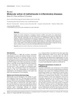
Fig. 1. Histochemical detection o f j ejunal an d lung mast c ells and immunoperoxidase l ocalization o f d uodenase i n bov ine intestine and l ung. Repre-
sentative positively s tained mast cells ar e indicated by large arrows. Panels ( a–e) show localization o f duodenase in bovine jejenum. In panel (a) ma st
cells surrounding crypts in the jejunal mucosa are toluidine blu e (pH 0.5)-p ositive (counterstained with eosin). Anti-
duodenase Ig stainin g is shown at low magnificatio n in panel (b), with ab undant stainin g of c ells with morphology and distribution similar to th at
shown for to luidin e blu e p anel ( a). Hi gher m agnification in panels (c–e), show s bovine j ejenum stain ed with control rabbit seru m, rabb it an ti-
duodenase Ig and rabbit anti-(sMCP-1) Ig, r espectively. A s imilar p attern o f s taining i s s een w ith a nti-duo denase Ig and anti-(sMCP-1) Ig, and no
staining is observed in the control. Sections of a bronchiole from bovine l ung infected with the lungworm Dic tyocaulus vi viparus are shown in panels
(f–h). I n panel (f), duodenase-positive cells are abund ant in the granulomatous reaction around the bronchiole. Panel (g) shows a n adjacent section
incubated with control serum. Panel (h) sh ows toluidine blue and eosin staining of a s ection adjacent to (f). Note the s imilar distribution of mast cells
in (f) a nd (h) and the presence of numerous eosinophils (small arrows) in the parasitized lung. In association with the accumulation of mas t cells
there i s increased fib rosis (*) an d smooth m uscle h ypertrophy (arrowhead). All of the tissues w ere fixed in 4% ( v/v) paraformaldehyde.
1174 A. D. Pemberton et al. (Eur. J. Biochem. 269) Ó FEBS 2002
at Phe8, a s has b een shown for duodenase [4]. Therefore, the
jejunal enzyme we purified was identified as duodenase, or a
highly similar variant of the enzyme.
Immunolocalization of duodenase
Toluidine blue staining identified abundant spindle or
stellate-shaped m ast cells in bovine jejunum samples. The se
cells were l ocate d principally in the lamina propria (Fig. 1a)
and submucosa (not shown). Immunostaining o f paraform-
aldehyde-fixed sections with rabbit anti-duodenase serum
and affin ity-purified rabbit anti-(sMCP-1) IgG detected cells
only in the lamina propria. These strongly staining cells
showed a similar distribution and morphology to those seen
with toluidine blue within the lamina propria (compare
Fig. 1a with Fig. 1b,d,e). The distribution of positive cells
after labelling with anti-duodenase Ig or anti-(sMCP-1) IgG
was very similar, and in neither instance was there any
labelling of submucosal tissues. Occasional intraepithelial
cells were weakly labelled (Fig. 1d), and the identity of these
toluidine blue negative cells was not confirmed. Tissues fixed
in neutral buffered formalin showed negligible mast cell
staining by comparison, and control rabbit serum was
negative regardless of the fixation procedure (Fig. 1c).
Lungworm-infected lung parenchyma showed the presence
of large numbers of eosinophils and toluidine blue-positive
mast c ells. An example of their distribution around a
bronchiole is shown in Fig. 1h, in which fibrosis and smooth
muscle hyperplasia was also evident. Numerous cells were
also lab elled with duodenase antiserum around bronchioles
(Fig. 1f) and within the alveolar septa (not shown). Their
size and distribution as observed in adjacent sections was
similar to that of toluidine blue-positive cells (compare
Fig. 1f,h). Control r abbit serum gave no labelling (Fig. 1g).
Duodenase induces DNA synthesis in pulmonary artery
fibroblasts
The effect of duodenase on DNA synthesis w as assessed
using [
3
H]thymidine incorporation in bovine primary pul-
monary artery fibroblasts. Treatment of cells for 24 h with
duodenase induced a concentration-dependent increase in
[
3
H]thymidine incorporation w hich was m aximal at 3 0 n
M
,
achieving a 5.5 ± 0.8-fold increase above control values
(Fig. 2 A). Pretreatment o f duodenase with soybean t rypsin
inhibitor ( 3 mg ÆmL
)1
, 1 5 min), an effective inhibitor of t his
enzyme [1] was found to inhibit completely the ability of this
enzyme to induce [
3
H]thymidine incorporation i n pulmo-
nary artery fibroblasts (Fig. 2B), confirming that the
proteolytic activity of duodenase is essential for induction
of DNA synthesis. Importantly, treatment of cells with
duodenase (30 n
M
) for 10 min followed by the addition of
soybean t rypsin inhibitor (3 mgÆmL
)1
, 1 5 min) induced
[
3
H]thymidine incorporation to a similar extent as addition
of duodenase alone (Fig. 2B), suggesting a rapid signalling
mechanism. Furthermore, conditioned media generated by
this method was used to assess whether duodenase could
cleave and release a cell surface molecule that could interact
Fig. 2. Duodenase induces DNA synthesis in pulmonary artery fibro-
blasts. (A) quiescent cells were treated with duodenase (3–100 n
M
)as
indicated f or 20 h prior to ad dition of [
3
H]thymidine (0.1 lCiÆwell
)1
):
incorporation was assessed after 4 h as detailed in Materials and
methods. (B) [
3
H]Thymidine incorporation tested in c ells treated with
duodenase ( duod, 3 0 n
M
)whichhadbeenpretreatedwithorwithout
soybean trypsin inhibitor (+ STI, 0.2 mgÆmL
)1
) for 15 min. To
examine a role for duo denase-induced release of a mitogenic f actor and
generation of con ditioned media, duodenase was a dded to cells for
10 min prior to addition of soybean trypsin inhibitor for 15 min,
media removed and r eplaced w ith f resh quiescent media (duod + STI
removed). This conditioned media was transferred to untreated cells
(cond. media) and [
3
H]thymidine incorporation assessed as before.
(C) [
3
H]Thymidine incorporation tested in cells treated with duodenase
(30 n
M
), PAR1 activating peptide (Ser-Phe-Leu-Leu-Arg-Asn,
100 l
M
), PAR2 activating peptide ( a, Ser-Leu-Ile-Gly-Arg-Leu; b, Ser-
Leu-Ile-Gly-Arg-Leu-NH
2
;both100l
M
) or P AR4 activating peptide
(Gly-Tyr-Pro-Gly-Lys-Phe, 100 l
M
). [
3
H]Thymidine incorporation
was assessed as detailed in Materials and methods. R esults are
expressed as m ean ± SEM-fold increase ov er control cells fro m four
separate exper iments, each performed i n triplicate.
Ó FEBS 2002 Duodenase induces DNA synthesis via PtdIns 3-kinase (Eur. J. Biochem. 269) 1175
with cell surface receptors or induce secretion o f a bioac tive
molecule to induce DNA synthesis. In these experiments,
addition of conditioned media to pulmonary artery fibro-
blasts had no significant effect on [
3
H]thymidine in corpor-
ation above control levels (Fig. 2B). Hence duodenase,
purified from bovine jejenum is mitogenic for bovine
pulmonary artery fibroblasts and this effect is dependent
on the direct proteolytic activity of th is enzyme.
As the PARs d escribed to date are activated by cleavage
of trypsin-like primary specificity, and as duodenase, (like
sMCP-1, which is also mitogenic in this system [8]), has a
trypsin-like component, a ctivating peptides selective for
PAR1, PAR2 and PAR4 were used to investigate whether
the mitogenic effect of duodenase was mediated v ia a
known PAR mechanism. Surprisingly, all PAR peptides
were unable to induce [
3
H]thymidine incorporation in
pulmonary artery fibroblasts (Fig. 2C). It should be noted
that two forms of the PAR2 activating peptide were
assessed, the f ree form a nd the a mido form, neither of which
showed ability to induce DNA synthesis ( Fig. 2C). Lack of
activation by these peptides is unlikely to be a c onsequence
of species differences in receptor sequences as Ser-Leu-Ile-
Gly-Arg-Leu (PAR2 activating peptide, mouse-derived
sequence, 100 l
M
) was reported to mobilize Ca
2+
in bovine
coronary artery smooth muscle cells [18]. The PAR1
activating peptide Ser-Phe-Leu-Leu-Arg-Asn (human-
derived sequence, 100 l
M
) activated phospholipase C in
bovine tracheal smooth muscle cells (T. R. W alker &
E. R. Chilvers, unpublished observations). Furthermore,
this PA R1 activating p eptide (100 l
M
) w as found to induce
aggregation o f isolated bovine platelets s imilar to that
induced by thrombin (T. R. Walker, unpublished observa-
tions).
Degradation of PAR2 model peptide
To further assess t he po tential interaction between duoden-
ase a nd PAR2, the ability of this enzyme to cleave a PAR2
substrate was investigated. Under the experimental condi-
tions used, the known P AR2 activators bovine t rypsin and
human mast cell tryptase rapidly cleaved the model PAR2
substrate PAR2(5–20) (t
½
¼ 3.5 and 3.4 min, respective -
ly). One cleavage product was resolved by HPLC and
identified by mass spectrometry a s Ser-Leu-Ile-Gly-Arg-
Leu-Asp-Thr-Pro (m/z ¼ 971) (the other product Gly-
Pro-Asn-Ser-Lys-Gly-Arg was not resolved under the
chromatographic conditions used). This confirms the
capacity of t rypsin and t ryptase to cleave at the appropriate
activation site. However, PAR2(5–20) was cleaved much
more slowly by duodenase (t
½
% 1200 min). Moreover, the
cleavage mixture exhibited HPLC peaks corresponding
both to the activation product (Ser-Leu-Ile-Gly-Arg-Leu-
Asp-Thr-Pro) a nd to other unidentified products, suggesting
multiple sites of cleavage of this substrate.
Together, these results suppo rt the hypothesis that
duodenase acts independently of the known trypsin/
tryptase-sensitive PAR2 receptor.
Duodenase induces GTPcS binding in pulmonary artery
fibroblast membranes
To establish the mechanism of action of duodenase,
[
35
S]GTPcS binding to fibroblast membranes was used as
an index of G protein activation. Duodenase (30 n
M
)
induced a 57.0 ± 2.3% increase in guanine nucleotide
binding to pulmonary artery fibroblast cell membranes
compared to controls, suggesting that the effects of
duodenase are indeed mediated through a G-protein-
coupled rec eptor. Pre-treatment of cells with pertussis toxin
(100 ngÆmL
)1
, 18 h) prior to cell fractionation and
membrane isolation inhibited [
35
S]GTPcS binding by
80.8 ± 10.3%, suggesting that the predominant G-protein
mediating this signal is a member o f the G
i/o
family (Fig. 3).
Intracellular signalling pathway underlying
duodenase-stimulated fibroblast proliferation
In order to identify a role for a downstream signalling
pathway that may mediate the effect of duodenase on
pulmonary artery fibroblasts, we examined a number of
diverse signalling pathways that have been implicated
in agonist-stimulated DNA synthesis in other cell
systems. Pulmonary artery fibroblasts preloaded with the
Ca
2+
-binding dye fura-2 were stimulated with duodenase
and fluorescence analy sed as an index of Ca
2+
mobilization.
As demonstrated in Fig. 4, duodenase at concentrations up
to 90 n
M
was unable to induce Ca
2+
mobilization. In
addition, thrombin, trypsin and chymotrypsin were also
unable to induce C a
2+
mobilization. However, addition of
bradykinin (5 l
M
) to these cells induced a rapid Ca
2+
transient indicating that these cells were responsive t o
activation through other G-protein-coupled receptors
(Fig. 4). As anticipated, this response to bradykinin could
be desensitized by prior exposure to the agonist (Fig. 4).
These results suggest that this group of proteases do not
appear to cause acute Ca
2+
mobilization or influx in these
cells. Of note, addition of a PAR2-activating peptide or
addition of thrombin, which will act t hrough PAR1, PAR3
and P AR4, all h ad no effect on Ca
2+
mobilization (Fig. 4).
These results demonstrate that Ca
2+
mobilization is
unlikely to be involved in mediating cell growth in
pulmonary artery fibroblasts.
Wortmannin (100 n
M
) and LY294002 (10 l
M
), two
structurally distinct and selective inhibitors of PtdIns
3-kinase, completely blocked duodenase-induced
[
3
H]thymidine incorporation, suggesting a key role for
PtdIns 3-kinase in this response (Fig. 5). In contrast,
PD98059, a MEK1 inhibitor, caused only a partial
Fig. 3. Effect of duodenase on [
35
S]GTPcS b inding. Pul monary artery
fibroblasts were untreated (open bars) or pretreated with pertussis
toxin (PTX, 100 ngÆmL
)1
, 18 h , hatch ed bars) p rior to ce ll lysis and
membrane isolation. [
35
S]GTPcS bin ding was carried out a s detailed i n
the Method s s ection, results are expressed as mean fold increase a bove
control ± SEM from three ex periments performed in triplicate.
1176 A. D. Pemberton et al. (Eur. J. Biochem. 269) Ó FEBS 2002
inhibition of DNA synthesis, r educing the response t o
duodenase by 54% ± 13% (Fig. 5), implying that acti-
vation of MEK1 and its downstream e ffectors may have a
modulatory role in duodenase-stimulated responses. Pre-
incubation of cells with the protein kinase C inhibitor
GF109203X, at a concentration previously shown to f ully
inhibit protein kinase C activity [19] again resulted in only
a modest reduction in duodenase-stimulated [
3
H]thymi-
dine incorporation (29% ± 1 0% inhibition from stimu-
lated control values), indicating that, although required
for a full mitogenic response, protein kinase C activation
does not ap pear to be critical for the initiation of this
response. Pretreatment of pulmonary artery fibroblasts for
18 h w ith pertussis toxin, which ADP-ribosylates the
a subunit of G
i
and G
o
resulting in blockade of G protein
activation, inh ibited duodenase-induced [
3
H]thymidine
incorporation by 52 ± 2.5%, suggesting involvement of
G
i
/G
o
in mediating this component of cell growth
(Fig. 5). In subsequent experiments, duodenase (30 n
M
)
was found to activate p85a-associated PtdIns 3-kinase in
pulmonary artery fibroblasts by 2.28 ± 0.14- fold above
control values, and pretreatment of these cells with
wortmannin (100 n
M
, 20 min) inhibited this activity to
below basal levels (Fig. 6). In combination with the major
inhibitory effects of wortmannin and LY294002 on
duodenase-stimulated [
3
H]thymidine incorporation, these
results indicate a key role for a G-protein-coupled
receptor/PtdIns 3-kinase p athway in mediating duoden-
ase-stimulated DNA synthesis.
Effect of inflammatory cytokines on duodenase-induced
DNA synthesis
Pretreatment of pulmonary artery fibroblasts with the
cytokines IL-1b and TNFa was undertaken to elucidate
whether th e effect of duodenase on DNA synthesis in our
model system could be augmented by factors which are
released at a site of inflammation. Furthermore, TNFa
has been reported to increase PAR2 expression and hence
would allow further insight into a potential role of PAR2
in mediating the effects of duodenase [20]. Exposure of
pulmonary artery fibroblasts to duodenase resulted in a
Fig. 5. Effect of signalling inhibitors on duodenase-induced DNA syn-
thesis. Pulmonary artery fibroblasts were pretreated with wortmannin
(100 n
M
, 20 m in) or LY294002 (10 l
M
, 20 m in), PD98059 (10 l
M
,
30 min), GF109203X (1 l
M
, 5 min ) or pertussis toxin ( PTX,
100 ngÆmL
)1
, 18 h) prior to addition of duodenase (30 n
M
).
[
3
H]Thymidine incorporation was assessed as indicated in Methods,
results are expressed as percentage mean ± S EM relative to untreated
cells st imulated with duoden ase. Results are from four independent
experiments e ach performed in triplicate.
Fig. 6. Duodenase a ctivates PtdIn s 3-kinase i n pulmonary artery fibro-
blasts. Pulmonary artery fibroblasts were incubated in the presence
(hatched bars) or absence (open bars) of wortmannin (100 n
M
)for
20 min prior to additio n of du odenase (30 n
M
, duod). Reactions
were terminated and PtdIns 3-kinase activity was assayed in p85a
immunoprecipitates as detailed in Materials and methods. Results are
expressed as mean c .p.m. ± SEM from a single experiment performed
in quadruplicate, representative of two others with s imilar results.
Fig. 4. Effect of duodenase on Ca
2+
mobilization. Pulmonary artery
fibroblasts preloaded with Fura-2 were stimulated with agonists as
indicated. Intracellular Ca
2+
was analysed and plotted over time as
indicated. Traces are representative of three separate experiments
which a ll gave ve ry similar results.
Ó FEBS 2002 Duodenase induces DNA synthesis via PtdIns 3-kinase (Eur. J. Biochem. 269) 1177
5.23 ± 0.47-fold increase in [
3
H]thymidine incorporation
above control levels (Table 1). While pretreatment of cells
with IL-1b (10 ngÆmL
)1
) alone for 24 h induced an
increase in DNA synthesis by 4 9 ± 12% (n ¼ 8,
p < 0.05), it also resu lted in a significant inhibition of
duodenase-induced [
3
H]thymidine incorporation relative
to IL-1b-treated control cells (Table 1). In contrast,
pretreatment with TNFa (10 ngÆmL
)1
) alone reduced the
level of [
3
H]thymidine incorporation by 69 ± 3%
(n ¼ 12, p < 0.05) (Table 1) but had no significant
effect on the relative magnitude of DNA synthesis
induced by duodenase: 6.19 ± 1.03-fold increase above
control values, respectively (Table 1).
DISCUSSION
Mast cells present within the intestinal mucosa of rodents
express subset-specific chymases which are thought to act as
part of the innate immune response against intestinal
nematodes by increasing epithelial permeability [21]. Similar
mucosal-specific mast cell subsets also e xist in the sheep and
goat intestine [21,22] and are typified by the expression of
sMCP-1 and goat mast cell proteinase-1, respectively.
Expulsion o f intestinal nematodes i n the sheep is associated
with simultaneous release of sMCP-1 into the gut lumen
and circulation [23].
The immunolocalization of duodenase to bovine intesti-
nal mucosal mast cells described h ere would suggest that it
too belongs to the ruminant mucosal mast cell proteinase
family, w hich are notable for their dual c hymase and
tryptase-like activities. It was possible to isolate duodenase
from bovine jejunum using methodology identical to that
employed for the purification of sMCP-1 from gastrointes-
tinal tissues. However, duodenase has previously been
localized only to the epithelial cells of Brunner’s glands
located in the duodenal wall [4]. This suggests either that
duodenase is present in both cell types, or that each site
produces distinct enzymes that are nonetheless highly
similar structurally, functionally and immunologically.
Lungworm infection in sheep is known to involve a
pronounced mastocytosis [24], and sMCP-1 is upregulated
in mast c ells recruited t o s ites of allergic lung inflammation
[25]. The current observation of abundant duodenase-
positive mast cells in lungworm-infec ted bovine lung shows
the potential for local duodenase release by mast cells
recruited to i nflammatory sites i n the bovine lung and i s
consistent with a putative role in tissue modelling.
In this study, we have shown t hat the similarity between
duodenase and sMCP-1 e xtends to the stimulation of
pulmonary artery fibroblasts, with both enzymes able to
induce DNA synthesis over a similar concentration range.
As soybean t rypsin inhibitor was able to completely inhibit
the duodenase effect, this demonstrates that the catalytic
activity is essential for its action. However, only a short
exposure to duodenase is required to induce maximal DNA
synthesis suggesting a rapid activation p rofile. Conditioned
media from duodenase and soybean trypsin inhibitor-
treated cells had no mitogenic effect, implying that
duodenase acts directly on fibroblasts and does not release
a mitogenic mediator from the cell or medium to act in an
autocrine or paracrine manner. Furthermore, duodenase
induces [
3
H]thymidine incorporation preferentially in sub-
confluent cell cultures suggesting t hat close cell–cell contac t
and intercellular activation is not a requirement for DNA
synthesis.
It is now recognized that the mitogenic effect of other
proteases such as thrombin and trypsin are mediated by
protease-activated receptors [12]. These are a family of
seven-transmembrane o r heptahelical receptors, which cou-
ple to heterotrimeric G-proteins to transduce their signal,
and are activated by cleavage of an extracellular portion of
the receptor close to the N-terminus, thus exposing a new
N-terminus that interacts with, and activates the receptor.
Receptors identified to date are PAR1, PAR2, PAR3 and
PAR4; each has a similar mechanism of action has a d istinct
sequence at its cleavage site. As a consequence, synthetic
peptides have been developed t hat mimic the newly exposed
N-terminus and a ct as specific activators [12]. Howe ver, no
selective ligand for PAR3 exists implying that this rece ptor
requires other structural interactions to achieve activation
[26]. Indeed, r ecent data suggest that PAR3 does not
mediate signal transduction directly but instead acts as a
cofactor for the clea vage and activation of PAR4 [27].
Thrombin has been shown t o cleave and a ctivate PAR1,
PAR3 and PAR4, whereas trypsin cleaves and activates
PAR2. As duodenase is capable of cleaving certain
substrates with trypsin-like primary specificity, we initially
hypothesized that induction of DNA synthesis by duoden-
ase is mediated through a PAR2 mechanism.
Surprisingly, we could find no evidence to support the
involvement of a classic PAR2 in mediating the mitogenic
effects of duodenase, specifically: (a) the synthetic peptide
Ser-Leu-Ile-Gly-Arg-Leu, which is specific for PAR2, was
unable to i nduce [
3
H]thymidine incorporation in fibroblasts,
and a similar lack of mimickery was evident for peptides
specific for PAR1 and PAR4; and (b) duodenase cleave d t he
model PAR2 substrate more slowly than either trypsin or
tryptase, and generated a very different array of peptides,
suggesting that duodenase may cleave PAR2 at different
sites. Activation of PAR3 by duodenase seems unlikely, as
this receptor has limited intrinsic signalling capacity [27] and
so far has only b een found to be activated by thrombin [12].
Schechter et al. [28] have described the action of mast cell
tryptase on keratinocytes, as acting through a subpopula-
tion of PAR2 receptors, suggesting the existence o f subtypes
Table 1. Effect of cytokines on duodenase-induced DNA synthesis.
Bovine p ulmonary artery fibroblasts were assessed for [
3
H]thymidine
incorporation induced by d u odenase (30 n
M
), following pretreatment
for 24 h with TNFa (10 n gÆmL
)1
)orIL-1b(10 ngÆmL
)1
)asindicated.
The values qu oted represent the ratio of [
3
H]thymidine incorporation
to the mean [
3
H]thymidine incorporation for the corresponding un -
treated control w ells in the s ame experiment. Results a re expressed as
mean ± SEM. Results in parentheses are corrected for the effects of
cytokines o n baseline c ell growth, and are expressed as the ratio o f
[
3
H]thymidine incorporation in proteinase-treated wells to that in
control wells for each cytokine treatment.
Untreated
a
+ TNF-a
a
+ IL-1b
b
Control 1.00 ± 0.03 0.31 ± 0.03 1.49 ± 0.12
Duodenase 5.23 ± 0.47 1.90 ± 0.32
(6.19 ± 1.03)
3.96 ± 0.57
(2.65 ± 0.38)*
a
n ¼ 12, over three separate experiments.
b
n ¼ 8, over two sepa-
rate experiments. * p > 0.001.
1178 A. D. Pemberton et al. (Eur. J. Biochem. 269) Ó FEBS 2002
of this receptor. In addition, it has been demonstrated that
regulation of intestinal ion transport in rat jejenum is
mediated by a P AR that, a lthough similar in many r espects
to PAR2, showed distinct and atypical orders of potency
when a range of peptide agonists were assessed [29]. These
reports and the data from this study, in particular the
pertussis toxin-sensitivity of DNA synthesis induction and
the ability o f duodenase to stimulate [
35
S]GTPcS binding to
pulmonary artery fibroblast membranes, would suggest that
the m itogenic action of duodenase is mediated via direct
interaction with a proteolytically activated G
i/o
-coupled
receptor. While the precise PAR subtype remains to be fully
identified, it may be an atypical P AR2 that is not activated
by existing classic PAR2 peptides. To date, no bo vine PAR
sequences have been published and analysis o f cleavage sites
on these receptors may reveal species-specific activation
motifs that are distinct from those in mouse, rat and
humans an d explain the lack of efficacy of current PAR2-
activating peptides in our model system.
A number of s ignalling pathways and intermediates such
as Ca
2+
mobilization, the E RK pathway, PtdIns 3-kinase
and protein kinase C h ave all been identified a s mediators of
proliferative signals in a variety of cell t ypes. I n pulmonary
artery fibroblasts, duodenase, trypsin, chymotrypsin,
thrombin and PAR2 peptides were unable to mobilize
Ca
2+
from intracellular stores. As duodenase induces DNA
synthesis for these cells, these data would app ear to
dissociate mobilization o f intrac ellular Ca
2+
from induction
of DNA synthesis, a situation very similar to that p reviously
demonstrated in bovine a irway s mooth muscle [19,30].
These results also parallel those described for the effects of
human tryptase on fibroblasts, where tryptase is mitogenic
for these cells but does not act via PAR2 or Ca
2+
-
dependent pathways [28]. Employing a range of selective
inhibitors, we investigated the r oles of PtdIns 3-kinase,
MEK1/ERK, p rotein kinase C a nd pertussis t oxin-sensitive
G-proteins. Only partial inhibition of DNA synthesis was
achieved with maximally effective concentrations of
PD98059 (MEK1 inhibitor) and G F109203X (protein
kinase C inhibitor) indicating that each of these pathways
has a modulatory r ather than a mandatory role to play in
mediating the proliferative response. In contrast, a recent
report has shown that tryptase induces DNA synthesis in
canine tracheal smooth muscle through an ERK1/2-depen-
dent mechanism, proliferation being inhibited completely by
PD98059 [31]. Moreover, in pulmonary artery fibroblasts,
inhibition of PtdIns 3-kinase by wortmannin or LY294002
inhibited completely duodenase-induced [
3
H]thymidine in-
corporation. This would suggest that activation of P tdIns 3-
kinase is the key regulatory step in the proliferative p athway
and that each of the other pathways interacts with this
pathway with the magnitude of the cellular response
determined by the integrated sum of each of these
components. Our data is supported by previous reports
demonstrating t hat thrombin a cts i n a PtdIns 3-kinase- a nd
p70
s6k
-dependent manner to induce DNA synthesis in
pulmonary artery fibroblasts [32]. In addition, this report
noted that downregulation of protein kinase C partially
attenuated thrombin-induced p70
s6k
activation, which
would concur with our findings that inhibition of protein
kinase C results in partial inhibition of DNA synthesis.
To date, identification of downstream signalling path-
ways for PARs have principally concentrated on PAR1 and
PAR2. P AR1 c ouples to members of the G
12/13
,G
q
and G
i
families, interacting with various signalling pathways
including phospholipase Cb, adenylyl cyclase, PtdIns
3-kinase and nonreceptor tyrosine kinases such as Src [33].
PAR2 activation is associated with MAPK activation,
phospholipase C activation and Ca
2+
mobilization. How-
ever, trypsin-induced MAPK activation was reported to
occur independently of PAR2 in bovine pulmonary artery
fibroblasts [34].
Inflammatory cytokines have previously been shown to
induce selective upregulation of P AR2 receptors without
affecting the thrombin rece ptor in human umbilical vein
endothelial cells [20]. To determine whether the prolifera-
tive response of pulmonary artery fi broblasts could be
modulated under inflammatory conditions, cells were
treated for 24 h with TNFa; this resulted in a reduction
in the level of [
3
H]thymidine incorporation under control
conditions, but had no significant influence on the relative
magnitude of this response to duoden ase. In contrast,
pretreatment of cells with IL-1b resulted in significant
inhibition of duodenase-induced DNA synthesis and an
enhanced level of baseline [
3
H]thymidine incorporation
under control conditions. These results suggest that these
cytokines cause the fibroblasts either to become refractory
to mitogens or to enter into S-phase more slowly over the
time period examined. It remains to be established whether
chronic exposure to TNFa and IL-1b would result in a
sensitization of these cells to mitogenic stimuli. These
results support further our hypothesis that duodenase is
not acting via a classical PAR2.
In summary, this study has demonstrated that duoden-
ase induces DNA synthesis in pulmonary artery fibro-
blasts and that this response may be mediated by an
atypical PAR, either an i soform of PAR2 or an uniden-
tified receptor. It is important to recognize that the
current study was undertaken in a fully homologous
system, using a bovine serine protease a nd bovine
pulmonary fibroblasts. This would indicate that the
proteolytic event and subsequent downstream signalling
and functional responses we have described m ay be an
important consequence o f duodenase release from Brun-
ner’s glands, or of mast cell activation in vivo. Indeed,
mast cell hyperplasia is known to be a prominent event in
many forms of chronic inflammation in the lung such as
cryptogenic fibrosing alveolitis, and fibroblast proliferation
is the most significant feature in the pathology of these
clinical conditions [9]. The precise nature and character-
ization of the receptor that mediates the effects of
duodenase requires further investigation.
ACKNOWLEDGEMENTS
This work was funded by the Norman Salvesen Emphysema Research
Trust, the Wellcome Trust, and the National Asthma Campaign (UK).
We thank Dr Joh n Huntley and Ms Anne Mackellar for providing the
bovine lung se ctions and Dr Jeremy B rown for h elp in p reparing Fig. 1.
REFERENCES
1. Zamolodchikova, T.S., Vorotyntseva, T.L. & Antonov, V.K.
(1995) Duodenase, a new serine protease of unusual specificity
from bovine duodenal mucosa. Purification and properties. Eur.
J. Biochem. 227, 866– 872.
Ó FEBS 2002 Duodenase induces DNA synthesis via PtdIns 3-kinase (Eur. J. Biochem. 269) 1179
2. McAleese, S .M., Pemberton, A.D., McGrath, M.E., Huntley, J.F.
& Miller, H.R.P. (1998) Sheep mast-cell proteinases-1 and -3:
cDNA cloning, prim ary structure and m olecular modellin g of the
enzymes and further s tudies on su bstrate sp ecificity. Biochem. J.
333, 801–809.
3. Pemberton, A.D., Huntley, J.F. & Miller, H.R.P. (1997) Sheep
mast cell proteinase-1: characterization a s a m ember of a new class
of dual-specific ruminant chymases. Biochem. J. 321, 665–670.
4. Zamolodchikova, T.S., Sokolova, E.A., Alexandrov, S.L.,
Mikhaleva, I.I., Prudchenko, I.A., Morozov, I.A., Kononenko,
N.V., Da Mirgorodskaya, O.A.U., Larionova, N.I., Pozdnev,
V.F. et al. (1997) Subcellular localization, substrate s pecificity and
crystallization o f duodenase, a potential a ctivator of enteropepti-
dase. Eur. J. Biochem. 249, 612– 621.
5. Jolly, S ., Coignoul, F., Gabriel, A. & Desmecht, D. (1999)
Detection of t ryptase i n b ovine mast c ells: c omparison o f e nzyme-
and immuno-histochemistry. J. Comp. Path. 120 , 269–279.
6. Pemberton, A.D., McAleese, S.M., Huntley, J.F., Collie, D.D.S.,
Scudamore, C.L., McEuen, A.R., Walls, A.F. & Miller, H.R.P.
(2000) cDNA sequence of two s heep mast cell tryptases a nd the
differential expression of tryptase and s heep mast cell proteinase-1
in lung, dermis and gastrointestinal tract. Clin. E xp Allergy. 30,
818–832.
7. Ruoss, S.J., Hartmann, T. & Caughey, G.H. (1991) Mast cell
tryptase is a mitogen for cultured fi broblasts. J. Clin. Invest. 88,
493–499.
8. Pemberton, A.D., Belham, C.M., Huntley, J.F., Plevin, R. &
Miller, H.R.P. ( 1997) Sheep mast cell proteinase-1, a serine pro-
teinase with both tryptase- and chymase-like p roperties, is inhib-
ited by plasma proteinase inhibitors an d i s m itogen ic fo r b ovine
pulmonary artery fib roblasts. Biochem. J. 323, 7 19–725.
9. Pesci., A., Bertorelli, G., Gabrielli, M. & Olivieri, D. (1993) Mast
cells i n fibrotic lung disorders. Chest 10 3, 989–996.
10. Vu, T K.H., Hung, D.T., Wheaton, V.I. & Coughlin, S .R. ( 1991)
Molecular cloning of a functional thrombin receptor reveals a
novel proteolytic mechanism of receptor activation. Cell 64,
1057–1068.
11. Hollenberg, M.D. (1999) Protease-activated receptors: PAR4 and
counting: how long is the course? Trends Pharmacol. Sci. 20,
271–273.
12. Coughlin, S.R. (1999) How t he protease thrombin tal ks t o cells.
Proc. N atl Acad. Sci. USA 96 , 11023–11027.
13. Parry, M.A., Myles, T., Tschopp, J . & Stone , S.R. (1996) Cleavage
of th e thrombin receptor: identification of potential activators and
inactivators. Bio c hem. J. 320, 335– 341.
14. Sture, G.H., Huntley, J.F., Mackellar, A. & Miller, H.R.P. (1995)
Ovine mast cell heterogeneity is defined by the distribution of
sheep mast cell proteinase . Vet Immunol. Immunopathol. 48 ,
275–285.
15. Freshney, R.I. (1983) Cul ture of A nimal Cells. A Manual of B asic
Techniques, pp. 99–118. Alan R . Liss I nc., New York.
16. Carter, A.N. & Downes, C.P. (1993) Signaling by neurotrophic
factors: activation o f pho spho inositide 3 -kinase b y n erve growth
factor. Neuroprotocols 3 , 107–118.
17. Mirgorodskaya, O., Kazanina, G., Mirgorodskaya, E.,
Vorotyntseva, T., Zamolodchikova, T. & Alexandrov, S. (1996)
A comp arative study of the specificity of m elittin hydrolysis by
duodenase, trypsin and plasmin. Protein Peptide Lett. 3 , 315–320.
18. Bretschneider, E., Kaufmann, R., Braun, M., Wittpoth, M.,
Glusa,E.,Nowak,G.&Schror,K.(1999)Evidenceforprotein-
ase-activated receptor-2 (PAR-2)-mediated mitogenesis in coro-
nary artery s mooth muscle cells. Br. J. Pharmacol. 126, 1735–1740.
19. Walker, T.R., Moore, S.M., Lawson, M.F., Panettieri J r, R.A. &
Chilvers, E.R. (1998) Platelet-derived growth factor-BB and
thrombin ac tivate phosphoinositide 3 -kinase and protein kinase B:
role in mediating airway smooth m uscle proliferation. Mol.
Pharm. 54, 1 007–1015.
20. Nystedt, S., Ramakrishnan, V. & Sundelin, J. (1996) The pro-
teinase-activated receptor 2 is induced by i nflammatory mediators
in human endothelial cells. Comparison with the t hrombin
receptor. J. Biol . Chem. 271, 1 4910–14915.
21. Miller, H .R.P., Huntley , J.F. & N ewlands, G.F.J. (1995) Mast cell
chymases in helminthosis and hypersensitivity. In Mast Cell
Proteases in Immunology and Biology (Caughey, G.H., ed.),
pp. 203 –235. Marcel Dekke r, New York.
22. Macaldowie, C.N., Mackellar, A. & Huntley, J .F. (1998) The
isolation and purification of a dual specific mast cell-derived
protease from parasitised caprine jejunal t issue. Res. Vet. Sci. 64,
17–24.
23. Huntley, J.F., G ibson, S ., Brown, D., Sm ith, W.D ., Jackson, F. &
Miller, H.R. (1987) Systemic release of a mast cell proteinase
following nematode infections in sheep. Parasite Immunol. 9,
603–614.
24. Mansfield,L.S.&Gamble,H.R.(1995)Alveolarmastocytosisand
eosinophilia in l ambs with naturally acquired nematode infections
of Protostrongylus rufescens and H aemonchu s contortus. Vet.
Immunol. I mmunopathol. 49 , 251–262.
25. Collie, D.D., McAldowie, C.N., Pemberton, A. D., Woodall, C.J.,
McLean, N., Hodgson, C. & Kennedy, M. (2001) Lo cal lung
responses following local l ung challenge with r ecombinant lu ng-
worm antigen i n systemically s ensitised s heep. Clin. Exp. Allergy
31, 1636–1647.
26. Ishihara, H ., Connolly, A.J., Zeng, D., Kahn, M .L., Zheng, Y.W.,
Timmons, C., Tram, T . & Coughlin, S.R. (1997) Protease-acti-
vated recep tor 3 is a second thromb in re ceptor i n hu mans. Nature
386, 502–506.
27. Nakanishi-Matsui, M ., Zheng, Y W., Sulciner, D.J., Weiss, E.J.,
Ludeman, M.J. & Coughlin, S.R. (2000) PAR3 is a cofactor f or
PAR4 activation by thrombin. Nature 404, 609–613.
28. Schechter, N.M., B rass, L.F., Lavker, R.M. & Jensen, P.J. (1998)
Reaction of mast cell proteases tryptase and chymase with pro-
tease activated receptors (PARs) on keratinocytes and fibroblasts.
J. Cell. P hysiol. 176, 365– 373.
29. Vergnolle, N., MacNaughton, W.K., Al-Ani, B., Saifeddine, M.,
Wallace, J.L. & Hollenberg, M.D. (1998) Proteinase-activated
receptor 2 (PAR2) -activating peptides: identification o f a receptor
distinct from PAR2 th at regulates intestinal transport. Proc. Natl
Acad. Sci. USA 95, 7766–7771.
30. Panettieri Jr, R .A., Hall, I.P., Maki, C .S. & Murray, R.K. (1995)
Alpha-thrombin increases cytosolic calcium and induces human
airway smooth muscle cell proliferation. Am. J. Respir. Cell Mol.
Biol. 13, 205 –216.
31. Brown, J .K., Jones, C.A., Rooney, L.A. & Caughey, G.H. (2001)
Mast cell tryptase activates extracellular-regulated kinases (p44/
p42) in airway smoo th-muscle c ells. Am. J . Respir. Cell Mol. Biol.
24, 146–154.
32. Belham, C.M., Scott, P.H., Twomey, D.P., Gould, G.W., Wads-
worth, R.M. & Ple vin, R. (1997 ) Evidence t hat thrombin-stimu-
lated DNA synthesis in pulmonary arterial fibroblasts involves
phosphatidylinositol 3-kinase-dep endent p70 ribosomal S6 kinase
activation. Cell Signal. 9, 109–116.
33. Coughlin, S .R. ( 2000) T hrombin s ignalling and protease-activated
receptors. Nature 407, 258–264.
34. Belham, C.M., Tate, R .J., Scott, P.H., P emberton, A.D., Miller,
H.R., Wadsworth, R.M., Gould, G.W. & Plevin, R. (1996)
Trypsin stimulates proteinase-activated receptor-2-dependent and
-independent activation of mitogen-act ivated protein kinases.
Biochem. J . 320, 9 39–946.
1180 A. D. Pemberton et al. (Eur. J. Biochem. 269) Ó FEBS 2002