Báo cáo Y học: Dietary bisphenol A prevents ovarian degeneration and bone loss in female mice lacking the aromatase gene (Cyp19 ) pptx
Bạn đang xem bản rút gọn của tài liệu. Xem và tải ngay bản đầy đủ của tài liệu tại đây (511.21 KB, 9 trang )
Dietary bisphenol A prevents ovarian degeneration and bone loss
in female mice lacking the aromatase gene (
Cyp19
)
Katsumi Toda
1
, Chisato Miyaura
2
, Teruhiko Okada
3
and Yutaka Shizuta
1
1
Department of Medical Chemistry, Kochi Medical School, Nankoku, Japan;
2
Department of Biochemistry, School of Pharmacy,
Tokyo University of Pharmacy and Life Science, Japan;
3
Department of Anatomy and Cell Biology, Kochi Medical School, Nankoku,
Japan
We previously generated mice lacking aromatase activity by
targeted disruption of Cyp19 (ArKO mice), and reported
phenotypes of the female mice, showing hemorrhage for-
mation and follicular depletion in the ovary, diminution in
uterine size, and bone loss. In the present study, we examined
the influence of dietary bisphenol A (BPA), a monomer used
for the production of polycarbonate and known to have
estrogenic activity, on these phenotypes of the ArKO mice.
When ArKO mice were fed chow diets supplemented with
0.1% or 1% (w/w) BPA for 5 months, they were protected
from ovarian degeneration, uterine diminution and bone
loss in a dose-dependent manner. Northern blot analyses of
ovarian RNA of ArKO mice showed differences in the
expression levels of insulin-like growth factor (IGF)-I, IGF-I
receptor, growth differentiation factor 9 and bone mor-
phogenetic protein 15 as compared with those in the ovaries
of wild-type mice. The differences in the expression levels
were restored by dietary BPA. In the ArKO uteri, expression
of progesterone receptor and vascular endothelial growth
factor mRNAs was diminished, and was restored by BPA to
the levels in wild-type mice. In contrast, BPA had little effect
on the ovarian, uterine and skeletal structures of wild-type
mice. In conclusion, estrogenic effects of BPA on the
reproductive tract as well as skeletal tissue were evident in
adult female ArKO mice. These results suggest that the
ArKO mouse is an animal model suitable for studying effects
of estrogenic chemicals as well as estrogen in vivo.
Keywords: ArKO mouse; bisphenol A; estrogens; IGF-I.
Estrogens are synthesized from androgens by three succes-
sive hydroxylation reactions which are catalyzed by the
enzyme aromatase (CYP19) [1]. In order to study the
physiological roles of estrogens in vivo, aromatase-knockout
(ArKO) mice were generated by targeted disruption of
Cyp19 [2–4]. These mice can be used also as a good animal
model for the postmenopausal woman. Female ArKO mice
are characterized by phenotypes such as follicular depletion
and hemorrhage formation in the ovaries, underdeveloped
uteri and immature mammary glands [2–5]. Female ArKO
mice also show osteopenia with increased bone turnover
[6,7]. Administration of 17b-estradiol (E2) protects the
ArKO mice from ovarian degeneration and bone loss [4,7].
ArKO mice were also used to study the roles of estrogens in
male mice, and the results demonstrated that estrogens are
critical for male reproductive ability and the development of
the potential for adult inter-male aggression [4,8–10].
Moreover, studies of ArKO mice strongly support the
notion that estrogens play important roles in lipid and
glucose metabolism [11,12].
Xenoestrogens, chemically synthesized nonsteroidal com-
pounds, have been reported to enter the body by ingestion
or adsorption and to exert estrogenic effects [13]. The effects
of these compounds are evaluated by determining the
responses of rodent uteri or testicular function [14–16].
Because estrogen plays important roles in the development
of uterine and breast cancer, exposure to xenoestrogens may
be a risk factor that affects cancer development in addition
to disturbing reproductive functions.
Bisphenol A (4,4¢-isopropylidenediphenol; BPA) is a class
of monomer widely used in the production of polycarbonate
plastic products. The level of human exposure to BPA is not
insignificant, as microgram amounts of BPA were reported
to be detectable in liquid from canned vegetables [17]. BPA
is considered as a xenoestrogen because it binds to estrogen
receptors with approximately 10 000 times less affinity than
E2 [18] and it exhibits estrogenic properties when studied in
in vitro assay systems. For instance, it stimulated the
production of vitellogenin in cultured trout hepatocytes [19]
and the growth of an MCF-7 human breast cancer cell line
[20]. BPA has also been shown to induce estrogen-depend-
ent b-galactosidase activity in an assay system using yeast
cells [21]. In vivo, the exposure of pregnant mice to low doses
of BPA accelerated the onset of puberty in pups [22].
However, it is still not known whether the effects of BPA
in vivo are due to its hormonal or its toxic effects. Because
endogenous E2 might affect the consequences of the
physiological actions of BPA in vivo,ArKOisauseful
animal model for characterization and evaluation of
Correspondence to: K. Toda, Department of Medical Chemistry,
Kochi Medical School, Nankoku, Kochi 783-8505, Japan.
Tel.:/Fax: +81 88 880 2316, E-mail:
Abbreviations: ArKO mice, aromatase knockout mice; BMD, bone
mineral density; BMP15, bone morphogenetic protein 15; BPA, bis-
phenol A; Cyp19, murine aromatase P450 gene; E2, 17b-estradiol;
FSH, follicle stimulating hormone; GAPDH, glyceraldehyde-3 phos-
phate dehydrogenase; GDF9, growth differentiation factor 9; IGF-I,
insulin like-growth factor-I; OVX, ovariectomized; pQCT, periferal
quantitative tomography; UGT, uridine diphosphate-glucuronosyl
transferase; VEGF, vascular endothelial growth factor.
(Received 23 November 2001, revised 18 February 2002, accepted 12
March 2002)
Eur. J. Biochem. 269, 2214–2222 (2002) Ó FEBS 2002 doi:10.1046/j.1432-1033.2002.02879.x
chemical compounds with putative estrogenic actions. The
objective of present study was to examine the in vivo
estrogenic effects of BPA on the female reproductive tract
and bone by using ArKO mice.
MATERIALS AND METHODS
Materials
A standard rodent chow (NMF) was obtained from
Oriental Yeast (Tokyo, Japan). BPA and E2 were from
Sigma-Aldrich. An ELISA kit for BPA was from Takeda
Pharmaceutical Co. Ltd. (Tokyo, Japan). All other chem-
icals were of analytical grade.
Animals
Animal care and experiments were carried out in accord-
ance with institutional animal regulations. All animals were
maintained on a 12-h light/dark cycle at 22–25 °Candgiven
water and rodent chow diet with or without BPA ad libitum.
The aromatase P450 gene (Cyp19) was disrupted by
homologous recombination [4]. In brief, an 87-base pair
(bp) fragment located within exon 9 of Cyp19 (the
nucleotide sequence position between +1124 and +1210
relative to the translational start site) was replaced with a
neomycin resistance gene derived from pMC1-neo. The
replacement caused a complete loss of aromatase activity as
shown by an in vitro expression study [4].
The chow diets supplemented with BPA (BPA-diet)
werepreparedbyimpregnationwithBPA,whichwas
dissolved in acetone. For example, 1 g BPA was dissolved
in 10 mL acetone and impregnated into 100 g rodent chow
to yield the chow diet supplemented with 1% (w/w) BPA.
Female wild-type and ArKO mice at 5 weeks of age were
divided into four diet groups: the first group was fed a
normal chow diet (0% BPA-diet; wild-type mice, n ¼ 4;
ArKO mice, n ¼ 5), the second group was fed a chow
diet supplemented with 0.1% BPA (0.1% BPA-diet; wild-
type mice, n ¼ 4; ArKO mice, n ¼ 5), the third group
was fed a chow diet supplemented with 1% BPA (1%
BPA-diet; wild-type mice, n ¼ 4; ArKO, mice n ¼ 4)
and the mice in the fourth group (ArKO mice n ¼ 5)
were given subcutaneous injections of E2 dissolved in
sesame oil (15 lgper25lL per mouse per injection) once
per week for 5 months. Mice were started on each diet at
5 weeks of age and sacrificed at 5 months of age for
examination. We repeated a series of the experiments and
obtained essentially same results.
Preparation and analysis of RNA
Uteri and ovaries were collected from each mouse and used
for preparation of total RNA according to the method of
Mirkes [23]. Northern blot analyses were performed using
15 lg of total RNA according to the method described [24].
Complementary DNA probes were prepared by PCR
amplification using oligo d(T)-primed cDNA derived from
ovarian RNA as a template with the following sets of
primers: insulin-like growth factor (IGF)-I (a 560-bp
fragment with sense primer: 5¢-GTCGTCTTCACACCTC
TTCTACCTG-3¢ and antisense primer: 5¢-CCCATCTTT
GTAATGTTATTGGACT-3¢), IGF-II (a 378-bp fragment
with sense primer: 5¢-AGCTTGTTGACACGCTTCAGT
TTGT-3¢ and antisense primer: 5¢-GTAACACGATCAG
GGGACGATGACG-3¢), IGF-I receptor (a 1387-bp
fragment with sense primer: 5¢-GGGGCCAAACTCAA
CCGTCTAAAC-3¢ and antisense primer:CGTAAGGC
TGTCTCTCATCAAAACT-3¢), bone morphogenetic
protein (BMP) 15 (a 1057-bp fragment with sense primer:
5¢-CCCTGGCAAGGAGATGAAGCAATGG-3¢ and
antisense primer: 5¢-GGGAAACCTGAGATAGCAACA
ACTT-3¢), growth differentiation factor (GDF) 9 (a 1299-bp
fragment with sense primer: 5¢-GCAAGAGCAGGCA
CCCAGCAACCAG-3¢ and antisense primer: 5¢-TTCCGT
CACATAAAACCACAGCACT-3¢), follicle stimulating
hormone (FSH) receptor (a 684-bp fragment with sense
primer: 5¢-TAGATGATGAACCCAGTTATGGAA-3¢
and antisense primer: 5¢-CCACAAAGGCCAGGGCGTT
GAGTA-3¢), progesterone receptor (a 723-bp fragment with
sense primer: 5¢-TGAACCACGCACTCCT-3¢ and anti-
sense primer: 5¢-GAATCAAAGCCATACTGT-3¢), and
vascular endothelial growth factor (VEGF) (a 612-bp
fragment with sense primer: 5¢-TCAAGCCGTCCTGTG
TGCCGCTGATGC-3¢ and antisense primer: 5¢-AGAAA
ATGGCGAATCCAGTCCCACGAG-3¢). The amplified
products were cloned into the EcoRV site of pBluescript
SKII(–) (Stratagene) and verified to be the expected products
by nucleotide sequence analysis. The inserted fragments
were radiolabeled by the random primer labeling procedure
using the Klenow fragment and used as hybridization
probes. The signals were quantified by using a Bioimage
Analyzer BAS2000 (Fuji) to determine relative intensity.
Histological examination
Ovaries and uteri were removed from the mice, fixed in 10%
phosphate-buffered formalin (pH 7.4) for 24 h, dehydrated,
and embedded in paraffin. Sections were cut 3-lmthickand
stained with hematoxylin & eosin.
Serum concentration of BPA
The concentration of BPA in serum was measured using an
ELISA kit for BPA according to the manufacturer’s
instructions. Blood ( 500 lL) was collected from the tail
of each mouse according to the method described [25] and
200 lL of serum was used for the determination of BPA
concentration. The rate of recovery of 50 ngÆmL
)1
BPA
added to untreated serum was 91.8% and the limit for
detection of BPA was 2.2 ngÆmL
)1
under the experimental
conditions used.
Radiographic analysis of the femur
Radiographs of femurs were taken with a soft X-ray
generator (model CMB-2; SOFTEX, Tokyo, Japan) [7].
The bone mineral density (BMD) of the femurs was
measured using a dual X-ray absorptiometer (model
DCS-600R; Aloka, Tokyo, Japan), as reported previously
[7]. Trabecular bone density of the femurs was measured by
peripheral quantitative tomography (pQCT) using a pQCT
system (model XCT Research SA+) with a version 5.4 soft-
ware (Stratec Medizintechnik GMBH., Pfzheim, Germany).
The position of the 500-lm slice was located 1.2 mm away
from the growth plate in the distal metaphysis.
Ó FEBS 2002 Effects of bisphenol A on ArKO females (Eur. J. Biochem. 269) 2215
Statistical analysis
Data were expressed as means ± SD. The significance of
the differences was analyzed using Student’s t-test using
INSTAT
(GraphPad Software, Inc., San Diego, CA, USA).
RESULTS
Serum levels of BPA
We first determined the serum concentrations of BPA in
mice fed chow diets supplemented with BPA. The levels of
BPA in serum were elevated in a dose-dependent manner in
the mice of both genotypes. No significant differences were
observed in the concentrations between the wild-type and
ArKO mice (Table 1). These data indicate that endogenous
estrogen does not influence the intake or the rate of
degradation of BPA.
Estrogenic effects of dietary BPA on the uteri of ArKO
mice
We reported previously that the body weights of female
ArKO mice increased significantly compared with those of
their wild-type littermates after 12 weeks of age [4,11]. The
body weights of ArKO mice fed the 1% BPA-diet were
significantly decreased as compared with those of untreated
ArKO mice, but the 0.1% BPA-diet did not influence the
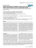
body weights of ArKO mice (Fig. 1A).
Diminution of uterine size is one of the typical pheno-
types observed in aromatase-deficient mice [2–4]. When
ArKO mice were fed BPA-diets, the uterine weight
increased significantly in a dose-dependent manner
(Fig. 1B). The uterine weight of ArKO mice fed 0.1% and
1% BPA-diets increased approximately 2.5-fold and five-
fold over that of the untreated ArKO mice, respectively. The
uterine weight of the ArKO mice fed the 1% BPA-diet was
comparable to that of the wild-type mice. In contrast, the
BPA-containing diets did not cause any alterations of the
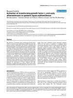
uterine weight in the wild-type mice. Histological examina-
tions showed that the uteri of ArKO mice exhibited atrophy
with suppressed proliferation of endometrium cells (Fig. 2)
[4]. Consumption of a BPA-diet resulted in proliferation of
the uterine endometrial as well as myometrial cells in ArKO
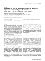
mice in a dose-dependent manner (Fig. 2). To examine the
effects of BPA on the expression of estrogen-responsive
genes in the uterus, Northern blot analysis was performed
using cDNA probes for progesterone receptor and VEGF.
While the expression of these genes in the uterus was
diminished in ArKO mice as compared with that in wild-
type mice, it was restored to the levels of the wild-type mice
by dietary BPA (Fig. 3). These results demonstrate that
dietary BPA activates the estrogen signaling pathway in the
uteri of ArKO mice, as does E2.
Estrogenic effects of dietary BPA on the ovaries
of ArKO mice
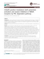
To examine the effects of dietary BPA on the ovaries of
ArKO mice, histological analysis was performed. Depletion
of follicles and formation of hemorrhagic cysts were evident
in the ovaries of untreated ArKO mice at 5 months of age
(Fig. 4D) as reported previously [4]. When the mice were fed
on BPA-diet, ovarian degeneration was suppressed in a
dose-dependent manner. With 0.1% BPA, no apparent
protective effects against follicular depletion in the ovary
were observed (Fig. 4E). In contrast, with 1% BPA, ArKO
mice were completely protected from hemorrhage forma-
tion and follicular loss in the ovaries (Fig. 4F). Nevertheless,
typical corpus lutea were not detectable. These histological
observations made in the ovaries of ArKO mice fed 1%
BPA are similar to what is seen in the ovaries of ArKO mice
treated with E2 [4]. The ovaries of wild-type mice fed BPA-
diets showed no obvious structural alterations (Fig. 4A–C).
Estrogenic effects of BPA on the ovaries were examined
by measuring the mRNA expression of genes for IGF-I,
IGF-II, IGF-I receptor, BMP15, GDF9 and FSH receptor,
which have been reported to be important for ovarian
function [26–34]. As shown in Fig. 5, the expression level of
the IGF-I gene was markedly elevated in the ArKO ovaries
(6.5-fold over the wild-type level). When the ArKO mice
were fed on BPA-diet, the expression was suppressed in a
dose-dependent manner. The expression of the IGF-I gene
was normalized in response to the treatment with E2 in
ArKO mice. BPA did not influence the expression of the
IGF-I gene in the ovaries of wild-type mice (Fig. 5). In
contrast, the levels of mRNA expression of the IGF-I
receptor, GDF9 and BMP15 were suppressed in the ovaries
of ArKO mice as compared with those of the wild-type mice
(relative intensities were 0.55 ± 0.06, 0.65 ± 0.02 and
0.86 ± 0.06, respectively). These expression levels were
increased by treatment with BPA in a dose-dependent
Table 1. Serum concentration of BPA. The concentration of BPA was
determined using 0.2 mL of serum of each mouse. Data are presented
as mean ± SD (n ¼ 4–5). No significant differences were observed
between wild-type and ArKO mice in each group.
Genotype
Concentration of BPA added to diet (ngÆmL
)1
)
0% 0.1% 1.0%
Wild-type 4.6 ± 1.7 166.1 ± 94.7 508.3 ± 104
ArKO 3.2 ± 1.9 84.3 ± 8.7 768.7 ± 204
Fig. 1. Effects of dietary BPA on body weight and uterine weight in wild-
type and ArKO mice. Body weight (A) and uterine wet weight (B) were
measured at 5 months of age. Wild-type and ArKO mice were fed
chow diet supplemented with 0%, 0.1% or 1% BPA. The data are
expressed as the mean ± SD. a, Significantly different from untreated
ArKO mice in panel A, P < 0.02; b, significantly different from
untreated wild-type mice in panel B, P < 0.001; c, significantly
different from untreated ArKO mice in panel B, P <0.001.
2216 K. Toda et al. (Eur. J. Biochem. 269) Ó FEBS 2002
manner (Fig. 5). Recovery of the expression of these genes
was also observed in the ovaries of ArKO mice treated with
E2. The levels of expression of IGF-II and FSH receptor
mRNAs in the ovaries were not affected by BPA (Fig. 5).
These results demonstrate that BPA regulates ovarian
expression of the IGF-I, IGF-I receptor, BMP15, and
GDF9 genes in vivo, as does E2.
Estrogenic effects of dietary BPA on bone mass
in ArKO mice
It is well known that estrogen is essential for the maintenance
of bone mass in rodents and humans. We reported that
ArKO mice exhibit marked bone loss due to increased bone
resorption, and that the treatment with E2 restored the bone
mass in ArKO mice [7]. To examine the effects of BPA on
bone mass in ArKO mice, the femur was subjected to
radiographic X-ray analysis and measurement of BMD. As
reported previously, the femoral BMD was markedly
reduced in ArKO mice and the loss of mineralized cancellous
bone was evident in the distal metaphysis of the femur in
ArKO mice (Fig. 6A). Dietary BPA prevented ArKO mice
from bone loss in a dose-dependent manner (Fig. 6A). In
pQCT analysis, the distincttrabecular bone could be detected
visually, seen as red and yellow, in wild-type mice, but the
trabeculae disappeared and the area was occupied by bone
marrow, seen as gray and black, in ArKO mice (Fig. 6B).
Consumption of a BPA-diet completely reversed the loss of
femoral trabecular bone in ArKO mice (Fig. 6B). BPA did
not affect femoral bone density in wild-type mice (Fig. 6).
DISCUSSION
Xenoestrogens are thought to interact with endogenous
estrogen through binding to estrogen receptors in target
tissues in vivo. ArKO mice appear to be a useful animal
model to study in vivo estrogenic actions of xenoestrogens,
because endogenous estrogen is absent in these mice, and
Fig. 2. Histology of the uteri of ArKO mice fed
diets supplemented with BPA. The uteri of
ArKO mice fed the 0% BPA-diet (A), 0.1%
BPA-diet (B), or 1% BPA-diet (C) and the
uterus of an untreated wild-type mouse (D)
were fixed and stained with hematoxylin &
eosin for histological analysis. Decreases in the
thickness of the endometrial and myometrial
cell layers in ArKO mice were prevented by
the diet supplemented with BPA in a dose-
dependent manner. Bar, 500 lm.
Fig. 3. Alterations in expression of progesterone receptor and VEGF
mRNAs in the uteri of ArKO mice fed diets supplemented with BPA.
Expression of progesterone receptor (A) VEGF (B) and glyceralde-
hyde-3-phosphate dehydrogenase (GAPDH) (C) mRNAs was ana-
lyzed by Northern blot hybridization using 15 lgtotalRNAfrom
uteri of wild-type and ArKO mice fed 0% BPA-diet, 0.1% BPA-diet or
1% BPA-diet. Signals of progesterone receptor and VEGF mRNAs
were analyzed using a radioactive image analyzer (BAS 2000) and
normalized relative to GAPDH mRNA levels to calculate the relative
intensity. The experiment was repeated at least twice for quantification
of the signals.
Ó FEBS 2002 Effects of bisphenol A on ArKO females (Eur. J. Biochem. 269) 2217
replacement with estrogen can prevent the mutant pheno-
types of ArKO mice [4,7,8,10].
In the present study, we examined in vivo estrogenic
effects of BPA, a kind of xenoestrogen, on ovarian
degeneration and bone loss of female ArKO mice. Because
these phenotypes have been reported to become evident in
aged ArKO mice [4,7], we treated the mice with dietary-
BPA for a relatively long time. When ArKO mice were fed a
0.1% BPA-diet for 5 months, bone loss was significantly
prevented and uterus size was increased, but ovarian
degeneration was not protected fully. With a 1% BPA-diet,
full estrogenic effects on these tissue-sites were observed.
Serum concentrations of ArKO mice fed 0.1% and 1%
BPA-diets were measured as 84 ngÆmL
)1
and 760 ngÆmL
)1
,
respectively. As BPA binds to estrogen receptors with
10 000-fold lower affinity than E2 in vitro [18], 84 ngÆmL
)1
and 760 ngÆmL
)1
BPA might be, respectively, equivalent to
the concentrations of 8.4 pgÆmL
)1
and 76 pgÆmL
)1
E2 in
terms of the binding ability to estrogen receptors in vitro.
Additionally, Nagel et al. reported that estrogenic activity
of BPA was potentiated in the presence of serum [35]. Thus
these observations strongly indicate that the estrogenic
potency of BPA is strictly paralleled with the serum
concentration of BPA in ArKO mice. The present study
also demonstrated that dietary BPA showed little influence
on reproductive organs and bone in female wild-type mice.
Metabolism of BPA apparently plays an important role in
modulating estrogenic activity in vivo [36]. The major
pathway for the metabolism of BPA is glucuronidation in
the liver, where the reaction is catalyzed by an isoform of
uridine diphosphate-glucuronosyl transferase (UGT) [37].
Thus the little influence observed in the wild-type mice
might be attributable to enhanced enzymatic activity of
UGT. Indeed, the levels of the activity and transcripts of a
certain isoform of UGT were reported to be down-regulated
by androgens [38], of which serum concentration in ArKO
females is about 10-fold higher than that in the wild-type
mice [4]. However, it is also plausible that endogenous
estrogens are a more dominant factor than BPA in the
target tissues of wild-type mice in vivo.
It was of interest that we detected low amounts of BPA in
serum of mice fed control diet (about 5 ngÆmL
)1
), which is
almost the limit of detection of the experimental conditions
used. Recently, similar amounts of BPA (between 0.6 and
1.5 ngÆmL
)1
) were detected by ELISA in serum of normal
humans [39]. It is not clear whether or not these amounts of
BPA are physiologically important.
In the ovaries, the intrafollicular IGF-I system is consi-
dered to play important roles in follicular selection, which
distinguishes follicles destined to ovulate from those
destined to succumb to atresia [26,28]. Furthermore,
targeted disruption of the IGF-I gene was reported to cause
infertility of female mice due to anovulation [27]. Such
studies thus demonstrate that IGF-I is essential for ovarian
function. Yet little is known about regulatory factors
involved in the ovarian expression of the IGF-I gene. In the
present study, we showed that the expression of IGF-I
mRNA was markedly elevated in the ovaries of ArKO mice,
and that the level of this expression was attenuated by
dietary BPA (Fig. 5). In contrast, the expression of IGF-I
receptor mRNA was suppressed in the ovaries of ArKO
mice, and elevated to the same level as in wild-type mice by
BPA. Treatment with E2 also restored the levels of
expression of IGF-I and IGF-I receptor mRNAs in
Fig. 4. Histology of the ovaries of ArKO mice fed diet supplemented with BPA. Wild-type and ArKO mice were fed 0% BPA-diet, 0.1% BPA-diet or
1% BPA-diet from 5 weeks of age until 5 months of age. Ovaries were collected from wild-type mice fed 0% (A), 0.1% (B) and 1% (C) BPA and
from ArKO mice fed 0% (D), 0.1% (E) and 1% (F) BPA and processed for histological analysis. The sections were stained with hematoxylin &
eosin. Note that many hemorrhagic cysts (Hr) were formed in the ovary of untreated ArKO mice (D). In contrast, hemorrhage formation was
suppressed and many follicles were observed in the ovaries of ArKO mouse fed the diet supplemented with 1% BPA (F), although no typical
corpora lutea (CL) are observed. Bar; 200 lm.
2218 K. Toda et al. (Eur. J. Biochem. 269) Ó FEBS 2002
the ovaries of ArKO mice, indicating that transcription of
IGF-I and its receptor genes are regulated by E2 in the
ovary. Nevertheless it is also plausible that estrogens affect
the expression of these genes through altering the levels of
testosterone or pituitary hormones in vivo. BMP15 and
GDF9, members of transforming growth factor b gene
superfamily, were reported to regulate the development and
maturation of ovarian follicles [40]. In the present study, we
showed suppression of the levels of expression of both
BMP15 and GDF9 mRNAs and elevation of the levels by
BPA as well as E2 in the ovaries of ArKO mice (Fig. 5).
These findings indicate that the levels of expression of
BMP15 and GDF9 in addition to IGF-I and its receptor
might be sensitive molecular markers to evaluate the
estrogenic effects of xenoestrogens in the ovaries of ArKO
mice in vivo.
Estrogen plays an important role not only in the
reproductive system but also in the regulation of bone
metabolism to maintain bone mass. In the present study,
dietary BPA was shown to prevent bone loss in ArKO mice
as does estrogen (Fig. 6). Ishimi et al. [41] have reported
that genistein, a typical phytoestrogen, acted like estrogen
and reversed the bone loss in ovariectomized (OVX) mice,
suggesting the beneficial effects of phytoestrogen for the
prevention of postmenopausal osteoporosis due to estrogen
deficiency. The effects of BPA on bone metabolism in OVX
Fig. 5. Alterations in gene expression in the ovaries of ArKO mice fed diets supplemented with BPA. The expression of IGF-I (A), IGF-II (B), FSH
receptor (C), IGF-I receptor (D), BMP15 (E), GDF9 (F) and GAPDH (G) mRNAs was analyzed by Northern blot hybridization using 15 lgof
total RNA from the ovaries of wild-type or ArKO mice. Mice were fed chow diet supplemented with 0%, 0.1%, or 1% BPA from 5 weeks of age
until 5 months of age. Signals of the respective mRNAs were analyzed using a radioactive image analyzer (BAS 2000) and normalized relative to
GAPDH mRNA levels to calculate the relative intensity. The total RNA of the ovaries from the ArKO mice supplemented with E2 was also
analyzed (E2). The experiment was repeated at least twice for quantification of the signals.
Ó FEBS 2002 Effects of bisphenol A on ArKO females (Eur. J. Biochem. 269) 2219
Fig. 6. Effects of dietary BPA on bone mass in wild-type and ArKO mice. Wild-type and ArKO mice were fed diets supplemented with 0%, 0.1% or
1% BPA from 5 weeks of age until 5 months of age. (A) Femurs were dissected from the mice, and BMD was measured in the total area of the
femur. *, Significantly different from 0% BPA group, P < 0.05. The data are expressed as the mean ± SEM. The upper panel shows soft X-ray
radiograms of the femurs collected from animals of each group. Note that there was marked bone loss in ArKO mice, and that the bone loss was
prevented by dietary BPA. (B) pQCT analysis of femoral distal metaphysis. Scanning was performed at a site 1.2 mm from the growth plate, and the
density of trabecular bone was determined visually as described in Materials and methods. The value of trabecular bone density (mg per cm
3
)is
shownineachpanel.
2220 K. Toda et al. (Eur. J. Biochem. 269) Ó FEBS 2002
mice are not known, and are now under investigation in our
laboratories.
The dosages of BPA, 0.1% and 1%, used in the present
study are extremely high compared with the levels of BPA
found in the environment. The amounts of BPA eluted from
a polycarbonate bottle by autoclaving were reported to be
10–15 n
M
[42]. One percent BPA is thus calculated to be
approximately 5 · 10
6
-fold higher than the concentration
released from bottles. Howdeshell et al. [22] have shown
that exposure of pregnant mice to environmental levels of
BPA (2.4 lgÆkg body weight
)1
) advanced the puberty of the
offspring pups. Assuming that the mean body weight of
adult ArKO mice is 30 g, and that they eat 3.5 ± 0.48 g
chow per day per mouse, then 1% BPA means 1.16 gÆkg
)1
body weight. This level is 5 · 10
5
-fold higher than the
environmental level of BPA reported by Howdeshell et al.
Therefore, 1% BPA, the dosage required to exert full
estrogenic effects in adult ArKO mice, seems to be extremely
high for an endocrine disrupter.
In summary, while the in vivo estrogenic effects of BPA
are still a subject prolific of controversy, especially at low
doses [35,43,44], our present in vivo study employing ArKO
female mice established that BPA acts as a nonsteroidal
estrogen without apparent toxic effects, but only at high
doses. This finding might imply that the enzyme activity of
aromatase is required to visualise the low-dose effects of
BPA in vivo. Furthrmore, our present study demonstrated
that the ArKO mouse is a useful animal model for studying
estrogenic effects of various compounds including xeno-
estrogens, phytoestrogens and nonsteroidal drugs in vivo.
ACKNOWLEDGEMENTS
We thank Y. Okada (Institute for Laboratory Animals at Kochi
Medical School) for technical assistance. This work was partially
supported by the grant-in Aid (13672305 for C. Miyaura) from the
Ministry of Education, Culture, Sports, Science and Technology of
Japan and (13670145 for K. Toda) from Japan society for the
promotion of science. This work was conducted as a part of research
projects of Japan Food Industrial Center.
REFERENCES
1. Simpson, E.R., Mahendroo, M.S., Means, G.D., Kilgore, M.W.,
Hinshelwood, M.M., Graham-Lorence, S., Amarneh, B., Ito, Y.,
Fisher, C.R., Michael, D., Mendelson, C.R. & Bulun, S.E. (1994)
Aromatase cytochrome P450, the enzyme responsible for estrogen
biosynthesis. Endocrinol. Rev. 15, 342–355.
2. Fisher, C.R., Graves, K.H., Parlow, A.F. & Simpson, E.R. (1998)
Characterization of mice deficient in aromatase (ArKO) because
of targeted disruption of the cyp19 gene. Proc. Natl Acad. Sci.
USA 95, 6965–6870.
3. Honda. S., Harada, N., Ito, S., Takagi, Y. & Maeda, S. (1998)
Disruption of sexual behavior in male aromatase-deficient mice
lackingexons1and2ofthecyp 19 gene. Biochem. Biophys. Res.
Commun. 252, 445–449.
4. Toda, K., Takeda, K., Okada, T., Akira, S., Saibara, T., Kaname,
T., Yamamura, K., Onishi, S. & Shizuta, Y. (2001) Targeted
disruption of the aromatase P450 gene (Cyp19) in mice and their
ovarian and uterine responses to 17b-oestradiol. J. Endocrinol.
170, 99–111.
5. Britt, K.L., Drummond, A.E., Cox, V.A., Dyson, M., Wreford,
N.G., Jones, M.E.E., Simpson, E.R. & Findlay, J.K. (2000) An
age-related ovarian phenotype in mice with targeted disruption of
the Cyp 19 (aromatase) gene. Endocrinology 141, 2614–2623.
6. Oz, O.K., Zerwekh, J.E., Fisher, C., Graves, K., Nanu, L., Mill-
saps, R. & Simpson, E.R. (2000) Bone has a sexually dimorphic
response to aromatase deficiency. J. Bone Miner. Res. 15, 507–
514.
7. Miyaura,C.,Toda,K.,Inada,M.,Ohshiba,T.,Matsumoto,C.,
Okada, T., Ito, M., Shizuta, Y. & Ito, A. (2001) Sex- and
age-related response to aromatase-deficiency in bone. Biochem.
Biophys. Res. Commun. 280, 1062–1068.
8. Toda,K.,Okada,T.,Takeda,K.,Akira,S.,Saibara,T.,Shiraishi,
M., Onishi, S. & Shizuta, Y. (2001) Oestrogen at the neonatal
stage is critical for reproductive ability of male mice as revealed by
supplementation with 17b-oestradiol to aromatase gene (Cyp19)
knockout mice. J. Endocrinol. 168, 455–463.
9. Robertson, K.M., O’Donnell, L., Jones, M.E.E., Meachem., S.J.,
Boon, W.C., Fisher, C.R., Graves, K.H., McLachlan, R.I. &
Simpson, E.R. (1999) Impairment of spermatogenesis in mice
lacking a functional aromatase (cyp19) gene. Proc. Natl Acad. Sci.
USA 96, 7986–7991.
10. Toda, K., Okada, T., Saibara, T., Onishi, S. & Shizuta, Y. (2001)
A loss of aggressive behaviour and its reinstatement by estrogen in
mice lacking the aromatase gene (Cyp19). J. Endocrinol. 168,
217–220.
11. Nemoto, Y., Toda, K., Ono, M., Fujikawa-Adachi, K., Saibara,
T., Onishi, S., Enzan, H., Okada, T. & Shizuta, Y. (2000) Altered
expression of fatty acid-metabolizing enzymes in aromatase-defi-
cient mice. J. Clin. Invest. 105, 1819–1825.
12. Jones,M.E.E.,Thorburn,A.W.,Britt,K.L.,Hewitt,K.N.,Wre-
ford, N.G., Proietto, J., Oz, O.K., Leury, B.J., Robertson, K.M.,
Yao, S. & Simpson, E.R. (2000) Aromatase-deficient (ArKO) mice
have a phenotype of increased adiposity. Proc. Natl Acad. Sci.
USA 97, 12735–12740.
13. Korach, K.S. (1993) Editorial: surprising places of estrogenic
activity. Endocrinology 132, 2277–2278.
14. Ashby, J. & Tinwell, H. (1998) Uterotrophic activity of bisphenol
A in the immature rat. Environ. Health Perspect. 106, 719–720.
15. Odum, J., Lefevre, P.A., Tittensor, S., Paton, D., Routledge, E.J.,
Beresford, N.A., Sumpter, J.P. & Ashby, J. (1997) The rodent
uterotropic assay: critical protocol features, studies with nonyl
phenols, comparison with a yeast estrogenicity assay. Regul.
Toxicol. Pharmacol. 25, 176–188.
16. Jensen, T.K., Toppari, J., Keiding, N. & Skakkebaek, N.E. (1995)
Do environmental estrogens contribute to the decline in male
reproductive health? Clin. Chem. 41, 1896–1901.
17. Brotons, J.A., Olea-Serrano, M.F., Villalobos, M., Pedrazza, V. &
Olea, N. (1995) Xenoestrogens released from lacquer coatings in
food cans. Environ. Health Perspect. 103, 608–612.
18. Kuiper, G.G., Lemmen, J.G., Carlsson, B., Corton, J.C.,
Safe, S.H., van der Saag, P.T., van der Burg, B. & Gustafsson, J.A.
(1998) Interaction of estrogenic chemicals and phytoestrogens
with estrogen receptor b. Endocrinology 139, 4252–4263.
19. Shilling, A.D. & Williams, D.E. (2000) Determining relative
estrogenicity by quantifying vitellogenin induction in rainbow
trout liver slices. Toxicol. Appl. Pharmacol. 164, 330–335.
20. Schafer, T.E., Lapp, C.A., Hanes, C.M., Lewis, J.B., Wataha, J.C.
& Schuster, G.S. (1999) Estrogenicity of bisphenol A and
bisphenol A dimethacrylate in vitro. J. Biomed. Mater. Res. 45,
192–197.
21. Rehmann, K., Schramm, K.W. & Kettrup, A.A. (1999)
Applicability of a yeast oestrogen screen for the detection of oes-
trogen-like activities in environmental samples. Chemosphere 38,
3303–3312.
22. Howdeshell, K.L., Hotchkiss, A.K., Thayer, K.A., Vandenbergh,
J.G. & vom Saal, F.S. (1999) Exposure to bisphenol A advances
puberty. Nature 401, 763–764.
23. Mirkes, P.E. (1985) Simultaneous banding of rat embryo DNA,
RNA, and protein in cesium trifluoroacetate gradients. Anal.
Biochem. 148, 376–383.
Ó FEBS 2002 Effects of bisphenol A on ArKO females (Eur. J. Biochem. 269) 2221
24. Sambrook, J., Fritsch, E.F. & Maniatis, T. (1989) Molecular
Cloning. A Laboratory Manual, 2nd edn. Cold Spring Harbor
Laboratory press, Cold Spring Harbor, NY.
25. Hogan, B., Beddington, R., Costantini, F. & Lacy, E. (1994) Tail
bleeding. In Manipulating the Mouse Embryo: A Laboratory
Manual,2ndedn.(Sambrook,J.,Fritsch,E.F.&Maniatis,T.,
eds), p. 323. Cold Spring Harbor Laboratory Press, Cold Spring
Harbor, NY.
26. Monget, P. & Bondy, C. (2000) Importance of the IGF system in
early folliculogenesis. Mol. Cell Endocrinol. 163, 89–93.
27. Baker,J.,Hardy,M.P.,Zhou,J.,Bondy,C.,Lupu,F.,Bellve,
A.R. & Efstratiadis, A. (1996) Effects of an igf1 gene mutation on
mouse reproduction. Mol. Endocrinol. 10, 903–918.
28. Adashi, E.Y., Resnick, C.E., Payne, D.W., Rosenfeld, R.G.,
Matsumoto, T., Hunter, M.K., Gargosky, S.E., Zhou, J. &
Bondy, C.A. (1997) The mouse intraovarian insulin-like growth
factor I system: departures from the rat paradigm. Endocrinology
138, 3881–3890.
29. Galloway, S.M., McNatty, K.P., Cambridge, L.M., Laitinen,
M.P.,Juengel,J.L.,Jokiranta,T.S.,McLaren,R.J.,Luiro,K.,
Dodds,K.G.,Montgomery,G.W.,Beattie,A.E.,Davis,G.H.&
Ritvos, O. (2000) Mutations in an oocyte-derived growth factor
gene (BMP15) cause increased ovulation rate and infertility in a
dosage-sensitive manner. Nature Genet. 25, 279–283.
30. Yan, Y., Wang, P., DeMayo, J., DeMayo, F.J., Elvin, J.A.,
Carino, C., Prasad, S.V., Skinner, S.S., Dunbar, B.S., Dube, J.L.,
Celeste, A.J. & Matzuk, M. (2001) Synergistic roles of bone
morphogenetic protein 15 and growth differentiation factor 9 in
ovarian function. Mol. Endocrinol. 15, 854–866.
31. Dong, J., Albertini, D.F., Nishimori, K., Kumar, T.R., Lu, N. &
Matuk, M.M. (1996) Growth differentiation factor 9 is required
during early ovarian folliculogenesis. Nature 383, 531–535.
32. Elvin, J.A., Yan, C., Wang, P., Nishimori, K. & Matzuk, M.M.
(1999) Molecular charactrization of the follicle defects in the
growth differentiation factor-9-deficient ovary. Mol. Endocrinol.
13, 1018–1034.
33. Dierich, A., Sairam, M.R., Monaco, L., Fimia, G.M., Gansmul-
ler, A., LeMeur, M. & Sassone-Corsi, P. (1998) Impairing follicle-
stimulating hormone (FSH) signaling in vivo: targeted disruption
of the FSH receptor leads to aberrant gametogenesis and hor-
monal imbalance. Proc. Natl Acad. Sci. USA 95, 13612–13617.
34. Abel, M.H., Wootton, A.N., Wilkins, V., Huhtaniemi, I., Knight,
P.G. & Charlton, H.M. (2000) The effect of a null mutation in the
follicle-stimulating hormone receptor gene on mouse reproduc-
tion. Endocrinology 141, 1795–1803.
35. Nagel, S.C., vom Saal, F.S., Thayer, K.A., Dhar, M.G., Boechler,
M. & Welshons, W.V. (1997) Relative binding affinity-serum
modified access (RBA-SMA) assay predicts the relative in vivo
bioactivity of the xenoestrogens bisphenol A and octylphenol.
Environ. Health Perspect. 105, 70–76.
36. Elsby, R., Ashby, J., Sumpter, J.P., Brooks, A.N., Pennie, W.D.,
Maggs, J.L., Lefevre, P.A., Odum, J., Beresford, N., Paton, D. &
Park, B.K. (2000) Obstacles to the prediction of estrogenicity from
chemical structure: assay-mediated metabolic transformation
and the apparent promiscuous nature of the estrogen receptor.
Biochem. Pharmacol. 60, 1519–1530.
37. Yokota, H., Iwano, H., Endo, M., Kobayashi, T., Inoue, H.,
Ikushiro, S. & Yuasa, A. (1999) Glucuronidation of the environ-
mental oestrogen bisphenol A by an isoform of UDP-
glucuronosyltransferase, UGT2B1, in the rat liver. Biochem. J.
340, 405–409.
38. Guillemette, C., Levesque, E., Beaulieu, M., Turgeon, D., Hum,
D.W. & Belanger, A. (1997) Differential regulation of two uridine
diphospho-glucuronosyltransferases, UGT2B15 and UGT2B17,
in human prostate LNCaP cells. Endocrinology 138, 2998–3005.
39. Takeuchi, T. & Tsutsumi, O. (2002) Serum bisphenol A con-
centrations showed gender differences, possibly linked to andro-
gen levels. Biochem. Biophys. Res. Commun. 291, 76–78.
40. Elvin, J.A., Yan, C. & Matzuk, M.M. (2000) Oocyte-expressed
TGF-b superfamily members in female fertility. Mol. Cell
Endocrinol. 159,1–5.
41. Ishimi, Y., Miyaura, C., Ohmura, M., Onoe, Y., Sato, T.,
Uchiyama, Y., Ito, M., Wang, X., Suda, T. & Ikegami, S. (1999)
Selective effects of genistein, a soybean isoflavone, on B-lympho-
poiesis and bone loss caused by estrogen deficiency. Endocrinology
140, 1893–1900.
42. Krishnan, A.V., Stathis, P., Permuth, S.F., Tokes, L. & Feldman,
D. (1993) Bisphenol-A: an estrogenic substance is released from
polycarbonate flasks during autoclaving. Endocrinology 132,
2279–2286.
43. Cagen, S.Z., Waechter, J.M. Jr, Dimond, S.S., Breslin, W.J.,
Butala, J.H., Jekat, F.W., Joiner, R.L., Shiotsuka, R.N., Veenstra,
G.E. & Harris, L.R. (1999) Normal reproductive organ develop-
ment in CF-1 mice following prenatal exposure to bisphenol A.
Toxicol. Sci. 50, 36–44.
44. Ashby, J., Tinwell, H. & Haseman, J. (1999) Lack of effects for low
dose levels of bisphenol A and diethylstilbestrol on the prostate
gland of CF1 mice exposed in utero. Regul. Toxicol. Pharmacol.
30, 156–166.
2222 K. Toda et al. (Eur. J. Biochem. 269) Ó FEBS 2002