Chitosan as a matrix of nanocomposites: A review on nanostructures, processes, properties, and applications
Bạn đang xem bản rút gọn của tài liệu. Xem và tải ngay bản đầy đủ của tài liệu tại đây (2.29 MB, 13 trang )
Carbohydrate Polymers 272 (2021) 118472
Contents lists available at ScienceDirect
Carbohydrate Polymers
journal homepage: www.elsevier.com/locate/carbpol
Review
Chitosan as a matrix of nanocomposites: A review on nanostructures,
processes, properties, and applications
Angelo Oliveira Silva, Ricardo Sousa Cunha, Dachamir Hotza,
Ricardo Antonio Francisco Machado *
Department of Chemical and Food Engineering (EQA), Federal University of Santa Catarina (UFSC), 88040-900 Florian´
opolis, SC, Brazil
A R T I C L E I N F O
A B S T R A C T
Chemical compounds studied in this article:
Chitosan (PubChem CID: 71853)
Chitin (PubChem CID: 6857375)
Polylactic acid (PubChem CID: 612)
Poly (vinyl alcohol) (PubChem CID:11199)
Poly (ethylene oxide) (PubChem CID: 174)
Poly (ethylene glycol) (PubChem CID: 174)
Iron Oxide (PubChem CID: 6432052)
Silicon dioxide (PubChem CID: 24261)
Halloysite (PubChem CID: 56841936)
Zinc oxide (PubChem CID: 14806)
Chitosan is a biopolymer that is natural, biodegradable, and relatively low price. Chitosan has been attracting
interest as a matrix of nanocomposites due to new properties for various applications. This study presents a
comprehensive overview of common and recent advances using chitosan as a nanocomposite matrix. The focus is
to present alternative processes to produce embedded or coated nanoparticles, and the shaping techniques that
have been employed (3D printing, electrospinning), as well as the nanocomposites emerging applications in
medicine, tissue engineering, wastewater treatment, corrosion inhibition, among others. There are several re
views about single chitosan material and derivatives for diverse applications. However, there is not a study that
focuses on chitosan as a nanocomposite matrix, explaining the possibility of nanomaterial additions, the inter
action of the attached species, and the applications possibility following the techniques to combine chitosan with
nanostructures. Finally, future directions are presented for expanding the applications of chitosan
nanocomposites.
Keywords:
Chitosan nanocomposites
Nanotechnology
3D printing
Scaffolds
Electrospinning
1. Introduction
Polysaccharides (starch, cellulose, chitin, hyaluronate…) are natural
polymeric biomaterials commonly employed in many biotechnological
fields. The use of biopolymers in life science is increasing due to their
advantages, such as high availability, biocompatibility, and biodegrad
ability. There is also the added advantage of being converted to a variety
of chemically or enzymatically modified derivatives for specific end uses
(Bakshi et al., 2020). One of the most versatile biomaterials is chitosan,
which finds potential application in food and nutrition, pharmaceuti
cals, biotechnology, material science, agriculture, and environmental
protection (Harish Prashanth & Tharanathan, 2007).
Due to its biocompatible nature, chitosan and its derivatives are used
extensively in water and waste treatment, medicine, electrochemical
fields (Riaz Rajoka et al., 2019). The versatile chitosan applications are
related to the 3B properties: biocompatibility, biodegradability, and
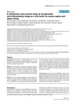
biomimetics (Bakhshayesh et al., 2019; Rizeq et al., 2019). Fig. 1 shows
the increased interest in chitosan materials and nanocomposites in the
last years.
With the advance of nanotechnological fields, organic and inorganic
nanofillers have been tested to produce chitosan nanocomposites pre
senting improved mechanical, chemical, thermal, and barrier properties
(Jafari et al., 2016; Rodrigues et al., 2020). Despite all that effort, there
is not a work in literature that systematically reviews the nanostructure
possibilities, and the related shaping processes required for the desired
application. Therefore, this work intends to fill this gap and present the
coming trends and challenges regarding chitosan as a matrix of
nanocomposites.
This paper is organized into three main sections: chemical structure
and synthesis of the chitosan matrix and specific nanofillers, the most
common shaping routes that have been employed, and the chemical and
biotechnological properties related to applications of chitosan
nanocomposites.
* Corresponding author.
E-mail address: (R.A.F. Machado).
/>Received 3 May 2021; Received in revised form 19 July 2021; Accepted 19 July 2021
Available online 22 July 2021
0144-8617/© 2021 Elsevier Ltd. This article is made available under the Elsevier license ( />
A.O. Silva et al.
60k
Carbohydrate Polymers 272 (2021) 118472
Chitosan
50k
Crustacean
Shell
+ Nanocomposites
2
Magnification of the publications
from 1934 to 1990
800
40k
Chitosan
20k
500
3
400
300
Deproteinaze
200
100
NaOH
4
0
10k
• Wash
• Crush
HCl
600
30k
1
Demineralize
700
Publications
Publications
Chitosan + Matrix
0
0
0
-197
-198
-199
1934
1981
1971
Year
Deacetylation
0
0
0
0
2
0
0
-202
-201
-199
-200
-198
-197
1991
1981
2001
2011
1971
1934
Chitin
5
Year
Chitosan
Fig. 1. Number of publications with search entries: “chitosan” (green), “chi
tosan” and “matrix” (yellow), “chitosan” and “matrix” and “nanocomposites”
(red). Total number of publications: 62563. Search date: March 15,
2021 (Scopus).
Fig. 3. Schematic overview of the main stages of production of chitin
and chitosan.
source of biomass for the industrial production of chitin and chitosan.
The chemical structure of the crustacean shell is composed of protein,
inorganic salts, chitin, and lipids. The synthesis process of chitosan
comes from a deacetylation reaction from chitin from the biomass
source. The chemical deacetylation reaction and the overview produc
tion of chitosan are presented in Fig. 3. Typically, the manufacturing
process follows these unit operations (Bakshi et al., 2020; Nasrollahza
deh et al., 2021; Riaz Rajoka et al., 2019):
(A)
• The raw material shells are washed, crushed, and ground to smaller
sizes with demineralization of some components, such as calcium
carbonate, by chemical extraction with dilute hydrochloric acid with
stirring at room temperature.
• After demineralization, deproteinization is performed by applying
dilute aqueous sodium hydroxide solution. Proteins can be recovered
by lowering the pH to 4.0 and then drying the precipitates.
• An additional decolorization step may be incorporated to remove
color. In this step, chitin is extracted as the main input material for
the production of chitosan.
• Chitosan is obtained by deacetylation from the chitin obtained, again
in sodium hydroxide but in an environment without oxygen and
sometimes by an enzymatic route. The three key reaction parameters
are alkali concentration, time, and temperature. Those factors define
the degree of deacetylation of the final material.
(B)
Fig. 2. Chemical structures of (A) chitin (R1 = H) and its derivatives; (B) chi
tosan (R1 = H, R2 = H, R3 = H) and their derivatives.
Adapted from Muanprasat and Chatsudthipong (2017).
2. Chitosan nanocomposites: structure and microstructure
Chitosan derivatives nanocomposites have earned high interest
especially due to their distinctive physical and chemical properties
(Fig. 4). Amine (NH2) and hydroxyl (OH) surface groups promote the
formation of several inter and intramolecular hydrogen bonds, which
allows the embedding of nanoparticles used as a filler. Chitosan has been
increasingly investigated as an eco-friendly, low-cost, sustainable, and
renewable nanocomposite.
2.1. Chitin and chitosan: chemical structure and synthesis
Chitin is a semi-crystalline homopolymer of β-(1 → 4)-linked Nacetyl-D-glucosamine. It is the second most abundant natural
biopolymer after cellulose (Bakshi et al., 2020). Chitosan, a partially
deacetylated product of chitin, is a copolymer consisting of β-(1 → 4)-2acetamido-D-glucose and β-(1 → 4)-2-amino-D-glucose units, where the
structures of both substances are presented in Fig. 2 (Muanprasat &
Chatsudthipong, 2017).
In Fig. 2, radicals R1, R2, and R3 correspond to hydrogen in plain
chitin and chitosan molecule. Those surface groups led to the amino
(NH2) and hydroxyl groups (OH), responsible for chitosan organic
modifications with several possibilities producing polymeric derivatives
of these compounds (Tharanathan & Kittur, 2003).
Crab and shrimp shell exoskeleton wastes are the raw material
2.2. Chitosan as a nanocomposite matrix
Besides the use of chitosan as a pure matrix biomaterial, with the
advance of nanotechnology, chitosan can be coupled with several kinds
of nanostructures, either embedded into the bulk material or deposited
on the surface.
Biopolymers, such as chitosan, as pure single materials may exhibit
2
A.O. Silva et al.
Carbohydrate Polymers 272 (2021) 118472
Hydrophilic and
Bioadhesive
High cristalinity
Insoluble in water
and organic solvents
Soluble in diluted
acetic acid
CHITOSAN
NANOCOMPOSITES
PROPERTIES
Biocompatibility and
Biodegradability
Antimicrobial
Chelating and
Complexing
Ionic conductivity
Fig. 4. Typical physical and chemical properties of chitosan-based nanocomposites.
either as a filler dispersed inside the whole matrix, and/or as a coating at
the material surface (Kankala et al., 2020) to outcome most of those
structure drawbacks. Several nanostructures of inorganic, organic,
metallic, or semiconducting nature can be applied and dispersed as
additives in chitosan, such as nanoparticles, nanosheets, nanorods,
nanocapsules, nanowires, and nanofibers, as shown in Table 1.
This chapter focuses on the addition of single nanostructures to
chitosan before shaping, and shows the materials that are commonly
employed, as well as the chemical modifications or reactions required.
Other recent engineering processes for insertions of nanostructures
within chitosan will be discussed further.
Table 1
Some nanomaterials applied as additives (fillers, coatings) in chitosan
nanocomposites.
Nanoadditive
Nanostructure
Filler/
coating
Dimensions
(particle
size/length)
in nm
References
Ag
Nanoparticles/
nanowires
Filler/
coating
20–100
Cellulose
Nanocrystals
Filler
100
Chitin
Nanofibers
Filler
50–500
Fe3O4
Nanoparticles
Filler/
coating
9.5–124
Nanoclay
Nanoparticles
Filler
<100
PVA
Nanocapsules
Filler
113
Pt
Nanoparticles
Filler
3–7
SiO2
Nanocapsules/
nanoparticles
Filler
6–50
ZnO
Nanoparticles
Filler
~100
Graphene
Nanosheets
Filler/
coating
<400
(Aziz, Abdullah,
et al., 2019; Aziz
et al., 2020; Aziz,
Brza, et al., 2019;
Kim et al., 2019;
Vunain et al.,
2016)
(Marín-Silva et al.,
2019)
(Jafari et al.,
2016)
(Barra et al., 2020;
Chatrabhuti &
Chirachanchai,
2013;
Heidarinasab
et al., 2016)
(Rodrigues et al.,
2020)
(Mishra et al.,
2017)
(Kankala et al.,
2020)
(Dakroury et al.,
2020; Wu et al.,
2019)
(Priyadarshi &
Negi, 2017;
Rodrigues et al.,
2020)
(Holder et al.,
2017; Jia, Gai,
et al., 2016)
2.2.1. Nanofillers
The most common way to produce nanocomposites using chitosan as
a matrix is to blend the nanofillers in a solution with dissolved chitosan,
cast the slurry, and let it dry at room temperature (Priyadarshi & Rhim,
2020). In some cases, a former step of nanoparticle reduction is needed
by means of a reducing agent, which is added into the slurry (Kim et al.,
2019). By this method, it is possible to disperse a nanostructured ma
terial or composite inside the whole chitosan matrix. This approach is
employed to promote better properties such as mechanical (Esmaeili
et al., 2019), thermal (Smirnova et al., 2019), barrier (Jafari et al.,
´mez P´
2016), magnetical (Go
erez et al., 2020; Hasan et al., 2020), or to
slow down the release of some active nanomaterial (Mishra et al., 2017;
Tripathi et al., 2011).
Nanoaddition of several compounds has been investigated such as
organic nanostructures (Marín-Silva et al., 2019; Smirnova et al., 2019),
oxide ceramics (Aziz, Brza, et al., 2019; Aziz et al., 2020), metallic
nanoparticles (Kim et al., 2019), alloys (Nivethaa et al., 2017) or com
binations of these materials (Mishra et al., 2017; Wu et al., 2019).
Therefore, there is an extended group of possibilities regarding
nanotechnological species applied together with chitosan as matrix
material. The interactions between the nanostructures and chitosan are
aimed to provide a suitable shaping process or promote a desired
physicochemical properties regarding some specific applications.
2.2.2. Nanocoatings and nanofilms
There are some particular cases when the simple blending with
nanostructures is not the most adequate way for producing chitosan
some major drawbacks such as poor mechanical strength, low thermal
stability, and poor barrier properties (Bakshi et al., 2020). Inorganic and
organic compounds in the nanoscale size have been added to chitosan
3
A.O. Silva et al.
Carbohydrate Polymers 272 (2021) 118472
Fig. 5. Examples of typical covalent grafting functionalization in chitosan at the chemical amino (NH2) and hydroxyl (OH) radicals.
Based on concepts from Erathodiyil and Ying (2011).
nanocomposites. Having the nanostructure only at the surface of the
chitosan matrix sometimes can be necessary, e.g. to promote an easy
releasing or leaching of the nanospecies (Chimisso et al., 2020).
Chitosan has the chemical ability to form complexes with transition
metals and post-transition ions (Priyadarshi & Rhim, 2020). Positively
or negatively charged nanostructures in the solution can easily elec
trostatically bind or assembly with chitosan using the layer-by-layer
approach. Zhou and Fu (2020) made a flame retardant wood attaching
wood, chitosan, phthalate, and metallic nanoparticles by controlling the
pH of each species solution. Chitosan, which tends to be positively
charged in acidic conditions was bound at the wood surface. Additions
of phthalate solution that is negatively charged followed, and further
phthalate with positively charged nanoparticles of TiO2 and ZnO were
added.
De Mesquita et al. (2010) have studied a layer-by-layer deposition of
chitosan and cellulose nanowhiskers producing a new biodegradable
nanocomposite. The authors discussed that the use of this technique
maximized the interaction between both components and allowed the
incorporation of a high amount of nanofillers.
Controlling the polyelectrolyte ions is essential to stabilize the
nanomaterial's surface when added to the chitosan matrix. The intro
duction of metallic nanoparticles into polyelectrolyte multilayers has
already proven to be achievable and promising.
Silver nanoparticles (AgNPs) and other noble metallic nanoparticles
can also be introduced in the chitosan matrix, due to the affinity in the
structure toward Ag+ ions, which is related to the amine and hydroxyl
groups (Kumar et al., 2020). This kind of reaction can let the material at
the surface of the nanocomposite.
To deposit the macromolecules on the chitosan surface, a chemical
covalent modification of its structure is eventually necessary, especially
to allow the immobilization of the functional biopolymer/network. The
surface may be modified by the introduction of functional groups that
can react with the polymer/nanostructure that has to be attached
(Chimisso et al., 2020). Chitosan presents amine (NH2) and hydroxyl
(OH) radical groups that can enhance covalent or protonation reactions
as shown in Fig. 5. The molecules applied can be used for nanoparticle
chemistry stabilization at the surface (Erathodiyil & Ying, 2011).
For example, the chitosan-modified graphene oxide nanosheet can
be shaped by covalent conjugation of the amide linkage between the
carboxylic groups of graphene and the amine groups of chitosan (Jia,
Gai, et al., 2016). To maintain the suspension stability of magnetic
nanoparticles with chitosan, the covalent bond is more stable compared
to other forces (Chatrabhuti & Chirachanchai, 2013). The epoxide
opening reaction was performed by Heidarinasab et al. (2016) to attach
magnetic nanoparticles, and provide the best conditions for the mag
netic nanocarrier delivery.
3. Manufacturing processes
3.1. Mixing and shaping
For the nanocomposites production, some considerations have to be
taken into account to assure complete mixing of chitosan with the
nanoadditives and solvents (Riaz Rajoka et al., 2019). According to the
chitosan solubility, a film formation can be either more easily performed
or become a difficult task (Sampath et al., 2016).
4
A.O. Silva et al.
Carbohydrate Polymers 272 (2021) 118472
Table 2
Comparison of some approaches for 3D printing chitosan derivative nanomaterials.
Process
Methods
Characteristics
Features
References
Traditional manufacturing
Solvent casting
Material dissolved in a solvent
(Bakshi et al., 2020)
Additive manufacturing
(AM)
Stereolithography (SLA)
UV light or electron-beam is used to start a
reaction
Same principle that traditional inkjet printers
Cannot be accurately
controlled
Non-biological
manufacturing
Biological manufacturing
Jet based printing
Laser-assisted
bioprinting
Nozzle-free deposition
(Seo et al., 2020)
(Ahmed et al., 2020; Sommer et al.,
2017)
(Bozuyuk et al., 2018).
Adapted from Wang et al. (2020).
Chitosan possesses a mild organic base structure behavior, capable to
produce salts in contact with weak acids. The most common experi
mental practice is to add small fractions of acetic acid in water to a
chitosan suspension so that salt formation and dissolution occur
concurrently (Roberts, 1992). Chitosan has a high hydrophilic behavior
and for that reason, it is practically insoluble in organic solvents,
although some organic solutions of chitosan in aqueous acetic acid can
tolerate the addition of large volumes of polar solvents without causing
precipitation of the polymer (John et al., 2019; Roberts, 1992). This kind
of chemical nature was very helpful for the chitosan nanocomposite
preparation by John et al. (2019). In this case, the presence of ethanol in
the solvent mixture chitosan/water in acetic acid promoted adequate
TiO2 nanoparticle nucleation and film formation.
Most of the studies about chitosan nanocomposites dispersion have
used simple magnetic mixing (Priyadarshi & Rhim, 2020). Some works
employed more effective dispersion equipment like mechanical stirrer
with ultrasonication (Celebi & Kurt, 2015). Some particular cases used
stronger mixing devices such as ultraturrax (Marín-Silva et al., 2019).
The main shaping technique for most of those nanocomposite systems
consists of a solution or solvent casting method (Bakshi et al., 2020)
which is the production of a dispersion phase in acetic acid, followed by
evaporation or a drying step (Martínez-Camacho et al., 2010).
parameters associated with the selection of compounds for bioinks (Lee
& Yeong, 2016). According to the applications, bioinks can be either
supporting or functional. The most commonly supporting bioink is a
hydrogel; a functional bioink (e.g., DNA and factor) is mainly used to
study intracellular delivery, gene diagnosis, and cell behaviors.
Chitosan is one of the best 3D printing matrix bioink candidates due
to its desirable physicochemical properties and essential features for cell
adhesion, extracellular matrix (ECM) deposition, and finally tissue
regeneration (Jiankang et al., 2009). However, the chitosan matrix
presents a common drawback characteristically of hydrogels, which is a
weak mechanical resistance (Whyte et al., 2019). For that reason, it is
usually blended or coated with other materials, including nano
materials, to improve mechanical properties (Semba et al., 2020).
Sommer et al. (2017) developed a modified oil-in-water emulsion con
taining chitosan with modified silica nanoparticles in the water phase.
The resulting ink provided good stability for the emulsion, and it was
ideal for 3D printing and displayed high yield stress, storage modulus,
and elastic recovery.
Chitosan as a raw biomaterial for 3D printing is mostly processed
using inkjet bioprinting approaches for bone implants and artificial skin
applications (Whyte et al., 2019). There has been an increased interest in
3D printing related to chitosan nanomaterials and their potential ap
plications in biomedical engineering including tissue engineering and
medical implants (Ahmed et al., 2020). Pahlevanzadeh et al. (2020)
elaborated a concise review about 3D printable chitosan, both single and
derivatives, aiming the development directions and prospect directions.
According to the authors, chitosan decomposes at typically lower tem
peratures, not higher than 220 ◦ C, thus the nanomaterials employed in
3D printing techniques ought to be either sintered under this tempera
ture, or a corresponding thermoresistance should be promoted. Never
theless, other limitations can be in a wide range considering the type of
different methods, their performance strategy, and the desired
application.
Normally, 3D robust shaping techniques such as stereolithography
(SLA), or Fused Deposition Modelling (FDM) are not possible with chi
tosan as the main material. However, Seo et al. (2020) have overcome
that limitation for SLA printing. In this case, 3D printing is based on
photopolymerization by a laser, so that SLA requires a polymer that can
be photo-crosslinked, such as hydroxybutyl methacrylated chitosan
(HBC-MA). HBC-MA was developed using chitosan as precursor mate
rial, which corresponds to a photocrosslinkable temperature-reversible
chitosan derivative, reacting sensitively to temperature changes. Cellu
lose nanofibrils were employed as nanoadditives being physically
confined in the photocrosslinked hydrogel to assist in the directionally
of volume expansion, and swelling rate. In conclusion, HBC-MA can be
regarded as a potential material for tissue engineering, and medical
applications.
Limited by their difficult solubility and non-melting properties,
chitosan 3D nanocomposites are hard to be directly manufactured by
Fused Deposition Modelling (FDM). Although the use of chitosan as
second material (Yu et al., 2020), or single modification have been re
ported (Elviri et al., 2017), until the present moment there is to the best
of our knowledge no printed chitosan nanocomposites fabricated by
3.2. 3D printing
Three-dimensional nanostructured composites have become a high
interest in numerous fields including biomedical engineering, energy
storage, and structural or functional materials (Sommer et al., 2017).
Additive manufacturing (AM), commonly known as 3D printing (3D), is
a compilation of techniques comprising non-biological and biological
approaches production of a physical object from a three-dimensional
model, where the most common routes for chitosan nanocomposite
derivatives are presented in Table 2.
The 3D printing techniques allow an elevated control of the geom
etry of any manufactured biological structure, that accurately corre
sponds to a computer-aided design (CAD) project (Elviri et al., 2017). An
example is a 3D system developed by Elviri et al. (2017) which corre
sponds to a simple, safe, and low-cost process avoiding the use of organic
solvents, the need for high processing temperature, or the difficulty in
the removal of dust, which is very typical of analogous techniques of
additive manufacturing. To be used in in vivo studies, the 3D printed
scaffold also should not lose its shape and strength after being soaked in
water for a long time (Gang et al., 2019).
Advanced biological scaffolds for tissue engineering can be easily
fabricated using a 3D printing technique, for example, robocast-assisted
deposition as tested by Cebe et al. (2020). Their work with a robocaster
allows precise control of micropatterning by determining the di
mensions of filaments, the size and shape of pores, and the percentage of
porosity of the scaffold.
Bioinks are the common raw materials used in a 3D printer for bio
logical applications (Sahranavard et al., 2020). For practical applica
tions, printability, fidelity, viscoelasticity, shear-thinning, yield stress,
creep, shelf life, cross-linking time, and cost are some of the essential
5
A.O. Silva et al.
Carbohydrate Polymers 272 (2021) 118472
(A)
(B)
DC High Voltage
Off
+
15.0 kV
Off
-
+
Polymeric Solution
On/Off
Volumetric Flow
On
10.0 kV
-
Polymeric Solution
1.0 mL/h
Syringe Pump
DC High Voltage
On
On/Off
Volumetric Flow
Metal
Collector
0.5 mL/h
Syringe Pump
Metal
Collector
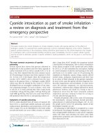
Fig. 6. Schematic view of typical nanofiber and nanoparticle manufacturing processes: (A) electrospinning and (B) electrospraying, respectively.
Producing chitosan nanofibers through the electrospinning process is
quite challenging because of the material's ionic nature (Cai et al.,
2016). The rigid structure prevents chain entanglements leading to the
jet break up (Kersani et al., 2020). In reason of that, most works prefer to
bind with other polymers or biopolymers such as Poly(vinyl alcohol)
(PVA) (Koosha et al., 2019; Sedghi et al., 2017), Poly(ethylene oxide)
(PEO) (Kersani et al., 2020), Polylactic acid (PLA) (Shan et al., 2014; Xu
et al., 2009), Polyethylene glycol (PEG) (Han et al., 2011).
Nanoparticles have been inserted before and after the electro
spinning process to improve material properties. Halloysite and carbon
nanotubes (Koosha et al., 2019; Liu et al., 2019) and silica nanoparticles
(Zhao et al., 2015) have been added for enhancing mechanical behavior.
Silver nanoparticles (Zhan et al., 2017) and Fe3O4 (Cai et al., 2016)
nanoparticles were also added to improve chitosan antimicrobial
activity.
There are few studies in the literature on the use of electrosprayed
chitosan as the matrix material in nanocomposites. Versatility and lowcost operation are some advantages that delineate a particular interest in
this field (Jayasinghe et al., 2006; Park et al., 2007; San Thian et al.,
2008). Chng et al. (2019) allowed a controlled and precise deposition in
dental implants that improved their properties, compared to conven
tional shaping techniques, since chitosan is a biopolymer that presents
compatibility with biological systems, it has been used as a matrix after
silane-based treatment to chemically bond the coated chitosan to the
substrate, maximizing the adhesion strength between the coating and a
surface. Yuan et al. (2019) produced microparticles containing chitosan
and nano-hydroxyapatite (NanoHap), as well as pharmacological spe
cies. These particles provided an effective way to long-term sustained
release activity.
FDM.
The advancements in the two-photon direct laser writing (TDLW)
technique, a derivation of laser-assisted bioprinted, allowed the 3D
fabrication of complex polymeric structures. Bozuyuk et al. (2018)
fabricated chitosan derivative microswimmers by two-photon-based 3D
printing of a natural polymer derivative of chitosan in the form of a
magnetic polymer nanocomposite. Amino groups presented on the
microswimmers are modified with doxorubicin employing a photo
cleavable linker. Chitosan imparts the microswimmers with biocom
patibility, and biodegradability for use in a biological setting. Their local
3D patterning has been performed with the use of versatile chemical
moieties and provided the possibility to embed nanoparticle additives
during the shaping step (Bozuyuk et al., 2018).
Another possibility for chitosan nanocomposites is by coating other
3D printed materials. Azadmanesh et al. (2021) performed an FDM
printing process with polylactic acid for scaffold production. After the
3DP, chitosan and copper carbon dots (Cu-CDs) were cross-linked with
the PLA scaffolds. This is an alternative approach to overcome the dif
ficulties regarding chitosan physical and chemical properties.
3.3. Electrospinning and electrospraying
The use of electrospinned and electrosprayed materials has expanded
in many important recent biotechnological fields such as food technol
ogy, tissue engineering, drug delivery, and wound dressing (Soares
et al., 2018). Electrospinning is a technique that aims to produce micro
and nanofibers mats from polymeric solutions or melt polymers (Xue
et al., 2019). Commonly regarded advantages of materials and com
posites made by electrospinning are high porosity, low pore size, and a
large surface area (Chahal et al., 2019).
In electrospinning, the charged polymer solution or melt overcomes
its surface tension under the action of a high-voltage electrostatic field to
form small jets, which are further accelerated and stretched, and finally
fall on the collector with solvent evaporation or melt cooling to form
fibers (Haider et al., 2018; Yarin et al., 2001). Typically, a syringe
comprises an electrospinning shaping apparatus with a needle attached
to the syringe tip, which is directed to a metallic base acting as a support
for the fiber mat collection (Haider et al., 2018; Yarin et al., 2001).
The needle and the metallic base are connected to a high voltage
power source through electrodes. An electrospinning device is generally
composed of a high-voltage power supply, a liquid supply device
(injector, etc.), and a collector (drum or metal plate, etc.) (Araldi da Silva
et al., 2021). A typical device for electrospinning is presented in Fig. 6.
In the case of electrospraying, a process similar to electrospinning,
micro, and nanoparticles (spheres or capsules) can be obtained from a
polymer in solution with a high conductive solvent (Soares et al., 2018).
3.4. Other techniques
As mentioned before, the main shaping technique for both chitosan
and chitosan nanocomposites is the solution or solvent casting method
(Bakshi et al., 2020). This method consists of the production of disper
sion phase using commonly acetic acid, at high quantities, as solvent
followed by evaporation or a drying step (Martínez-Camacho et al.,
2010). This technique is limited to a specific material design structure
(flat thin film deposited generally on a glass surface) generally as a final
dense material (Esmaeili et al., 2019).
Moreover, chitosan is still also a natural-based polymer material and
for that reason might be submitted to common polymer shaping pro
cesses such as thermoforming, injection molding, compression molding,
rotational molding, extrusion, blow molding, among others. Extruded
chitosan nanocomposites were prepared successfully by Amouzgar et al.
(2017) and Choo et al. (2016). In the first work, they studied the use of
6
A.O. Silva et al.
Carbohydrate Polymers 272 (2021) 118472
4.1. Waste and wastewater treatment
Water and Wastewater
treatment
Scaffolds and Tissue
Engineering
Clinical Applications
Waste and wastewater treatment is an important concern worldwide.
The necessity of access to quality potable water and effluents disposal
has emerged in many material developments. Chitosan both as a single
material and as composites has gained significant interest in multilayer
coatings for improving corrosion resistance (Nasrollahzadeh et al.,
2021), and wastewater treatment (Mohammadzadeh Pakdel & Peigh
ambardoust, 2018; Thirugnanasambandham et al., 2013).
Crini et al. (2017) described the use of chitosan in the process of
ultrafiltration (UF) where a large variety of metal ions can be adsorbed
and selectively separated. Other authors also investigate and demon
strated good results using chitosan nanocomposites in heavy metal
remediation (Al-Sherbini et al., 2019; Cheraghipour & Pakshir, 2020;
Kenawy et al., 2019).
Due to their chemical structure, chitosan nanomaterials have been
employed in the removal of pollutants (Nasrollahzadeh et al., 2021)
such as heavy metal ions Cu(II) and Cr(VI) (Anush et al., 2020), Cr (VI)
(Reis et al., 2021), Co (III) (Abdelbasir et al., 2021), iron (Shehap et al.,
2021), dyes (Krishna et al., 2021; Mostafa et al., 2020a; Rashid et al.,
2018; Rebekah et al., 2020; Reghioua et al., 2021; Sathiyavimal et al.,
2020), antibiotics and pesticides (Asgari et al., 2020).
The industrial use of organic dyes in many industries is environ
mentally hazardous, toxic, and carcinogenic. Chitosan derivative
nanocomposites emerge as a potential material to promote an efficient
and sustainable dye removal (Rashid et al., 2018). Recently, some
research works have been studied according to the combination of
different ceramic nanostructures or nanoclays with chitosan aimed to
remove different types of organic dyes (da Silva et al., 2021; Krishna
et al., 2021; Mostafa et al., 2020b).
The use of chitosan nanocomposites was explored by Asgari et al.
(2020) for antibiotic removal from wastewater. Chitosan was tested
together with Fe3O4 magnetic nanoparticles, which were capable to
retain spontaneous metronidazole from water in industrial and hospital
wastewater.
The chitosan capability of binding semiconductors metals can create
photocatalysts nanocomposites (Huang & Peng, 2021). Those new
nanomaterials can improve the quality of degradation of organic pol
lutants (Midya et al., 2020) or dye removal by the Fenton process (Ali
mard, 2019).
The strong heavy metal adsorption capacity by chitosan and their
derivatives can be related to multifunctional surface chemical groups,
high hydrophilicity, high chemical reactivity, and polymer flexible
structure (Vunain et al., 2016). Gupta et al. (2012) described charac
teristics of chitosan related to the polymer molecular chain, which
contains plenty of amino and hydroxyl groups at the surface. Those
chemical groups can produce stables chelates binding with several metal
ions, such as Hg2+, Ni2+, Cu2+, Pb2+, Zn+2, Cd+2. Zhu et al. (2021) in a
recent study emphasize that when chitosan is applied as an adsorbent
directly, the specific surface area is modest, therefore the adsorption
capacity is low. Those properties effectiveness of single chitosan mate
rial can be effectively improved by shaping it into nanofibers. However,
other properties such as mechanical, stability, and reusability still de
mand to be further improved. That lack of research might be fulfilled
with the advanced knowledge in nanocomposites using chitosan as the
matrix. Furthermore, the majority of heavy metal adsorption research
using chitosan is still on the laboratory scale, and it has some obstacles to
their scale-up and practical implementation in the treatment of heavy
metals present in industrial wastewater.
Corrosion Inhibition
Food Packaging
Energy Storage
DNA Extraction
Fig. 7. Chart of distribution of chitosan nanocomposites applications in
this review.
chitosan with nanoactivated carbon; and in the second one, the selected
structures were halloysite nanotubes. In both works, an additional ul
trasonic dispersion was required for a successful extrusion process.
There is no report until the present moment on the use of thermo
forming, injection molding, rotational molding, compression molding,
and blow molding with chitosan nanocomposites. In the case of blow
molding and rotational molding, no report was found on the use of
chitosan on this material as a matrix too. However, injection molding,
thermoforming, and compression molding have been applied to the
single use of chitosan material or using chitosan as an additive in other
polymeric matrices.
4. Chitosan nanocomposites: properties and applications
Due to all the application possibilities regarding chitosan nano
composites, recent and important applications will be presented.
Because of the material versatility, as seen in Fig. 7, the literature has
been focused on chitosan nanocomposites mainly in water and waste
water treatment, tissue engineering, biomedical applications, and
corrosion inhibition.
Table 3
Examples of chitosan porous materials and composites according to shaping
methods.
Material/
composite
Shaping
Pore size
(μm)
Porosity
(%)
References
Chitosan
Freeze-drying
60–90
88–97
Freeze-drying and
Particulate leaching
Liquid hardening
7–500
60–90
200–500
80
Gas foaming CO2
30–40
>30
3D printing
3.5–9
52
Melt molding and
NaCl leaching
Mold casting/
Infrared dehydration
Electrospinning
>100
58.3–91.2
0.2
0.5–2.5
Not
informed
>50
Freeze-drying
50–150
<80
Freeze gelation
150–300
Not
informed
(Nwe et al.,
2009)
(Lim et al.,
2011)
(Hsieh et al.,
2007)
(Ji et al.,
2011)
(Intini et al.,
2018)
(Li et al.,
2004)
(Xie et al.,
2009)
(Kim et al.,
2013)
(Ying et al.,
2020)
(Oudadesse
et al., 2020)
Chitosan/
PLA
Chitosan/
PLGAa
Chitosan/
NanoHap
Chitosan/
Nanoglass
a
4.2. Scaffolds for tissue engineering
Tissue and organ failures caused by injury, aging accounts, or dis
eases are an important concern worldwide (Abbasian et al., 2019).
Tissue engineering has become an important research field for clinical or
biomedical applications regarding chitosan nanocomposites. Chitosan is
Poly lactic-co-glycolic acid.
7
A.O. Silva et al.
Carbohydrate Polymers 272 (2021) 118472
4.3. Other biomedical applications
Freeze-drying
Besides the use of chitosan scaffolds for tissue engineering, chitosan
as a single and composite nanomaterial has been studied in other related
biomedical fields such as wound healing, drug delivery, dietary sup
plement, biosensors, among others. Accordingly, chitosan has been
applied to help to solve several human health issues: blood clotting, fast
skin burn healing, food allergies and intolerance control, weight loss and
cholesterol control, gene therapy, and cancer treatment, among others
(Rizeq et al., 2019).
The most common pharmaceutical ability of chitosan was noticed
especially inside fat and cholesterol-burning supplements, throughout
the material ability to reduce weight gain by electrostatically attach to
fatty acids released by digestion in the small intestine (Shariatinia,
2019). May et al. (2020) explained the chitosan mechanism during the
digestion of full cream milk, they noticed that the lipids present selfassembled into characteristics stable liquid crystalline nanostructures.
However, a presence of a high concentration of chitosan was able to
reduce fatty acids and prevent the self-assembly of lipids.
In some conditions, chitosan can be suitable for the wound healing
process due to antimicrobial properties linked with low mammalian cell
toxicity (Matica et al., 2019). For external skin wound healing, a
nanocomposite containing silver nanoparticles and chitosan was tested
by Ding et al. (2017). The film material needed to attach over the skin
demanded a higher mechanical strength and was cross-linked with
genipin (GB), resulting in a loss of chitosan active sites and consequently
a decrease in antimicrobial response, which was replaced by the addi
tion of silver nanoparticles. The treatment of internal wounds is also
possible with chitosan nanocomposites. The work of Sundaram et al.
(2019) has exploited the use of an injectable nanocomposite containing
chitosan and nanobioglass. Their work, tested in vivo, provides a fast a
secure blood clotting achieved by a synergistic effect between chitosan
and nanoglass when in contact with blood.
Biosensors are defined as chemical sensors capable to recognize the
properties of biological components, and chitosan derivatives are ideal
candidates for several biosensing applications (Wang et al., 2016).
Erdem et al. (2014) tried to attach DNA aptamer immobilized grapheneoxide nanostructures to chitosan. In this case, chitosan is attached to a
pencil graphite electrode (PGE) surface The biosensor was developed
and tested for the selective and sensitive detection of lysozyme (LYS),
where low quantities could be a marker for some health problems.
Novel kinds of cancer and other diseases treatment are possible with
the use of chitosan nanocomposites, particularly by the means of drug
delivery. Kankala et al. (2020) successfully fabricated a versatile drug
delivery platform by coating platinum (Pt) nanoparticles embedded
chitosan polymer composite layer, over Zn-doped siliceous frameworks
loaded with doxorubicin, a drug used to combat multidrug resistance
(MDR) in cancer treatment. This chitosan nanoderivative substantially
facilitated deep tissue penetration efficacy due to the Pt nanometric
sizes. Also, the Pt nanoparticles synergistically participated in the in
crease of the inhibition effect of the chemotherapeutic agent. The in vivo
findings suggested that the innovative organic-inorganic nanohybrid
material loaded with combinatorial therapeutics could be an alternative
approach over conventional clinical strategies against cancer.
Liquid hardening
Porosity (%)
Melt molding
and NaCl leaching
Freeze-drying and
Particulate leaching
3D printing
Electrospinning
Gas foaming CO2
Pore size (µm)
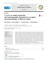
Fig. 8. Schematic diagram of different shaping techniques using porous chi
tosan materials as scaffolds for tissue engineering as a function of porosity and
pore size.
considered an ideal kind of material for use in tissue engineering mainly
due to its desired biological characteristics such as biocompatibility,
biodegradability, bioinertness (Abinaya et al., 2019). Chitosan's chem
ical structure can resemble major components of bone and cartilaginous
tissue, promoting cell adhesion (Oudadesse et al., 2020).
A particular area in tissue engineering, especially linked to chitosan
material is the production of scaffolds. Scaffolds are porous materials
specially engineered to promote desirable cellular interaction, allowing
the formation of new functional tissues (Whyte et al., 2019).
The major difficulty related to scaffold design manufacturing is to
produce high porosity, controlled pore size, and pore interconnectivity
from natural-based polymeric raw materials. Biobased materials such as
chitosan are heat sensitive, which limits the use of several scaffold
shaping techniques (Sampath et al., 2016). Pure chitosan scaffolds with
controlled porosity are mainly produced for freeze-drying approaches or
combinations, although electrospinning has been increasingly employed
(Vandghanooni & Eskandani, 2019).
Due to the chitosan possibility of chemical crosslink, the use of chi
tosan composites with other synthetic biopolymers, such as PLA, and the
use of nanoparticles, such as nano-hydroxyapatite, broaden the chitosan
shaping possibilities (Bulanov et al., 2020; Sampath et al., 2016). The
additives also must be specially selected, to maintain the biocompati
bility from single chitosan (Abbasian et al., 2019). Table 3 shows some
shaping techniques of chitosan materials and composites, used as porous
scaffolds for tissue engineering.
In this context, Fig. 8 shows a relationship between porosity and pore
size of chitosan materials, obtained through different shaping tech
niques, as listed in Table 3. It is noted that, depending on the use of a
particular technique or combination of techniques, different porous
systems can be obtained, which shows the versatility of this raw
material.
Therefore, the application of chitosan single or composite materials
in scaffolds for tissue engineering have been encouraged due to their
physical, chemical, and biological properties such as (Vandghanooni &
Eskandani, 2019).
•
•
•
•
•
4.4. Corrosion inhibition
Chitosan is a suitable polysaccharide for application as an effective
corrosion inhibitor for many metallic substrates (Qasim et al., 2019;
Umoren & Eduok, 2016). Chitosan shows a strong capability of adhesion
to a metal surface allow this polymer to be coated on metals to provide a
protective barrier. Chitosan's anti-corrosion ability is derived from its
own molecular structure. The chitosan molecule bears electron donate
rich hydroxyl and amino groups capable of metal surface bonding, and
subsequent corrosion inhibition via coordinate bonding, as these elec
trons are donated to the empty or partially occupied metallic orbitals
Cytocompatibility (in vitro or in vivo)
Crosslinking to improve mechanical and barrier properties
Mild processing conditions
Antibacterial effect related to the chitosan cationic structure
Excellent interactions with adhesive proteins and receptors
8
A.O. Silva et al.
Carbohydrate Polymers 272 (2021) 118472
nanocomposites has been developed for food packaging as active anti
microbial polymer or a biocidal nanocomposite (Kaur et al., 2020;
Priyadarshi & Rhim, 2020; Virgili et al., 2021), energy storage (Hassan
et al., 2014) for fuel cell application, a catalyst for some specific
chemical and electrochemical reactions (Nasri et al., 2020), and DNA
´mez P´
extraction or separation (Go
erez et al., 2020). In those cases,
different nanomaterials/chitosan combinations have been employed.
The low cost and versatility of chitosan, as well as the broad chemical
and biochemical features, can be turned into future industrial products.
Table 4
Examples of nanomaterials and deposition methods on metallic substrates in
systems containing chitosan as a matrix.
Metal
substrates
Nanoparticles
applied
Deposition method
References
1020
Carbon
steel
1020
Carbon
steel
1020
Carbon
steel
Ag
AZ91 Mg
alloy
C3003
aluminun
alloy
Cu
Mg
Mg, Hydroxyapatite
Spin coating
(Sutha et al., 2015)
Mo
Electrophoretic
deposition
(Oliveira et al.,
2021)
W
Electrophoretic
deposition
(Oliveira et al.,
2020)
Ag
Bioactive glass
Electrodeposition
Electrophoretic
deposition
Solvent
evaporation
(Pan et al., 2020)
(Alaei et al., 2020)
SiO2
Cerium oxide
Layer-by-layer
Electrodeposition
Mg
Mg
Layer-by-layer
Layer-by-layer
Mg
Graphene oxide
Graphene oxide,
Heparin
ZnO
(Bahari et al., 2020)
(Jia, Xiong, et al.,
2016)
(Gao et al., 2019)
(Gao et al., 2020)
Mild steel
Ti
TiO2
Au
Dip coating
Electrodeposition
Ti
Graphene oxide
Ti
Halloysite
Ti
Hydroxyapatite,
Halloysite
Mg, Sr, Halloysite
Electrophoretic
deposition
Electrophoretic
deposition
Electrophoretic
deposition
Electrodeposition
Ti
Ti-13Nb13Zr alloy
TiO2
Ti-6Al-4V
Hydroxyapatite,
Bioactive glass,
Fe3O4
Bioactive glass
Ti6Al7Nb
Ag, Au
Dip coating
Electrophoretic
deposition
Electrophoretic
deposition
Solvent
evaporation
5. Concluding remarks and future perspectives
In the present review, nanocomposites having chitosan as a matrix
were presented and discussed regarding their chemical structure,
shaping processes, properties, and applications. There is a remarkable
increase of studies about binding or embedding nanostructures into a
chitosan matrix. The nanoadditives presented here are mostly used to
provide better mechanical, thermal, and chemical features among other
desired properties, such as antimicrobial activity or drug delivery. The
surface structure of chitosan also provides a possibility of nanoparticle
nucleation, and stabilization of noble metals due to electron donation of
the amino reactant group. Self-assembled chitosan binding capability,
such as the layer-by-layer approach, also provides an adequate way to
assure dispersion of ionic nanostructures.
Due to some shapes or coatings required for specific applications,
sophisticated processing methods have been increasingly developed.
Electrospinning and 3D printing, for example, start to become very
popular in the production of chitosan nanocomposites instead of tradi
tional methods like solvent casting.
The present review exposes some lack of research that might be
further developed in the literature, such as the development of 3D
printable chitosan nanocomposites for FDM, the use of chitosan as a
nanofiber matrix for instance in wastewater treatment, and the feasi
bility of adapting techniques like injection molding and thermoforming
for chitosan nanocomposites. The combination of nanotechnology with
innovative shaping and surface deposition techniques broadens the
horizon of chitosan applications, such as biomedical, food packaging,
energy storage, and wastewater treatment.
(Lai et al., 2021)
(Bakhsheshi-rad
et al., 2021)
(John et al., 2019)
(Farghali et al.,
2015)
(Shi et al., 2016)
(Molaei et al., 2016)
(Molaei &
Yousefpour, 2018)
(Chozhanathmisra
et al., 2018)
(Singh et al., 2021)
(Mahlooji et al.,
2019)
(Kleszcz et al., 2021)
Acknowledgments
(Umoren & Eduok, 2016). The easiness of chemical functionalization of
chitosan with nanospecies is a specific characteristic that can provide an
improvement on mechanical properties, adhesiveness, and barrier ef
fect, which enhances the capability for corrosion protection (AshassiSorkhabi & Kazempour, 2020).
Chitosan materials have been applied mostly on steel (Ashassi-Sor
khabi & Kazempour, 2020; Wei et al., 2020), magnesium (Li et al.,
2018), titanium, aluminum (Bouali et al., 2020), copper (Jmiai et al.,
2017), and alloys for improving anti-corrosion and biodegradability
properties, as well as for providing higher biocompatibility. Table 4
presents some works on the use of chitosan nanocomposites for anticorrosion purposes.
Besides, other works have shown properties that enabled anti-fouling
and anti-corrosion properties simultaneously in metallic substrates
(Idumah et al., 2020). Noble metallic nanocomposites employing chi
tosan as matrix material have been studied for mild steel coating pro
tection even in chilled water circuits or in aggressive chloride media
with promising results (Fetouh et al., 2020; Srivastava et al., 2019).
Smart coatings were also obtained by chitosan, which can also be
applied in the production of sensors (Carneiro et al., 2015; Zouaoui
et al., 2020).
˜o de Aperfeiỗoamento de
The authors are grateful to the Coordenaỗa
Pessoal de Nível Superior (CAPES, Brazil, grant number 01) and Con
´gico (CNPq,
selho Nacional de Desenvolvimento Científico e Tecnolo
Brazil) for financial support (grant numbers: 06316/2007-2 and
311270/2017-4).
References
Abbasian, M., Massoumi, B., Mohammad-rezaei, R., Samadian, H., & Jaymand, M.
(2019). Scaffolding polymeric biomaterials: Are naturally occurring biological
macromolecules more appropriate for tissue engineering? International Journal of
Biological Macromolecules, 134, 673–694. />ijbiomac.2019.04.197
Abdelbasir, S. M., El-Shewaikh, A. M., El-Sheikh, S. M., & Ali, O. I. (2021). Novel
modified chitosan nanocomposites for Co(II) ions removal from industrial
wastewater. Journal of Water Process Engineering, 41. />jwpe.2021.102008
Abinaya, B., Prasith, T. P., & Ashwin, B. (2019). Chitosan in surface modification for
bone tissue engineering applications. Advanced Science News, 14(1900171), 1–10.
/>Ahmed, J., Mulla, M., & Maniruzzaman, M. (2020). Rheological and dielectric behavior
of 3D-printable chitosan/graphene oxide hydrogels. ACS Biomaterials Science &
Engineering, 6(1), 88–99. />Alaei, M., Atapour, M., & Labbaf, S. (2020). Electrophoretic deposition of chitosanbioactive glass nanocomposite coatings on AZ91 Mg alloy for biomedical
applications. In Progress in organic coatings. />porgcoat.2020.105803
Alimard, P. (2019). Fabrication and kinetic study of Nd-Ce doped Fe3O4-chitosan
nanocomposite as catalyst in Fenton dye degradation. Polyhedron, 171, 98–107.
/>
4.5. Other applications
Besides the above-mentioned applications, research on chitosan
9
A.O. Silva et al.
Carbohydrate Polymers 272 (2021) 118472
Celebi, H., & Kurt, A. (2015). Effects of processing on the properties of chitosan/cellulose
nanocrystal films. Carbohydrate Polymers, 133, 284–293. />carbpol.2015.07.007
Chahal, S., Kumar, A., Shahitha, F., & Hussian, J. (2019). Development of biomimetic
electrospun polymeric biomaterials for bone tissue engineering. A review
biomaterials for bone tissue engineering . A review. Journal of Biomaterials Science,
Polymer Edition, 30(14), 1308–1355. />09205063.2019.1630699
Chatrabhuti, S., & Chirachanchai, S. (2013). Single step coupling for multi-responsive
water-based chitin/chitosan magnetic nanoparticles. Carbohydrate Polymers, 97(2),
441–450. />Cheraghipour, E., & Pakshir, M. (2020). Process optimization and modeling of Pb(II) ions
adsorption on chitosan-conjugated magnetite nano-biocomposite using response
surface methodology. Chemosphere, 260, Article 127560. />chemosphere.2020.127560
Chimisso, V., Maffeis, V., Hürlimann, D., Palivan, C. G., & Meier, W. (2020). Selfassembled polymeric membranes and nanoassemblies on surfaces: Preparation,
characterization, and current applications. Macromolecular Bioscience, 20(1), 1–19.
/>Chng, E. J., Watson, A. B., Suresh, V., Fujiwara, T., Bumgardner, J. D., &
Gopalakrishnan, R. (2019). Adhesion of electrosprayed chitosan coatings using
silane surface chemistry. Thin Solid Films, 692(April), Article 137454. https://doi.
org/10.1016/j.tsf.2019.137454
Choo, C. K., Kong, X. Y., Goh, T. L., Ngoh, G. C., Horri, B. A., & Salamatinia, B. (2016).
Chitosan/halloysite beads fabricated by ultrasonic-assisted extrusion-dripping and a
case study application for copper ion removal. Carbohydrate Polymers, 138, 16–26.
/>Chozhanathmisra, M., Govindaraj, D., Karthikeyan, P., Pandian, K., Mitu, L., &
Rajavel, R. (2018). Fabrication of bilayer coating of poly(3,4ethylenedioxythiophene)-Halloysite/Chitosan and Mg2+/Sr2+-doped HAP on
titanium alloy for biomedical implant applications: Physicochemical and in vitro
biological performances studies. Journal of Chemistry, 2018. />10.1155/2018/9813827
Crini, G., Morin-Crini, N., Fatin-Rouge, N., D´
eon, S., & Fievet, P. (2017). Metal removal
from aqueous media by polymer-assisted ultrafiltration with chitosan. Arabian
Journal of Chemistry, 10, S3826–S3839. />arabjc.2014.05.020
da Silva, J. C. S., Franỗa, D. B., Rodrigues, F., Oliveira, D. M., Trigueiro, P., Silva
Filho, E. C., & Fonseca, M. G. (2021). What happens when chitosan meets bentonite
under microwave-assisted conditions? Clay-based hybrid nanocomposites for dye
adsorption. Colloids and Surfaces A: Physicochemical and Engineering Aspects, 609.
/>Dakroury, G. A., Abo-Zahra, S. F., Hassan, H. S., & Fathy, N. A. (2020). Utilization of
silica–chitosan nanocomposite for removal of152+154Eu radionuclide from aqueous
solutions. Journal of Radioanalytical and Nuclear Chemistry, 323(1), 439–455. https://
doi.org/10.1007/s10967-019-06951-6
De Mesquita, J. P., Donnici, C. L., & Pereira, F. V. (2010). Biobased nanocomposites from
layer-by-layer assembly of cellulose nanowhiskers with chitosan. Biomacromolecules,
11(2), 473–480. />Ding, L., Shan, X., Zhao, X., Zha, H., Chen, X., Wang, J., & Yu, G. (2017). Spongy bilayer
dressing composed of chitosan–Ag nanoparticles and chitosan–Bletilla striata
polysaccharide for wound healing applications. Carbohydrate Polymers, 157,
1538–1547. />Elviri, L., Foresti, R., Bergonzi, C., Zimetti, F., Marchi, C., Bianchera, A., & Bettini, R.
(2017). Highly defined 3D printed chitosan scaffolds featuring improved cell growth.
Biomedical Materials (Bristol), 12(4), 1–12. />aa7692
Erathodiyil, N., & Ying, J. Y. (2011). Functionalization of inorganic nanoparticles for
bioimaging applications. Accounts of Chemical Research, 44(10), 925–935. https://
doi.org/10.1021/ar2000327
Erdem, A., Eksin, E., & Muti, M. (2014). Chitosan-graphene oxide based aptasensor for
the impedimetric detection of lysozyme. Colloids and Surfaces B: Biointerfaces, 115,
205–211. />Esmaeili, M., Rajabi, L., & Bakhtiari, O. (2019). Preparation and characterization of
chitosan-boehmite nanocomposite membranes for pervaporative ethanol
dehydration. Journal of Macromolecular Science, Part A: Pure and Applied Chemistry,
56(11), 1022–1034. />Farghali, R. A., Fekry, A. M., Ahmed, R. A., & Elhakim, H. K. A. (2015). Corrosion
resistance of Ti modified by chitosan–gold nanoparticles for orthopedic
implantation. International Journal of Biological Macromolecules, 79, 787–799.
/>Fetouh, H. A., Hefnawy, A., Attia, A. M., & Ali, E. (2020). Facile and low-cost green
synthesis of eco-friendly chitosan-silver nanocomposite as novel and promising
corrosion inhibitor for mild steel in chilled water circuits. Journal of Molecular
Liquids, 319. />Gang, F., Yan, H., Ma, C., Jiang, L., Gu, Y., Liu, Z., & Sun, X. (2019). Robust magnetic
double-network hydrogels with self-healing, MR imaging, cytocompatibility and 3D
printability. Chemical Communications, 55(66), 9801–9804. />10.1039/c9cc04241e
Gao, F., Hu, Y., Gong, Z., Liu, T., Gong, T., Liu, S., & Pan, C. (2019). Fabrication of
chitosan/heparinized graphene oxide multilayer coating to improve corrosion
resistance and biocompatibility of magnesium alloys. Materials Science and
Engineering C, 104. />Gao, F., Hu, Y., Li, G., Liu, S., Quan, L., Yang, Z., & Pan, C. (2020). Layer-by-layer
deposition of bioactive layers on magnesium alloy stent materials to improve
Al-Sherbini, A.-S. A., Ghannam, H. E. A., El-Ghanam, G. M. A., El-Ella, A. A., &
Youssef, A. M. (2019). Utilization of chitosan/Ag bionanocomposites as eco-friendly
photocatalytic reactor for bactericidal effect and heavy metals removal. Heliyon, 5
(6), Article e01980. />Amouzgar, P., Chan, E. S., & Salamatinia, B. (2017). Effects of ultrasound on
development of Cs/NAC nano composite beads through extrusion dripping for
acetaminophen removal from aqueous solution. Journal of Cleaner Production, 165,
537–551. />Anush, S., Chandan, H. R., Gayathri, B., Asma, M. N., Vishalaksi, B., & Kalluraya, B.
(2020). Graphene oxide functionalized chitosan-magnetite nanocomposite for
removal of cu(II) and Cr(VI) from waste water. International Journal of Biological
Macromolecules, 164, 4391–4402. />Araldi da Silva, B., de Sousa Cunha, R., Val´
erio, A., De Noni Junior, A., Hotza, D., &
G´
omez Gonz´
alez, S. Y. (2021). Electrospinning of cellulose using ionic liquids: An
overview on processing and applications. European Polymer Journal, 147. https://doi.
org/10.1016/j.eurpolymj.2021.110283
Asgari, E., Sheikhmohammadi, A., & Yeganeh, J. (2020). Application of the Fe3O4chitosan nano-adsorbent for the adsorption of metronidazole from wastewater:
Optimization, kinetic, thermodynamic and equilibrium studies. International Journal
of Biological Macromolecules, 164, 694–706. />ijbiomac.2020.07.188
Ashassi-Sorkhabi, H., & Kazempour, A. (2020). Chitosan, its derivatives and composites
with superior potentials for the corrosion protection of steel alloys: A comprehensive
review. Carbohydrate Polymers, 237, Article 116110. />carbpol.2020.116110
Azadmanesh, F., Pourmadadi, M., Zavar Reza, J., Yazdian, F., Omidi, M., &
Haghirosadat, B. F. (2021). Synthesis of a novel nanocomposite containing chitosan
as a three-dimensional printed wound dressing technique: Emphasis on gene
expression. Biotechnology Progress, e3132, 1–13. />Aziz, S. B., Abdullah, R. M., Kadir, M. F. Z., & Ahmed, H. M. (2019). Non suitability of
silver ion conducting polymer electrolytes based on chitosan mediated by barium
titanate (BaTiO3) for electrochemical device applications. Electrochimica Acta, 296,
494–507. />Aziz, S. B., Brza, M. A., Mohamed, P. A., Kadir, M. F. Z., Hamsan, M. H.,
Abdulwahid, R. T., & Woo, H. J. (2019). Increase of metallic silver nanoparticles in
Chitosan:AgNt based polymer electrolytes incorporated with alumina filler. Results in
Physics, 13(102326), 1–10. />Aziz, S. B., Karim, W. O., & Ghareeb, H. O. (2020). The deficiency of chitosan:AgNO3
polymer electrolyte incorporated with titanium dioxide filler for device fabrication
and membrane separation technology. Journal of Materials Research and Technology.
/>Bahari, H. S., Ye, F., Carrillo, E. A. T., Leliopoulos, C., Savaloni, H., & Dutta, J. (2020).
Chitosan nanocomposite coatings with enhanced corrosion inhibition effects for
copper. International Journal of Biological Macromolecules, 162, 1566–1577. https://
doi.org/10.1016/j.ijbiomac.2020.08.035
Bakhshayesh, R. A. D., Asadi, N., Alihemmati, A., Nasrabadi, H. T., Montaseri, A.,
Davaran, S., & Abedelahi, A. (2019). An overview of advanced biocompatible and
biomimetic materials for creation of replacement structures in the musculoskeletal
systems: Focusing on cartilage tissue engineering. Journal of Biological Engineering, 9.
/>Bakhsheshi-rad, H. R., Hamzah, E., Ying, W. S., Razzaghi, M., Sharif, S., Ismail, A. F., &
Berto, F. (2021). Improved bacteriostatic and anticorrosion effects of
polycaprolactone/chitosan coated magnesium via incorporation of zinc oxide.
Materials, 14, 1930–1945. />Bakshi, P. S., Selvakumar, D., Kadirvelu, K., & Kumar, N. S. (2020). Chitosan as an
environment friendly biomaterial – A review on recent modifications and
applications. International Journal of Biological Macromolecules, 150, 1072–1083.
/>Barra, A., Alves, Z., Ferreira, N. M., Martins, M. A., Oliveira, H., Ferreira, L. P., &
Ferreira, P. (2020). Biocompatible chitosan-based composites with properties
suitable for hyperthermia therapy. Journal of Materials Chemistry B, 8(6), 1256–1265.
/>Bouali, A. C., Serdechnova, M., Blawert, C., Tedim, J., Ferreira, M. G. S., &
Zheludkevich, M. L. (2020). Layered double hydroxides (LDHs) as functional
materials for the corrosion protection of aluminum alloys: A review. Applied
Materials Today, 21, Article 100857. />Bozuyuk, U., Yasa, O., Yasa, I. C., Ceylan, H., Kizilel, S., & Sitti, M. (2018). Lighttriggered drug release from 3D-printed magnetic chitosan microswimmers. ACS
Nano, 12(9), 9617–9625. />Bulanov, E., Silina, N., Lelet, M., Knyazev, A., Smirnova, L., Aleynik, D., & Charykova, I.
(2020). Study of physicochemical properties of nanohydroxyapatite–chitosan
composites. Bulletin of Materials Science, 43(1), 1–6. />s12034-020-2065-0
Cai, N., Li, C., Han, C., Luo, X., Shen, L., Xue, Y., & Yu, F. (2016). Tailoring mechanical
and antibacterial properties of chitosan/gelatin nanofiber membranes with Fe3O4
nanoparticles for potential wound dressing application. Applied Surface Science, 369,
492–500. />Carneiro, J., Tedim, J., & Ferreira, M. G. S. (2015). Chitosan as a smart coating for
corrosion protection of aluminum alloy 2024: A review. Progress in Organic Coatings,
89, 348–356. />Cebe, T., Ahuja, N., Monte, F., Awad, K., Vyavhare, K., Aswath, P., & Varanasi, V. (2020).
Novel 3D-printed methacrylated chitosan-laponite nanosilicate composite scaffolds
enhance cell growth and biomineral formation in MC3T3 pre-osteoblasts. Journal of
Materials Research, 35(1), 58–75. />
10
A.O. Silva et al.
Carbohydrate Polymers 272 (2021) 118472
corrosion resistance and biocompatibility. Bioactive Materials, 5(3), 611–623.
/>´
G´
omez P´erez, A., Gonz´
alez-Martínez, E., Díaz Aguila,
C. R., Gonz´
alez-Martínez, D. A.,
Gonz´
alez Ruiz, G., García Artalejo, A., & Yee-Madeira, H. (2020). Chitosan-coated
magnetic iron oxide nanoparticles for DNA and rhEGF separation. Colloids and
Surfaces A: Physicochemical and Engineering Aspects, 591(October 2019), Article
124500. />Gupta, N., Kushwaha, A. K., & Chattopadhyaya, M. C. (2012). Adsorptive removal of Pb2
+, Co2+ and Ni2+ by hydroxyapatite/chitosan composite from aqueous solution.
Journal of the Taiwan Institute of Chemical Engineers, 43(1), 125–131. />10.1016/j.jtice.2011.07.009
Haider, A., Haider, S., & Kang, I. K. (2018). A comprehensive review summarizing the
effect of electrospinning parameters and potential applications of nanofibers in
biomedical and biotechnology. Arabian Journal of Chemistry, 11(8), 1165–1188.
/>Han, J., Zhang, J., Yin, R., Ma, G., Yang, D., & Nie, J. (2011). Electrospinning of methoxy
poly(ethylene glycol)-grafted chitosan and poly(ethylene oxide) blend aqueous
solution. Carbohydrate Polymers, 83(1), 270–276. />carbpol.2010.07.057
Harish Prashanth, K. V., & Tharanathan, R. N. (2007). Chitin/chitosan: Modifications and
their unlimited application potential-an overview. Trends in Food Science and
Technology, 18(3), 117–131. />Hasan, I., Bhatia, D., Walia, S., & Singh, P. (2020). Removal of malachite green by
polyacrylamide-g-chitosan γ-Fe2O3 nanocomposite-an application of central
composite design. Groundwater for Sustainable Development, 11(100378), 1–12.
/>Hassan, S., Suzuki, M., & El-Moneim, A. A. (2014). Synthesis of MnO2–chitosan
nanocomposite by one-step electrodeposition for electrochemical energy storage
application. Journal of Power Sources, 246, 68–73. />jpowsour.2013.06.085
Heidarinasab, A., Ahmad Panahi, H., Faramarzi, M., & Farjadian, F. (2016). Synthesis of
thermosensitive magnetic nanocarrier for controlled sorafenib delivery. Materials
Science and Engineering C, 67, 42–50. />Holder, S. L., Lee, C. H., & Popuri, S. R. (2017). Simultaneous wastewater treatment and
bioelectricity production in microbial fuel cells using cross-linked chitosan-graphene
oxide mixed-matrix membranes. Environmental Science and Pollution Research, 24
(15), 13782–13796. />Hsieh, W. C., Chang, C. P., & Lin, S. M. (2007). Morphology and characterization of 3D
micro-porous structured chitosan scaffolds for tissue engineering. Colloids and
Surfaces B: Biointerfaces, 57(2), 250–255. />colsurfb.2007.02.004
Huang, C., & Peng, B. (2021). Photocatalytic degradation of patulin in apple juice based
on nitrogen-doped chitosan-TiO2 nanocomposite prepared by a new approach. Lwt,
140. />Idumah, C. I., Obele, C. M., Emmanuel, E. O., & Hassan, A. (2020). Recently emerging
nanotechnological advancements in polymer nanocomposite coatings for anticorrosion, anti-fouling and self-healing. Surfaces and Interfaces, 21, Article 100734.
/>Intini, C., Elviri, L., Cabral, J., Mros, S., Bergonzi, C., Bianchera, A., & McConnell, M.
(2018). 3D-printed chitosan-based scaffolds: An in vitro study of human skin cell
growth and an in-vivo wound healing evaluation in experimental diabetes in rats.
Carbohydrate Polymers, 199, 593–602. />carbpol.2018.07.057
Jafari, H., Pirouzifard, M. K., Khaledabad, M. A., & Almasi, H. (2016). Effect of chitin
nanofiber on the morphological and physical properties of chitosan/silver
nanoparticle bionanocomposite films. International Journal of Biological
Macromolecules, 92, 461–466. />Jayasinghe, S. N., Qureshi, A. N., & Eagles, P. A. M. (2006). Electrohydrodynamic jet
processing: An advanced electric-field-driven jetting phenomenon for processing
living cells. Small, 2(2), 216–219. />Ji, C., Annabi, N., Khademhosseini, A., & Dehghani, F. (2011). Fabrication of porous
chitosan scaffolds for soft tissue engineering using dense gas CO2. Acta Biomaterialia,
7(4), 1653–1664. />Jia, J., Gai, Y., Wang, W., & Zhao, Y. (2016). Green synthesis of biocompatiable chitosangraphene oxide hybrid nanosheet by ultrasonication method. Ultrasonics
Sonochemistry, 32, 300–306. />Jia, Z., Xiong, P., Shi, Y., Zhou, W., Cheng, Y., Zheng, Y., & Wei, S. (2016). Inhibitor
encapsulated, self-healable and cytocompatible chitosan multilayer coating on
biodegradable Mg alloy: A pH-responsive design. Journal of Materials Chemistry B, 4
(14), 2498–2511. />Jiankang, H., Dichen, L., Yaxiong, L., Bo, Y., Hanxiang, Z., Qin, L., & Yi, L. (2009).
Preparation of chitosan-gelatin hybrid scaffolds with well-organized microstructures
for hepatic tissue engineering. Acta Biomaterialia, 5(1), 453–461. />10.1016/j.actbio.2008.07.002
Jmiai, A., El Ibrahimi, B., Tara, A., Oukhrib, R., El Issami, S., Jbara, O., & Hilali, M.
(2017). Chitosan as an eco-friendly inhibitor for copper corrosion in acidic medium:
Protocol and characterization. Cellulose, 24(9), 3843–3867. />10.1007/s10570-017-1381-z
John, S., Salam, A., Baby, A. M., & Joseph, A. (2019). Corrosion inhibition of mild steel
using chitosan/TiO2 nanocomposite coatings. Progress in Organic Coatings, 129
(December 2018), 254–259. />Kankala, R. K., Liu, C. G., Yang, D. Y., Wang, S. B., & Chen, A. Z. (2020). Ultrasmall
platinum nanoparticles enable deep tumor penetration and synergistic therapeutic
abilities through free radical species-assisted catalysis to combat cancer multidrug
resistance. Chemical Engineering Journal, 383(August 2019), Article 123138. https://
doi.org/10.1016/j.cej.2019.123138
Kaur, J., Sood, K., Bhardwaj, N., Arya, S. K., & Khatri, M. (2020). Nanomaterial loaded
chitosan nanocomposite films for antimicrobial food packaging. Materials Today:
Proceedings, 28, 1904–1909. />Kenawy, I. M. M., Eldefrawy, M. M., Eltabey, R. M., & Zaki, E. G. (2019). Melamine
grafted chitosan-montmorillonite nanocomposite for ferric ions adsorption: Central
composite design optimization study. Journal of Cleaner Production, 241. https://doi.
org/10.1016/j.jclepro.2019.118189
Kersani, D., Mougin, J., Lopez, M., Degoutin, S., Tabary, N., Cazaux, F., & Martel, B.
(2020). Stent coating by electrospinning with chitosan/poly-cyclodextrin based
nanofibers loaded with simvastatin for restenosis prevention. European Journal of
Pharmaceutics and Biopharmaceutics, 150, 156–167. />ejpb.2019.12.017
Kim, J. S., Lim, J. K., & Park, J. S. (2019). Enhancement of mechanical stability and ionic
conductivity of chitosan-based solid polymer electrolytes using silver nanowires as
fillers. Bulletin of the Korean Chemical Society, 40(9), 898–905. />10.1002/bkcs.11844
Kim, S. J., Yang, D. H., Chun, H. J., Chae, G. T., Jang, J. W., & Shim, Y. B. (2013).
Evaluations of chitosan/poly(D,L-lactic-co-glycolic acid) composite fibrous scaffold
for tissue engineering applications. Macromolecular Research, 21(8), 931–939.
/>Kleszcz, K., Hebda, M., Kyzioł, A., Krawiec, H., & Kyzioł, K. (2021). Towards prevention
of biofilm formation: Ti6Al7Nb modified with nanocomposite layers of chitosan and
Ag/Au nanoparticles. Applied Surface Science, 557. />apsusc.2021.149795
Koosha, M., Raoufi, M., & Moravvej, H. (2019). One-pot reactive electrospinning of
chitosan/PVA hydrogel nanofibers reinforced by halloysite nanotubes with
enhanced fibroblast cell attachment for skin tissue regeneration. Colloids and Surfaces
B: Biointerfaces, 179, 270–279. />Krishna, L. S., Soontarapa, K., Vinaykumar, Marella, R. K., & Karthik, K. (2021).
Preparation of novel chitosan polymeric nanocomposite as an efficient material for
the removal of Acid Blue 25 from aqueous environment. International Journal of
Biological Macromolecules, 168, 760–768. />ijbiomac.2020.11.133
Kumar, S., Mukherjee, A., & Dutta, J. (2020). Chitosan based nanocomposite films and
coatings: Emerging antimicrobial food packaging alternatives. Trends in Food Science
& Technology, 97, 196–209. />Lai, X., Hu, J., Ruan, T., Zhou, J., & Qu, J. (2021). Chitosan derivative corrosion inhibitor
for aluminum alloy in sodium chloride solution: A green organic/inorganic hybrid.
Carbohydrate Polymers, 265. />Lee, J. M., & Yeong, W. Y. (2016). Design and printing strategies in 3D bioprinting of cellhydrogels: A review. Advanced Healthcare Materials, 5(22), 2856–2865. https://doi.
org/10.1002/adhm.201600435
Li, L., Ding, S., & Zhou, C. (2004). Preparation and degradation of PLA/Chitosan
composite materials. Journal of Applied Polymer Science, 91(1), 274–277. https://doi.
org/10.1002/app.12954
Li, L.-Y., Cui, L.-Y., Zeng, R.-C., Li, S.-Q., Chen, X.-B., Zheng, Y., & Kannan, M. B. (2018).
Advances in functionalized polymer coatings on biodegradable magnesium alloys –
A review. Acta Biomaterialia, 79, 23–36. />actbio.2018.08.030
Lim, J. I., Lee, Y. K., Shin, J. S., & Lim, K. J. (2011). Preparation of interconnected porous
chitosan scaffolds by sodium acetate particulate leaching. Journal of Biomaterials
Science, Polymer Edition, 22(10), 1319–1329. />092050610X504783
Liu, Y., Wang, S., Lan, W., & Qin, W. (2019). Fabrication of polylactic acid/carbon
nanotubes/chitosan composite fibers by electrospinning for strawberry preservation.
International Journal of Biological Macromolecules, 121, 1329–1336. />10.1016/j.ijbiomac.2018.09.042
Mahlooji, E., Atapour, M., & Labbaf, S. (2019). Electrophoretic deposition of bioactive
glass – Chitosan nanocomposite coatings on Ti-6Al-4V for orthopedic applications.
Carbohydrate Polymers, 226. />Marín-Silva, D. A., Rivero, S., & Pinotti, A. (2019). Chitosan-based nanocomposite
matrices: Development and characterization. International Journal of Biological
Macromolecules, 123, 189–200. />Martínez-Camacho, A. P., Cortez-Rocha, M. O., Ezquerra-Brauer, J. M., GracianoVerdugo, A. Z., Rodriguez-F´
elix, F., Castillo-Ortega, M. M., & Plascencia-Jatomea, M.
(2010). Chitosan composite films: Thermal, structural, mechanical and antifungal
properties. Carbohydrate Polymers, 82(2), 305–315. />carbpol.2010.04.069
Matica, M. A., Aachmann, F. L., Tøndervik, A., Sletta, H., & Ostafe, V. (2019). Chitosan as
a wound dressing starting material: Antimicrobial properties and mode of action.
International Journal of Molecular Sciences, 20(1). />ijms20235889
May, K. L., Tangso, K. J., Hawley, A., Boyd, B. J., & Clulow, A. J. (2020). Interaction of
chitosan-based dietary supplements with fats during lipid digestion. Food
Hydrocolloids, 108(March), Article 105965. />foodhyd.2020.105965
Midya, L., Sarkar, A. N., Das, R., Maity, A., & Pal, S. (2020). Crosslinked chitosan
embedded TiO2 NPs and carbon dots-based nanocomposite: An excellent
photocatalyst under sunlight irradiation. International Journal of Biological
Macromolecules, 164, 3676–3686. />Mishra, S. K., Teotia, A. K., Kumar, A., & Kannan, S. (2017). Mechanically tuned
nanocomposite coating on titanium metal with integrated properties of biofilm
inhibition, cell proliferation, and sustained drug delivery. Nanomedicine:
Nanotechnology, Biology, and Medicine, 13(1), 23–35. />nano.2016.08.010
11
A.O. Silva et al.
Carbohydrate Polymers 272 (2021) 118472
Mohammadzadeh Pakdel, P., & Peighambardoust, S. J. (2018). Review on recent
progress in chitosan-based hydrogels for wastewater treatment application.
Carbohydrate Polymers, 201, 264–279. />carbpol.2018.08.070
Molaei, A., Amadeh, A., Yari, M., & Reza Afshar, M. (2016). Structure, apatite inducing
ability, and corrosion behavior of chitosan/halloysite nanotube coatings prepared by
electrophoretic deposition on titanium substrate. Materials Science and Engineering C,
59, 740–747. />Molaei, A., & Yousefpour, M. (2018). Electrophoretic deposition of
chitosan–bioglass®–hydroxyapatite–halloysite nanotube composite coating. Rare
Metals, 1–8. />Mostafa, M. H., Elsawy, M. A., Darwish, M. S. A., Hussein, L. I., & Abdaleem, A. H.
(2020a). Microwave-assisted preparation of Chitosan/ZnO nanocomposite and its
application in dye removal. Materials Chemistry and Physics, 248(December 2019).
/>Mostafa, M. H., Elsawy, M. A., Darwish, M. S. A., Hussein, L. I., & Abdaleem, A. H.
(2020b). Microwave-assisted preparation of Chitosan/ZnO nanocomposite and its
application in dye removal. Materials Chemistry and Physics, 248. />10.1016/j.matchemphys.2020.122914
Muanprasat, C., & Chatsudthipong, V. (2017). Chitosan oligosaccharide: Biological
activities and potential therapeutic applications. Pharmacology and Therapeutics, 170,
80–97. />Nasri, A., Jaleh, B., Khazalpour, S., Nasrollahzadeh, M., & Shokouhimehr, M. (2020).
Facile synthesis of graphitic carbon nitride/chitosan/au nanocomposite: A catalyst
for electrochemical hydrogen evolution. International Journal of Biological
Macromolecules, 164, 3012–3024. />Nasrollahzadeh, M., Sajjadi, M., Iravani, S., & Varma, R. S. (2021). Starch, cellulose,
pectin, gum, alginate, chitin and chitosan derived (nano)materials for sustainable
water treatment: A review. Carbohydrate Polymers, 251(May 2020), Article 116986.
/>Nivethaa, E. A. K., Dhanavel, S., Narayanan, V., & Stephen, A. (2017). Chitosan stabilized
Ag-Au nanoalloy for colorimetric sensing and 5-fluorouracil delivery. International
Journal of Biological Macromolecules, 95, 862–872. />ijbiomac.2016.10.066
Nwe, N., Furuike, T., & Tamura, H. (2009). The mechanical and biological properties of
chitosan scaffolds for tissue regeneration templates are significantly enhanced by
chitosan from Gongronella butleri. Materials, 2(2), 374–398. />10.3390/ma2020374
Oliveira, J. A. M., de Santana, R. A. C., & Wanderley Neto, A.d. O. (2020).
Characterization of the chitosan-tungsten composite coating obtained by
electrophoretic deposition. Progress in Organic Coatings, 143, Article 105631. https://
doi.org/10.1016/j.porgcoat.2020.105631
Oliveira, J. A. M., de Santana, R. A. C., & Wanderley Neto, A.d. O. (2021).
Electrophoretic deposition and characterization of chitosan-molybdenum composite
coatings. Carbohydrate Polymers, 255, Article 117382. />carbpol.2020.117382
Oudadesse, H., Najem, S., Mosbahi, S., Rocton, N., Refifi, J., El Feki, H., & Lefeuvre, B.
(2020). Development of hybrid scaffold: Bioactive glass nanoparticles/chitosan for
tissue engineering applications. Journal of Biomedical Materials Research - Part A,
(February 2019), 1–10. />Pahlevanzadeh, F., Emadi, R., Valiani, A., Kharaziha, M., Poursamar, S. A., BakhsheshiRad, H. R., & Berto, F. (2020). Three-dimensional printing constructs based on the
chitosan for tissue regeneration: State of the art, developing directions and prospect
trends. Materials, 13(2663), 1–39. />Pan, J., Zhang, Z., Zhan, Z., Xiong, Y., Wang, Y., Cao, K., & Chen, Y. (2020). In situ
generation of silver nanoparticles and nanocomposite films based on
electrodeposition of carboxylated chitosan. Carbohydrate Polymers, 242. https://doi.
org/10.1016/j.carbpol.2020.116391
Park, J. U., Hardy, M., Kang, S. J., Barton, K., Adair, K., Mukhopadhyay, D. K., &
Rogers, J. A. (2007). High-resolution electrohydrodynamic jet printing. Nature
Materials, 6(10), 782–789. />Priyadarshi, R., & Negi, Y. S. (2017). Effect of varying filler concentration on zinc oxide
nanoparticle embedded chitosan films as potential food packaging material. Journal
of Polymers and the Environment, 25(4), 1087–1098. />s10924-016-0890-4
Priyadarshi, R., & Rhim, J. W. (2020). Chitosan-based biodegradable functional films for
food packaging applications. Innovative Food Science and Emerging Technologies, 62
(102346), 1–20. />Qasim, A., Khan, M. S., Lal, B., & Shariff, A. M. (2019). A perspective on dual purpose gas
hydrate and corrosion inhibitors for flow assurance. Journal of Petroleum Science and
Engineering, 183(December 2018), Article 106418. />petrol.2019.106418
Rashid, S., Shen, C., Yang, J., Liu, J., & Li, J. (2018). Preparation and properties of
chitosan–metal complex: Some factors influencing the adsorption capacity for dyes
in aqueous solution. Journal of Environmental Sciences, 66, 301–309. />10.1016/j.jes.2017.04.033
Rebekah, A., Bharath, G., Naushad, M., Viswanathan, C., & Ponpandian, N. (2020).
Magnetic graphene/chitosan nanocomposite: A promising nano-adsorbent for the
removal of 2-naphthol from aqueous solution and their kinetic studies. International
Journal of Biological Macromolecules, 159, 530–538. />ijbiomac.2020.05.113
Reghioua, A., Barkat, D., Jawad, A. H., Abdulhameed, A. S., Al-Kahtani, A. A., &
Alothman, Z. A. (2021). Parametric optimization by Box-Behnken design for
synthesis of magnetic chitosan-benzil/ZnO/Fe3O4nanocomposite and textile dye
removal. Journal of Environmental Chemical Engineering, 9(3). />10.1016/j.jece.2021.105166
Reis, E. D. S., Gorza, F. D. S., Pedro, G. D. C., MacIel, B. G., Da Silva, R. J.,
Ratkovski, G. P., & De Melo, C. P. (2021). (Maghemite/Chitosan/Polypyrrole)
nanocomposites for the efficient removal of Cr (VI) from aqueous media. Journal of
Environmental Chemical Engineering, 9(1). />jece.2020.104893
Riaz Rajoka, M. S., Zhao, L., Mehwish, H. M., Wu, Y., & Mahmood, S. (2019). Chitosan
and its derivatives: Synthesis, biotechnological applications, and future challenges.
Applied Microbiology and Biotechnology, 103(4), 1557–1571. />10.1007/s00253-018-9550-z
Rizeq, B. R., Younes, N. N., Rasool, K., & Nasrallah, G. K. (2019). Synthesis ,
bioapplications, and toxicity evaluation of chitosan-based nanoparticles.
International Journal of Molecular Sciences, 20, 1–24.
Roberts, G. A. F. (1992). Chitin chemistry. Sereal Untuk.
Rodrigues, C., de Mello, J. M. M., Dalcanton, F., Macuvele, D. L. P., Padoin, N.,
Fiori, M. A., & Riella, H. G. (2020). Mechanical, thermal and antimicrobial properties
of chitosan-based-nanocomposite with potential applications for food packaging.
Journal of Polymers and the Environment, 28(4), 1216–1236. />10.1007/s10924-020-01678-y
Sahranavard, M., Zamanian, A., Ghorbani, F., & Shahrezaee, M. H. (2020). A critical
review on three dimensional-printed chitosan hydrogels for development of tissue
engineering. Bioprinting, 17(e00063), 1–9. />e00063
Sampath, U. G. T. M., Ching, Y. C., Chuah, C. H., Sabariah, J. J., & Lin, P. C. (2016).
Fabrication of porous materials from natural/synthetic biopolymers and their
composites. Materials, 9(12), 1–32. />San Thian, E., Ahmad, Z., Huang, J., Edirisinghe, M. J., Jayasinghe, S. N., Ireland, D. C.,
& Best, S. M. (2008). The role of electrosprayed apatite nanocrystals in guiding
osteoblast behaviour. Biomaterials, 29(12), 1833–1843. />biomaterials.2008.01.007
Sathiyavimal, S., Vasantharaj, S., Kaliannan, T., & Pugazhendhi, A. (2020). Ecobiocompatibility of chitosan coated biosynthesized copper oxide nanocomposite for
enhanced industrial (Azo) dye removal from aqueous solution and antibacterial
properties. Carbohydrate Polymers, 241. />carbpol.2020.116243
Sedghi, R., Shaabani, A., Mohammadi, Z., Samadi, F. Y., & Isaei, E. (2017).
Biocompatible electrospinning chitosan nanofibers: A novel delivery system with
superior local cancer therapy. Carbohydrate Polymers, 159, 1–10. />10.1016/j.carbpol.2016.12.011
Semba, J. A., Mieloch, A. A., & Rybka, J. D. (2020). Bioprinting introduction to the stateof-the-art 3D bioprinting methods, design, and applications in orthopedics.
Bioprinting, 18(e00070), 1–17. />Seo, J. W., Shin, S. R., Park, Y. J., & Bae, H. (2020). Hydrogel production platform with
dynamic movement using photo-crosslinkable/temperature reversible chitosan
polymer and stereolithography 4D printing technology. Tissue Engineering and
Regenerative Medicine, 17(4), 423–431. />Shan, X., Li, F., Liu, C., & Gao, Q. (2014). Electrospinning of chitosan/poly(lactic acid)
nanofibers: The favorable effect of nonionic surfactant. Journal of Applied Polymer
Science, 131(22), 1–8. />Shariatinia, Z. (2019). Pharmaceutical applications of chitosan. Advances in Colloid and
Interface Science, 263, 131–194. />Shehap, A. M., Nasr, R. A., Mahfouz, M. A., & Ismail, A. M. (2021). Preparation and
characterizations of high doping chitosan/MMT nanocomposites films for removing
iron from ground water. Journal of Environmental Chemical Engineering, 9. https://
doi.org/10.1016/j.jece.2020.104700
Shi, Y. Y., Li, M., Liu, Q., Jia, Z. J., Xu, X. C., Cheng, Y., & Zheng, Y. F. (2016).
Electrophoretic deposition of graphene oxide reinforced chitosan–hydroxyapatite
nanocomposite coatings on Ti substrate. Journal of Materials Science: Materials in
Medicine, 27(3), 1–13. />Singh, S., Singh, G., & Bala, N. (2021). Electrophoretic deposition of Fe3O4 nanoparticles
incorporated hydroxyapatite-bioglass-chitosan nanocomposite coating on AZ91 Mg
alloy. Materials Today Communications, 26. />mtcomm.2020.101870
Smirnova, V. E., Dresvyanina, E. N., Kolbe, K. A., Popova, E. N., Saprykina, N. N., &
Yudin, V. E. (2019). Thermomechanical analysis of composite films based on
chitosan and chitin nanofibrils. Russian Journal of Applied Chemistry, 92(11),
1506–1512. />Soares, R. M. D., Siqueira, N. M., Prabhakaram, M. P., & Ramakrishna, S. (2018).
Electrospinning and electrospray of bio-based and natural polymers for biomaterials
development. Materials Science & Engineering. C, Materials for Biological Applications,
92, 969–982. />Sommer, M. R., Alison, L., Minas, C., Tervoort, E., Rühs, P. A., & Studart, A. R. (2017). 3D
printing of concentrated emulsions into multiphase biocompatible soft materials.
Soft Matter, 13(9), 1794–1803. />Srivastava, M., Srivastava, S. K., Nikhil, Ji, G., & Prakash, R. (2019). Chitosan based new
nanocomposites for corrosion protection of mild steel in aggressive chloride media.
International Journal of Biological Macromolecules, 140, 177–187. />10.1016/j.ijbiomac.2019.08.073
Sundaram, M. N., Amirthalingam, S., Mony, U., Varma, P. K., & Jayakumar, R. (2019).
Injectable chitosan-nano bioglass composite hemostatic hydrogel for effective
bleeding control. International Journal of Biological Macromolecules, 129, 936–943.
/>Sutha, S., Dhineshbabu, N. R., Prabhu, M., & Rajendran, V. (2015). Mg-doped
hydroxyapatite/chitosan composite coated 316L stainless steel implants for
biomedical applications. Journal of Nanoscience and Nanotechnology, 15(6),
4178–4187. />
12
A.O. Silva et al.
Carbohydrate Polymers 272 (2021) 118472
Xie, F., Li, Q. F., Gu, B., Liu, K., & Shen, G. X. (2009). In vitro and in vivo evaluation of a
biodegradable chitosan–pla composite peripheral nerve guide conduit material.
Microsurgery, 504–506. />Xu, J., Zhang, J., Gao, W., Liang, H., Wang, H., & Li, J. (2009). Preparation of chitosan/
PLA blend micro/nanofibers by electrospinning. Materials Letters, 63(8), 658–660.
/>Xue, J., Wu, T., Dai, Y., & Xia, Y. (2019). Electrospinning and electrospun nanofibers:
Methods, materials, and applications. Chemical Reviews, 119(8), 5298–5415. https://
doi.org/10.1021/acs.chemrev.8b00593
Yarin, A. L., Koombhongse, S., & Reneker, D. H. (2001). Taylor cone and jetting from
liquid droplets in electrospinning of nanofibers. Journal of Applied Physics, 90(9),
4836–4846. />Ying, R., Wang, H., Sun, R., & Chen, K. (2020). Preparation and properties of a highly
dispersed nano-hydroxyapatite colloid used as a reinforcing filler for chitosan.
Materials Science and Engineering C, 110(October 2019), Article 110689. https://doi.
org/10.1016/j.msec.2020.110689
Yu, Z., Wang, Y., Zheng, J., Xiang, Y., Zhao, P., Cui, J., & Li, D. (2020). Rapidly fabricated
triboelectric nanogenerator employing insoluble and infusible biomass materials by
fused deposition modeling. Nano Energy, 68, 104382–104389. />10.1016/j.nanoen.2019.104382
Yuan, X., Zhang, M., Wang, Y., Zhao, H., & Sun, D. (2019). Using co-axial electrospray
deposition to eliminate burst release of simvastatin from microparticles and to
enhance induced osteogenesis. Journal of Biomaterials Science, Polymer Edition, 30(5),
355–375. />Zhan, F., Sheng, F., Yan, X., Zhu, Y., Jin, W., Li, J., & Li, B. (2017). Enhancement of
antioxidant and antibacterial properties for tannin acid/chitosan/tripolyphosphate
nanoparticles filled electrospinning films: Surface modification of sliver
nanoparticles. International Journal of Biological Macromolecules, 104, 813–820.
/>Zhao, Q., Li, K., Sun, H., Sui, H., Zhang, Y., Liang, H., & Xiaofeng, W. (2015). Composite
mesoporous silica nanoparticle/chitosan nanofibers for bone tissue engineering. RSC
Advances. />Zhou, L., & Fu, Y. (2020). Flame-retardant wood composites based on immobilizing with
chitosan/sodium. Coatings, 10(296), 1–12. />coatings10030296
Zhu, F., Zheng, Y. M., Zhang, B. G., & Dai, Y. R. (2021). A critical review on the
electrospun nanofibrous membranes for the adsorption of heavy metals in water
treatment. Journal of Hazardous Materials, 401, 1–22. />jhazmat.2020.123608
Zouaoui, F., Bourouina-Bacha, S., Bourouina, M., Jaffrezic-Renault, N., Zine, N., &
Errachid, A. (2020). Electrochemical sensors based on molecularly imprinted
chitosan: A review. TrAC, Trends in Analytical Chemistry, 130, Article 115982.
/>
Tharanathan, R. N., & Kittur, F. S. (2003). Chitin - The undisputed biomolecule of great
potential. Critical Reviews in Food Science and Nutrition, 43(1), 61–87. https://doi.
org/10.1080/10408690390826455
Thirugnanasambandham, K., Sivakumar, V., & Prakash Maran, J. (2013). Application of
chitosan as an adsorbent to treat rice mill wastewater—Mechanism, modelling and
optimization. Carbohydrate Polymers, 97(2), 451–457. />carbpol.2013.05.012
Tripathi, S., Mehrotra, G. K., & Dutta, P. K. (2011). Chitosan-silver oxide nanocomposite
film: Preparation and antimicrobial activity. Bulletin of Materials Science, 34(1),
29–35. />Umoren, S. A., & Eduok, U. M. (2016). Application of carbohydrate polymers as
corrosion inhibitors for metal substrates in different media: A review. Carbohydrate
Polymers, 140, 314–341. />Vandghanooni, S., & Eskandani, M. (2019). International Journal of Biological
Macromolecules Electrically conductive biomaterials based on natural
polysaccharides: Challenges and applications in tissue engineering. International
Journal of Biological Macromolecules, 141, 636–662. />ijbiomac.2019.09.020
Virgili, A. H., Laranja, D. C., Malheiros, P. S., Pereira, M. B., Costa, T. M. H., &
Menezes, E. W. (2021). Nanocomposite film with antimicrobial activity based on
gold nanoparticles, chitosan and aminopropylsilane. Surface and Coatings Technology,
415. />Vunain, E., Mishra, A. K., & Mamba, B. B. (2016). Dendrimers, mesoporous silicas and
chitosan-based nanosorbents for the removal of heavy-metal ions: A review.
International Journal of Biological Macromolecules, 86, 570–586. />10.1016/j.ijbiomac.2016.02.005
Wang, Y., Gao, M., Wang, D., Sun, L., & Webster, T. J. (2020). Nanoscale 3D bioprinting
for osseous tissue manufacturing. International Journal of Nanomedicine, 15, 215–226.
/>Wang, Z., Yu, J., Gui, R., Jin, H., & Xia, Y. (2016). Carbon nanomaterials-based
electrochemical aptasensors. Biosensors and Bioelectronics, 79, 136–149. https://doi.
org/10.1016/j.bios.2015.11.093
Wei, H., Heidarshenas, B., Zhou, L., Hussain, G., Li, Q., & Ostrikov, K.(. K.). (2020).
Green inhibitors for steel corrosion in acidic environment: State of art. Materials
Today Sustainability, 10. />Whyte, D. J., Rajkhowa, R., Allardyce, B., & Kouzani, A. Z. (2019). Bioprinting a review
on the challenges of 3D printing of organic powders. Bioprinting, 16(e00057), 1–10.
/>Wu, C., Zhu, Y., Wu, T., Wang, L., Yuan, Y., Chen, J., & Pang, J. (2019). Enhanced
functional properties of biopolymer film incorporated with curcurmin-loaded
mesoporous silica nanoparticles for food packaging. Food Chemistry, 288(October
2017), 139–145. />
13