Production of levan from Bacillus subtilis var. natto and apoptotic effect on SH-SY5Y neuroblastoma cells
Bạn đang xem bản rút gọn của tài liệu. Xem và tải ngay bản đầy đủ của tài liệu tại đây (1.11 MB, 7 trang )
Carbohydrate Polymers 273 (2021) 118613
Contents lists available at ScienceDirect
Carbohydrate Polymers
journal homepage: www.elsevier.com/locate/carbpol
Production of levan from Bacillus subtilis var. natto and apoptotic effect on
SH-SY5Y neuroblastoma cells
Amanda Mota Vieira a, Farrah Zahed b, Alessandre Carmo Crispim c, Edson de Souza Bento c,
Rafael de Freitas Oliveira Franỗa d, Irapuan Oliveira Pinheiro a, Luis A. Pardo b, Bruno
Melo Carvalho a, *
a
Biological Sciences Institute, University of Pernambuco (ICB/UPE), Recife, Brazil
Max Planck Institute for Experimental Medicine, Gă
ottingen, Germany
Institute of Chemistry and Biotechnology, Federal University of Alagoas, Maceio, Brazil
d
Department of Virology, Aggeu Magalh˜
aes Institute (IAM/FIOCRUZ Pernambuco), Recife, Brazil
b
c
A R T I C L E I N F O
A B S T R A C T
Keywords:
Exopolysaccharide
Neoplastic cells
Live-cell analysis
Levan is a high-valued polysaccharide of fructose produced by several microbial species. These polysaccharides
have been described as effective therapeutic agents in some human disease conditions, such as cancer, heart
diseases and diabetes. The objective of this study was to examine the effect of levan (β-(2 → 6)-fructan) produced
through sucrose fermentation by B. subtilis var. natto on the proliferation rate, cytotoxicity, and apoptosis of
human neuroblastoma SH-SY5Y cells. It was obtained 41.44 g/L of levan in 18 h by biotechnological fermen
tation and SH-SY5Y cells were exposed to 1000 μg/mL of levan. The treatment with 1000 μg/mL of levan induced
apoptosis in SH-SY5Y cancer cells by the significant increase in Annexin V/7-AAD and caspase 3/7 activation,
but did not decrease proliferation or triggered a cytotoxic effect. 1000 μg/mL levan treatment is a promising
therapeutic strategy for SH-SY5Y neuroblastoma cells.
1. Introduction
Cancer is a disease characterized by the abnormal growth of cells,
which can invade adjoining parts of the body and/or spread to other
organs, affecting almost any part of the body. According to the World
Health Organization, cancer is one of the leading causes of death glob
ally, and it was responsible for 10 million deaths in 2020 (World Health
Organization, 2020). This disease rarely occurs before the age of 20
years, but childhood cancers do exist, raising a range of medical, psy
chological, ethical, and societal concerns (Steliarova-foucher et al.,
2017).
Neuroblastoma (NB) is the most common neoplasm during infancy,
usually diagnosed in the first year of life. Prevention and screening are
not possible, due to the formation of this tumor during sympathetic
nervous system development (Cheung & Dyer, 2013). The tumorigenesis
of neuroblastoma involves both embryonic and oncogenic factors, as this
is a highly heterogeneous and complex disease (Kågedal, 2009). Many
factors, such as age and stage of the disease at diagnosis, and molecular,
cellular, and genetic characteristics of the tumor, determine whether it
will spontaneously regress or metastasize and become highly malignant
(Cheung & Dyer, 2013).
The standard treatment for NB is based on the combination of
chemotherapeutic drugs such as doxorubicin, vincristine, cyclophos
phamide, and cisplatin; however, chemoresistance occurs and the tumor
becomes highly aggressive and metastatic (Tibullo et al., 2018). In
search of new therapeutic approaches, several compounds from pro
cesses have been developed and tested in the SH-SH5Y neuroblastoma
cell line (Biedler, Roffler-tarlov, Schachner, & Freedman, 1978).
Levan are fructose polysaccharides that are produced by plants and
many microorganisms. Consisting almost solely of fructosyl residues
linked via the β-2, 6 carbons, these fructan molecules are packed into
nano-sized, spherical forms, providing them with a remarkably low
intrinsic viscosity (Arvidson, Rinehart, & Gadala-Maria, 2006). Micro
organisms, such as Bacillus subtilis (Veerapandian, Ramiah, & Varadhan,
2020), Bacillus aryabhattai (Nasir et al., 2020), Brachybacterium pheno
liresistens (Moussa, Al-qaysi, Thabit, & Kadhem, 2017), Gluconobacter
ăvels, Kosciow, Kniewel, Jakob, & Deppenmeier, 2020) can
strains (Ho
produce levan from sucrose, syrups, or molasses in submerged cultures,
* Corresponding author at. Av. Gov. Agamenon Magalh˜
aes - Santo Amaro, Recife, PE 50100-010, Brazil.
E-mail address: (B.M. Carvalho).
/>Received 15 June 2021; Received in revised form 20 August 2021; Accepted 23 August 2021
Available online 27 August 2021
0144-8617/© 2021 Elsevier Ltd. This article is made available under the Elsevier license ( />
A.M. Vieira et al.
Carbohydrate Polymers 273 (2021) 118613
and possible levan structural variability may be induced by different
production conditions and molecular weight (Hundschell, Jakob, &
Wagemans, 2020). Levansucrase EC 2.4.10 are fructosyltransferases
enzyme (E.C.2.4.1.9) that catalyzes β-(2,6)-levan synthesis through su
crose hydrolysis to glucose and fructose, and also catalyzes formation of
fructooligosaccharides (FOS). It has been considered one of the most
important enzymes in levan's biotechnological field, with high levels of
efficiency (using 100 g/L of sucrose as substrate in production medium,
it was obtained 47 g/L of levan yield) (Ragab et al., 2019). The microbial
source of levansucrase determines the molecular weight, degree of
branching (Runyon, Nilsson, Ulmius, & Castro, 2014), diameter,
intrinsic viscosity, stability, and functionalities such as immunogenic
activity and adhesive strength. Since the discovery of the molecular
versatility of levan, researchers have been attracted by the potential
health benefits of these natural product.
Regarding anticancer activity, levan molecular weight execute an
important role (Calazans, Lima, de Franỗa, & Lopes, 2000), although
researchers have not yet reached a consensus on the ideal molecular
weight for the treatment. The antineoplastic activity of levan has been
widely investigated for the potential in treating hepatocellular and
gastric carcinomas (Yoon, Yoo, Cha, & Lee, 2004) (Abdel-fattah, Gamaleldeen, Helmy, & Esawy, 2012a; Cabral de Melo, Borsato, Macedo, &
Celligoi, 2015; Dahech, Belghith, Belghith, & Mejdoub, 2012; Esawy
et al., 2013; Sarilmiser & Oner, 2014); however, information regarding
the effects of levan in other cancer cell lines is lacking. Thus, the aim of
this study was to produce levan by microorganism B. subtilis var. natto
and evaluate the effects of levan on cellular proliferation, cytotoxicity,
and apoptosis processes in an aggressive neuroblastoma cell line (SHSY5Y).
Columbus, Ohio, EUA) and a dissolved oxygen probe (Type In PRO®;
Mettler Toledo). Pre-cultured medium (600 mL) were inoculated into
2400 mL of levan production medium and cultured for 18 h. Agitation
was provided by a three-blade impeller operated at 950 rpm. Aeration
was provided by a sprinkler, and the aeration rate was maintained at 3
vvm. pH was maintained at 7.0 during cultivation and was monitored
according to previous pH optimization studies (Chidambaram et al.,
2019; Gojgic-Cvijovic et al., 2019; Ragab et al., 2019). To maintain the
pH, 2 N NaOH and 2 N HCl were automatically added to the culture
broth. The fermentation temperature was maintained using a recirculating water bath at 37 ◦ C. During fermentation, samples were
extracted hourly to evaluate bacterial growth by turbidimetry at λ =
660 nm. Levan production were performed in duplicate.
2.4. Sucrose concentration
Sucrose concentration was determined using the High Performance
Liquid Chromatography Serie 1200 (Agilent Technologies, Waldbronn,
Germany) using a 300 × 7.8 mm Aminex HPX-87H column, with 9 μm
particle size (Bio-Rad, Hercules, CA, USA). The mobile phase used was 5
mM sulfuric acid at a flow rate of 0.6 mL/min and a temperature of
20 ◦ C. Sucrose was used for the standard curve (1 g/L–5 g/L), and
integration of the peak area was performed using ChemStation Rev.
B.04.02.98 software (Agilent Technologies).
2.5. Levan molecular weight
Levan was isolated from the cell-free culture fluid by centrifugation
at 18,500 ×g at 4 ◦ C, followed by 0.22 μm filtration, and precipitated
using 70% v/v of cold ethanol. The precipitate was redispersed in 1 L of
˜o Paulo,
distilled water and dried in a Spray dryer MSDi 1.0 (LabMaq, Sa
Brazil), at a flow rate of 0.8 L/h, with a 140 ◦ C inlet temperature and a
113.3 ◦ C outlet temperature. At the end of the operation, the powder
was weighed, and the levan concentration was estimated in g/L. The
average molecular weight (Mw), number average molecular weight
(Mn), and molecular weight of the highest peak (Mp) polydispersity
index (PDI = Mw/Mn) of levan was determined using a highperformance size-exclusion chromatography system (GPC), models
BOM007, INJ 003, and CRL008 (Waters, Santa Clara, CA, USA) coupled
with four serially connected Shodex columns (SB 806 M HQ, SB 804 HQ,
SB 803 HQ, and SB 802 HQ). The polysaccharide sample was dissolved
in ultrapure water and filtered through a 0.22 μm Millipore filter
(Merck, Newark, NJ, USA) before injection. The mobile phase was 0.1 M
sodium nitrate at a flow rate of 0.4 mL/min at room temperature, and
the Waters 2414 refractive index was used for detection. Pullulan
(Shodex, Tokyo, Japan) was used as a standard for the correlation curve
(MP 5800, MP 12,200, MP 23,700, MP 48,000, MP 44,200, MP 100,000,
MP 186,000, and MP 1,660,000 g/mol). Finally, data acquisition and
processing were performed using Waters Empower 2 software (Walters,
Santa Clara, CA, USA).
2. Material and methods
2.1. Microorganisms and reagents for culture medium
B. subtilis var. natto was purchased from GEM Cultures (Ft Bragg, CA,
USA). B. subtilis var. natto was kept on Nutrient Agar (NA) medium,
containing: agar (15 g/L), beef extract (3 g/L), sodium chloride (5 g/L),
peptone (5 g/L). And also nutrient broth (NB) was composed of beef
extract (3 g/L), peptone (1.5 g/L), and NaCl (5 g/L) were purchased
from Becton Dickinson (BD) (Franklin Lakes, New Jersey, USA).
MgSO4⋅7H2O, NaH2.PO4.2 H2O, and NaH2⋅PO4⋅12H2O were obtained
from Sigma Chemicals (San Luis, Missouri, USA). All reagents used had a
high level of purity (≥98% of purity grade).
2.2. Inoculum preparation
B. subtilis var. natto was maintained at 4 ◦ C on NA medium and
subcultured every 15 days. For activation, bacteria were cultured on NA
at 37 ◦ C, pH 7.4, overnight, then colonies were transferred into 5 mL of
nutrient broth (pH 7.4) and incubated at 37 ◦ C for 24 h, with agitation at
150 rpm. Cell growth was determined by turbidimetry at λ = 660 nm
using a spectrophotometer (Bel Photonics, S˜
ao Paulo, Brazil) and the
inoculum was 1% (v/v). After incubation, cells were transferred to a 60
mL Erlenmeyer flask for 24 h, shaking at 150 rpm. Following, 540 mL of
levan production medium, a modification of that previously described
by da Costa et al. (da Costa, 2005), composed of sucrose (250 g/L), urea
(2 g/L), yeast extract (5 g/L), KH2PO4 (1 g/L), K2HPO4 (8 g/L),
MgSO4⋅7H2O (1 g/L), FeSO4.7H2O (0.1 g/L), CuSO4⋅5H2O (0.0088 g/L),
MnSO4⋅H2O (0.0076 g/L), and ZnSO4⋅7H2O (0.01 g/L), was added under
the same conditions.
2.6.
1
H and
13
C NMR spectroscopy
Chemical structure of levan was confirmed by NMR. All NMR spectra
were measured on a AvanceTM 600 spectrometer (Bruker, Madison,
WI), 600.09 MHz for 1H and 150.89 MHz for 13C nuclei at 25 ◦ C, using a
5 mm PABBO probe. Spectra were obtained at 298 K in D2O (10 mg/mL),
and the residual H1 chemical shift signal of D2O was used as reference
for chemical shift in the hydrogen spectrum. Homo- and heteronuclear
two-dimensional (2D) spectra H–H COSY (correlation spectroscopy),
H–C HSQC (Heteronuclear Single-Quantum Coherence) and H–C
HMBC (Heteronuclear Multiple Bond Correlation) applying standard
Brucker's pulse sequences were used for full assignment of the signals.
TOPSPIN 3.2 (Bruker) was used for data acquisition and processing.
2.3. Levan production by batch fermentation
Fermentation was carried out in a fully instrumented and computercontrolled 5-L SL-135/E stirred tank bioreactor (Solab, S˜
ao Paulo,
Brazil), equipped with a pH probe (Type In PRO® 3255; Mettler Toledo,
2
A.M. Vieira et al.
Carbohydrate Polymers 273 (2021) 118613
2.7. Cell culture
mean ± SEM. Cell fluorescence was represented by the green object
fluorescence (GOF) unit. The Student's t-test and one-way analysis of
variance (ANOVA) were used to compare differences between the
groups. Values of p < 0.05 were considered statistically significant.
SH-SY5Y neuroblastoma cancer cells were purchased from the
American Type Culture Collection (ATCC, Rockville, MD, USA) and
maintained in culture with Dulbecco's Modified Eagle Medium (1×) +
GlutaMAX (GIBCO-Invitrogen, Carlsbad, CA, USA) supplemented with
15% FCS and antibiotic/antimycotic (1% penicillin/streptomycin), in a
humidified atmosphere at 37 ◦ C and 5% CO2. The medium was changed
as needed, and cells were sub-cultured upon reaching ~85% confluence.
3. Result and discussion
3.1. Evaluation of the levan produced by B. subtilis var. natto
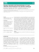
The growth curve of B. subtilis var. natto and sucrose concentration in
media changed during fermentation, as shown in Fig. 1. The microor
ganisms remained in the lag phase for the first 4 h, grew linearly for 6 h,
and declined from the tenth hour until the end of levan production
(Fig. 1). In medium with an initial 250 g/L of sucrose, the substrate was
completely consumed after 14 h of fermentation. The final levan con
centration after drying was estimated at 41.44 g/L. Similar findings
were previously reported using fermentation with the same microor
ganism and initial sucrose levels, reaching 61 g/L at 24 h (Wu, Chou, &
Shih, 2013). In addition, using Bacillus licheniformis NS03, which is of
the same genus, approximately 53 g/L of levan was produced after 48 h
of fermentation (Gojgic-Cvijovic et al., 2019). Taking into account the
total concentration of levan and the duration of fermentation time,
B. subtilis var. natto effectively produced levan in bioreactor.
2.8. Cell proliferation assay
Cellular proliferation rate was measured using a live-cell imaging
system (IncuCyte ZOOM, Essen Bioscience, Birmingham, UK) and the
corresponding software application. Cells (1.0 × 10⁵) were seeded into
96-well plates, and levan treatment was initiated after 24 h, in triplicate.
Levan concentration ranged from 200 μg/mL to 1000 μg/mL, and im
ages were recorded hourly over 1–5 days. Proliferation rate (%) was
determined as cell confluence over time every hour.
2.9. Caspase 3/7 activity assay
Apoptotic processes dependent on caspase 3/7 activation were
evaluated by live-cell imaging (IncuCyte ZOOM®; Essen Bioscience)
using the CellPlayer96-Well Kinetic Caspase-3/7 reagent containing the
caspase substrate DEVD coupled to the DNA intercalating dye NucView
488 (Incucyte Caspase 3/7 Green reagent; Essen Bioscience) at a con
centration of 2 μM, which was added along with cell culture medium to
the different levan test concentrations (200, 600, 800, and 1000 μg/mL)
in 96-well plates. The wells were analyzed until they reached 80%–90%
cell confluence. The number of apoptotic cells per well was determined
in triplicate, as the number of green positive signals (in the green
channel; 488 nm)/mm2 over time.
3.2. Levan molecular weight description
Levan generated in this study showed a bimodal distribution of two
distinct Mw; the larger proportion had a lower Mw (8.8 kDa), and the
smaller portion had a higher Mw (~2201 kDa), and both had a relatively
low polydispersity index (around 1.3) (Supplementary data, Fig. S1;
Supplementary data, Table S1).
Levan's physicochemical characteristics and biological potential are
ă
determined by the microorganism and production conditions (Oner,
´ndez, & Combie, 2016; Tanaka, Oi, & Yamamoto, 1980). A narrow
Herna
polydispersity index is important for the suitability to various applica
tions, and values <2.0 are considered low (Wolff et al., 2000). However,
it is challenging to precisely determine this characteristic, due to the
lack of suitable techniques and standards (Raessler, Wissuwa, Breul,
Unger, & Grimm, 2008) (Barclay, Ginic-markovic, Cooper, & Petrovsky,
2010), and most studied physical parameter (molecular weight) and
correlate to some biological application.
Another aspect to be considered is the enzyme that microorganisms
synthesize levan. Levansucrase enzyme generate levan as a mixture of
low- and high-molecular-weight fractions, making possible a wide range
of molecular weights (Srikanth, Reddy, Siddartha, Ramaiah, & Uppu
luri, 2015). Furthermore, the molecular weight of the enzyme itself can
influence in levan structure and consequently, in its properties.
2.10. Cytotoxicity assay
IncuCyte ZOOM® (Essen Bioscience) was used to measure levan
cytotoxicity, as well as CellPlayer 96-Well Kinetic CellTox™ (Promega,
Madison, USA), containing asymmetric cyanine dye as a fluorescent
marker that enters dead cells and fluoresces upon binding to DNA,
providing a signal that is directly proportional to cytotoxicity. Medium
and dye at a concentration of 2 μM were added at the same time as levan
(200, 600, 800, and 1000 μg/mL). The total number of cytotoxic dead
cells was counted in the green channel (488 nm) over time and calcu
lated as the number of green positive signal/mm2.
2.11. Annexin V apoptosis assay
Apoptosis assays were assessed by FACS Aria III flow cytometry (BD
Biosciences, Heidelberg, Germany) according to the manufacturer's
protocol, using the Annexin V-FITC Apoptosis kit (Thermo-Fisher,
Darmstadt, Germany). SH-SY5Y cells (105 cells per well) were seeded in
24 well plates, in triplicate, and incubated with 1000 μg/mL of levan for
96 h at 37 ◦ C and 5% CO2 (three independent experiments). Thereafter,
the cells were collected and centrifuged (380 ×g for 5 min) at room
temperature, washed three times with PBS, and then resuspended in
binding buffer. The cells were then stained with Annexin V and 7-amino
actinomycin D (7 ADD) as dead cell marker, and incubated for 15 min at
37 ◦ C in the dark. The cells were analyzed by flow cytometry using FITC
and PerCP-Cy5.5 channels, and 3.0 × 10⁴ events were acquired. Late
apoptotic cells were determined by double marking of the
fluorochromes.
2.12. Statistical analysis
Fig. 1. Kinetic and molecular characteristics of levan production by B. subtilis
var. natto. Growth (-■-) and sucrose concentration (-●-) during fermentation.
All experiments were performed in triplicate. Data are expressed as
3
A.M. Vieira et al.
Carbohydrate Polymers 273 (2021) 118613
Levansucrase with low molecular weight has an optimal enzymatic ac
tivity at pH 8.2 and 45 ◦ C (Salama et al., 2019); and these parameters
may not reflect in highest levan yield considering the microorganism
and production conditions (Ua-Arak, Jakob, & Vogel, 2017). Taking this
into account, the levan molecular weight itself will depend on the
microorganism used, the production medium, production conditions
and the specific aspects of the enzyme being produced. All these com
plex factors may not be in harmony, being extremely important to
characterize the molecular weight of levan in each batch (Ua-Arak,
Jakob, & Vogel, 2017). Levan produced by the genus Bacillus essentially
consist of a mix but with high molecular weight predominating (Calaư
zans, Lima, de Franỗa, & Lopes, 2000; Dahech, Belghith, Belghith, &
Mejdoub, 2012; Yoo, Yoon, Cha, & Lee, 2004). In contrast to literature,
the levan produced in the present study by B. subtilis var. natto it was
observed predominance of levan with low molecular weight (Supple
mentary data, Table S1). Also, some authors have reported that high
molecular weights (>500 kDa) are more associated with antitumor acư
tivity (Calazans, Lima, de Franỗa, & Lopes, 2000; Tanaka, Oi, & Yama
moto, 1980). However, the relationship between levan molecular
weight and antineoplastic activity needs further investigation. As well as
considering the complete fermentation process (microorganism,
enzyme, production medium and condition) in able to optimize pro
duction on industrial scale.
3.3. NMR levan characterization
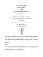
Fig. 2. . 1H–13C correlated NMR spectrum of B. subtilis var. natto levan.
NMR spectroscopy was used to confirm the chemical structural of
levan produced by B. subtilis var. natto. In 13C spectra of levan (Sup
plementary data, Fig. S2), six main peaks are visible (104.2, 80.3, 76.5,
75.3, 63.4, 60.1 ppm) with positions similar reported by most of levan,
despite the producing microorganism, characterized for NMR (Table 1).
The 13C NMR, DEPT 90 and DEPT 135 were performed to report the
structure and their respective signals positions. The absence of an
intense signal, at 104.2 ppm, present in 13C NMR spectrum, but not in
DEPT90 and 135 spectra was evidence for quaternary carbon of fructose
compounds (Supplementary data, Fig. S2) disclosed by other authors
(Cai, Liu, Li, & Lu, 2019; Duymaz et al., 2019; Magri et al., 2020). The
spectrum of 1H–13C HSQC NMR (Fig. 2) showed all expected correla
tions for levan with the correlated signals (Table 2), and these dis
placements are associated with corresponding to the resonance signals
from β-2,6 fructofuranose or levan (Aramsangtienchai, Kongmon,
Pechroj, & Srisook, 2020).
Additionally, the 1H NMR spectrum of levan (Supplementary data,
Fig. S3) showed seven main proton signals in the ring proton region
3.5–4.1 ppm, implying that there are no anomeric protons in this fructan
(4.20 to 5.40 ppm), as described previously (Aramsangtienchai, Kong
mon, Pechroj, & Srisook, 2020). Finally, COSY spectrum, allowed to
visualize the correlation between the vicinal and geminal hydrogens
(Supplementary data, Fig. S4).
Table 1
Chemical shifts in the
13
Table 2
1
H, 13C, HSQC, HMBC and COSY NMR data (δ — ppm; J — Hz) for levan.
H/
C
δ 13C
(ppm)
δ 1H (ppm)
HMBC δ
13
C
3.58 (H-1, d, J = 12.80 Hz);
3.67 (H-1, d, J = 12.80 Hz)
76.5,
104.3
4.09 (H-3, t, J = 17.6 Hz)
4.00 (H-4, t, J = 18,0 Hz)
75.3
63.5, 76.5
1
60.1
2
3
4
104.3
76.5
75.3
5
80.3
3.86 (H-4, t, J = 21,0 Hz)
6
63.4
3.62 (H-6, dt, J = 72.5 Hz, J =
15.5 Hz);
COSY δ
1
H
4.00
4.09,
3.86
4.00,
3.62
s, singlet; d, doublet; t, triplet.
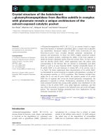
3.4. Effect of levan on proliferation in SH-SY5Ycells
Four different concentrations of levan (200, 600, 800, and 1000 μg/
mL) were used to generate a concentration-effect curve for the effect of
levan on cell proliferation. The proliferation rate (%) was not altered at
any of the concentrations during 96 h of treatment (Fig. 3). In other
studies, levan showed an antiproliferative effect in other cancer cell
lines, none of them related to neuroblastoma (Patel & Agrawal, 2019;
Poli et al., 2009; Sarilmiser & Oner, 2014; Tanaka, Oi, & Yamamoto,
C NMR spectra of levan produced by B. subtilis var. natto compared to other sources.
Chemical shifts (ppm)
Reference
Carbon 2
Carbon 5
Carbon 3
Carbon 4
Carbon 6
Carbon 1
This study
(Gonz´
alez-Garcinu˜
no et al., 2019)
(Duymaz et al., 2019)
(Taylan, Yilmaz, & Dertli, 2019)
(De Vuyst et al., 2014)
(Bounaix et al., 2010)
(Bao et al., 2012)
(Chidambaram et al., 2019)
104.2
104.2
104.0
104.1
104.2
104.7
105.6
104.2
80.3
80.3
80.1
80.1
80.3
80.9
81.7
80.2
76.5
76.3
76.2
76.1
76.3
76.8
77.0
76.3
75.3
75.2
75.0
75.1
75.2
75.8
76.7
na
63.4
63.4
63.3
63.2
63.4
64.0
64.8
63.2
60.1
59.8
59.8
59.7
59.9
60.4
61.4
59.8
na – not available.
4
A.M. Vieira et al.
Carbohydrate Polymers 273 (2021) 118613
3.6. Levan apoptotic effect in SH-SY5Ycells
In order to investigate the role of levan in neoplastic progression, we
used the human neuroblastoma (NB) SH-SY5Y cell line. Neuroblastoma
is aggressive, metastatic, and one of the most common cancers in infants
(Goodman, Gurney, Smith, & Olshan, 1999; Gurney, Smith, & Ross,
1999; Khan, Pandian, Ramraj, Aravindan, & Herman, 2015). SH-SY5Y
cells are regularly used in nervous system research for Parkinson's,
Alzheimer's, Huntington disease, and other neurodegenerative disorders
(Li, Peng, Deng, Li, & Tian, 2020; Rakshit et al., 2020; Schilling et al.,
2019; Yeo et al., 2018). The kinetics of activation of the apoptosis
executor caspase 3/7 was evaluated at different concentrations for 96 h
(Fig. 5). Levan led to a significant increase in the number of cells posi
tively labeled for active caspase3/7 in SH-SY5Y cells after 72 h of
treatment with 1000 μg/mL compared with the untreated control group
(Control 232.25 GOF ± 11.33 vs. 1000 μg/mL Levan-treated 275.36
GOF ± 11.67). To confirm this result, we evaluated apoptotic processes
using flow cytometry in triplicate, with negative and positive controls
(Fig. 6a). We observed that the number of cells treated with 1000 μg/mL
levan at 96 h and labeled with 3/7 caspase was significantly increased
(Control 13.63 GOF ± 1.24 vs. Treated 39.46 GOF ± 3.66) compared to
the control group. The number of double positive apoptotic cells
(Fig. 6b) confirmed that levan induced apoptosis in SH-SY5Y cells,
increasing the number of double-labeled cells in the late stage of
apoptosis. Taking into account molecular weight of levan (around 8.8
kDa), it can be speculated that apoptotic effect on neuroblastoma cells
may be due to increased penetration in the cell by the size of the poly
saccharide. We demonstrated that levan induced apoptosis by the
annexin/7ADD double stain in NB cells and that these cells had a sig
nificant increase in caspase 3/7 activation for up to 72 h. Other authors
found similar results using B. subtilis NRC1aza and Halomonas smyrnensis
AAD6T, but levan's therapeutical application was tested in HepG2 and
human breast cancer (MCF-7) cells, respectively. Levan treatment of
100 μg/mL and 1000 μg/mL, separately, activates caspase 3 in different
cancer lines (Abdel-fattah, Gamal-eldeen, Helmy, & Esawy, 2012a;
Queiroz et al., 2017; Sarilmiser & Oner, 2014). Considering that levan is
a carbohydrate and its specific effect on apoptosis, the mechanism used
to activate this pathway is not yet known. It can be speculated that levan
activates the apoptosis cascade in neuroblastoma cells through an
intermediary, a membrane protein that functions as a levan transporter
or cellular signal transducer (Brodie & Blumberg, 2003; Carneiro & ElDeiry, 2020). Further experiments are needed to support this theory.
Fig. 3. Proliferation rate of SH-SY5Y cells after treatment with different con
centrations of levan. Control group (-●-), 200 μg/mL levan (-■-), 600 μg/mL
levan (-▴-), 800 μg/mL levan (-▾-), and 1000 μg/mL levan (-◆-). Results are
expressed as mean ± SEM.
1980; Yoon, Yoo, Cha, & Lee, 2004). The evaluation in most of the
literature is performed using the MTT of other colorimetric test, while in
the present study, proliferation was evaluated in real time using a livecell analysis system, which is an extremely sensitive method.
3.5. Cytotoxic effect of levan in SH-SY5Ycells
A kinetic concentration-effect curve was developed for levan treat
ment and showed that levan did not exert cytotoxicity at up to 96 h of
treatment (Fig. 4) under any of the experimental conditions. In several
studies, it has been reported that levan exhibit selective cytotoxic ac
tivity, affecting some cell lines more than others, especially in human
hepatocellular carcinoma (HepG2) cells. In those studies, the cytotoxic
effect was quantified through the MTT colorimetric assay (Abdel-fattah,
Gamal-eldeen, Helmy, & Esawy, 2012a; Sarilmiser & Oner, 2014). Be
sides, levan are not cytotoxic when used at 1000 μg/mL against healthy
strains of human fibroblasts, osteoblasts, and murine macrophages
(Domz˙ ał-Kędzia et al., 2019; Gonz´
alez-Garcinu˜
no et al., 2017).
Fig. 5. Kinetic activation of caspase 3/7 in SH-SY5Y cells treated with levan for
up to 96 h. Control group (-●-), 200 μg/mL levan (-■-), 600 μg/mL levan (-▴-),
800 μg/mL levan (-▾-), and 1000 μg/mL levan (-◆-). Caspase 3/7 was analyzed
by the recognition motif (DEVD) to a DNA intercalating dye, and real-time
quantification pictures were captured and analyzed. Data are expressed as the
mean of arbitrary fluorescence units ± SEM. Replicates, n = 3.
Fig. 4. Levan cytotoxic effect on SH-SY5Y cells after treatment with different
concentrations of levan. Control group (-●-), 200 μg/mL levan (-■-), 600 μg/
mL levan (-▴-), 800 μg/mL levan (-▾-), and 1000 μg/mL levan (-◆-). Results are
expressed as mean ± SEM.
5
A.M. Vieira et al.
Carbohydrate Polymers 273 (2021) 118613
Fig. 6. Evaluation of Annexin V/7ADD activation in SH-SY5Y cells. Dot plot of analysis by cytometry (a), bar graph of levan apoptotic activity in SH-SY5Y cells after
96 h of treatment (b). Results are expressed as mean ± SEM (three independent experiments). Statistical analysis: Two-way ANOVA followed by Student's t-test, *p <
0.05 compared with control.
4. Conclusion
Bacillus subtilis NRC1aza. Carbohydrate Polymers, 89(2), 314–322. />10.1016/j.carbpol.2012.02.041
Aramsangtienchai, P., Kongmon, T., Pechroj, S., & Srisook, K. (2020). Enhanced
production and immunomodulatory activity of levan from the acetic acid bacterium,
Tanticharoenia sakaeratensis. International Journal of Biological Macromolecules, 163,
574–581. />Arvidson, S. A., Rinehart, B. T., & Gadala-Maria, F. (2006). Concentration regimes of
solutions of levan polysaccharide from Bacillus sp. Carbohydrate Polymers, 65(2),
144–149. />Bao, Q. H., Liu, W. J., Yu, J., Wang, W. H., Qing, M. J., Chen, X., … Zhang, H. P. (2012).
Isolation and identification of cultivable lactic acid bacteria in traditional yak milk
products of Gansu Province in China. Journal of General and Applied Microbiology, 58
(2), 95–105. />Barclay, T., Ginic-markovic, M., Cooper, P., & Petrovsky, N. (2010). Inulin — A versatile
polysaccharide with multiple pharmaceutical and food chemical uses. Journal of
Excipients and Food Chemistry, 1(3), 27–50.
Biedler, J. L., Roffler-tarlov, S., Schachner, M., & Freedman, L. S. (1978). Multiple
neurotransmitter synthesis by human neuroblastoma cell lines and clones. Cancer
Research, 38(November 1972), 3751–3757.
Bounaix, M. S., Gabriel, V., Robert, H., Morel, S., Remaud-Sim´
eon, M., Gabriel, B., &
Fontagn´e-Faucher, C. (2010). Characterization of glucan-producing Leuconostoc
strains isolated from sourdough. International Journal of Food Microbiology, 144(1),
1–9. />Brodie, C., & Blumberg, P. M. (2003). Regulation of cell apoptosis by protein kinase c δ.
Apoptosis, 8(1), 19–27. />Cabral de Melo, F. C. B., Borsato, D., Macedo, F. C. D., & Celligoi, C. (2015). Study of
levan productivity from Bacillus subtilis Natto by surface response methodology and
its antitumor activity against HepG2 cells using metabolomic approach. Pakistan
Journal of Pharmaceutical Sciences, 28(December), 1917–1926.
Cai, G., Liu, Y., Li, X., & Lu, J. (2019). New levan-type exopolysaccharide from Bacillus
amyloliquefaciens as an antiadhesive agent against enterotoxigenic Escherichia coli.
Journal of Agricultural and Food Chemistry, 67(28), 8029–8034. />10.1021/acs.jafc.9b03234
Calazans, G. M. T., Lima, R. C., de Franỗa, F. P., & Lopes, C. E. (2000). Molecular weight
and antitumour activity of Zymomonas mobilis levans. International Journal of
Biological Macromolecules, 27(4), 245–247. Retrieved from .
nih.gov/pubmed/10921850.
Carneiro, B. A., & El-Deiry, W. S. (2020). Targeting apoptosis in cancer therapy. Nature
Reviews Clinical Oncology, 17(7), 395–417. />Cheung, N. K. V., & Dyer, M. A. (2013). Neuroblastoma: Developmental biology, cancer
genomics and immunotherapy. Nature Reviews Cancer, 13(6), 397–411. https://doi.
org/10.1038/nrc3526
Chidambaram, J. S. C. A., Veerapandian, B., Sarwareddy, K. K., Mani, K. P.,
Shanmugam, S. R., & Venkatachalam, P. (2019). Studies on solvent precipitation of
levan synthesized using Bacillus subtilis MTCC 441. Heliyon, 5(9), Article e02414.
/>da Costa, C. S. (2005). Produỗ
ao de protease por Bacillus firmus via batelada alimentada
utilizando-se perfis constantes e exponencial de alimentaỗ
ao. Universidade Federal de
Pernambuco.
Dahech, I., Belghith, K. S., Belghith, H., & Mejdoub, H. (2012). Partial purification of a
Bacillus licheniformis levansucrase producing levan with antitumor activity.
International Journal of Biological Macromolecules, 51(3), 329–335. />10.1016/j.ijbiomac.2012.04.030
De Vuyst, L., Van Kerrebroeck, S., Harth, H., Huys, G., Daniel, H. M., & Weckx, S. (2014).
Microbial ecology of sourdough fermentations: Diverse or uniform? Food
Microbiology, 37, 11–29. />Dom˙zał-Kędzia, M., Lewi´
nska, A., Jaromin, A., Weselski, M., Pluskota, R., &
Łukaszewicz, M. (2019). Fermentation parameters and conditions affecting levan
In the current study, levan was produced by B. subtilis var. natto, and
41.44 g/L of yield was obtained after purification, consisting of mostly
low molecular weight units. Additionally, our results indicated that
treatment of a neuroblastoma cell line (SH-SY5Y) with 1000 μg/mL of
levan exhibited no effect on proliferation and lacked the cytotoxicity.
However, levan showed apoptotic effect in neuroblastoma cells, by
caspase 3/7 activation which increases over time. Nonetheless, further
investigations on purification parameters, application in other
neoplastic cell lines with description of the action mechanism of levan
are needed, together with in vivo testing.
CRediT authorship contribution statement
Amanda Mota Vieira: Conceptualization, Methodology, Formal
analysis, Investigation, Data curation, Writing – original draft, Writing –
review & editing. Farrah Zahed: Conceptualization, Methodology,
Formal analysis, Investigation. Alessandre Carmo Crispim: Formal
analysis, Investigation, Writing – original draft. Edson de Souza Bento:
Supervision, Formal analysis, Investigation, Writing original draft.
Rafael de Freitas Oliveira Franỗa: Supervision. Irapuan Oliveira
Pinheiro: Supervision. Luis A. Pardo: Supervision, Writing – original
draft. Bruno Melo Carvalho: Conceptualization, Formal analysis,
Writing – original draft, Writing – review & editing, Supervision, Project
administration.
Acknowledgments
o de Aperfeiỗoaư
This study was financed in part by the Coordenaỗa
mento de Pessoal de Nớvel Superior - Brasil (CAPES) - Finance Code 001
and PDSE scholarship - 88881.132835/2016-01 (AMV). The authors
` Ciˆencia e Tecnologia do Estado de
also thank the Fundaỗ
ao de Amparo a
Pernambuco (FACEPE) grant APQ-0411-2.07/18 and Conselho
´gico (CNPq) for
Nacional de Desenvolvimento Científico e Tecnolo
financial support of this work.
Appendix A. Supplementary data
Supplementary data to this article can be found online at https://doi.
org/10.1016/j.carbpol.2021.118613.
References
Abdel-fattah, A. M., Gamal-eldeen, A. M., Helmy, W. A., & Esawy, M. A. (2012a).
Antitumor and antioxidant activities of levan and its derivative from the isolate
6
A.M. Vieira et al.
Carbohydrate Polymers 273 (2021) 118613
International Journal of Biological Macromolecules, 102, 565–570. />10.1016/j.ijbiomac.2017.04.035
Raessler, M., Wissuwa, B., Breul, A., Unger, W., & Grimm, T. (2008). Determination of
water-extractable nonstructural carbohydrates, including inulin, in grass samples
with high-performance anion exchange chromatography and pulsed amperometric
detection. Journal of Agriculture and Food Chemistry, 56, 7649–7654. />10.1021/jf800973v
Ragab, T. I. M., Malek, R. A., Elsehemy, I. A., Farag, M. M. S., Salama, B. M., Abd ELBaseer, M. A., … Esawy, M. A. (2019). Scaling up of levan yield in Bacillus subtilis M
and cytotoxicity study on levan and its derivatives. Journal of Bioscience and
Bioengineering, 127(6), 655–662. />Rakshit, J., Mallick, A., Roy, S., Sarbajna, A., Dutta, M., & Bandyopadhyay, J. (2020).
Iron-induced apoptotic cell death and autophagy dysfunction in human
neuroblastoma cell line SH-SY5Y. Biological Trace Element Research, 193, 138–151.
Runyon, J. R., Nilsson, L., Ulmius, M., & Castro, A. (2014). Characterizing changes in
levan physicochemical properties in different pH environments using asymmetric
flow field-flow fractionation. Analytical and Bioanalytical Chemistry, 406, 1597–1605.
/>Salama, B. M., Helmy, W. A., Ragab, T. I. M., Ali, M. M., Taie, H. A. A., & Esawy, M. A.
(2019). Characterization of a new efficient low molecular weight Bacillus subtilis
NRC 16 levansucrase and its levan. Journal of Basic Microbiology, 59(10), 1004–1015.
/>Sarilmiser, H. K., & Oner, E. T. (2014). Investigation of anti-cancer activity of linear and
aldehyde-activated levan from Halomonas smyrnensis AAD6 T. Biochemical
Engineering Journal, 92, 28–34. />Schilling, J., Broemer, M., Atanassov, I., Duernberger, Y., Vorberg, I., Dieterich, C., …
Krauß, S. (2019). Deregulated splicing is a major mechanism of RNA-induced
toxicity in Huntington’s disease. Journal of Molecular Biology, 431(9), 1869–1877.
/>Srikanth, R., Reddy, C. H. S. S. S., Siddartha, G., Ramaiah, M. J., & Uppuluri, K. B.
(2015). Review on production, characterization and applications of microbial levan.
Carbohydrate Polymers, 120, 102–114. />carbpol.2014.12.003
Steliarova-foucher, E., Colombet, M., Ries, L. A. G., Moreno, F., Dolya, A., Bray, F., …
Shin, H. Y. (2017). Articles international incidence of childhood cancer, 2001–10: A
population-based registry study. Lancet Oncology, 18, 719–731. />10.1016/S1470-2045(17)30186-9
Tanaka, T., Oi, S., & Yamamoto, T. (1980). The molecular structure of low and high
molecular weight levans synthesized by levansucrase. Journal of Biochemistry, 87(1),
297–303.
Taylan, O., Yilmaz, M. T., & Dertli, E. (2019). Partial characterization of a levan type
exopolysaccharide (EPS) produced by Leuconostoc mesenteroides showing
immunostimulatory and antioxidant activities. International Journal of Biological
Macromolecules, 136, 436–444. />Tibullo, D., Giallongo, C., Puglisi, F., Tomassoni, D., Camiolo, G., Cristaldi, M., …
Bramanti, V. (2018). Effect of lipoic acid on the biochemical mechanisms of
resistance to bortezomib in SH-SY5Y neuroblastoma cells. Molecular Neurobiology,
55, 3344–3350. />Ua-Arak, T., Jakob, F., & Vogel, R. F. (2017). Fermentation pH modulates the size
distributions and functional properties of Gluconobacter albidus TMW 2.1191 levan.
Frontiers in Microbiology, 8(MAY), 1–11. />Veerapandian, B., Ramiah, S., & Varadhan, S. (2020). Levan production from sucrose
using chicken feather peptone as a low cost supplemental nutrient source.
Carbohydrate Polymers, 227(August 2019), Article 115361. />j.carbpol.2019.115361
Wolff, D., Czapla, S., Heyer, A. G., Radosta, S., Mischnick, P., & Springer, J. (2000).
Globular shape of high molar mass inulin revealed by static light scattering and
viscometry. Polymer, 41, 8009–8016.
World Health Organization. (2020). Cancer. Retrieved May 3, 2021, from https://www.
who.int/news-room/fact-sheets/detail/cancer.
Wu, F.-C., Chou, S.-Z., & Shih, I.-L. (2013). Factors affecting the production and
molecular weight of levan of Bacillus subtilis natto in batch and fed-batch culture in
fermenter. Journal of the Taiwan Institute of Chemical Engineers, 44(6), 846–853.
/>Yeo, S., Sung, B., Hong, Y., Van Den Noort, M., Bosch, P., Lee, S., … Lim, S. (2018).
Decreased expression of serum- and glucocorticoid-inducible kinase 1 (SGK1)
promotes alpha-synuclein increase related with down-regulation of dopaminergic
cell in the Substantia Nigra of chronic MPTP-induced Parkinsonism mice and in SHSY5Y cells. Gene, 1(2017), 1–21. />Yoo, S. H., Yoon, E. J., Cha, J., & Lee, H. G. (2004). Antitumor activity of levan
polysaccharides from selected microorganisms. International Journal of Biological
Macromolecules, 34, 37–41. />Yoon, E. J., Yoo, S. H., Cha, J., & Lee, H. G. (2004). Effect of levan’s branching structure
on antitumor activity. International Journal of Biological Macromolecules, 34(3),
191–194. />
production and its potential applications in cosmetics Marta. Bioorganic Chemistry
Journal, 93, Article 102787. />Duymaz, B. T., Erdiler, F. B., Alan, T., Aydogdu, M. O., Inan, A. T., Ekren, N., …
Gunduz, O. (2019). 3D bio-printing of levan/polycaprolactone/gelatin blends for
bone tissue engineering: Characterization of the cellular behavior. European Polymer
Journal, 119(August), 426–437. />Esawy, M. A., Abdel-fattah, A. M., Ali, M. M., Helmy, W. A., Salama, B. M., Taie, H. A. A.,
… Awad, G. E. A. (2013). Levansucrase optimization using solid state fermentation
and levan biological activities studies. Carbohydrate Polymers, 96(1), 332–341.
/>Gojgic-Cvijovic, G. D., Jakovljevic, D. M., Loncarevic, B. D., Todorovic, N. M.,
Pergal, M. V., Ciric, J., … Vrvic, M. M. (2019). Production of levan by Bacillus
licheniformis NS032 in sugar beet molasses-based medium. International Journal of
Biological Macromolecules, 121, 142–151. />ijbiomac.2018.10.019
´ Tabernero, A., Domínguez, A.,
´ Miguel, A., Martin, E. M.,
Gonz´
alez-garcinu˜
no, A.,
Tabernero, A., … Martin, E. M. (2017). Levan and levansucrases: Polymer, enzyme,
micro- organisms and biomedical applications. Biocatalysis and Biotransformation,
2422(April). />´ Tabernero, A., Marcelo, G., Sebasti´
Gonz´
alez-Garcinu˜
no, A.,
an, V., Arruebo, M.,
Santamaría, J., & Martín del Valle, E. (2019). Differences in levan nanoparticles
depending on their synthesis route: Microbial vs cell-free systems. International
Journal of Biological Macromolecules, 137, 62–68. />ijbiomac.2019.06.128
Goodman, M. T., Gurney, G. J., Smith, M. A., & Olshan, A. (1999). Sympathetic nervous
system tumors. In Cancer incidence and survival among children and adolescents: United
States SEER program 1975–1995 (pp. 65–72). National Cancer Institute.
Gurney, J., Smith, M., & Ross, J. (1999). Cancer among infants. In Cancer incidence and
survival among children and adolescents: United States SEER program 19751995 (pp.
149156). National Cancer Institute.
Hă
ovels, M., Kosciow, K., Kniewel, J., Jakob, F., & Deppenmeier, U. (2020). High yield
production of Levan-type fructans by Gluconobacter japonicus LMG 1417.
International Journal of Biological Macromolecules, 164, 295–303. />10.1016/j.ijbiomac.2020.07.105
Hundschell, C. S., Jakob, F., & Wagemans, A. M. (2020). Molecular weight dependent
structure of the exopolysaccharide levan. International Journal of Biological
Macromolecules, 161, 398–405. />Kågedal, B. (2009). Detecting minimal residual disease in neuroblastoma: Still a ways to
go. Clinical Chemistry, 55(7), 1268–1270. />clinchem.2009.127308
Khan, F. H., Pandian, V., Ramraj, S., Aravindan, S., & Herman, T. S. (2015).
Reorganization of metastamiRs in the evolution of metastatic aggressive
neuroblastoma cells. BMC Genomics, 1–17. />Li, L. H., Peng, W. N., Deng, Y., Li, J. J., & Tian, X. R. (2020). Action of trichostatin A on
Alzheimer’s disease-like pathological changes in SH-SY5Y neuroblastoma cells.
Neural Regeneration Research, 15(2), 293–301. />Magri, A., Oliveira, M. R., Baldo, C., Tischer, C. A., Sartori, D., Mantovani, M. S., &
Celligoi, M. A. P. C. (2020). Production of fructooligosaccharides by Bacillus subtilis
natto CCT7712 and their antiproliferative potential. Journal of Applied Microbiology,
128(5), 1414–1426. />Moussa, T. A. A., Al-qaysi, S. A. S., Thabit, Z. A., & Kadhem, S. B. (2017). Microbial levan
from Brachybacterium phenoliresistens: Characterization and enhancement of
production. Process Biochemistry, 57, 9–15. />procbio.2017.03.008
Nasir, A., Sattar, F., Ashfaq, I., Lindemann, S. R., Chen, M., Ende, W., … Anwar. (2020).
Production and characterization of a high molecular weight levan and
fructooligosaccharides from a rhizospheric isolate of Bacillus aryabhattai. LWT - Food
Science and Technology, 123(October 2019), 18. />lwt.2020.109093
ă
Oner,
E. T., Hern´
andez, L., & Combie, J. (2016). Review of Levan polysaccharide: From a
century of past experiences to future prospects. Biotechnology Advances.. https://doi.
org/10.1016/j.biotechadv.2016.05.002
Patel, P., & Agrawal, Y. (2019). Preparation and in-vitro evaluation of levan micelles: A
polyfructan based nano-carrier for breast cancer targeted delivery. In Drug delivery
letters (pp. 97107). Bentham Science Publishers.
ă
Poli, A., Kazak, H., Gürleyendaǧ, B., Tommonaro, G., Pieretti, G., Oner,
E. T., &
Nicolaus, B. (2009). High level synthesis of levan by a novel Halomonas species
growing on defined media. Carbohydrate Polymers, 78(4), 651–657. />10.1016/j.carbpol.2009.05.031
Queiroz, E. A. I. F., Fortes, Z. B., da Cunha, M. A. A., Sarilmiser, H. K., Barbosa
ă
Dekker, A. M., Oner,
E. T., … Khaper, N. (2017). Levan promotes antiproliferative
and pro-apoptotic effects in MCF-7 breast cancer cells mediated by oxidative stress.
7