5 nội soi chẩn đoán và điều trị XHTH trên không do giãn vỡ TMTQ ESGE 2021
Bạn đang xem bản rút gọn của tài liệu. Xem và tải ngay bản đầy đủ của tài liệu tại đây (1.7 MB, 33 trang )
Published online: 2021-02-10
Guidelines
Authors
Ian M. Gralnek 1, 2, Adrian J. Stanley3, A. John Morris 3, Marine Camus 4, James Lau 5, Angel Lanas 6, Stig B. Laursen7 ,
Franco Radaelli8, Ioannis S. Papanikolaou 9, Tiago Cỳrdia Gonỗalves10, 11, 12, Mario Dinis-Ribeiro13, 14, Halim Awadie 1 ,
Georg Braun15, Nicolette de Groot16, Marianne Udd 17, Andres Sanchez-Yague 18, 19, Ziv Neeman2, 20, Jeanin E. van
Hooft 21
Institutions
1 Institute of Gastroenterology and Hepatology, Emek
Medical Center, Afula, Israel
2 Rappaport Faculty of Medicine, Technion-Israel
Institute of Technology, Haifa, Israel
3 Department of Gastroenterology, Glasgow Royal
Infirmary, Glasgow, UK
4 Sorbonne University, Endoscopic Unit, Saint Antoine
Hospital Assistance Publique Hopitaux de Paris, Paris,
France
5 Department of Surgery, Prince of Wales Hospital, The
Chinese University of Hong Kong, Hong Kong SAR,
China
6 Digestive Disease Services, University Clinic Hospital,
University of Zaragoza, IIS Aragón (CIBERehd), Spain
7 Department of Gastroenterology, Odense University
Hospital, Odense, Denmark
8 Department of Gastroenterology, Valduce Hospital,
Como, Italy
9 Hepatogastroenterology Unit, Second Department of
Internal Medicine – Propaedeutic, Medical School,
National and Kapodistrian University of Athens, Attikon
University General Hospital, Athens, Greece
10 Gastroenterology Department, Hospital da Senhora da
Oliveira, Guimarães, Portugal
11 School of Medicine, University of Minho, Braga/
Guimarães, Portugal
12 ICVS/3B’s–PT Government Associate Laboratory,
Braga/Guimarães, Portugal
13 Center for Research in Health Technologies and
Information Systems (CINTESIS), Faculty of Medicine,
Porto, Portugal
14 Gastroenterology Department, Portuguese Oncology
Institute of Porto, Portugal
15 Medizinische Klinik 3, Universitätsklinikum Augsburg,
Augsburg, Germany.
16 Red Cross Hospital Beverwijk, Beverwijk, The
Netherlands
300
17 Gastroenterological Surgery, University of Helsinki and
Helsinki University Hospital, Helsinki, Finland
18 Gastroenterology Unit, Hospital Costa del Sol,
Marbella, Spain
19 Gastroenterology Department, Vithas Xanit
International Hospital, Benalmadena, Spain
20 Diagnostic Imaging and Nuclear Medicine Institute,
Emek Medical Center, Afula, Israel
21 Department of Gastroenterology and Hepatology,
Leiden University Medical Center, Leiden, The
Netherlands
published online 10.2.2021
Bibliography
Endoscopy 2021; 53: 300–332
DOI 10.1055/a-1369-5274
ISSN 0013-726X
© 2021. European Society of Gastrointestinal Endoscopy
All rights reserved.
This article ist published by Thieme.
Georg Thieme Verlag KG, Rüdigerstraße 14,
70469 Stuttgart, Germany
Supplementary material
Supplementary material is available under
/>
Corresponding author
Ian M. Gralnek, MD MSHS, Rappaport Faculty of Medicine,
Technion-Israel Institute of Technology, Institute of
Gastroenterology and Hepatology, Emek Medical Center,
Afula, Israel 18101
Gralnek Ian M et al. Endoscopic diagnosis and … Endoscopy 2021; 53: 300–332 | © 2021. European Society of Gastrointestinal Endoscopy. All rights reserved.
This document was downloaded for personal use only. Unauthorized distribution is strictly prohibited.
Endoscopic diagnosis and management of nonvariceal upper
gastrointestinal hemorrhage (NVUGIH): European Society
of Gastrointestinal Endoscopy (ESGE) Guideline – Update 2021
MAIN RECO MMENDAT IONS
7 ESGE suggests that in patients with persistent bleeding
1 ESGE recommends in patients with acute upper gastro-
refractory to standard hemostasis modalities, the use of a
topical hemostatic spray/powder or cap-mounted clip
should be considered.
Weak recommendation, low quality evidence.
2 ESGE recommends that in patients with acute UGIH who
are taking low-dose aspirin as monotherapy for secondary
cardiovascular prophylaxis, aspirin should not be interrupted. If for any reason it is interrupted, aspirin should be restarted as soon as possible, preferably within 3–5 days.
Strong recommendation, moderate quality evidence.
8 ESGE recommends that for patients with clinical evidence
of recurrent peptic ulcer hemorrhage, use of a cap-mounted
clip should be considered. In the case of failure of this second
attempt at endoscopic hemostasis, transcatheter angiographic embolization (TAE) should be considered. Surgery
is indicated when TAE is not locally available or after failed
TAE.
Strong recommendation, moderate quality evidence.
9 ESGE recommends high dose proton pump inhibitor (PPI)
endoscopy since as compared to early endoscopy, patient
outcomes are not improved.
Strong recommendation, high quality evidence.
therapy for patients who receive endoscopic hemostasis
and for patients with FIIb ulcer stigmata (adherent clot)
not treated endoscopically.
(a) PPI therapy should be administered as an intravenous
bolus followed by continuous infusion (e. g., 80 mg then
8 mg/hour) for 72 hours post endoscopy.
(b) High dose PPI therapies given as intravenous bolus dosing (twice-daily) or in oral formulation (twice-daily) can be
considered as alternative regimens.
Strong recommendation, high quality evidence.
5 ESGE recommends for patients with actively bleeding ul-
10 ESGE recommends that in patients who require ongoing
3 ESGE recommends that following hemodynamic resuscitation, early (≤ 24 hours) upper gastrointestinal (GI) endoscopy should be performed.
Strong recommendation, high quality evidence.
4 ESGE does not recommend urgent (≤ 12 hours) upper GI
cers (FIa, FIb), combination therapy using epinephrine injection plus a second hemostasis modality (contact thermal
or mechanical therapy).
Strong recommendation, high quality evidence.
6 ESGE recommends for patients with an ulcer with a nonbleeding visible vessel (FIIa), contact or noncontact thermal
therapy, mechanical therapy, or injection of a sclerosing
agent, each as monotherapy or in combination with epinephrine injection.
Strong recommendation, high quality evidence.
SOURCE AND SCOPE
This Guideline is an official statement from the European
Society of Gastrointestinal Endoscopy (ESGE). It is an update of the previously published 2015 ESGE Clinical
Guideline addressing the role of gastrointestinal endoscopy in the diagnosis and management of acute nonvariceal upper gastrointestinal hemorrhage (NVUGIH). The
evidence statements and recommendations specifically
pertaining to endoscopic hemostasis therapies are limited to peptic ulcer hemorrhage. Endoscopic hemostasis
therapy recommendations for nonulcer NVUGIH etiologies, can be found in the 2015 ESGE Guideline.
This document was downloaded for personal use only. Unauthorized distribution is strictly prohibited.
intestinal hemorrhage (UGIH) the use of the Glasgow–
Blatchford Score (GBS) for pre-endoscopy risk stratification.
Patients with GBS ≤ 1 are at very low risk of rebleeding, mortality within 30 days, or needing hospital-based intervention and can be safely managed as outpatients with outpatient endoscopy.
Strong recommendation, moderate quality evidence.
anticoagulation therapy following acute NVUGIH (e. g.,
peptic ulcer hemorrhage), anticoagulation should be resumed as soon as the bleeding has been controlled, preferably within or soon after 7 days of the bleeding event, based
on thromboembolic risk. The rapid onset of action of direct
oral anticoagulants (DOACS), as compared to vitamin K antagonists (VKAs), must be considered in this context.
Strong recommendation, low quality evidence.
Introduction
The most common causes of acute upper gastrointestinal hemorrhage (UGIH) are nonvariceal. These include gastric and
duodenal peptic ulcers, mucosal erosive disease of the esophagus/stomach/duodenum, malignancy, Mallory–Weiss syndrome, Dieulafoy lesion, “other” diagnosis, or no identifiable
cause [1]. This ESGE Guideline focuses on the pre-endoscopic,
endoscopic, and post-endoscopic management of patients presenting with acute nonvariceal upper gastrointestinal hemorrhage (NVUGIH), specifically peptic ulcer hemorrhage.
Gralnek Ian M et al. Endoscopic diagnosis and … Endoscopy 2021; 53: 300–332 | © 2021. European Society of Gastrointestinal Endoscopy. All rights reserved.
301
Guidelines
APA
APC
ASA
AUROC
DAPT
CHADS2
CI
DOAC
ESGE
FFP
GBS
GI
GRADE
HR
ICU
INR
IRR
NBVV
antiplatelet agent
argon plasma coagulation
American Society of Anesthesiologists
area under receiver operating characteristic
dual antiplatelet therapy
congestive heart failure, hypertension, age
≥ 75 years, diabetes mellitus, and previous
stroke or transient ischemic attack [risk score]
confidence interval
direct oral anticoagulant
European Society of Gastrointestinal
Endoscopy
fresh frozen plasma
Glasgow–Blatchford Score
gastrointestinal
Grading of Recommendations Assessment,
Development and Evaluation
hazard ratio
intensive care unit
international normalized ratio
incident rate ratio
nonbleeding visible vessel
Methods
ESGE commissioned this Guideline (ESGE Guideline Committee
chair, J.V.H.) and appointed a guideline leader (I.M.G.). The
guideline leader established four task forces based on the statements of the previous 2015 Guideline [2], each with its own leader (M.C., A.J.S., J.M., J.L.).
Key questions (Table 1 s, see online-only in Supplementary
material) were prepared by the coordinating team (I.M.G., M.
C., A.S., J.M., J.L.) according to the PICO format (patients, interventions, controls, outcomes) and divided amongst the four
task forces. Given this is an update of the 2015 ESGE Clinical
Guideline on NVUGIH, each task force performed a structured
systematic literature search using key words (Table 2 s) in
English-language articles limited from January 1, 2014 to January 31, 2020, in Ovid MEDLINE, Embase, Google Scholar, and
the Cochrane Database of Systematic Reviews. Additional topic-specific searches on timing of endoscopy and role of capmounted clips for hemostasis in peptic ulcer hemorrhage were
conducted up to August 31, 2020. The hierarchy of studies included in this evidence-based guideline was, in decreasing order of evidence level, published systematic reviews/metaanalyses, randomized controlled trials (RCTs), prospective and
retrospective observational studies, and case series. New evidence on each key question was summarized in evidence tables
(Table 3 s), using the Grading of Recommendations Assessment, Development and Evaluation (GRADE) system [3]. Grading of the evidence depends on the balance between the benefits and risk or burden of any health intervention. Further
details on ESGE guideline development have been previously
reported [4].
302
NGT
nasogastric tube
NNT
number needed to treat
NVUGIH nonvariceal upper gastrointestinal
hemorrhage
OR
odds ratio
OTS
over-the-scope
PCC
prothrombin complex concentrate
PCI
percutaneous coronary intervention
PICO
patients, interventions, controls, outcomes
PNED
Progetto Nazionale Emorragia Digestive
PPI
proton pump inhibitor
PUB
peptic ulcer bleeding
RBC
red blood cell
RCT
randomized controlled trial
RD
risk difference
RR
relative risk or risk ratio
TAE
transcatheter angiographic embolization
TTS
through-the-scope
TXA
tranexamic acid
UGIH
upper gastrointestinal hemorrhage
VKA
vitamin K antagonist
The results of the literature search and answers to PICO
questions were presented to all guideline group members during two online face-to-face meetings conducted on June 27 and
28, 2020. Subsequently, drafts were made by each task force
leader and distributed between the task force members for revision and online discussion. In September 2020, a draft prepared by I.M.G. and the four task force leaders was sent to all
guideline group members. After agreement of all members
was obtained, the manuscript was reviewed by two independent external reviewers. The manuscript was then sent for further comments to the 49 ESGE member societies and individual
members. It was then submitted to the journal Endoscopy for
publication. The final revised manuscript was agreed upon by
all the authors. This ESGE Guideline was issued in 2021 and will
be considered for update in 2025. Any interim updates will be
noted on the ESGE website: />
Evidence statements and Recommendations
Evidence statements and Recommendations are grouped according to the different task force topics: pre-endoscopy management (task forces 1 and 2), intraendoscopy management
(task force 3), and postendoscopy management (task force 4).
Each statement is followed by the strength of evidence based
on GRADE and the discussion of the evidence that occurred
during the two 3-hour online face-to-face meetings. ▶ Table 1
summarizes all recommendations in this updated guideline.
Gralnek Ian M et al. Endoscopic diagnosis and … Endoscopy 2021; 53: 300–332 | © 2021. European Society of Gastrointestinal Endoscopy. All rights reserved.
This document was downloaded for personal use only. Unauthorized distribution is strictly prohibited.
A BB R E VI AT I ONS
▶ Table 1 Summary of Guideline statements and recommendations.
Pre-endoscopy management
Initial patient evaluation and hemodynamic resuscitation
1
ESGE recommends immediate assessment of hemodynamic status in patients who present with acute upper gastrointestinal hemorrhage
(UGIH), with prompt intravascular volume replacement initially using crystalloid fluids if hemodynamic instability exists.
Strong recommendation, low quality evidence.
2
ESGE recommends, in hemodynamically stable patients with acute UGIH and no history of cardiovascular disease, a restrictive RBC transfusion strategy with a hemoglobin threshold of ≤ 7 g/dL prompting RBC transfusion. A post-transfusion target hemoglobin concentration of
7–9 g/dL is desired.
Strong recommendation, moderate quality evidence.
3
ESGE recommends in hemodynamically stable patients with acute UGIH and a history of acute or chronic cardiovascular disease, a more
liberal RBC transfusion strategy with a hemoglobin threshold of ≤ 8 g/dL prompting RBC transfusion. A post transfusion target hemoglobin
concentration of ≥ 10 g/dL is desired.
Strong recommendation, low quality evidence.
Patient risk stratification
4
ESGE recommends in patients with acute UGIH the use of the Glasgow–Blatchford Score (GBS) for pre-endoscopy risk stratification. Patients
with GBS ≤ 1 are at very low risk of rebleeding, mortality within 30 days, or needing hospital-based intervention and can be safely managed as
outpatients with outpatient endoscopy.
Strong recommendation, moderate quality evidence.
Management of antithrombotic agents (antiplatelet agents and anticoagulants)
5
ESGE recommends that in patients with acute UGIH who are taking low dose aspirin as monotherapy for primary cardiovascular prophylaxis,
aspirin should be temporarily interrupted. Aspirin can be re-started after careful re-evaluation of its clinical indication.
Strong recommendation, low quality evidence.
6
ESGE recommends that in patients with acute UGIH who are taking low dose aspirin as monotherapy for secondary cardiovascular
prophylaxis, aspirin should not be interrupted. If for any reason it is interrupted, aspirin should be re-started as soon as possible, preferably
within 3–5 days.
Strong recommendation, moderate quality evidence.
7
ESGE recommends that in patients with acute UGIH who are taking dual antiplatelet therapy (DAPT) for secondary cardiovascular
prophylaxis, aspirin should not be interrupted. The second antiplatelet agent should be interrupted, but re-started as soon as possible,
preferably within 5 days. Cardiology consultation is suggested.
Strong recommendation, low quality evidence.
8
ESGE does not recommend routine platelet transfusion for patients with acute NVUGIH who are taking antiplatelet agents.
Strong recommendation, low quality evidence.
9
ESGE does not recommend the use of tranexamic acid in patients with acute NVUGIH.
Strong recommendation, high quality evidence.
10
ESGE recommends that in patients with acute UGIH taking vitamin K antagonists (VKAs), that the anticoagulant be withheld.
Strong recommendation, low quality evidence
11
ESGE recommends that in patients with acute UGIH taking vitamin K antagonists (VKAs) who have hemodynamic instability, low dose vitamin
K supplemented with intravenous prothrombin complex concentrate (PCC), or fresh frozen plasma (FFP) if PCC is not available, should be
administered. However, this should not delay endoscopy or if required, endoscopic hemostasis.
Strong recommendation, low quality evidence.
12
ESGE recommends that in patients with acute UGIH taking direct oral anticoagulants (DOAC), the anticoagulant should be withheld and
endoscopy not delayed. In patients with severe ongoing bleeding, use of a DOAC reversal agent or intravenous PCC should be considered.
Strong recommendation, low quality evidence.
Proton pump inhibitor (PPI) therapy
13
ESGE suggests that pre-endoscopy high dose intravenous proton pump inhibitor (PPI) therapy be considered in patients presenting with
acute UGIH, to downstage endoscopic stigmata and thereby reduce the need for endoscopic therapy; however, this should not delay early
endoscopy.
Weak recommendation, high quality evidence.
Gralnek Ian M et al. Endoscopic diagnosis and … Endoscopy 2021; 53: 300–332 | © 2021. European Society of Gastrointestinal Endoscopy. All rights reserved.
303
This document was downloaded for personal use only. Unauthorized distribution is strictly prohibited.
Red blood cell (RBC) transfusion strategy
Guidelines
Somatostatin and somatostatin analogues
14
ESGE does not recommend the use of somatostatin, or its analogue octreotide, in patients with NVUGIH.
Strong recommendation, low quality evidence.
Nasogastric/orogastric tube aspiration and lavage
15
ESGE does not recommend the routine use of nasogastric or orogastric aspiration/lavage in patients presenting with acute UGIH.
Strong recommendation, moderate quality evidence.
16
ESGE does not recommend routine prophylactic endotracheal intubation for airway protection prior to upper endoscopy in patients with
acute UGIH.
Strong recommendation, high quality evidence.
17
ESGE recommends prophylactic endotracheal intubation for airway protection prior to upper endoscopy only in selected patients with acute
UGIH (i. e., those with ongoing active hematemesis, agitation, or encephalopathy with inability to adequately control the airway).
Strong recommendation, low quality evidence.
Prokinetic medications
18
ESGE recommends pre-endoscopy administration of intravenous erythromycin in selected patients with clinically severe or ongoing active
UGIH.
Strong recommendation, high quality evidence.
Endoscopic management
Timing of upper GI endoscopy
1
ESGE recommends adopting the following definitions regarding the timing of upper GI endoscopy in acute UGIH relative to the time of
patient presentation: urgent ≤ 12 hours, early ≤ 24 hours, and delayed > 24 hours.
Strong recommendation, moderate quality evidence.
2
ESGE recommends that following hemodynamic resuscitation, early (≤ 24 hours) upper GI endoscopy should be performed.
Strong recommendation, high quality evidence.
3
ESGE does not recommend urgent (≤ 12 hours) upper GI endoscopy since as compared to early endoscopy, patient outcomes are not
improved.
Strong recommendation, high quality evidence.
4
ESGE does not recommend emergent (≤ 6 hours) upper GI endoscopy since this may be associated with worse patient outcomes.
Strong recommendation, moderate quality evidence.
5
ESGE recommends that the use of antiplatelet agents, anticoagulants, or a predetermined international normalized ratio (INR) cutoff level,
should not be used to define or guide the timing of upper GI endoscopy in patients with acute UGIH.
Strong recommendation, low quality evidence.
On-call GI endoscopy resources
6
ESGE recommends the availability of both an on-call GI endoscopist proficient in endoscopic hemostasis and on-call nursing staff with
technical expertise in the use of endoscopic devices, to allow performance of endoscopy on a 24/7 basis.
Strong recommendation, low quality evidence.
Endoscopic diagnosis
7
ESGE recommends the Forrest (F) classification be used in all patients with peptic ulcer hemorrhage to differentiate low risk and high risk
endoscopic stigmata.
Strong recommendation, high quality evidence.
8
ESGE recommends that peptic ulcers with spurting or oozing bleeding (FIa and FIb respectively) or with a nonbleeding visible vessel (FIIa)
receive endoscopic hemostasis because these lesions are at high risk for persistent bleeding or recurrent bleeding.
Strong recommendation, high quality evidence.
9
ESGE suggests that peptic ulcers with an adherent clot (FIIb) be considered for endoscopic clot removal. Once the clot is removed, any
identified underlying active bleeding (FIa or FIb) or nonbleeding visible vessel (FIIa) should receive endoscopic hemostasis.
Weak recommendation, moderate quality evidence.
10
ESGE does not recommend endoscopic hemostasis in patients with peptic ulcers having a flat pigmented spot (FIIc) or clean base (FIII), as
these stigmata have a low risk of adverse outcomes. In selected clinical settings these patients may have expedited hospital discharge.
Strong recommendation, moderate quality evidence.
11
ESGE does not recommend the routine use of Doppler endoscopic probe in the evaluation of endoscopic stigmata of peptic ulcer bleeding.
Strong recommendation, low quality evidence.
304
Gralnek Ian M et al. Endoscopic diagnosis and … Endoscopy 2021; 53: 300–332 | © 2021. European Society of Gastrointestinal Endoscopy. All rights reserved.
This document was downloaded for personal use only. Unauthorized distribution is strictly prohibited.
Endotracheal intubation
12
ESGE does not recommend the routine use of capsule endoscopy technology in the evaluation of acute UGIH.
Strong recommendation, low quality evidence.
13
FIa, FIb (active bleeding)
(a) ESGE recommends for patients with actively bleeding ulcers (FIa, FIb), combination therapy using epinephrine injection plus a second
hemostasis modality (contact thermal or mechanical therapy).
Strong recommendation, high quality evidence.
(b) ESGE suggests that in selected actively bleeding ulcers (FIa,FIb), specifically those > 2 cm in size, with a large visible vessel > 2 mm, or
located in a high-risk vascular area (e. g., gastroduodenal, left gastric arteries), or in excavated/fibrotic ulcers, endoscopic hemostasis using a
cap-mounted clip should be considered as first-line therapy.
Weak recommendation, low quality evidence.
14
FIIa (nonbleeding visible vessel)
ESGE recommends for patients with an ulcer with a nonbleeding visible vessel (FIIa), contact or noncontact thermal therapy, mechanical
therapy, or injection of a sclerosing agent, each as monotherapy or in combination with epinephrine injection.
Strong recommendation, high quality evidence.
15
ESGE does not recommend that epinephrine injection be used as endoscopic monotherapy. If used, it should be combined with a second
endoscopic hemostasis modality.
Strong recommendation, high quality evidence.
16
ESGE recommends that persistent bleeding be defined as ongoing active bleeding refractory to standard hemostasis modalities.
Strong recommendation, high quality evidence.
17
ESGE suggests that in patients with persistent bleeding refractory to standard hemostasis modalities, the use of a topical hemostatic
spray/powder or cap-mounted clip should be considered.
Weak recommendation, low quality evidence.
18
ESGE recommends that in patients with persistent bleeding refractory to all modalities of endoscopic hemostasis, transcatheter
angiographic embolization (TAE) should be considered. Surgery is indicated when TAE is not locally available or after failed TAE.
Strong recommendation, moderate quality evidence.
19
ESGE suggests considering the use of hemostatic forceps as an alternative endoscopic hemostasis option in peptic ulcer hemorrhage.
Weak recommendation, moderate quality evidence.
Post-endoscopy management
Proton pump inhibitor (PPI) therapy
1
ESGE recommends high dose PPI therapy for patients who receive endoscopic hemostasis and for patients with FIIb ulcer stigmata (adherent
clot) not treated endoscopically.
(a) PPI therapy should be administered as an intravenous bolus followed by continuous infusion (e. g., 80 mg then 8 mg/hour) for 72 hours
post endoscopy.
(b) High dose PPI therapies given as intravenous bolus dosing (twice-daily) or in oral formulation (twice-daily) can be considered as alternative regimens.
Strong recommendation, high quality evidence.
Second-look endoscopy
2
ESGE does not recommend routine second-look endoscopy as part of the management of NVUGIH.
Strong recommendation, high quality evidence.
Management of recurrent bleeding
3
ESGE recommends that recurrent bleeding be defined as bleeding following initial successful endoscopic hemostasis.
Strong recommendation, high quality evidence.
4
ESGE recommends that patients with clinical evidence of recurrent bleeding should receive repeat upper endoscopy with hemostasis if
indicated.
Strong recommendation, high quality evidence.
5
ESGE recommends that in the case of failure of this second attempt at endoscopic hemostasis, transcatheter angiographic embolization
(TAE) should be considered. Surgery is indicated when TAE is not locally available or after failed TAE.
Strong recommendation, high quality evidence.
Gralnek Ian M et al. Endoscopic diagnosis and … Endoscopy 2021; 53: 300–332 | © 2021. European Society of Gastrointestinal Endoscopy. All rights reserved.
305
This document was downloaded for personal use only. Unauthorized distribution is strictly prohibited.
Endoscopic therapy for peptic ulcer hemorrhage
Guidelines
6
ESGE recommends that for patients with clinical evidence of recurrent peptic ulcer hemorrhage, use of a cap-mounted clip should be
considered. In the case of failure of this second attempt at endoscopic hemostasis, transcatheter angiographic embolization (TAE) should be
considered. Surgery is indicated when TAE is not locally available or after failed TAE.
Strong recommendation, moderate quality evidence.
7
ESGE recommends, in patients with NVUGIH secondary to peptic ulcer, investigation for the presence of Helicobacter pylori in the acute
setting (at index endoscopy) with initiation of appropriate antibiotic therapy when H. pylori is detected.
Strong recommendation, high quality evidence.
8
ESGE recommends re-testing for H. pylori in those patients with a negative test at index endoscopy.
Strong recommendation, high quality evidence.
9
ESGE recommends documentation of successful H. pylori eradication.
Strong recommendation, high quality evidence.
Dual antiplatelet therapy and PPI co-therapy
10
ESGE recommends that in patients who have had acute NVUGIH and require ongoing dual antiplatelet therapy (DAPT), PPI should be given as
co-therapy.
Strong recommendation, moderate quality evidence.
Re-starting anticoagulation therapy (vitamin K antagonists [VKAs], direct oral anticoagulants [DOACs])
11
ESGE recommends that in patients who require ongoing anticoagulation therapy following acute NVUGIH (e. g., peptic ulcer hemorrhage),
anticoagulation should be resumed as soon as the bleeding has been controlled, preferably within or soon after 7 days of the bleeding event,
based on thromboembolic risk. The rapid onset of action of direct oral anticoagulants (DOACS), as compared to vitamin K antagonists (VKAs),
must be considered in this context.
Strong recommendation, low quality evidence.
12
ESGE recommends PPIs for gastroduodenal prophylaxis in patients requiring ongoing anticoagulation and with a history of NVUGIH.
Strong recommendation, low quality evidence.
Pre-endoscopy management
Initial patient evaluation and hemodynamic
resuscitation
RECO MMENDATION
ESGE recommends immediate assessment of hemodynamic status in patients who present with acute upper
gastrointestinal hemorrhage (UGIH), with prompt intravascular volume replacement initially using crystalloid
fluids if hemodynamic instability exists.
Strong recommendation, low quality evidence.
The goals of hemodynamic resuscitation are to correct intravascular hypovolemia, restore adequate tissue perfusion, and
prevent multiorgan failure. Early intensive hemodynamic resuscitation of patients with acute UGIH has been shown to significantly decrease mortality [5]. However, uncertainty remains regarding the optimal rate of fluid resuscitation (aggressive vs.
restrictive) [6–9]. A small RCT, including 51 participants presenting with acute UGIH and hemorrhagic shock, suggested
that as compared to a conventional fluid resuscitation strategy,
a restrictive fluid resuscitation regimen combined with an inotropic pharmacologic agent (dopamine hydrochloride) led to
fewer adverse events [6]. A meta-analysis of 11 studies, including 3 studies specifically on UGIH, reported significant reductions in mortality (risk ratio [RR] 0.67, 95 %CI 0.56–0.81;
P < 0.001), postoperative complications (multiorgan dysfunction syndrome, RR 0.37, 95 %CI 0.21–0.66, P < 0.001, and
306
acute respiratory distress syndrome, RR 0.35, 95 %CI 0.21–
0.6; P < 0.001) in those patients receiving limited fluid resuscitation [8]. However, most of the patients in this
meta-analysis suffered from trauma, and it is unclear
whether the results can be extrapolated to patients with
acute UGIH.
Moreover, there is ongoing uncertainty regarding the ideal
crystalloid fluid type to be used in hemodynamic resuscitation
for acute UGIH, either saline 0.9 % sodium chloride or balanced
crystalloids [10–12]. The selection of fluid type in critically ill
patients requires careful consideration, based on safety, effects
on patient outcomes, and costs. In both a large RCT and a metaanalysis of critically ill patients (most without UGIH), as compared to saline, use of a balanced crystalloid solution (e. g., lactated Ringer’s solution) was shown to reduce both mortality
and major adverse renal events [11, 12]. However, there remains a lack of evidence for the subgroup of patients presenting with acute UGIH.
Gralnek Ian M et al. Endoscopic diagnosis and … Endoscopy 2021; 53: 300–332 | © 2021. European Society of Gastrointestinal Endoscopy. All rights reserved.
This document was downloaded for personal use only. Unauthorized distribution is strictly prohibited.
Helicobacter pylori
Red blood cell (RBC) transfusion strategy
RECO MMENDATION
ESGE recommends, in hemodynamically stable patients
with acute UGIH and no history of cardiovascular disease,
a restrictive red blood cell (RBC) transfusion strategy with
a hemoglobin threshold of ≤ 7 g/dL prompting RBC transfusion. A post-transfusion target hemoglobin concentration of 7–9 g/dL is desired.
Strong recommendation, moderate quality evidence.
acute coronary syndrome in patients managed with a restrictive RBC transfusion strategy was significantly increased (RR
1.78, 95 %CI 1.18–2.70, P = 0.01). The authors concluded that
until adequately powered, high quality RCTs become available
for patients with cardiovascular disease, a more liberal hemoglobin threshold (> 8 g/dL) to prompt RBC transfusion should
be used for patients with both acute or chronic cardiovascular
disease.
Patient risk stratification
RECO MMENDATION
ESGE recommends, in hemodynamically stable patients
with acute UGIH and a history of acute or chronic cardiovascular disease, a more liberal RBC transfusion strategy
with a hemoglobin threshold of ≤ 8 g/dL prompting RBC
transfusion. A post-transfusion target hemoglobin concentration of ≥ 10 g/dL is desired.
Strong recommendation, low quality evidence.
A restrictive red blood cell (RBC) transfusion strategy is considered standard of care in non-massive, acute UGIH [13–15]. A
meta-analysis of five RCTs comprising 1965 patients with acute
UGIH reported that, as compared to a liberal RBC transfusion
strategy, a restrictive RBC transfusion strategy was associated
with significantly lower mortality (RR 0.65, 95 %CI 0.44–0.97)
and reduced rebleeding (RR 0.58, 95 %CI 0.40–0.84) [16]. This
was true for patients with both variceal or nonvariceal bleeding.
However, the hemoglobin thresholds that prompted RBC transfusion differed between RCTs and most of the data used in the
meta-analysis came from two large RCTs, which could affect
generalizability [13, 14].
A meta-analysis of 31 RCTs comprising 12 587 anemic
patients with a variety of underlying comorbidities found that
a restrictive RBC transfusion strategy did not adversely affect
patient outcomes. In-hospital mortality was lower with a restrictive strategy, but 30-day mortality was not significantly
different (RR 0.97, 95 %CI 0.81–1.16) [17]. The most common
hemoglobin thresholds used to prompt RBC transfusion were
≤ 7 g/dL or ≤ 8 g/dL for the restrictive RBC transfusion strategy
and ≤ 9 g/dL or ≤ 10 g/dL for the liberal transfusion strategy. Despite limited data, this meta-analysis concluded that a restrictive RBC transfusion strategy appeared to be safe in patients
with underlying cardiovascular disease. However, there were
no available data for patients with acute coronary syndrome.
In a separate meta-analysis examining the effects of a restrictive versus liberal RBC transfusion strategy on outcomes in
patients with cardiovascular disease not undergoing cardiac
surgery (11 RCTs including 3033 patients with cardiovascular
disease), Docherty et al. found that it may not be safe to use a
hemoglobin threshold of < 8 g/dL to prompt RBC transfusion in
patients with ongoing acute coronary syndrome or chronic cardiovascular disease [18]. The authors reported that the risk of
ESGE recommends, in patients with acute UGIH, the use
of the Glasgow–Blatchford Score (GBS) for pre-endoscopy risk stratification. Patients with GBS ≤ 1 are at very
low risk of rebleeding, mortality within 30 days, or needing hospital-based intervention and can be safely managed as outpatients with outpatient endoscopy.
Strong recommendation, moderate quality evidence.
Three risk stratification scores have been primarily studied in
patients presenting with acute UGIH: the Glasgow-Blatchford
Score (GBS), the pre-endoscopy Rockall Score, and the AIMS65
[19–21]. Risk stratification of patients presenting with acute
UGIH can assist the triage of patients to in-hospital versus outof-hospital management. Our updated systematic literature
search identified several recent studies that provide additional
evidence supporting pre-endoscopy risk stratification and identification of low risk patients. A retrospective study of 2305
consecutive patients admitted for suspected UGIH demonstrated that a GBS ≤ 1 identified a significantly higher proportion of true low risk patients compared with a GBS = 0 (24.4 %
vs. 13.6 %, P < 0.001) [22]. A systematic review assessed the
predictive value of pre-endoscopy risk scores for 30-day serious
adverse events (the composite outcome included 30-day mortality, recurrent bleeding, and need for intervention) [23].
Overall, the predictive value of the GBS was superior (sensitivity
and specificity of 0.98 and 0.16, respectively, as compared to
0.93 and 0.24, respectively, for the pre-endoscopy Rockall
score, and 0.79 and 0.61, respectively, for the AIMS65). In a
prospective, international cohort study including 3012 patients, Stanley et al. evaluated the accuracy of the Rockall preendoscopy and complete scores, and the AIMS65, GBS, and
Progetto Nazionale Emorragia Digestive (PNED) [24]. The GBS
was reported to have the highest accuracy (AUROC 0.86) for
predicting need for hospital-based intervention (RBC transfusion, endoscopic treatment, arterial embolization, surgery) or
death. Moreover, a GBS ≤ 1 was the optimal threshold to predict
patient survival without need for hospital-based intervention,
with a sensitivity of 98.6 % and specificity of 34.6 %. However,
none of the evaluated risk scores were able to predict other
outcomes with acceptable ability (AUROC ≤ 0.80).
The sensitivity of a risk stratification score (e. g., detecting
patients at high risk) is important so as not to incorrectly classify high risk patients as low risk when deciding on early hospital
discharge. In contrast, risk score specificity is less crucial, since
Gralnek Ian M et al. Endoscopic diagnosis and … Endoscopy 2021; 53: 300–332 | © 2021. European Society of Gastrointestinal Endoscopy. All rights reserved.
307
This document was downloaded for personal use only. Unauthorized distribution is strictly prohibited.
RECOMMENDATION
low specificity results in more low risk patients being admitted
to hospital, but not in high risk patients being prematurely discharged. Moreover, the use of a validated risk stratification
score (such as the GBS) with early discharge of low risk patients
can reduce the need for endoscopy services, hospital admission, and resource utilization, without increasing patient risk.
Two prospective studies found that implementation of GBS = 0
as a standard for non-admission was associated with a positive
clinical effect in terms of reduced rates of hospital admission
(15 % of all acute UGIH patients), shorter length of hospital
stay (6 vs. 19 hours), and reduced resource utilization among
the low risk patients [25, 26]. It should be noted that when the
GBS is used to identify very low risk patients, discharged patients should be informed of the limited risk of recurrent bleeding and should be advised to maintain contact with the discharging hospital.
Pre-endoscopy management of antithrombotic
agents (antiplatelet agents and anticoagulants)
RECO MMENDATION
ESGE recommends that in patients with acute UGIH who
are taking low dose aspirin as monotherapy for primary
cardiovascular prophylaxis, aspirin should be temporarily
interrupted. Aspirin can be restarted after careful re-evaluation of its clinical indication.
Strong recommendation, low quality evidence.
RECO MMENDATION
ESGE recommends that in patients with acute UGIH who
are taking low dose aspirin as monotherapy for secondary
cardiovascular prophylaxis, aspirin should not be interrupted. If for any reason it is interrupted, aspirin should
be restarted as soon as possible, preferably within 3–5
days.
Strong recommendation, moderate quality evidence.
RECO MMENDATION
ESGE recommends that in patients with acute UGIH who
are taking dual antiplatelet therapy (DAPT) for secondary
cardiovascular prophylaxis, aspirin should not be interrupted. The second antiplatelet agent should be interrupted, but restarted as soon as possible, preferably within 5
days. Cardiology consultation is suggested.
Strong recommendation, low quality evidence.
Patients with NVUGIH (e. g., peptic ulcer hemorrhage) who
take antiplatelet agents face a serious clinical challenge since
the risk of maintaining the antiplatelet agent to avoid thrombotic events must be balanced against the risk of persistent or recurrent bleeding. Both events are associated with increased
mortality. Thus, it is important to know whether the indication
308
for antiplatelet therapy is for primary or secondary cardiovascular prophylaxis. Primary prophylaxis is defined as use of antiplatelet agents by individuals who are free of, but at potential risk
of developing cardiovascular disease. Secondary prophylaxis is
the use of antiplatelet agents to prevent a second event in individuals who have had a myocardial infarction or certain types of
cerebrovascular event. The evidence here however is limited
and mostly restricted to low dose aspirin monotherapy. In the
only published RCT, 156 recipients of low dose aspirin for secondary cardiovascular prophylaxis who had peptic ulcer bleeding with high risk endoscopic stigmata were randomized after
endoscopic therapy to receive continuous aspirin or placebo
[27]. At 8-week follow-up, all-cause mortality was significantly
lower in the patients randomized to aspirin than in those receiving placebo (1.3 % vs. 12.9 %; i. e., a difference of 11.6 percentage points, 95 %CI 3.7–19.5 percentage points; hazard ratio
[HR] 0.20), with the difference being attributable to cardiovascular, cerebrovascular, or gastrointestinal complications. In a
retrospective analysis of 118 low dose aspirin users who had
been treated for peptic ulcer bleeding and who were followed
up for a median of 2 years, 47 (40 %) patients stopped their aspirin [28]. Those who discontinued aspirin and those who continued aspirin had similar mortality rates (31 %). However, in
the subgroup of patients with cardiovascular comorbidities,
those who discontinued aspirin had an almost fourfold increase
in the risk of death or an acute cardiovascular event (P < 0.01).
Three more recent observational studies reported similar results. One study reported on 544 patients with peptic ulcer
bleeding, of whom 74 (13.6 %) were taking antithrombotic
agents [29]. The HR for a thrombotic event when antithrombotic agents were discontinued was 10.9 (95 %CI 1.3–89.7). No
significant differences in recurrent bleeding events were observed between the two groups. A similar conclusion was reported in another retrospective cohort study [30]. Using Cox
regression analysis, the investigators showed that the HR for recurrent bleeding was 2.98 (95 %CI 0.67–8.36) in patients who
continued their antithrombotic agent(s) (85.5 % aspirin). However, the HR for death or acute cardiovascular disease in those
who stopped taking antithrombotic agents was 5.21 (95 %CI
1.03–26.3). Lastly, Siau et al. evaluated outcomes in 118 patients with acute upper GI bleeding who had their antithrombotic therapy stopped at hospital discharge [31]. These authors
reported that cessation of antithrombotic therapy was associated with increased mortality (HR 3.3, 95 %CI 1.1–10.3), increased thrombotic events (HR 5.8, 95 %CI 1.3–26.4), and overall increased adverse events (HR 3.0, 95 %CI 1.3–6.7). However,
there was no significant increase in post-hospital discharge
bleeding rates. These observational studies appear to concur
with the only available RCT on this topic [27].
The optimal timing for the resumption of aspirin and/or
other antiplatelet agents in the setting of acute NVUGIH (e. g.,
peptic ulcer hemorrhage) has not been adequately studied. A
meta-analysis reported that the time interval to develop acute
coronary syndrome after antithrombotic discontinuation is estimated to be within 1 week, and to be within 2 weeks for a cerebrovascular event [32]. In the updated Asia-Pacific working
group consensus on nonvariceal upper gastrointestinal
Gralnek Ian M et al. Endoscopic diagnosis and … Endoscopy 2021; 53: 300–332 | © 2021. European Society of Gastrointestinal Endoscopy. All rights reserved.
This document was downloaded for personal use only. Unauthorized distribution is strictly prohibited.
Guidelines
RECOMMENDATION
ESGE does not recommend routine platelet transfusion
for patients with acute NVUGIH who are taking antiplatelet agents.
Strong recommendation, low quality evidence.
RECOMMENDATION
ESGE does not recommend the use of tranexamic acid in
patients with acute NVUGIH.
Strong recommendation, high quality evidence.
There is no high quality evidence supporting the benefit of
routine platelet transfusion in patients who have acute UGIH
while taking antiplatelet agents. Moreover, endoscopic hemostasis appears safe in patients with thrombocytopenia [37].
Zakko et al. reported that platelet transfusion in patients with
GI bleeding taking antiplatelet medication(s), and in the absence of thrombocytopenia, did not reduce rebleeding, but
was associated with higher mortality [38]. However, it would
appear reasonable to consider platelet transfusion in patients
taking antiplatelet medication(s) and with thrombocytopenia
who have severe bleeding.
Several small studies and meta-analyses [39–42] have suggested benefit from use of tranexamic acid (TXA) in GI bleeding. However, a recent international multicenter RCT (the
HALT-IT study), comparing TXA versus placebo in acute GI
bleeding, reported no mortality benefit from TXA. Mortality,
defined as death due to bleeding within 5 days of randomization, was 4 % (222 patients) in the TXA group and 4 % (226) in
the placebo group (RR 0.99, 95 %CI 0.82–1.18). Moreover TXA
was associated with a higher number of venous thromboembolic events (48 [0.8 %] vs. 26 [0.4 %]; RR 1.85, 95 %CI 1.15–2.98)
[43].
RECOMMENDATION
ESGE recommends that, in patients with acute UGIH taking vitamin K antagonists (VKAs) the anticoagulant be
withheld.
Strong recommendation, low quality evidence.
Gralnek Ian M et al. Endoscopic diagnosis and … Endoscopy 2021; 53: 300–332 | © 2021. European Society of Gastrointestinal Endoscopy. All rights reserved.
309
This document was downloaded for personal use only. Unauthorized distribution is strictly prohibited.
bleeding, it was recommended that in patients with peptic
ulcer hemorrhage, antithrombotic agents could be restarted
the same day or not be interrupted at all if endoscopy demonstrates a Forrest III (clean base) ulcer [33]. A recent retrospective cohort study, including 871 GI bleeding patients, of whom
25 % had peptic ulcer hemorrhage and all of whom were taking
antithrombotic medications (52.5 % antiplatelet agents),
showed that at long-term follow-up (mean 24.9 months),
resumption of either antiplatelet or anticoagulant therapy was
associated with a higher risk of rebleeding, but a lower risk of an
ischemic event or death [34]. Moreover, the investigators
reported that when compared to late resumption of antithrombotic therapy, early resumption (≤ 7 days) following the bleeding episode showed no difference in mortality, a lower rate of
ischemic events (13.6 % vs. 20.4 %), yet a significantly higher
rate of GI rebleeding (30.6 % vs. 23.1 %; P = 0.04).
After 5 days of aspirin interruption, 50 % of circulating platelets are new and therefore able to produce thromboxane which
plays a key role in thrombotic events [35]. Therefore, aspirin
can be temporarily interrupted and resumed within a 5-day
window in patients considered at high risk for recurrent bleeding. Overall, there is good evidence to maintain, or at least to
only temporarily interrupt and then quickly resume aspirin
therapy after aspirin interruption in patients with known
cardiovascular disease who develop peptic ulcer hemorrhage.
To date, no studies have specifically investigated outcomes
of the interruption and/or timing of resumption of non-aspirin
antiplatelet agents in patients with peptic ulcer hemorrhage.
Moreover, the data that are available are limited to the use of
aspirin for secondary cardiovascular prophylaxis. Therefore, recommendations to withhold aspirin that has been prescribed
for primary cardiovascular prophylaxis in patients who develop
peptic ulcer hemorrhage is based solely on clinical judgment. In
such patients, the risk of persistent or recurrent bleeding
should outweigh the risk of a cardiovascular event. However,
in a recent study of 95 patients taking low dose aspirin for primary cardiovascular prevention who developed peptic ulcer hemorrhage, 18 (18.9 %) subsequently had a cardiovascular event
during follow-up. This suggests that the actual cardiovascular
risk and aspirin indication for these patients should be more
adequately assessed before interrupting aspirin for longer periods of time [34].
No studies have evaluated the best management strategy
for patients taking dual antiplatelet therapy (DAPT) who develop peptic ulcer hemorrhage. In general, patients taking DAPT
have in the recent past undergone a percutaneous coronary intervention (PCI) with stent placement and are at high risk of
stent thrombosis if antiplatelet agents are interrupted [36].
Therefore, in patients with a recent PCI and stent placement
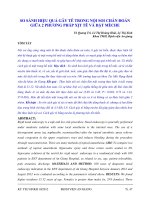
and NVUGIH, a cardiologist should be consulted and maintenance of both antiplatelet agents be considered if the risk of rebleeding is thought to be low. ▶ Fig. 1 a, b outlines the management of antiplatelet therapy in patients with acute NVUGIH.
RECO MMENDATION
ESGE recommends that, in patients with acute UGIH taking vitamin K antagonists (VKAs) who have hemodynamic
instability, low dose vitamin K supplemented with intravenous prothrombin complex concentrate (PCC), or fresh
frozen plasma (FFP) if PCC is not available, should be administered. However, this should not delay endoscopy or,
if required, endoscopic hemostasis.
Strong recommendation, low quality evidence.
RECO MMENDATION
ESGE recommends that, in patients with acute UGIH taking direct oral anticoagulants (DOACs), the anticoagulant
should be withheld and endoscopy not delayed. In patients with severe ongoing bleeding, use of a DOAC reversal agent or intravenous PCC should be considered.
Strong recommendation, low quality evidence.
The management of patients taking anticoagulants (VKAs,
DOACs) who develop acute UGIH (e. g., peptic ulcer hemorrhage) is clinically challenging since anticoagulant management must be addressed both prior to and following upper
endoscopy [44]. Unfortunately, no studies have specifically addressed the optimal timing of endoscopy in patients receiving
anticoagulants. Furthermore, since the pharmacokinetics and
pharmacodynamic profiles of VKAs and DOACs are different,
management is different. DOACs (factor Xa and thrombin inhibitors) have a rapid onset of action and a much shorter half-life
than VKA, and routine tests for anticoagulant activity are lacking [45].
The anticoagulant effect of VKA is measured using the international normalized ratio (INR). Studies have shown that endoscopy outcomes in VKA-anticoagulated patients were similar in
patients with normal INR compared with those with elevated
INR at hospital admission, or in those where INR was corrected
to a value < 2.5 prior to endoscopy [44, 46–48]. More recent
observational studies provide additional supporting evidence.
Nagata et al. reported that in patients with acute upper (47 %)
or lower GI bleeding, early endoscopy (within 24 hours) in anticoagulant users (n = 157) was not associated with an increased
risk of rebleeding (13.4 % vs. 15.9 %, P = 0.52) or thromboembolic events (5.7 % vs. 3.2 %, P = 0.68) when compared to matched controls not taking anticoagulants [49]. An INR > 2.5 was
seen in 22.9 % of the anticoagulant users at the time of endoscopy. However rapid INR correction was associated with an increased risk of thromboembolism, as suggested in other studies [50, 51]. Another small study also suggested that the INR
level did not affect rebleeding or endoscopy outcomes [52].
However, Peloquin et al. reported that in 134 patients with GI
bleeding and a supratherapeutic INR of ≥ 3.5, therapeutic endoscopic intervention was less likely to be effective as the INR increased [53].
310
Reversal of the anticoagulant effect of VKAs in patients with
acute UGIH can be achieved with low dose vitamin K, however,
this takes time since the INR only starts to decrease within 2–4
hours and normalizes within 24 hours. Moreover, the anticoagulant reversal effect of vitamin K persists as compared to prothrombin complex concentrate (PCC) or fresh frozen plasma
(FFP) [54]. Sin et al. reported that four-factor PCC appears to
be associated with a significant thromboembolic risk; however
it remains a useful agent for warfarin reversal [55]. That same
study also suggested that in patients requiring reversal of warfarin anticoagulation, lack of concomitant vitamin K may contribute to “INR rebound,” therefore concomitant low dose vitamin K may be appropriate in this situation. However, given the
limited data, caution must be exercised when giving vitamin K
since its persisting effect can impede re-coagulation efforts.
Limitations of FFP include the requirement for a higher volume
load to achieve a reversal effect, slower onset of action compared with PCC, and requirement for blood group typing. In addition, recent evidence suggests that use of FFP is associated with
increased mortality in patients undergoing endoscopy for
NVUGIH [56–58]. Three- or four-factor PCC or FFP can be used
when the reversal of anticoagulation is urgent because of patient hemodynamic instability or life-threatening massive
bleeding, irrespective of INR values. Recombinant factor VIIa is
currently not recommended because of its high cost and higher
risk of thromboembolism [59].
Patients who develop acute UGIH while taking DOACs must
follow a similar protocol of early endoscopy and reversal of
anticoagulation in cases of hemodynamic instability or lifethreatening bleeding. However, there are particular considerations because of DOAC’s specific pharmacodynamics and the
availability of antidotes which rapidly block its anticoagulation
effects. It is important to know the time of the last DOAC dose,
since most DOACs have an 8–12-hour half-life and their effect
usually disappears within 24 hours. Hemodialysis is effective to
remove dabigatran from plasma and can help to prevent rebleeding [60]. PCC has also been shown to be effective for reversal of anticoagulation in patients with acute UGIH who are
taking DOACs [61, 62]. However, the best potential therapeutic
options rely on the availability of DOAC reversal agents that
should be used in cases of life-threatening acute UGIH. The
risk of thromboembolism with use of reversal agents is a concern, but very few data are available [63–67]. Idarucizumab is
a specific antidote for dabigatran and works effectively within
minutes. Thromboembolism and rebound effects have been reported in 6.8 % and 23 % of patients, respectively [63]. Other
DOAC antidotes are being investigated but are not yet on the
market [66, 67].
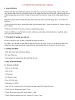
▶ Fig. 2 outlines management of anticoagulant therapy in
patients with acute NVUGIH.
Gralnek Ian M et al. Endoscopic diagnosis and … Endoscopy 2021; 53: 300–332 | © 2021. European Society of Gastrointestinal Endoscopy. All rights reserved.
This document was downloaded for personal use only. Unauthorized distribution is strictly prohibited.
Guidelines
Acute UGIH in a patient taking antiplatelet agent/s (APA/s)
Upper GI endoscopy demonstrates nonvariceal source of hemorrhage, e.g. peptic ulcer
High risk endoscopic stigmata diagnosed
(FIa, FIb, FIIa, FIIb – active spurting/oozing bleeding, nonbleeding visible ulcer, adherent clot)
APA* used for secondary prophylaxis (known cardiovascular disease)
1 Patient on low dose aspirin alone
(a) Continue low dose aspirin without interruption
(b) If aspirin has been interrupted, resume within 3–5 days
(c) Second-look endoscopy should be at the discretion of the
endoscopist, prior to restarting aspirin
This document was downloaded for personal use only. Unauthorized distribution is strictly prohibited.
Low dose aspirin used for primary prophylaxis
(a) Continue to withhold low dose aspirin
(b) Resume low dose aspirin after careful
re-evaluation of its clinical indication
2 Patient on dual antiplatelet therapy (DAPT)
(a) Continue low dose aspirin without interruption
(b) The second APA should be restarted as soon as possible, preferably
within 5 days.
Cardiology consultation regarding timing of restarting second APA
is suggested
(c) Second-look endoscopy should be at the discretion of the
endoscopist, prior to restarting second APA
a
Acute UGIH in a patient taking APA(s)
UGI endoscopy demonstrates nonvariceal source of hemorrhage, e.g. peptic ulcer
Low risk endoscopic stigmata diagnosed
(FIIc, FIII – flat pigmented spot, clean-base ulcer)
Low dose aspirin used for primary prophylaxis
(a) Continue to withhold low dose aspirin
(b) Resume low dose aspirin after careful
re-evaluation of its clinical indication
APA* used for secondary prophylaxis (known cardiovascular disease)
1 Patient on low dose aspirin alone
(a) Continue low dose aspirin without interruption
2 Patient on dual antiplatelet therapy (DAPT)
(a) Continue DAPT without interruption
b
▶ Fig. 1 Management of antiplatelet therapy in patients with acute nonvariceal upper gastrointestinal hemorrhage (NVUGIH) with a high risk,
and b low risk stigmata, diagnosed at endoscopy. *In patients using a nonaspirin antiplatelet agent (APA) as monotherapy (e. g. thienopyridine
alone), low dose aspirin may be substituted for an interval period provided there is no contraindication or allergy to aspirin. Cardiology consultation is suggested for further APA recommendations. UGIH, upper gastrointestinal hemorrhage.
Gralnek Ian M et al. Endoscopic diagnosis and … Endoscopy 2021; 53: 300–332 | © 2021. European Society of Gastrointestinal Endoscopy. All rights reserved.
311
Acute UGIH in patient taking anticoagulation
(e.g., VKA, DOAC)
1 Withhold anticoagulant at time of patient
presentation
2 In patients taking VKA and with hemodynamic
instability, low dose vitamin K supplemented
with intravenous PCC, or FFP if PCC not available,
should be administered
3 In patients taking DOAC and with severe ongoing
bleeding, use of a DOAC reversal agent or intravenous
PCC should be considered
4 Upper GI endoscopy and if required, endoscopic
hemostasis, should not be delayed
Upper GI endoscopy demonstrates nonvariceal
source of hemorrhage
1 Anticoagulation should be resumed as soon as the
bleeding has been controlled, preferably within or
soon after 7 days of the bleeding event based on
thromboembolic risk
2 Rapid onset of action of DOAC, as compared to VKA,
must be considered in this context
3 Use of validated scores that estimate thrombotic risk
(e.g., CHA2DS2-VASc) and bleeding risk (e.g.,
HAS-BLED) can be used to help guide clinical decision
making
▶ Fig. 2 Management of anticoagulants in acute nonvariceal upper
gastrointestinal hemorrhage (NVUGIH) before and after upper GI
endoscopy. UGIH, upper gastrointestinal hemorrhage; VKA, vitamin
K antagonist; DOAC, direct oral anticoagulant; PCC, prothrombin
complex concentrate; FFP, fresh frozen plasma; GI, gastrointestinal.
Pre-endoscopy proton pump inhibitor (PPI) therapy
the time of index endoscopy, and thereby reduces the need for
endoscopic hemostasis, PPIs provide no significant impact on
patient outcomes, including recurrent hemorrhage, need for
surgery, or mortality [68]. In the 2015 ESGE NVUGIH guideline,
initiation of high dose intravenous PPI was recommended for
patients presenting with acute UGIH awaiting upper endoscopy, without delaying early endoscopy [1]. This was a strong
recommendation based upon high quality evidence. However,
the lack of a significant impact of pre-endoscopy PPI therapy
on clinically relevant patient outcomes in acute NVUGIH has recently led to revised recommendations from several international evidence-based guideline bodies. In 2018, the Asia-Pacific working group consensus revised their earlier support for
routine pre-endoscopy intravenous PPI administration in acute
UGIH [33]. Since there is no proven impact on patient outcomes
and costs are increased, the working group members voted to
reject the indiscriminate use of pre-endoscopy intravenous PPIs
in patients presenting in a stable condition with symptoms suggestive of acute UGIH. However, the working group noted that
when endoscopy facilities or expertise in acute UGIH are not
available within 24 hours, the downgrading of stigmata of recent hemorrhage and reducing the need for urgent endoscopy
by use of intravenous PPIs could be justified. In 2019, the International Consensus Group on NVUGIH recommended that
“pre-endoscopic PPI therapy may be considered to downstage
the endoscopic lesion and decrease the need for endoscopic intervention but should not delay endoscopy” [15]. This was the
same as their earlier recommendation in 2010 [69]. Lastly, the
recently published United Kingdom consensus care bundle for
early clinical management of acute UGIH did not recommend
use of PPI prior to endoscopy [70].
Considering the available evidence, ESGE now “suggests”
that pre-endoscopy, high dose intravenous PPI “be considered”
in patients presenting with acute UGIH. This change is reflective of the lack of high level evidence for the impact of preendoscopy PPI on clinically relevant patient outcomes and remains consistent with other recent NVUGIH guideline recommendations.
Somatostatin and somatostatin analogues
RECO MMENDATION
ESGE suggests that pre-endoscopy high dose intravenous
proton pump inhibitor (PPI) therapy be considered in patients presenting with acute UGIH, to downstage endoscopic stigmata and thereby reduce the need for endoscopic therapy; however, this should not delay early
endoscopy.
Weak recommendation, high quality evidence.
In the systematic literature search (from January 2014 to
January 2020) for this updated NVUGIH guideline, we were unable to identify any systematic reviews, meta-analyses, RCTs, or
observational studies evaluating pre-endoscopy PPI administration in patients presenting with acute UGIH. Although preendoscopy PPI therapy significantly reduces the prevalence of
high risk endoscopic stigmata in peptic ulcer hemorrhage at
312
RECOMMENDATION
ESGE does not recommend the use of somatostatin, or its
analogue octreotide, in patients with NVUGIH.
Strong recommendation, low quality evidence.
Somatostatin, and its analogue octreotide, inhibit both acid
and pepsin secretion while also reducing gastroduodenal mucosal blood flow [71]. However, they are not recommended in
NVUGIH (e. g., peptic ulcer bleeding), either before endoscopy
or as an adjunctive therapy following endoscopy, since published data show little or no benefit. A recently published retrospective cohort study including 180 patients with acute
NVUGIH continues to show no significant differences in outcomes between patients receiving combination therapy (PPI
plus octreotide infusion) and those receiving PPI alone (hospital
Gralnek Ian M et al. Endoscopic diagnosis and … Endoscopy 2021; 53: 300–332 | © 2021. European Society of Gastrointestinal Endoscopy. All rights reserved.
This document was downloaded for personal use only. Unauthorized distribution is strictly prohibited.
Guidelines
Nasogastric/orogastric tube aspiration and lavage
RECO MMENDATION
ESGE does not recommend the routine use of nasogastric
or orogastric aspiration/lavage in patients presenting
with acute UGIH.
Strong recommendation, moderate quality evidence.
A recent retrospective study and a review both concluded
that nasogastric tube (NGT) aspiration does not differentiate upper from lower GI bleeding in patients with melena [73, 74].
Moreover, a randomized, single-blind, noninferiority study comparing NGT placement (with aspiration and lavage) to no NGT
placement (n = 140 in each arm), failed to show that NGT aspiration could accurately predict the presence of a high risk lesion
requiring endoscopic therapy (39 % vs. 38 %, respectively) [75].
In addition, adverse events (pain, nasal bleeding, or failure of
NGT placement) occurred in 34 % and there were no observed
differences in rebleeding rates or mortality.
Endotracheal intubation
RECO MMENDATION
ESGE does not recommend routine prophylactic endotracheal intubation for airway protection prior to upper
endoscopy in patients with acute UGIH.
Strong recommendation, high quality evidence.
RECO MMENDATION
ESGE recommends prophylactic endotracheal intubation
for airway protection prior to upper endoscopy only in
selected patients with acute UGIH (i. e., those with ongoing active hematemesis, agitation, or encephalopathy
with inability to adequately control their airway).
Strong recommendation, low quality evidence.
It has been posited that prophylactic endotracheal intubation prior to upper endoscopy in unselected patients with acute
UGIH could protect the patient’s airway from potential aspiration of gastric contents and prevent cardiorespiratory adverse
events. However, three recent systematic reviews/meta-analyses
and a small retrospective case series show that prophylactic
endotracheal intubation before upper endoscopy in patients
with acute UGIH may be associated with a higher risk of aspiration and pneumonia, longer hospital stays, and potentially
higher mortality [76–79]. In a meta-analysis by Almashhrawi
et al., the authors reported that in patients with acute UGIH
who received prophylactic endotracheal intubation prior to
upper endoscopy, pneumonia within 48 hours was identified in
20 of 134 patients (14.9 %) as compared with 5 of 95 patients
(5.3 %) not prophylactically intubated (P = 0.02, OR 3.13) [78].
Despite observed trends, no significant differences were found
for aspiration (P = 0.11) or mortality (P = 0.18). Alshamsi et al.,
in their meta-analysis including 10 observational studies (n =
6068 patients), reported that prophylactic endotracheal intubation was associated with a significant increase in aspiration
(OR 3.85, 95 %CI 1.46–10.25; P = 0.01), pneumonia (OR 4.17,
95 %CI 1.82–9.57; P < 0.001) and hospital length of stay (mean
difference 0.86 days, 95 %CI 0.13–1.59; P = 0.02) [77]. However, there was no observed effect on mortality (OR 1.92, 95 %
CI 0.71–5.23; P = 0.20). Chaudhuri et al. included 7 observational studies (n = 5662 patients) in their meta-analysis and
found that prophylactic endotracheal intubation was associated with significantly higher rates of pneumonia (OR 6.58, 95 %
CI 4.91–8.81), longer hospital length of stay (mean difference,
0.96 days, 95 %CI 0.26–1.67), and increased mortality (OR
2.59, 95 %CI 1.01–6.64) [76]. However, because of the observational design of the included studies, the data should be considered to be of low quality.
Prokinetic medications
RECOMMENDATION
ESGE recommends pre-endoscopy administration of intravenous erythromycin in selected patients with clinically
severe or ongoing active UGIH.
Strong recommendation, high quality evidence.
In patients with acute UGIH, the quality of the endoscopic
examination can be adversely affected by poor visibility in the
upper GI tract due to blood, clots and fluids. It is reported that
in 3 % to 19 % of UGIH cases, no obvious cause of bleeding is
identified [80, 81]. This may in part be related to the presence
of blood and clots impairing endoscopic visualization. Prokinetics may improve gastric mucosa visualization by inducing gastric emptying. Most studies assessing the use of pre-endoscopy
prokinetics in UGIH have used erythromycin. Insufficient data
were found to make recommendations for the use of metoclopramide [82–84].
Five published meta-analyses have evaluated the role of prokinetic agent infusion prior to upper GI endoscopy in patients
presenting with acute UGIH [82–86]. The most recently published meta-analysis (n = 598 patients) by Rahman et al.,
showed that erythromycin infusion prior to upper endoscopy
significantly improved gastric mucosa visualization (OR 4.14,
95 %CI 2.01–8.53; P < 0.01), reduced the need for a secondlook endoscopy (OR 0.51, 95 %CI 0.34–0.77; P < 0.01), and reduced the length of hospital stay (mean difference –1.75, 95 %
CI –2.43 to –1.06; P < 0.01) [86]. However other relevant outcomes, such as duration of the procedure, units of blood transfused, and need for emergency surgery showed no significant
differences. Mortality was not assessed.
A single intravenous dose of erythromycin appears to be safe
and generally well tolerated, with no adverse events reported in
Gralnek Ian M et al. Endoscopic diagnosis and … Endoscopy 2021; 53: 300–332 | © 2021. European Society of Gastrointestinal Endoscopy. All rights reserved.
313
This document was downloaded for personal use only. Unauthorized distribution is strictly prohibited.
and intensive care unit [ICU] median length of stay, respectively, 6.1 vs. 4.9 days, P = 0.25, and 2.3 vs. 1.9 days, P = 0.24; rebleeding 9 % vs. 12 %, P = 0.63; RBC units transfused 3 vs. 2
units, P = 0.43; and mortality 6.7 % vs. 5.6 %, P = 1.00) [72].
Guidelines
Endoscopic management
Timing of upper GI endoscopy
RECO MMENDATION
ESGE recommends adopting the following definitions
regarding the timing of upper GI endoscopy in acute
UGIH relative to the time of patient presentation: urgent
≤ 12 hours, early ≤ 24 hours, and delayed > 24 hours.
Strong recommendation, moderate quality evidence.
RECO MMENDATION
ESGE recommends that following hemodynamic resuscitation, early (≤ 24 hours) upper GI endoscopy should be
performed.
Strong recommendation, high quality evidence.
RECO MMENDATION
ESGE does not recommend urgent (≤ 12 hours) upper GI
endoscopy since as compared to early endoscopy, patient
outcomes are not improved.
Strong recommendation, high quality evidence.
RECO MMENDATION
ESGE does not recommend emergent (≤ 6 hours) upper GI
endoscopy since this may be associated with worse patient outcomes.
Strong recommendation, moderate quality evidence.
314
RECOMMENDATION
ESGE recommends that the use of antiplatelet agents,
anticoagulants, or a predetermined international normalized ratio (INR) cutoff level, should not be used to define
or guide the timing of upper GI endoscopy in patients
with acute UGIH.
Strong recommendation, low quality evidence.
In patients with acute NVUGIH, upper GI endoscopy performed within 24 hours or after 24 hours of patient presentation are the commonly accepted definitions for “early” and
“delayed” endoscopy [90–95]. Urgent upper GI endoscopy in
the setting of acute UGIH has been variably defined as endoscopy performed between 6–12 hours of patient presentation
[91, 96, 97]. There is no consensus definition of emergent
endoscopy.
Early endoscopy (≤ 24 hours from the time of patient presentation) is associated with lower in-hospital mortality, shorter
length of stay, and lower total hospital costs, and should be
performed in patients with acute UGIH [92–94]. A beneficial
role of urgent endoscopy (≤ 12 hours from the time of patient
presentation) however, is not routinely demonstrated as published studies show conflicting results. While one recent study
concluded that urgent endoscopy was an independent predictor of lower mortality [96], other studies have shown that urgent endoscopy was a predictor of worse patient outcomes
[90, 97], or that clinical outcomes were not significantly different between urgent and early endoscopy [91]. Moreover, in a
well-executed large RCT by Lau et al., the investigators reported that, at 30-day follow-up, as compared to “early” upper
endoscopy (mean time to endoscopy 24.7 ± 9.0 hours), “urgent” upper endoscopy (mean time to endoscopy 9.9 ± 6.1
hours) performed in patients at high risk for further bleeding
or death, was not associated with significantly lower rates of
further bleeding (7.8 % vs. 10.9 %; HR 1.46, 95 %CI 0.83–2.58)
or lower mortality (6.6 % vs. 8.9 %; HR 1.35, 95 %CI 0.72–2.54)
[98]. Lastly, in a large prospective cohort study from Denmark,
including 12 601 patients admitted to hospital with peptic ulcer
bleeding, emergent endoscopy (performed < 6 hours from the
time of patient presentation) was associated with higher inhospital and 30-day mortality, particularly in hemodynamically
unstable patients or in patients with an American Society of Anesthesiologists (ASA) score ≥ 3 [99]. In those patients, optimizing hemodynamic resuscitation and adequately attending to
comorbidities prior to endoscopy may improve outcomes.
Although antiplatelet and anticoagulant therapies are usually
interrupted or discontinued in patients with acute UGIH, it is
now realized that complete reversal of the antithrombotic effect of those drugs is not necessary for performance of diagnostic and therapeutic endoscopy. One study evaluated the risk of
rebleeding in patients receiving anticoagulants and concluded
that an INR > 2.5 was not a risk factor for rebleeding in patients
with acute UGIH [49]. This finding, combined with the fact that
the antithrombotic effect of DOACs is not measured by INR, has
led to the recommendation to avoid using a predetermined INR
Gralnek Ian M et al. Endoscopic diagnosis and … Endoscopy 2021; 53: 300–332 | © 2021. European Society of Gastrointestinal Endoscopy. All rights reserved.
This document was downloaded for personal use only. Unauthorized distribution is strictly prohibited.
the meta-analyses. Most studies that reported a significant improvement in endoscopic visualization with pre-endoscopic erythromycin infusion did include patients admitted to the intensive care unit because of acute UGIH with clinical evidence of
active bleeding or hematemesis. These are the patients most
likely to benefit from erythromycin infusion prior to endoscopy.
The dose of erythromycin most commonly used is 250 mg, infused 30–120 minutes prior to upper GI endoscopy. A costeffectiveness study found that pre-endoscopy erythromycin
infusion in UGIH was cost-effective, primarily because of a
reduction in the need for second-look endoscopy [87].
It should be noted that there have been difficulties accessing
erythromycin in many countries. Furthermore, there are some
contraindications to its administration. These include patient
sensitivity to macrolide antibiotics and presence of a prolonged
QT interval. Drug interactions such as erythromycin-induced
digoxin toxicity have been reported to occur when erythromycin is repeatedly administrated, although the risk appears to be
very low [88]. In addition, the combination of simvastatin and
erythromycin may increase the risk of rhabdomyolysis [89].
On-call GI endoscopy resources
RECO MMENDATION
ESGE recommends the availability of both an on-call GI
endoscopist proficient in endoscopic hemostasis and oncall nursing staff with technical expertise in the use of
endoscopic devices, to allow performance of endoscopy
on a 24/7 basis.
Strong recommendation, low quality evidence.
Although a retrospective study from Japan concluded that
the clinical outcomes of patients who underwent emergency
endoscopic hemostasis for acute UGIH outside regular hours
did not differ from those of patients treated during regular
hours [100], two systematic reviews/meta-analyses found
otherwise [95, 101]. Xia et al. reported that NVUGIH patients
who were admitted out of hours had significantly higher mortality and received less timely endoscopy [95]. Shih and colleagues showed that the “weekend effect” was associated with increased mortality in UGIH patients, particularly in patients with
NVUGIH [101].
Endoscopic diagnosis
RECO MMENDATION
ESGE recommends the Forrest (F) classification be used in
all patients with peptic ulcer hemorrhage to differentiate
low risk and high risk endoscopic stigmata.
Strong recommendation, high quality evidence.
RECO MMENDATION
ESGE recommends that peptic ulcers with spurting or
oozing bleeding (FIa or FIb, respectively) or with a nonbleeding visible vessel (FIIa) receive endoscopic hemostasis because these lesions are at high risk for persistent
bleeding or recurrent bleeding.
Strong recommendation, high quality evidence.
RECO MMENDATION
ESGE suggests that peptic ulcers with an adherent clot
(FIIb) be considered for endoscopic clot removal. Once
the clot is removed, any identified underlying active
bleeding (FIa or FIb) or nonbleeding visible vessel (FIIa)
should receive endoscopic hemostasis.
Weak recommendation, moderate quality evidence.
RECOMMENDATION
ESGE does not recommend endoscopic hemostasis in patients with peptic ulcers having a flat pigmented spot
(FIIc) or clean base (FIII), as these stigmata have a low
risk of adverse outcomes. In selected clinical settings
these patients may have expedited hospital discharge.
Strong recommendation, moderate quality evidence.
RECOMMENDATION
ESGE does not recommend the routine use of Doppler
endoscopic probe in the evaluation of endoscopic stigmata of peptic ulcer bleeding.
Strong recommendation, low quality evidence.
RECOMMENDATION
ESGE does not recommend the routine use of capsule
endoscopy technology in the evaluation of acute UGIH.
Strong recommendation, low quality evidence.
The Forrest (F) classification was developed more than 40
years ago to standardize the endoscopic characterization of
peptic ulcers [102]. The Forrest classification is defined as follows: FIa spurting hemorrhage, FIb oozing hemorrhage, FIIa
nonbleeding visible vessel, FIIb adherent clot, FIIc flat pigmented spot, and FIII clean base ulcer. This classification has been
used in numerous studies to identify patients at risk of persistent ulcer bleeding, recurrent ulcer bleeding, and mortality.
Most of these studies have shown that the presence of an ulcer
endoscopically classified as FIa or FIb is an independent risk factor for persistent bleeding or recurrent bleeding [103]. A potential limitation of the Forrest classification is that recognition
and identification of endoscopic stigmata and interobserver
agreement may be less than optimal, although data are conflicting [104, 105].
The classification of FIb as a high risk stigma following endoscopic therapy is controversial. It is apparent that FIb stigmata
require endoscopic hemostasis as there is active bleeding (i. e.,
oozing hemorrhage), but the response to endoscopic treatment may be different compared to that with other high risk
endoscopic stigmata of hemorrhage (FIa, FIIa, and in some
cases FIIb), specifically in peptic ulcer rebleeding rates. An RCT
including 388 patients comparing PPI or placebo following successful endoscopic treatment of FIb ulcers found no apparent
benefit on rebleeding rates with the addition of PPI (5.4 % vs.
4.9 %; OR 1.11, 95 %CI 0.42–2.95) [106]. In the placebo group,
FIb ulcers had a lower risk of rebleeding (4.9 %) compared to FIa
(22.5 %), FIIb (17.6 %), and FIIa (11.3 %). Studies using a Doppler
endoscopic probe have shown rebleeding rates from FIb ulcers
following endoscopic therapy to be lower than the rebleeding
rates of FIa, FIIa and FIIb ulcers. This has led some to consider
Gralnek Ian M et al. Endoscopic diagnosis and … Endoscopy 2021; 53: 300–332 | © 2021. European Society of Gastrointestinal Endoscopy. All rights reserved.
315
This document was downloaded for personal use only. Unauthorized distribution is strictly prohibited.
cutoff value to define the timing of endoscopy in the setting of
acute UGIH.
a reassessment of the risk stratification of endoscopic stigmata
of recent hemorrhage as follows: “high risk,” FIa, FIIa, and FIIb;
“medium risk,” FIb and FIIc; and “low risk,” FIII [106, 107]. A
prospective study, that included two patient cohorts with 87
high risk stigmata (FIa, FIIa, FIIb) ulcers and 52 medium risk
stigmata (FIb, FIIc) ulcers, demonstrated significantly higher
Doppler signal-positive arteries in high risk stigmata ulcers
compared to the medium risk stigmata ulcers, before endoscopic hemostasis (87.4 % vs. 42.3 %, P < 0.001) as well as after
endoscopic hemostasis (27.4 % vs. 13.6 %), and significantly
higher 30-day rebleeding rates (28.6 % vs. 0 %, P = 0.04). In addition, for spurting bleeding (FIa) versus oozing bleeding (FIb),
baseline Doppler endoscopic probe arterial flow was 100 % versus 46.7 %, residual blood flow detected after endoscopic hemostasis was 35.7 % versus 0 %, and 30-day rebleed rates were
28.6 % versus 0 % (all P < 0.05) [107]. However, given the low
numbers of patients included in this study, larger size studies
are needed before considering a change in endoscopic stigmata risk classification.
In addition to the Forrest classification, there are additional
endoscopic features of peptic ulcers that can predict adverse
outcomes and/or endoscopic treatment failure and recent publications continue to support this [108, 109]. These endoscopic
features include large size of ulcer (> 2 cm), large size of nonbleeding visible vessel, and ulcer location on the posterior duodenal wall or the proximal lesser curvature of the stomach.
The persistence of a positive Doppler probe signal following
endoscopic hemostasis has been shown to predict recurrent
bleeding [110]. The results of available studies have been disparate and limited by their methodology, the older endoscopic
hemostasis therapies used, and the small numbers of patients
included. However, two recent studies have used a throughthe-scope (TTS) Doppler probe to guide endoscopic hemostasis. In an RCT with a subgroup of 86 patients with peptic ulcer
bleeding, 53 were classified as “high risk” (FIa, FIIa, FIIb) and
23 as “medium risk” (FIb, FIIc). Patients were randomly assigned to standard endoscopic hemostasis or Doppler probeguided hemostasis with repeat intervention until the Doppler
signal was completely obliterated. Total rebleeding rates were
significantly lower in the Doppler probe-guided hemostasis
group (11.1 % vs. 26.3 %, P = 0.02) but there were no significant
differences in other outcomes [111]. In a study comprising 60
patients with FIa, FIb, and FIIa ulcers that were “assigned by
chance” to standard endoscopic hemostasis (n = 25) or Doppler
probe-guided intervention (n = 35) until the Doppler signal was
obliterated, the Doppler probe-guided hemostasis group
showed significantly lower rates for rebleeding (52 % vs. 20 %,
P = 0.013) and surgery (2 % vs. 26 %, P = 0.02) [112]. A costminimization analysis suggests a per-patient cost-saving
with the use of the Doppler endoscopic probe in patients
with peptic ulcer bleeding, but cost-savings may be dependent on and limited to specific healthcare settings [113].
Since publication of the previous ESGE NVUGIH Guideline,
five additional studies have been published that evaluate the
role of capsule endoscopy technology (e. g., video capsule
endoscopy, magnetically assisted capsule endoscopy, telemetric sensor capsule) in acute UGIH, namely one RCT, three
316
prospective cohort studies, and one retrospective case series
[114–118]. In the only RCT, Marya et al. reported on 87 patients with nonhematemesis GI hemorrhage who were randomized to early video capsule endoscopy or to “standard of care”
whereby the gastroenterologist chose which procedures to perform and when to perform them based on the patient’s presentation [114]. A source of GI bleeding was located in 64.3 % of
the patients in the early video capsule endoscopy arm and in
31.1 % of the patients in the standard of care arm (P < 0.01).
Moreover, the likelihood of endoscopic location of bleeding
over time was greater for patients receiving early video capsule
endoscopy (adjusted hazard ratio 2.77, 95 %CI 1.36–5.64).
Overall, patients who received capsule endoscopy technology
to evaluate their GI bleeding were more likely to undergo therapeutic procedures (e. g., balloon enteroscopy, colonoscopy, or
surgery) than patients with standard of care treatment. Thus,
capsule endoscopy technology may be helpful in the setting of
acute UGIH, as it may assist in the clinical management plan.
However, because data continue to be limited, including on
costs and on availability of technology, the exact role for capsule endoscopy modalities in evaluating patients presenting
with acute UGIH remains unknown. Additional high level studies are needed to further assess the diagnostic role of capsule
endoscopy in this patient population.
Endoscopic therapy for peptic ulcer hemorrhage
RECOMMENDATION
FIa, FIb (active bleeding)
(a) ESGE recommends for patients with actively bleeding
ulcers (FIa, FIb), combination therapy using epinephrine
injection plus a second hemostasis modality (contact
thermal or mechanical therapy) .
Strong recommendation, high quality evidence.
(b) ESGE suggests that in selected actively bleeding ulcers (FIa, FIb), specifically those > 2 cm in size, with a large
visible vessel > 2 mm, or located in a high risk vascular
area (e. g., gastroduodenal, left gastric arteries), or in
excavated/fibrotic ulcers, endoscopic hemostasis using a
cap-mounted clip should be considered as first-line
therapy.
Weak recommendation, low quality evidence.
RECOMMENDATION
FIIa (nonbleeding visible vessel)
ESGE recommends, for patients with an ulcer with a nonbleeding visible vessel (FIIa), contact or noncontact thermal therapy, mechanical therapy, or injection of a sclerosing agent, each as monotherapy or in combination
with epinephrine injection.
Strong recommendation, high quality evidence.
Gralnek Ian M et al. Endoscopic diagnosis and … Endoscopy 2021; 53: 300–332 | © 2021. European Society of Gastrointestinal Endoscopy. All rights reserved.
This document was downloaded for personal use only. Unauthorized distribution is strictly prohibited.
Guidelines
ESGE does not recommend that epinephrine injection be
used as endoscopic monotherapy. If used, it should be
combined with a second endoscopic hemostasis modality.
Strong recommendation, high quality evidence.
RECO MMENDATION
ESGE recommends that “persistent bleeding” be defined
as ongoing active bleeding refractory to standard hemostasis modalities.
Strong recommendation, high quality evidence.
RECO MMENDATION
ESGE suggests that in patients with persistent bleeding
refractory to standard hemostasis modalities, the use of
a topical hemostatic spray/powder or cap-mounted clip
should be considered.
Weak recommendation, low quality evidence.
RECO MMENDATION
ESGE recommends that in patients with persistent bleeding refractory to all modalities of endoscopic hemostasis,
transcatheter angiographic embolization (TAE) should be
considered. Surgery is indicated when TAE is not locally
available or after failed TAE.
Strong recommendation, moderate quality evidence.
Endoscopic hemostasis can be achieved using injection,
thermal, and/or mechanical modalities, and it has been well
demonstrated that any endoscopic hemostasis therapy is
superior to pharmacotherapy alone in patients with FIa, FIb
and FIIa ulcers [119, 120]. Meta-analyses show that thermal devices (contact and noncontact), injectable agents other than
epinephrine (i. e., sclerosing agents, thrombin/fibrin glue), and
clips are all effective methods for achieving durable hemostasis, with no single modality being superior [119–123]. Epinephrine injection therapy is effective at achieving primary hemostasis, but inferior to other endoscopic hemostasis monotherapies or combination therapy in preventing ulcer rebleeding
[119, 120, 122]. Therefore, current evidence-based guidelines
recommend that if epinephrine is used to treat peptic ulcer
bleeding with high risk stigmata, it should only be used in combination with a second endoscopic hemostasis modality and
not as monotherapy [1, 15].
▶ Fig. 3 a–c presents an algorithm, stratified according to
the Forrest classification of endoscopic stigmata, for the endoscopic management of NVUGIH secondary to peptic ulcer.
Two recent meta-analyses support the superiority of combination endoscopic therapy (injection plus thermal therapy, and
injection plus mechanical therapy) over epinephrine injection
monotherapy in peptic ulcers with high risk stigmata [124,
125]. Baracat et al. performed a systematic review and metaanalysis of 28 RCTs that included 2988 adults with high risk
peptic ulcer endoscopic stigmata. These authors reported
that injection therapy alone, as compared to injection plus
thermal therapy was inferior in terms of ulcer rebleeding (risk
difference [RD] –0.08, 95 %CI –0.14 to –0.02) and need for
emergency surgery (RD –0.06, 95 %CI –0.12 to 0.00). Moreover, they reported that injection therapy alone, as compared
to injection plus mechanical therapy was also inferior in terms
of rebleeding (RD –0.10, 95 %CI –0.018 to –0.03) and need
for surgery (RD –0.11, 95 %CI –0.18 to –0.04) [124]. No significant difference in mortality between hemostasis modalities was observed. In a network meta-analysis, Shi et al. reported that the addition of mechanical therapy following
epinephrine injection significantly reduced the probability of
rebleeding and surgery (OR 0.19, 95 %CI 0.07–0.52 and OR
0.10, 95 %CI 0.01–0.50, respectively), while the addition of
thermal therapy only reduced ulcer rebleeding rates (OR
0.30, 95 %CI 0.10–0.91) [125].
With respect to noncontact thermal therapy (e. g., argon
plasma coagulation [APC]), limited data from three previous
small RCTs suggest that in peptic ulcer hemorrhage, APC may
provide similar efficacy to injection of a sclerosing agent (polidocanol) or contact thermal therapy (heater probe) [119].
More recently, a single RCT (noninferiority design) compared
combination endoscopic therapies using epinephrine injection
plus APC versus epinephrine injection plus soft coagulation
using hemostatic forceps [126]. That study included 151 patients with high risk stigmata gastroduodenal ulcers (FIa, FIb,
FIIa). The authors reported similar outcomes between APC and
hemostatic forceps for rates of primary hemostasis (96.0 % vs.
96.1 %, P = 1.00), 7-day ulcer rebleeding (4.0 % vs. 6.6 %,
P = 0.72) and 30-day ulcer rebleeding rates (6.7 % vs. 9.2 %,
P = 0.56).
Clinicians must distinguish between two clinical scenarios in
NVUGIH: persistent bleeding and recurrent bleeding. Persistent
bleeding is defined as ongoing active bleeding (spurting, arterial pulsatile bleeding, or oozing) that is present at the end of index endoscopy and refractory to standard hemostasis modal-
▶ Fig. 3 Algorithm for the endoscopic management of nonvariceal upper gastrointestinal hemorrhage (NVUGIH) secondary to peptic ulcer,
stratified by Forrest classification endoscopic stigmata: a FIa, FIb, FIIa; b FIIb; c FIIc, FIII. 1Use of a large single-channel or double-channel
therapeutic upper gastrointestinal endoscope is recommended. 2Large-size 10-Fr probe recommended for contact thermal therapy. 3Absolute
alcohol, polidocanol, or ethanolamine injected in limited volumes. 4The benefit of endoscopic hemostasis may be greater in patients at higher
risk for recurrent bleeding, e. g., with older age, comorbidities, in-hospital UGIH. GI, gastrointestinal; PPI, proton pump inhibitor, TAE, transcatheter angiographic embolization.
Gralnek Ian M et al. Endoscopic diagnosis and … Endoscopy 2021; 53: 300–332 | © 2021. European Society of Gastrointestinal Endoscopy. All rights reserved.
317
This document was downloaded for personal use only. Unauthorized distribution is strictly prohibited.
RECO MMENDATION
Guidelines
Performance of upper GI endoscopy1
Performance of upper GI endoscopy1
High risk endoscopic stigmata
FIa (active spurting, pulsatile arterial bleeding)
FIb (active oozing)
FIIa (nonbleeding visible vessel)
FIIb (adherent clot)
Perform endoscopic hemostasis
FIa and FIb stigmata
Combination therapy
using dilute epinephrine
injection + a second
hemostasis modality
(thermal2, mechanical or
sclerosant injection3)
FIIa stigmata
Thermal2, mechanical,
or sclerosant injection3
as monotherapy or in
combination with dilute
epinephrine injection
▪ High dose PPI (intravenous bolus + continuous
infusion or minimum twice-daily intravenous bolus
dosing for 72 hours or oral dosing)
▪ May start clear liquids soon after endoscopy
▪ Test for Helicobacter pylori at index endoscopy, treat
if positive; document H. pylori eradication
▪ If negative H. pylori test at index endoscopy, repeat
testing within 4 weeks following the acute bleeding
episode to confirm initial test was true negative
If clot removal/endoscopic hemostasis performed:
▪ Dilute epinephrine injection circumferential to base
of clot followed by clot removal using cold polyp
snare guillotine technique
▪ If underlying high risk stigmata identified after clot
removal, apply endoscopic hemostasis as described
for FIa, FIb, FIIa stigmata
▪ High dose PPI (intravenous bolus + continuous
infusion or minimum twice-daily intravenous bolus
dosing for 72 hours or oral dosing)
▪ May start clear liquids soon after endoscopy
▪ Test for H. pylori, treat if positive; document H. pylori
eradication
▪ If negative H. pylori test at index endoscopy, repeat
testing within 4 weeks following the acute bleeding
episode to confirm initial test was true negative
If clinical evidence of rebleeding, repeat endoscopy with
endoscopic hemostasis if indicated;
If endoscopic hemostasis still unsuccessful, refer for TAE
if locally available, otherwise refer for surgery
If clinical evidence of rebleeding, repeat endoscopy with
endoscopic hemostasis if indicated;
If endoscopic hemostasis still unsuccessful, refer for TAE
if locally available, otherwise refer for surgery
a
b
Performance of upper GI endoscopy1
Low risk stigmata
FIIc (flat pigmented spot)
FIII (clean base)
No endoscopic hemostasis required
In select clinical settings, these patients may have expedited hospital discharge
▪
▪
▪
▪
Start oral PPI
Start regular diet
Test for H. pylori, treat if positive; document H. pylori eradication
If negative H. pylori test at index endoscopy, repeat testing within 4 weeks following the acute bleeding episode to confirm
initial test was true negative
c
318
Gralnek Ian M et al. Endoscopic diagnosis and … Endoscopy 2021; 53: 300–332 | © 2021. European Society of Gastrointestinal Endoscopy. All rights reserved.
This document was downloaded for personal use only. Unauthorized distribution is strictly prohibited.
Consider performing clot removal followed by
endoscopic hemostasis of underlying high risk stigmata4
OR
Medical management with high dose PPI (intravenous
bolus + continuous infusion for 72 hours or
minimum twice-daily intravenous bolus dosing for
72 hours or oral dosing)
approach was TC-325 plus traditional endoscopic hemostasis
(5.8 % less effective and $635 more costly per patient). The limitations of topical sprays/powders are that they only bind to
sites with active bleeding and usually wash away within 12–24
hours; thus they are a temporary measure.
The role of cap-mounted clips (e.g, the Over the Scope Clip
[OTSC], Ovesco, Tübingen, Germany; and the Padlock system,
Steris Endoscopy, Mentor, Ohio, USA) in treating NVUGIH,
used as first-line and second-line (e. g., rescue/salvage) therapy, continues to evolve. In a retrospective case series evaluating over-the-scope (OTS) clip technology as first-line treatment
in NVUGIH (the FLETRock study), Wedi et al. reported on 118
patients with NVUGIH, including 60 patients (50.8 %) defined
as high risk based upon a Rockall risk score ≥ 8 [135]. Primary
clinical success (hemostasis by OTS clipping alone) was
achieved in 107 patients (90.8 %) and secondary clinical success
(hemostasis by OTS clipping in combination with adjunctive
measures) in 7 patients (1.7 %). In 7.5 % of clip applications,
the bleeding could not be stopped and treatment was defined
as clinical failure. Patients with Forrest Ia active bleeding were
at higher risk of rebleeding (11/31 patients, 35.5 %). Manta et
al., in a multicenter retrospective study, also reported on the
outcomes of 286 patients (74.8 % with NVUGIH) who were
treated with OTS clipping as first-line endoscopic hemostasis
therapy [136]. Of the 214 patients with NVUGIH, technical success was achieved in 208 (97.2 %), including 202/208 (97.1 %)
achieving hemostasis with OTS clipping as monotherapy. Early
rebleeding, within 24 hours, occurred in 9 patients (4.5 %), and
no delayed bleeding (within 30 days) was reported. Technical
failure of OTS clipping occurred in 6 patients, in ulcers located
in the gastric fundus or posterior wall of the duodenal bulb.
Brandler et al. reported an additional retrospective case series
of 67 patients (60 patients with NVUGIH, including 49 due to
peptic ulcer, 11 with Forrest Ia active bleeding) with bleeding
lesions defined by the authors as being at “high risk of adverse
outcome” (visible vessel > 2 mm; ulcer location in high risk vascular region, including gastroduodenal, left gastric arteries; penetrating, excavated or fibrotic ulcer with high risk stigmata)
[137]. OTS clipping was performed as first-line therapy in 49
patients. The authors reported 100 % technical success, OTS
clipping success (no bleeding related to OTS clipping requiring
re-intervention) in 52 patients (81.3 %), and true success (no
bleeding within 30 days) in 46 patients (71.8 %). They reported
no adverse events.
In a systematic review and meta-analysis, Chandrasekar et al.
examined the effectiveness of cap-mounted clip technology in
achieving “definitive hemostasis” in GI bleeding, defined as successful primary hemostasis without rebleeding during the follow-up period (median 56 days) [138]. This meta-analysis included 21 studies (1 RCT, 20 observational) with 851 patients
(687 with UGIH). In those patients with UGIH, OTS clipping was
used as first-line endoscopic therapy in 75.8 % and definitive hemostasis was achieved in 86.6 % (95 %CI 81.9–91.3). The rebleeding rate in patients with UGIH was 11.0 % (95 %CI 6.8 %–
15.2 %). The OTSC failure rate for UGIH was 6.2 % (95 %CI 3.1 %–
9.2 %) and 16.9 % (95 %CI 9.3 %–24.5 %) for first- and second-line
therapy, respectively. It must be noted that this meta-analysis is
Gralnek Ian M et al. Endoscopic diagnosis and … Endoscopy 2021; 53: 300–332 | © 2021. European Society of Gastrointestinal Endoscopy. All rights reserved.
319
This document was downloaded for personal use only. Unauthorized distribution is strictly prohibited.
ities. This is also referred to as “failed primary endoscopic
hemostasis” [1]. Few RCTs have compared alternative treatment modalities in the management of patients with persistent
ulcer bleeding. Meta-analyses and retrospective case series
comparing transcatheter arterial embolization (TAE) and surgery suggest that patient outcomes following either approach
are similar [127–129]. TAE, however, is associated with a higher
failure rate in the control of bleeding [127–129]. A populationbased cohort study compared outcomes in 282 patients (97
TAE and 185 surgery) and found a 34 % lower mortality among
those in the TAE group (adjusted HR 0.66, 95 %CI 0.46–0.96).
However, similarly to other cohort studies, rebleeding was
higher after TAE (HR 2.48, 95 %CI 1.33–4.62), whereas following surgery adverse events were significantly higher (32.2 % vs.
8.3 %, P < 0.001) [130].
Since publication of the original ESGE NVUGIH guideline in
2015, several additional studies have reported on the clinical
efficacy of topical hemostatic agents (e. g., TC-325, Endoclot,
and Inha University-Endoscopic Wound Dressing [UI-EWD]) in
patients with GI bleeding secondary to peptic ulcer bleeding.
These include case series, a multicenter patient registry, a pilot
RCT, and a cost–effectiveness analysis [131–134]. A multicenter (12 sites) patient registry evaluated the effectiveness of
TC-325 in upper and lower GI bleeding (167/314 [53 %] due to
peptic ulcer) [132]. In the subgroup of peptic ulcer hemorrhage
(most common stigmata, FIb), the authors reported an overall
hemostasis rate of 86 %, an overall rebleeding rate of 12.7 %,
and 7-day and 30-day all-cause mortality of 16.2 % and 24.6 %,
respectively. These data however should be interpreted with
caution because of the inherent limitations of a patient registry
that included lack of randomization or sequential patient selection, multiple bleeding indications (with GI bleeding secondary
to malignancy being over-represented in the cohort), along
with patient selection bias and self-reported or unverified outcomes. In addition, a pilot RCT evaluated the clinical efficacy of
TC-325 with/without epinephrine injection versus through-thescope (TTS) clipping with/without epinephrine injection, in 39
patients with active NVUGIH (the majority of cases due to peptic ulcer, and 35/39 [89.7 %] with FIb oozing bleeding) [133].
The authors reported that primary hemostasis was achieved in
all TC-325 cases and in 90 % of the mechanical therapy group (P
= 0.49). There was no difference in rebleeding, need for surgery, or mortality rates between the groups. This was a small
pilot study with a limited number of patients enrolled, and
thus not adequately powered to show a statistically significant
difference between groups. Moreover, five patients in the TC325 group required additional endoscopic intervention at the
time of second-look endoscopy, while none in the clipping
group required such therapy (P = 0.04). These results should
not be extrapolated to FIa bleeding lesions. Lastly, a decision
analysis model compared the cost–effectiveness of traditional
endoscopic hemostasis therapies alone, TC-325 alone, or these
therapies in combination, when treating acute NVUGIH [134].
The authors reported that traditional endoscopic hemostasis
complemented by TC-325 was more efficacious (97 % avoiding
rebleeding) and less expensive than comparator treatments
(mean cost per patient $ 9150). The second most cost-effective
limited, as all included studies but one were observational in
design. Other observational studies have also reported on
the efficacy and safety of OTSC used as either first-line or
second-line hemostasis treatment, with similar findings
[139–144].
Very recently, the first blinded RCT evaluating the efficacy
and safety of a cap-mounted clip (OTS clip, n = 25) versus standard endoscopic hemostasis therapy (TTS clip or contact thermal
therapy using multipolar electrocoagulation, n = 28) for firstline treatment of acute peptic ulcer or Dieulafoy bleeding was
published by Jensen et al. [145]. The investigators reported
that compared to standard endoscopic hemostasis, there was
both significantly less recurrent bleeding within 30 days (1/25
[4.0 %] vs. 8/28 [28.6 %], P = 0.017) and fewer adverse events
(0/25 [0 %] vs. 4/28 [14.3 %], P = 0.049) in the OTS clip group.
There were no observed differences in need for surgery or mortality. However, a number of methodological limitations to this
study must be noted, including the relatively limited number of
patients, the inclusion of Dieulafoy lesions in addition to peptic
ulcers, and the use of unconventional definitions of “major”
endoscopic stigmata of recent hemorrhage that are not widely
adopted.
In a multicenter RCT from Europe and Asia (the STING
study), Schmidt et al. reported on 66 patients with recurrent
peptic ulcer hemorrhage following initially successful endoscopic hemostasis, who were randomly assigned to undergo
hemostasis with either OTS clipping (n = 33) or standard endoscopic therapy (using TTS clips, n = 31, or contact thermal therapy plus injection with dilute epinephrine, n = 2) [146]. By perprotocol analysis, persistent ulcer bleeding was observed in 14
patients (42.4 %) in the standard therapy group and 2 patients
(6.0 %) in the OTS clip group (P = 0.001). Recurrent ulcer bleeding within 7 days occurred in 5 patients (16.1 %) in the standard
therapy group versus 3 patients (9.1 %) in the OTS clip group (P
= 0.47). Further bleeding occurred in 19 patients (57.6 %) in the
standard therapy group and in 5 patients (15.2 %) in the OTS
clip group (absolute difference 42.4 %, 95 %CI 21.6 %–63.2 %; P
= 0.001). During 30 days of follow-up, 1 patient (3.0 %) in the
standard therapy group and 1 patient (3.0 %) in the OTS clip
group required surgery (P = 0.99), 2 patients (6.3 %) died in the
standard therapy group and 4 patients (12.1 %) died in the
OTSC group (P = 0.67).
To date, almost all evidence on the efficacy of OTS clipping is
derived from case series or case series compared with historical
controls. Randomized trials directly comparing topical agents
and OTS clips/clamps with traditional hemostasis therapies are
required to better define their true efficacies and safety in both
first-line and second-line endoscopic management of acute
RECO MMENDATION
ESGE suggests considering the use of hemostatic forceps
as an alternative endoscopic hemostasis option in peptic
ulcer hemorrhage
Weak recommendation, moderate quality evidence.
NVUGIH, especially peptic ulcer bleeding.
320
In 2015, the previously published ESGE guideline on NVUGIH
reported on two small studies that compared the efficacy of
mechanical therapy versus hemostatic forceps in peptic ulcer
hemorrhage [147, 148]. The first was an RCT conducted in 96
patients with high risk bleeding gastric ulcers; it showed that
use of monopolar, soft coagulation hemostatic forceps was as
effective as mechanical therapy [147]. The second study was a
prospective cohort study including 50 patients in whom use of
bipolar hemostatic forceps was more effective than endoscopic
clipping, for both initial hemostasis (100 % vs. 78.2 %, P < 0.05)
and preventing recurrent bleeding (3.7 % vs. 22.2 %, P not significant) [148]. More recently, three additional RCTs have evaluated the efficacy of hemostatic forceps in peptic ulcer hemorrhage. Nunoe et al. reported on 111 patients with peptic ulcer
hemorrhage; compared to contact thermal therapy (i. e., heater
probe), hemostatic forceps achieved a significantly higher rate
of primary hemostasis (96 % vs. 67 %, P < 0.001) and lower ulcer
rebleeding rates (0 vs. 12 %) [149]. Kim et al, included 151 patients and failed to show any significant difference in rates of
primary hemostasis, rebleeding, adverse events, or mortality
between argon plasma coagulation (APC) and hemostatic forceps [150]. Finally, Toka et al. compared epinephrine injection
plus hemostatic forceps to epinephrine injection plus mechanical therapy using TTS clips, in 112 patients, and demonstrated
that as compared to mechanical therapy, hemostatic forceps
achieved significantly higher rates of primary hemostasis
(98.2 % vs. 80.4 %, P = 0.004) and significantly lower ulcer rebleeding (3.6 % vs. 17.7 %, P = 0.04) [151].
Box 1 presents a description of the endoscopic hemostatic
modalities.
Post-endoscopy management
Proton pump inhibitor therapy
RECOMMENDATION
ESGE recommends high dose proton pump inhibitor (PPI)
therapy for patients who receive endoscopic hemostasis,
and for patients with FIIb ulcer stigmata (adherent clot)
not treated endoscopically.
(a) PPI therapy should be administered as an intravenous
bolus followed by continuous infusion (e. g., 80 mg then
8 mg/hour) for 72 hours post endoscopy.
(b) High dose PPI therapies given as intravenous bolus
dosing (twice-daily) or in oral formulation (twice-daily)
can be considered as alternative regimens.
Strong recommendation, high quality evidence.
Previously published evidence-based guidelines on NVUGIH
recommended that PPI therapy, given as an 80 mg intravenous
bolus followed by 8 mg/hour continuous infusion, be used to
decrease ulcer rebleeding and mortality in patients with high
risk endoscopic stigmata who had undergone successful endoscopic hemostasis [1, 15]. Meta-analyses of RCTs comparing
low dose (80 mg/day or lower) to high dose PPI (> 80 mg/day),
suggest that patient-centered outcomes were similar following
Gralnek Ian M et al. Endoscopic diagnosis and … Endoscopy 2021; 53: 300–332 | © 2021. European Society of Gastrointestinal Endoscopy. All rights reserved.
This document was downloaded for personal use only. Unauthorized distribution is strictly prohibited.
Guidelines
Injection therapy
The primary mechanism of action of injection therapy is local tamponade resulting from a volume effect. Diluted
epinephrine (1:10 000 or 1:20 000 with normal saline injected in 0.5–2-ml aliquots in and around the ulcer base) may
also have a secondary effect that produces local vasoconstriction. Sclerosing agents such as ethanol, ethanolamine,
and polidocanol produce hemostasis by causing direct tissue injury and thrombosis. Another class of injectable
agents are tissue adhesives including thrombin, fibrin, and
cyanoacrylate glues, which are used to create a primary
seal at the site of bleeding.
Endoscopic injection is performed using needles which
consist of an outer sheath and an inner hollow-core needle
(19–25 gauge). The endoscopist or nursing assistant retracts the needle into the plastic sheath for safe passage
through the working channel of the endoscope. When the
catheter is passed out of the working channel and placed
near the site of bleeding, the needle is extended out of the
sheath and the solution injected into the mucosa using a
syringe attached to the catheter handle.
Thermal therapy
Thermal devices are divided into contact and noncontact
modalities. Contact thermal devices include heater probes
that generate heat directly, multipolar/bipolar electrocautery probes that generate heat indirectly by passage of an
electrical current through the tissue, and monopolar/bipolar hemostatic forceps. Noncontact thermal devices include argon plasma coagulation. Heat generated from
these devices leads to edema, coagulation of tissue proteins, vasoconstriction, and indirect activation of the coagulation cascade, resulting in a hemostatic bond. Contact
thermal probes also use local tamponade (mechanical pressure of the probe tip directly onto the bleeding site) combined with heat or electrical current to coagulate blood
vessels, a process known as “coaptive coagulation.”
Heater probes (available in 7-Fr and 10-Fr sizes) consist of a
Teflon-coated hollow aluminum cylinder with an inner
heating coil combined with a thermocoupling device at
the tip of the probe to maintain a constant energy output
(measured in joules, commonly delivering 15–30 J). Multipolar/bipolar electrocautery contact probes deliver thermal energy by completion of an electrical local circuit (no
grounding pad required) between two electrodes on the
tip of the probe as current flows through nondesiccated tissue. As the targeted tissue desiccates, there is a decrease in
electrical conductivity, limiting the maximum temperature
and depth and area of tissue injury. An endoscopistcontrolled foot pedal activates the heater probe, controls
the delivery of the energy (measured in watts) and provides
waterjet irrigation. The standard setting for use in achieving hemostasis in peptic ulcer bleeding is 15–20 watts,
which is delivered in 8–10-second applications (commonly
referred to as tamponade stations).
Monopolar/bipolar hemostatic forceps are contact thermal
devices widely used in the treatment of blood vessels or active bleeding during endoscopic submucosal dissection
(ESD) and third-space endoscopy (e. g., peroral endoscopic
myotomy [POEM]). However, studies evaluating the utility
and safety of hemostatic forceps in the treatment of peptic
ulcer bleeding are limited. Technically, hemostatic forceps
are applied differently during treatment of bleeding in ESD/
POEM and peptic ulcers. In ESD/POEM, the vessel is grasped
and gently retracted by the forceps, then soft coagulation
is applied. In the treatment of peptic ulcer bleeding, soft
coagulation is applied directly by contacting the bleeding
point with the closed tip of the hemostatic forceps. Potential disadvantages of hemostatic forceps should be considered, including a reduced coagulation effect in the presence of blood, clots, or water between the tip of the forceps and the bleeding point. Moreover, because of the
monopolar nature of some hemostatic forceps, the mode
of the cardiac device needs to be adjusted in patients with
pacemakers and implantable cardioverter-defibrillators.
Argon plasma coagulation (APC), a noncontact thermal
modality, uses high frequency, monopolar alternating current that is conducted to the target tissue without mechanical contact, resulting in coagulation of superficial tissue.
The electrons flow through a stream of electrically activated ionized argon gas, from the probe electrode to the target, causing tissue desiccation at the surface. As the tissue
surface loses its electrical conductivity, the plasma stream
shifts to adjacent nondesiccated (conductive) tissue, which
again limits the depth of tissue injury. If the APC catheter is
not near the target tissue, there is no ignition of the gas
and depression of the foot pedal results only in flow of inert
argon gas. Coagulation depth is dependent on the generator power setting, duration of application, and distance
from the probe tip to the target tissue (optimal distance
2–8 mm).
Mechanical therapy
Endoscopic mechanical therapies include clips (throughthe-scope [TTS] and cap-mounted) and band ligation devices. TTS endoscopic clips are deployed directly onto a
bleeding site and typically slough off within days to weeks
after placement. Clips are available in a variety of jaw
lengths and opening widths. The delivery catheter consists
of a metal cable within a sheath enclosed within a Teflon
catheter. After insertion of the catheter through the working channel of the endoscope, the clip is extended out of
the sheath. The clip is then positioned over the target area
and opened with the plunger handle. A rotation mechanism
on the handle is available on some commercially available
clips and this allows the endoscopist to change the orientation of the clip at the site of bleeding. The jaws of the clip
Gralnek Ian M et al. Endoscopic diagnosis and … Endoscopy 2021; 53: 300–332 | © 2021. European Society of Gastrointestinal Endoscopy. All rights reserved.
321
This document was downloaded for personal use only. Unauthorized distribution is strictly prohibited.
BOX 1 ENDOSCOP IC HEMOSTASIS TOOL BOX
are applied with pressure and closed onto the target tissue
by using the device handle. Some clips may be opened,
closed, and repositioned, whereas others are permanently
deployed and released upon clip closure. Similarly, some
clips are automatically released on deployment, while others require repositioning of the plunger handle to release
the deployed clip from the catheter. Hemostasis is achieved
by mechanical compression of the bleeding site.
Currently two types of cap-mounted clip devices are commercially available for use in GI bleeding: the Ovesco Over
The Scope Clip (OTSC) system (Ovesco Endoscopy, Tübingen, Germany) and the Padlock system (Steris Endoscopy,
Mentor, Ohio, USA). These devices are similar in that they
both utilize an applicator cap preloaded with a nitinol clip
(either bearclaw-shaped with teeth or hexagonal in shape
with circumferentially placed inner prongs) that fits onto
the tip of the endoscope. However, there are some differences between these systems. In the Ovesco OTSC system,
the applicator cap, with the preloaded nitinol clip, is affixed
to the tip of the endoscope and incorporates a clip-release
thread, which is pulled retrogradely through the working
channel of the endoscope and fixed onto a handwheel
mounted on the working channel access port of the endoscope. The clip is released by the endoscopist’s turning the
handwheel, in a manner similar to deploying a variceal ligation band. In contrast, the Padlock system deploys its hexagonally shaped clip using its “Lock-it” releasing mechanism. This is installed on the handle of the endoscope and
connects to the clip by a linking cable delivery system on
the outside of the endoscope. Thus, unlike the OTSC system, the Padlock does not take up the endoscope’s working
channel. The clips of both systems may remain attached to
tissue for weeks. Deployment of a cap-mounted clip requires accurate positioning and adequate retraction of tissue into the cap of the device (either by suction or use of a
retractor/anchoring device) before the clip can be properly
deployed. Clipping of lesions located in difficult anatomic
positions, such as the proximal lesser curvature of the
stomach and the anatomic transition from the first to second part of the duodenum, can be technically challenging.
Finally, endoscopic band ligation devices, commonly used
in esophageal variceal bleeding, have also been reported
for treatment of NVUGIH (e. g., Dieulafoy lesions). These involve the placement of elastic bands over tissue to produce
mechanical compression and tamponade.
322
Topical therapy
Topical agents are increasingly being used for nonvariceal
upper gastrointestinal hemorrhage (NVUGIH). Advantages
of noncontact, spray catheter delivery of hemostatic
agents include ease of use, lack of need for precise lesion
targeting, access to lesions in difficult locations, and the
ability to treat a larger surface area. One example of a topical agent is TC-325, also known as Hemospray (Cook Medical, Winston-Salem, North Carolina, USA), which is a proprietary, inorganic, absorbent powder that rapidly concentrates clotting factors at the bleeding site, forming a coagulum. Hemospray is applied using a hand-held device consisting of a pressurized CO2 canister, a TTS delivery catheter, and a reservoir for the powder cartridge. The powder is
delivered by the endoscopist by pushing a button in 1–2second bursts until hemostasis is achieved. The maximum
amount of TC-325 that can be safely administered during
a single treatment session has not yet been established.
The coagulum typically sloughs within 3 days and is naturally eliminated.
Other topical hemostatic sprays/powders include EndoClot, Ankaferd Blood Stopper, and Inha University-Endoscopic Wound Dressing (UI-EWD). EndoClot (EndoClot
Plus, Santa Clara, California, USA) consists of absorbable
modified polymers and is intended to be used as an adjuvant hemostatic agent to control bleeding in the GI tract.
It is a biocompatible, nonpyogenic, starch-derived compound that rapidly absorbs water from serum and concentrates platelets, red blood cells, and coagulation proteins at
the bleeding site to accelerate the clotting cascade. Hemostatic sprays/powders derived from plant products/extracts
have also been evaluated, such as Ankaferd Blood Stopper
(Ankaferd Health Products, Istanbul, Turkey). This topical
agent promotes formation of a protein mesh that acts as
an anchor for erythrocyte aggregation without significantly
altering coagulation factors or platelets. It is delivered onto
the bleeding site via an endoscopic spray catheter until an
adherent coagulum is formed. The particles are subsequently cleared from the bleeding site within hours to days.
Finally, UI-EWD (NextBiomedical, Incheon, South Korea) is a
biocompatible natural polymer in powder form using aldehyded dextran and succinic acid-modified L-lysine that is
converted to an adhesive gel when in contact with water.
The hemostatic powder is delivered via a spray catheter
placed through the endoscope’s working channel.
It should be noted that the overall efficacy of topical agents
in brisk arterial bleeding (FIa) may be limited because of
the rapid “wash-away” effect of the hemostatic agent by
ongoing blood flow.
Gralnek Ian M et al. Endoscopic diagnosis and … Endoscopy 2021; 53: 300–332 | © 2021. European Society of Gastrointestinal Endoscopy. All rights reserved.
This document was downloaded for personal use only. Unauthorized distribution is strictly prohibited.
Guidelines
RECO MMENDATION
ESGE does not recommend routine second-look endoscopy as part of the management of NVUGIH.
Strong recommendation, high quality evidence.
Routine second-look endoscopy is defined as a scheduled repeat endoscopic assessment of a previously diagnosed bleeding lesion usually performed within 24 hours following the index endoscopy [1]. This strategy employs repeat endoscopy regardless of the type of bleeding lesion, perceived rebleeding
risk, or clinical signs of rebleeding. However, second-look
endoscopy should be reserved for selected patients considered
to be at high risk of recurrent bleeding. Previous studies have
failed to demonstrate either a clinical or economic benefit of
routine second-look endoscopy [157, 158]. More recently, two
RCTs from Asia both reported no benefit of routine secondlook endoscopy in peptic ulcer hemorrhage [159, 160]. Chiu et
al. showed similar rates of rebleeding within 30 days, in 10/153
(6.5 %) in a PPI infusion group and in 12/152 (7.9 %) in a secondlook endoscopy group (P = 0.646). Moreover, ICU stay, transfusion requirements, need for surgery, and mortality were also
not different between the groups. However, patients in the
second-look endoscopy group were discharged from hospital 1
day earlier (P < 0.001) [159]. Park et al. found a higher rate of rebleeding within 30 days in those patients who underwent routine second-look endoscopy (16/158 (10.2 %) vs. 9/161 (4.5 %),
P = 0.13) [160]. Thus, second-look endoscopy should be reserved for selected patients considered to be at high risk of recurrent bleeding. This includes patients in whom at index
endoscopy there was an actively bleeding lesion, poor endoscopic visualization or an incomplete examination, or failure to
identify a definitive source of hemorrhage, or when endoscopic
hemostasis was considered by the endoscopist to be suboptimal.
Management of recurrent bleeding
RECOMMENDATION
ESGE recommends that recurrent bleeding be defined as
bleeding following initial successful endoscopic hemostasis.
Strong recommendation, high quality evidence.
RECOMMENDATION
ESGE recommends that patients with clinical evidence of
recurrent bleeding should receive repeat upper endoscopy, including hemostasis if indicated.
Strong recommendation, high quality evidence.
RECOMMENDATION
ESGE recommends that in the case of failure of this second attempt at endoscopic hemostasis, transcatheter
angiographic embolization (TAE) should be considered.
Surgery is indicated when TAE is not locally available or
after failed TAE.
Strong recommendation, high quality evidence.
RECOMMENDATION
ESGE recommends that for patients with clinical evidence
of recurrent peptic ulcer hemorrhage, use of a capmounted clip should be considered. In the case of failure
of this second attempt at endoscopic hemostasis, transcatheter angiographic embolization (TAE) should be considered. Surgery is indicated when TAE is not locally available or after failed TAE.
Strong recommendation, moderate quality evidence.
As previously stated, recurrent bleeding is defined as bleeding following initial successful endoscopic hemostasis [161].
Clinical evidence for recurrent bleeding is commonly defined
as follows: recurrent hematemesis or bloody nasogastric aspirate after index endoscopy; recurrent tachycardia or hypo-
Gralnek Ian M et al. Endoscopic diagnosis and … Endoscopy 2021; 53: 300–332 | © 2021. European Society of Gastrointestinal Endoscopy. All rights reserved.
323
This document was downloaded for personal use only. Unauthorized distribution is strictly prohibited.
intermittent PPI administration (given either as intravenous bolus dosing or orally) [152, 153]. In their meta-analysis of 13
RCTs of high risk bleeding ulcers treated with endoscopic hemostasis, Sachar et al. compared intermittent PPI dosing (oral
or intravenous) with the post-hemostasis PPI regimen of 80 mg
intravenous bolus followed by 8 mg/hour continuous infusion
[154]. The authors reported that the RR for recurrent ulcer
bleeding within 7 days for intermittent infusion of PPI versus
bolus plus continuous infusion of PPI was 0.72 (upper boundary
of one-sided 95 %CI, 0.97), with an absolute risk difference of
–2.64. RRs for other outcomes, including radiologic/surgical intervention and mortality, showed no differences between infusion regimens. These meta-analytic data indicate that intermittent PPI therapy may be comparable to intravenous bolus plus
continuous PPI infusion following endoscopic hemostasis.
Given the pharmacodynamic profile of PPIs, consideration
should be given to use of a higher dose of PPI (80 mg or more)
given either intravenously or orally at least twice-daily [155].
These data appear to be supported by the results from an RCT
(double-dummy, placebo-controlled design) that randomly assigned patients with peptic ulcer hemorrhage to high dose continuous infusion of esomeprazole versus 40 mg of oral esomeprazole twice-daily for 72 hours (118 vs. 126 patients, respectively) following endoscopic hemostasis [156]. In that study, recurrent ulcer bleeding at 30 days was reported in 7.7 % and
6.4 % of patients, respectively (difference −1.3 percentage
points, 95 %CI −7.7 to 5.1 percentage points) [156]. However,
it must be pointed out this study was conducted in an all-Asian
population, was not a noninferiority study design, was stopped
prematurely because of difficulty in patient recruitment, and
lacks sufficient sample size to detect any small difference
between low dose and high dose PPI regimens.
Guidelines
324
Helicobacter pylori
RECOMMENDATION
ESGE recommends, in patients with NVUGIH secondary to
peptic ulcer, investigation for the presence of Helicobacter
pylori in the acute setting (at index endoscopy) with initiation of appropriate antibiotic therapy when H. pylori is
detected.
Strong recommendation, high quality evidence.
RECOMMENDATION
ESGE recommends re-testing for H. pylori in those patients with a negative test at index endoscopy.
Strong recommendation, high quality evidence.
RECOMMENDATION
ESGE recommends documentation of successful H. pylori
eradication.
Strong recommendation, high quality evidence.
The value and cost–effectiveness of H. pylori eradication in
patients with peptic ulcer bleeding is well established [166–
168]. An updated Cochrane database systematic review, including 55 RCTs, that evaluated the benefits of eradication
therapy in H. pylori-associated peptic ulcer was published by
Ford and colleagues [169]. In duodenal ulcers, eradication therapy was found superior to both ulcer-healing drugs and no
treatment. Furthermore, eradication therapy prevented recurrence of both gastric and duodenal ulcers more effectively
compared to no treatment. However, results of this systematic
review did not demonstrate superiority of eradication therapy
in gastric ulcer healing and prevention of duodenal ulcer recurrence compared to ulcer-healing medications.
The consequences of delayed testing for H. pylori and initiation of eradication therapy in patients with peptic ulcer hemorrhage have been highlighted by several retrospective studies
[170–172]. In the first study, a total of 1920 patients with peptic ulcer hemorrhage were classified into two groups depending on the time of initial eradication therapy administration
after ulcer diagnosis. Results revealed that the late eradication
group (with late being defined as a time lag ≥ 120 days after initial diagnosis) had an increased risk of re-hospitalization due to
complicated recurrent ulcer compared to patients receiving
earlier eradication therapy (HR 1.52, 95 %CI 1.13–2.04; P =
0.006) [170]. Another study of 830 peptic ulcer hemorrhage
patients similarly displayed that adherence to the recommended H. pylori testing strategy (endoscopic biopsy, stool antigen
testing or serology for H. pylori within 60 days of index endoscopy) correlated with a lower risk of hospital ICU admission
(90 % of non-ICU patients tested vs. 66 % of ICU patients, P <
0.001; adjusted OR 0.42, 95 %CI 0.27–0.66) and a decreased
compound risk of rebleeding or mortality 14–365 days after
Gralnek Ian M et al. Endoscopic diagnosis and … Endoscopy 2021; 53: 300–332 | © 2021. European Society of Gastrointestinal Endoscopy. All rights reserved.
This document was downloaded for personal use only. Unauthorized distribution is strictly prohibited.
tension after achieving hemodynamic stability; melena and/or
hematochezia following normalization of stool color; or a reduction in hemoglobin ≥ 2 g/dL after a stable hemoglobin value
has been attained [1, 15, 33].
In the management of patients with recurrent peptic ulcer
bleeding after successful initial endoscopic control, an RCT
comparing repeat endoscopic therapy with surgery showed
that 35/48 (73 %) of patients randomized to endoscopic retreatment had long-term control of their peptic ulcer bleeding,
avoided surgery, and had a lower rate of adverse events as compared to the surgery-treated patients. The remaining 13 patients underwent salvage surgery because of failed repeat
endoscopic hemostasis (n = 11) or perforation due to contact
thermal therapy (n = 2). It is generally recommended that patients with clinical evidence of recurrent bleeding undergo repeat endoscopy and further endoscopic treatment if indicated
[162].
ESGE suggests the use of either a cap-mounted clip or a
topical hemostasis spray/powder when there is recurrent
bleeding and standard endoscopic treatments fail to control
the bleeding. As previously detailed, limited RCT data suggest
cap-mounted clipping may become the first-line hemostasis
therapy in recurrent peptic ulcer hemorrhage [146].
In registries and case series, the success rate of primary hemostasis with the use of a topical hemostasis powder approaches 95 %. In the GRAPHE (Groupe de Recherche Avancé des
Praticiens Hospitaliers en Endoscopie) registry, which included
202 patients with various upper GI bleeding etiologies (peptic
ulcer in 75 patients [37.1 %], tumor in 61 [30.2 %], postendoscopic therapy in 35 [17.3 %], or other in 31 [15.3 %]), the
primary hemostasis success rate using a topical powder (TC325) was 96.5 % [163]. The topical powder was used as a salvage therapy in 108 patients (53.5 %). The rate of further bleeding was high, 26.7 % by day 8 and 33.5 % by day 30. In a Spanish
multicenter retrospective study of 261 patients, of whom 219
(83.9 %) presented with acute UGIH (most common causes
were peptic ulcer [28 %], malignancy [18.4 %], and therapeutic
endoscopy-related GIB [17.6 %]), TC-325 was used as rescue
therapy in 191 patients (73.2 %) with a primary hemostasis success rate of 93.5 % (95 %CI 90 %–96 %). Failure at post-endoscopy days 3, 7, and 30 was 21.1 %, 24.6 %, and 27.4 %, respectively [164]. It must be noted that following successful application of a topical hemostatic powder such as TC-325, a follow-up
treatment plan is required (e. g. second-look endoscopy or referral for TAE).
There is some evidence from an RCT that in patients predicted to be at high risk of further peptic ulcer bleeding following
endoscopic hemostasis, prophylactic TAE may reduce recurrent
bleeding [165]. In a subgroup analysis, prophylactic TAE in patients with ulcers 15 mm or more in size significantly reduced
the rebleeding risk from 12/52 (23.1 %) to 2/44 (4.5 %) (P =
0.027). The number needed to treat with prophylactic TAE to
prevent one ulcer rebleed was 5.