RG-I galactan side-chains are involved in the regulation of the water-binding capacity of potato cell walls
Bạn đang xem bản rút gọn của tài liệu. Xem và tải ngay bản đầy đủ của tài liệu tại đây (1.58 MB, 7 trang )
Carbohydrate Polymers 227 (2020) 115353
Contents lists available at ScienceDirect
Carbohydrate Polymers
journal homepage: www.elsevier.com/locate/carbpol
RG-I galactan side-chains are involved in the regulation of the water-binding
capacity of potato cell walls
Michiel T. Klaassena,b, Luisa M. Trindadea,
a
b
T
⁎
Wageningen University & Research, Plant Breeding, P.O. Box 386, 6700 AJ Wageningen, the Netherlands
Aeres University of Applied Sciences, Department of Applied Research, P.O. Box 374, 8250 AJ Dronten, the Netherlands
A R T I C LE I N FO
A B S T R A C T
Chemical compounds studied in this article:
Pectin
Rhamnogalacturonan I, β-(1→4)-D-galactan
β-galactosidase
Potato cell walls (PCW) are a low value by-product from the potato starch industry. Valorisation of PCW is
hindered by its high water-binding capacity (WBC). The composition of polysaccharides and interactions between these entities, play important roles in regulating the WBC in the cell wall matrix. Here, we show that in
vivo exo-truncation of RG-I β-(1→4)-D-galactan side-chains decreased the WBC by 6–9%. In contrast, exotruncation of these side-chains increased the WBC by 13% in vitro. We propose that degradation of RG-I galactan
side-chains altered the WBC of PCW, due to cell wall remodelling and loosening that affected the porosity. Our
findings reinforce the view that RG-I galactan side-chains play a role in modulating WBC, presumably by affecting polysaccharide architecture (spacing) and interactions in the matrix. Better understanding of structurefunction relationships of pectin macromolecules is needed before cell wall by-products may be tailored to render
added-value in food and biobased products.
Keywords:
Water-binding capacity (WBC)
Potato cell walls (PCW)
Pectin
Rhamnogalacturonan I (RG-I)β-(1→4)-Dgalactan
β-galactosidase
1. Introduction
Plant cell walls consist of a matrix of polysaccharides and minor
amounts of (glyco)proteins. Besides surrounding and protecting the
inner cell compartments, cell walls fulfil numerous functions in plant
development. These functions include cell differentiation, organogenesis, adhesion, expansion and wall mechanical strength (Aldington &
Fry, 1993; Cosgrove, 2000; M. C. McCann & Roberts, 1994; Satoh,
1998). Between species, plant cell walls display a high degree of diversity in composition and structure, where water is a major integral
component (Brett & Hillmann, 1985).
Potato tubers are mainly composed of parenchyma cells, with typical thin primary cell walls (Lisinska & Leszczynski, 1989; McDougall,
Morrison, Stewart, & Hillman, 1996). Tuber skin (periderm) largely
consists of cork phellem (Lisinska & Leszczynski, 1989). Cell walls of
interior tuber tissues are composed of cellulose (30%) and hemicellulose (11% xyloglucan and 3% mannan), that hold together a vast
quantity of pectic polysaccharides (56%) (Vincken et al., 2000). These
pectic polysaccharides are rich in rhamnogalacturonan I (RG-I), a
branched heteropolymer that accounts for 50–75% of total pectin in
potato tubers (Oomen et al., 2003; Vincken et al., 2000). The RG-I
backbone polymer consists of repeating disaccharide units of L-rhamnose and D-galacturonic acid: [-α-L-Rhap-(1→4)-α-D-GalAp-(1→2)]
⁎
(McNeil, Darvill, & Albersheim, 1980). Neutral β-(1→4)-D-galactose
(galactan) and α-(1→5)-L-arabinose (arabinan) side-chains are attached
at the O-4 positions of rhamnose moieties on the RG-I backbone
(Carpita & Gibeaut, 1993; Schols & Voragen, 1994). Potato RG-I galactan side-chains may be substituted by short chains of galactan or
arabinan, also known as type I arabinogalactan (Carpita & Gibeaut,
1993; Øbro, Harholt, Scheller, & Orfila, 2004; Ridley, O’Neill, &
Mohnen, 2001). RG-I galactan side-chains are abundant in potato,
where they account for 28–36% of the cell wall (Øbro et al., 2004;
Vincken et al., 2000).
Many endeavours have been made to define the biological roles of
RG-I galactan side-chains in different species. However, their definitive
functions remain a matter of debate. These structures have been suggested to function and maintain open pores in the cell wall matrix
through spatial separation of cellulose microfibrils (McCartney,
Ormerod, Gidley, & Knox, 2000; Baron-Epel, Gharyal, & Schindler,
1988; Roach et al., 2011). Potato RG-I galactan side-chains have been
implicated to associate with cellulose in-vitro (Zykwinska, Ralet,
Garnier, & Thibault, 2005). More recently, these interactions have been
shown to be more abundant than previously thought (Wang, Zabotina,
& Hong, 2012; Wang, Park, Cosgrove, & Hong, 2015). It has also been
suggested that RG-I galactan side-chains affect the bio-mechanical
properties of cell walls (Dick‐Perez, Wang, Salazar, Zabotina, & Hong,
Corresponding author.
E-mail addresses: , (M.T. Klaassen), (L.M. Trindade).
/>Received 6 June 2019; Received in revised form 11 September 2019; Accepted 19 September 2019
Available online 23 September 2019
0144-8617/ © 2019 The Author(s). Published by Elsevier Ltd. This is an open access article under the CC BY-NC-ND license
( />
Carbohydrate Polymers 227 (2020) 115353
M.T. Klaassen and L.M. Trindade
Table 1
Tuber and raw PCW properties of the β-GAL lines and control.
Line
Tuber yield
(g FW per plant)
Tuber dry matter content
(% FW)
Starch content in tubers
(% DM)
Raw PCW content in tubers
(% DM)
Starch content in raw PCW
(% DM)
β-GAL-7
β-GAL-14
β-GAL-27
Control (WT)
228
236
207
236
25.5
25.5
22.2
25.9
74.7
74.7
74.0
75.3
1.49
1.46
2.72
1.93
33.0
32.7
29.9
29.9
±
±
±
±
79
36
60
82
±
±
±
±
0.9
0.3
1.6 *
0.7
±
±
±
±
2.8
1.7
2.6
2.2
±
±
±
±
0.15
0.32
0.48 *
0.40
±
±
±
±
2.9
2.1
1.5
1.8
Data (mean ± SD). Data for tuber yield and tuber dry matter content were collected from five biological replicates (N = 5). Starch content was measured in samples
from three biological replicates (N = 3), each measured in four technical replicates (N = 4). PCW = potato cell walls. β-GAL = β-galactosidase transgenic line. FW
= fresh weight. DM = dry matter. Control (WT) = wild type (untransformed Karnico). Asterisks denote significant differences between the β-GAL lines and control
at α = 0.05, derived from one-way ANOVA post-hoc (LSD). SD = standard deviation.
Table 2
Monosaccharide composition (μg/g) of PCW from the β-GAL lines and control.
μg/g PCW (dry basis)
PCW sample
Rha
β-GAL-7
β-GAL-14
β-GAL-27
Control (WT)
Control (WT) after starch degradation
349
330
319
309
427
±
±
±
±
±
34
18
18
25
26
Ara
Gal
801 ± 66
934 ± 41
961 ± 40
981 ± 50
1250 ± 58
1053
1580
2141
3150
3931
GlcΔ
±
±
±
±
±
73
44
55
83
97
6671
6795
6582
6497
4758
Man
±
±
±
±
±
353
122
168
161
161
326
320
299
303
300
Xyl
±
±
±
±
±
33
17
16
19
14
363
446
380
365
477
GalA
±
±
±
±
±
36
24
20
26
31
2367
2514
2255
2249
2625
±
±
±
±
±
180
109
142
116
130
Gal:Rha
HG:RG-I
3
4.8
6.7
10.2
9.2
2.9
3.3
3
3.2
2.6
Data (mean ± SD). β-GAL = β-galactosidase transgenic line. PCW = potato cell walls. Control = untransformed control (wild type). Rha = rhamnose; Ara =
arabinose; Gal = galactose; Man = mannose; Xyl = xylose; GalA = galacturonic acid. Gal:Rha = ratio galactose to rhamnose (i.e. RG-I galactan side-chain length).
HG:RG-I = ratio homogalacturonan (HG) to RG-I. Δ = includes glucose in starch. Data were collected from three (N = 3) biological replicates, each measured in two
(N = 2) technical replicates. SD = standard deviation.
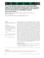
Fig. 1. Electron micrographs of coalesced potato cell walls (PCW) from the wild type control (untransformed Karnico) (A) before starch
degradation showing both entrapped and loose
starch granules and (B) after starch degradation that removed all starch granules. Scanning
election microscopy (SEM) was used to capture
the micrographs of the lyophilized PCW samples. The white arrows show the presence and
absence of starch granules in the PCW samples.
2012; Larsen et al., 2011; McCartney et al., 2000; Tang, Belton, Ng, &
Ryden, 1999; Ulvskov et al., 2005), presumably by modulating the
hydration capacity of the cell wall matrix. Under hydrated conditions,
RG-I galactan side-chains are highly mobile and enhance interactions
with water (Ha, Viëtor, Jardine, Apperley, & Jarvis, 2005; Larsen et al.,
2011). Water embedded in the cell wall matrix is thought to maintain
spatial structures and pores (Makshakova, Faizullin, Mikshina,
Gorshkova, & Zuev, 2018). Intermolecular forces (determined by the
architecture and composition of the cell wall), may also bind or entrap
water through dipole interactions (Labuza, 1968). Dipole interactions
(e.g. hydrogen bonds) arise from hydrophilic pectin groups that include
hydroxyl, carboxyl and amide entities (Matveev, Grinberg, &
Tolstoguzov, 2000). Moreover, pH, (an)ions and drying conditions that
disrupt polymer organization (Moore, Farrant, & Driouich, 2008), have
been implicated to affect interactions of cell walls with water (Renard,
Crépeau, & Thibault, 1994; Serena & Knudsen, 2007).
It is well established that pectins are important swelling components
of the primary cell wall (Thakur, Singh, Handa, & Rao, 1997). Based on
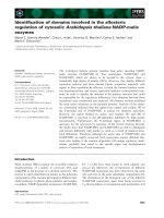
Fig. 2. WBC of PCW from the β-GAL lines and wild type control (untransformed
Karnico). Data (mean ± SD) were collected from three biological replicates (N
= 3), each measured in four technical replicates (N = 4). Asterisks denote
significant differences at α = 0.05 between the β-GAL lines and the control,
derived from one-way ANOVA post-hoc (LSD) tests. SD = standard deviation.
2
Carbohydrate Polymers 227 (2020) 115353
M.T. Klaassen and L.M. Trindade
starch synthase (GBSS) promoter. The tetraploid (2n = 4x = 48) starch
variety Karnico (Averis Seeds, Valthermond, The Netherlands), served
as the genetic background of the β-GAL lines and control (wild type).
The potato plants were grown in pots in an outdoor screen cage (Unifarm, Wageningen UR, Wageningen, The Netherlands), during the potato growing season in The Netherlands (April to September, 2016). At
the end of the growing season, tubers were harvested from 50 individual plants of the β-GAL lines and the control. Tuber fresh weight
and dry matter content were determined directly after harvest. Tuber
dry matter content was determined, after drying the samples at 105oC
until constant weight. The harvested tubers were stored for 5 weeks at
5oC prior to the extraction of raw PCW.
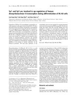
Fig. 3. WBC of de-starched PCW from the wild type control (untransformed
Karnico) after β-galactosidase treatment in-vitro compared to the control (blank
treatment: no enzyme). Data (mean ± SD) were derived from three biological
replicates (N = 3), each measured in one technical replicate (N = 1). The
asterisk denotes a significant difference at α = 0.05 (two-samples Student’s t
test) relative to the control. SD = standard deviation.
2.2. Extraction of raw PCW
To extract raw PCW, tubers were processed in a set-up that mimicked the industrial potato starch recovery process as reported earlier
(Ramasamy, Lips, Bakker, Gruppen, & Kabel, 2014). Tuber batches
(3–5 kg) were processed sequentially. Prior to milling, the tubers were
washed in a rotating drum to remove traces of soil. Milling was performed by using a spinning cylindrical teeth grinder (type: RU 40–260,
Nivoba, Veendam, The Netherlands). To inhibit enzymatic browning of
the slurry, 0.1% (v/w) of 10% sodium metabisulphite (w/v) was added
during the milling process. After milling, the slurry was filtered four
times over a 90 μm centrifugal sieve (Larssons, Bromolla, Sweden). This
step was carried out to wash out free starch granules and to acquire raw
PCW samples. Raw PCW samples were lyophilized and used for further
experiments.
2.3. Starch degradation
Fig. 4. Monosaccharides released in the (not hydrolysed) supernatant from destarched PCW from the wild type control (untransformed Karnico) after β-galactosidase treatment in-vitro compared to the control (blank treatment: no
enzyme). Data (mean ± standard deviation) were derived from three biological replicates (N = 3) and one technical replicate (N = 1).
To acquire PCW with a low (or no) starch content, residual starch
was hydrolysed enzymatically according to the following protocol.
Samples of 5 g PCW (dry matter) were mixed in a 500 mL sodium
acetate buffer solution (0.2 M, pH = 5.6) and homogenized with a
Ultra-turrax disperser. To gelatinize starch and inactivate endogenous
enzymes, the mixtures were heated for 30 min at 80oC. Next, the mixtures were incubated and gently stirred (120 rpm) for 4 h at 40oC, together with a dose of 2000 U α-amylase from porcine pancreas (SigmaAldrich, St. Louis, MO, USA). Afterwards, the pH was lowered to 4.6 by
adding acetic acid (glacial: 99.9%). Next, 500 U amyloglucosidase of
Rhizopus sp. (Megazyme, Bray, Ireland) was added. The mixtures were
incubated for 2 h at 40oC under gentle stirring conditions (120 rpm).
Subsequently, the polysaccharides were precipitated using ethanol 70%
(v/v). After 15 min of precipitation, the residues were collected and
repeatedly subjected to another two cycles of heating and enzymatic
degradation. The final residues were lyophilized to acquire de-starched
PCW.
NMR spectroscopy and enzymatic studies, pectic side-chains have been
pointed out to regulate the hydration capacity of plant cell walls
(Belton, 1997; Funami et al., 2011; Larsen et al., 2011; Ramasamy,
Gruppen, & Kabel, 2015; Ramaswamy, Kabel, Schols, & Gruppen,
2013). To the best of our knowledge however, the specific truncation of
RG-I galactan side-chains in cell walls has not been studied in regard to
the water-binding capacity (WBC). In this study, we evaluated the effects of in-vivo and in-vitro exo-truncation of RG-I β-(1→4)-D-galactan
side-chains on the WBC of potato cell walls (PCW).
2. Materials and methods
2.1. Plant material
2.4. Scanning electron microscopy (SEM)
Three transgenic potato lines (β-GAL) expressing β-(1,4)-galactosidase from chickpea (Cicer arietinum) were used (Martín et al., 2005).
Expression of β-galactosidase was driven by the potato granule-bound
To inspect for starch in PCW, samples were visualized microscopically by using scanning electron microscopy (SEM) (Phenom™,
Table 3
Monosaccharide composition (μg/g) of β-galactosidase treated PCW (in vitro) and control.
μg / g PCW dry basis
PCW sample
Rha
Ara
Gal
Glc
Man
Xyl
GalA
Gal:Rha
HG:RG-I
β-galactosidase (in-vitro)
Control
128 ± 21
124 ± 22
747 ± 70
751 ± 55
3831 ± 265
3891 ± 343
4509 ± 367
4415 ± 355
315 ± 34
321 ± 14
499 ± 40
500 ± 15
607 ± 133
592 ± 132
29.9
31.4
1.9
1.9
Data (mean ± SD). SD = standard deviation. PCW = potato cell walls from the untransformed control (wild type). β-galactosidase (in vitro) = hydrolysed PCW
residue after β-galactosidase (A. niger) treatment in vitro. Control = hydrolysed blank treatment (no enzyme). Rha = rhamnose; Ara = arabinose; Gal = galactose;
Man = mannose; Xyl = xylose; GalA = galacturonic acid. Gal:Rha = ratio galactose to rhamnose (i.e. RG-I galactan side-chain length). HG:RG-I = ratio homogalacturonan (HG) to RG-I. Data were derived from three (N = 3) biological replicates and one (N = 1) technical replicate.
3
Carbohydrate Polymers 227 (2020) 115353
M.T. Klaassen and L.M. Trindade
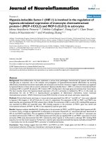
Fig. 5. A simplified conceptual model for the
primary cell wall matrix of PCW, consisting of
RG-I, RG-I galactan side-chains, cellulose, xyloglucan (XyG) and homogalacturonan (HG).
The presence of calcium ions induce cooperative binding of free carboxyl groups from
smooth HG stretches, to form hydrophilic gelling zones. (A) A remodelled, compressed and
stiffer network due to in-vivo expression of βgalactosidase that shortened RG-I galactan
side-chains (direct effect) and modified xyloglucan structures as an indirect effect. These
effects most likely altered arrangements and
interactions between the cell wall components,
potentially reducing the WBC. (B) The network
of the wild type control, showing longer RG-I
galactan side-chains that maintain open pores
that embody free water. (C) A loosened and
more spacious network (increased porosity)
due to β-galactosidase treatment in-vitro
showing shorter RG-I galactan side-chains that
potentially affected intermolecular interactions. Further opening of the apoplastic space
may have increased the WBC.
FEI, Eindhoven, The Netherlands) as previously described (Xu et al.,
2017).
Galactan side − chain length of RG − I =
Ratio HG to RG − I =
2.5. In-vitro β-galactosidase treatment
De-starched PCW samples (300 mg, dry matter) were treated with β(1,4)-galactosidase (EC 3.2.1.23, A. niger) (Megazyme, Bray, Ireland).
Incubations were carried out in 30 mL sodium acetate buffer solutions
(0.1 M) at a pH of 4.5 for 48 h at 40oC. Gentle stirring took place during
incubation (60 rpm). Doses of β-galactosidase (600 U) were added at
the start and again after 24 h during the incubation process.
galactose
rhamnose
galacturonic acid − rhamnose
2 * rhamnose
(1)
(2)
2.8. Water-binding capacity (WBC)
The water-binding capacity of PCW was quantified according to a
modified centrifugation method (Pustjens et al., 2012). PCW samples
(250 mg, dry matter) were added to 30 mL deionized water and stirred
for 5 min at 500 rpm. Next, the samples were centrifuged using nylon
centrifugal filters to remove bound water (pore size: 0.45 μm, F2519-4,
Thermo Fischer Scientific, Waltham, MA, USA). After centrifugation
(1328 × g for 5 min), the wet PCW weight was measured gravimetrically. To quantify the dry weight of the samples, wet samples were
lyophilized for 48 h. All steps were carried out at room temperature.
The water-binding capacity (WBC) was expressed in millilitre (mL)
water per gram (g) dry PCW (Thibault, Renard, & Guillon, 2000). WBC
was calculated as follows:
2.6. Starch content
Starch content (w/w) was determined using a commercially available starch quantification kit (R-Biopharm AG, Darmstadt, Germany).
2.7. Monosaccharide composition
The PCW samples were pre-hydrolysed for 60 min at 30oC using
72% (w/w) sulphuric acid. Subsequently, the mixtures were diluted to a
4% (w/w) sulphuric acid concentration using deionized water. The
samples were further hydrolysed for 180 min at 100oC. Afterwards, the
samples were centrifuged at 15,000 × g for 15 min and the supernatant
phases were collected. Dilutions were made for determining the content
of glucose (dilution factor 100) and the contents of rhamnose, arabinose, galactose, mannose, xylose and uronic acids (dilution factor 7).
The monosaccharide contents were quantified using high-performance
anion exchange chromatography, with pulsed amperometric detection
(HPAEC-PAD) and HPLC-Dionex™ ICS-5000+ DC (Thermo Fischer
Scientific, Waltham, MA, USA). HPAEC-PAD runs were carried out
using a Dionex CarboPac™ PA1 guard column (2 × 250 mm). Eluent
solutions were used as solvents: 0.1 M NaOH, 1 M sodium acetate
(NaAc) in 0.1 M NaOH and deionized water. Volumes of 2.5–5 μL
passed through the system at a flow rate of 250 μL per minute at 30oC.
Recovery standards were employed to correct for monosaccharide
losses, due to destruction by acid hydrolysis (Sluiter et al., 2008). Galacturonic acid content was inferred from total uronic acids using
HPAEC-PAD. The length of RG-I galactan side-chains and ratio of
homogalacturonan (HG) to RG-I were calculated as follows (Huang
et al., 2017):
Water − binding capacity (WBC ) =
wet PCW weight (g )
dry PCW weight (g )
(3)
3. Results and discussion
3.1. Plant performance
To assess the potential impact of β-galactosidase on yield, several
tuber properties of the β-GAL lines were compared to the control.
Earlier work by Martín et al. (2005) showed that β-galactosidase expression levels were high, moderate and low in β-GAL lines 7, 14 and 27
respectively. Tuber yield (fresh weight) was not affected (Table 1); although the yield of β-GAL line 27 was lower than the control, but not
statistically significant. For line β-GAL-27, tuber dry matter content was
lower (P < 0.05), whilst the total amount of extracted PCW was higher
(P < 0.05) relative to the control. These effects were not observed for
the other two β-GAL lines. No significant differences were observed for
starch content in the tubers and raw PCW. Our findings are in line with
earlier studies, reporting that in-vivo expression of β-galactosidase does
not (clearly) impair tuber yield, nor did it induce noticeable phenotypic
changes (Huang et al., 2017; Mayer & Hillebrandt, 1997; Meyer, Dam,
& Lærke, 2009).
4
Carbohydrate Polymers 227 (2020) 115353
M.T. Klaassen and L.M. Trindade
interactions that ultimately affected the WBC. Xyloglucans bind tightly
to cellulose microfibrils, thereby reducing the elasticity of the cell wall
(Abasolo et al., 2009). Therefore, interactions between cellulose, xyloglucan and RG-I galactan side-chains may be crucial to maintain a
functional cell wall. For instance, to create sufficient space in the matrix
to allow water and electrolytes to manoeuvre through (apoplastic
transport). These indirect effects may influence the properties of the cell
wall as suggested earlier (Cosgrove, 2016). Although at this point, no
direct link can be made between truncated RG-I galactan side-chains invivo and reduced WBC.
The WBC increased by 13% after β-galactosidase treatment in-vitro
(Fig. 3). This coincided with a minor release of galactose (1.98% w/w)
after quantification of the monosaccharides in the supernatant phase
(Fig. 4). Physical barriers in the cell wall matrix may have limited the
accessibility of the enzyme to effectively cleave off more galactose
molecules from the non-reducing ends of the galactan chains
(Zykwinska, Thibault, & Ralet, 2007). The levels of galacturonic acid,
rhamnose and arabinose were reduced in PCW (residue) samples from
both the enzyme treatment and the blank control (Table 3), when
compared to PCW used as input material (Table 2). Numerous pectic
fragments from PCW are soluble (Meyer et al., 2009; Ramasamy, 2014),
therefore solubilisation of arabinans and stretches of RG-I and HG may
have caused these changes in our samples. A clear difference between
galactose content in PCW from the enzyme treated and the control residues was not observed (Table 3). The relatively low release of galactose (1.98% w/w) by β-galactosidase in-vitro may underlie this observation (Fig. 4). Both solubilisation of pectic fragments and
degradation of galactan side-chains in-vitro may have distorted the
mediation of intermolecular interactions between cell wall structures
that could have resulted in a different organization of the matrix. For
instance, the water-holding (and swelling) capacity of insoluble cell
wall fractions from wheat flour were increased by in-vitro xylanase
degradation (Gruppen, Kormelink, & Voragen, 1993). The authors
proposed that the minor degradation of xylans may have loosened cell
wall structures, consequently allowing greater swelling and water retention in the cell wall matrix. Changes in the cell wall organization
may affect the porosity and packing of polysaccharides that may extend
the wall (Fujino & Itoh, 1998; Vincken et al., 2003). An alternative
explanation to the increased WBC by β-galactosidase in-vitro, may be
related to the electro-charge of the cell wall matrix. The predominant
cleavage of neutral RG-I galactan side-chains may have increased the
proportion of negatively-changed pectins that stimulated the formation
of gelling zones in the cell wall (Sørensen et al., 2000; Willats, Knox, &
Mikkelsen, 2006). The potential increased abundance of gelling zones
may have affected the WBC. Although we observed that truncated RG-I
galactan side-chains in-vitro increased the WBC of PCW, a causal link
between cannot be established at this point.
3.2. Monosaccharide composition
To assess potential modifications of the cell wall composition due to
β-galactosidase activity, monosaccharides were quantified in PCW
samples of the β-GAL lines and compared to the control. Galactose levels (μg/g) were clearly reduced by 32–67% for the β-GAL lines,
whereas the levels of rhamnose were slightly higher (Table 2). Huang
et al. (2016) showed that the transgene most strongly reduced the
length of galactan side-chains in the hot buffer soluble solids (HBSS)
extracts, where the non-bound HBSS sub-species was strongly affected.
As described in Section 3.3, β-galactosidase activity in-vivo may have
induced other (pleiotropic) effects by modifying the composition and
architecture of other cell wall polysaccharides such as xyloglucan.
Changes in galactose and arabinose were in line with previous studies
regarding these β-GAL lines (Huang et al., 2016, 2017; Martín et al.,
2005). Our findings confirmed that in-vivo expression of β-galactosidase
strongly reduced galactose levels in PCW of the β-GAL lines. The length
of RG-I galactan side-chains were shortened, as shown by the reduced
ratio of galactose to rhamnose by 34–71%. Structural modification of
the ratio homogalacturonan (HG) to rhamnogalacturonan I (RG-I) was
not observed.
In PCW from the wild type control, starch granules were present as
loose entities or were entrapped in partially erupted or intact cells
(Fig. 1). After enzymatic starch degradation, the granules were not
visible anymore and the starch content was reduced from 29.9% to
1.9% (w/w).
3.3. β-galactosidase affected WBC in-vivo and in-vitro
To study the effect of RG-I galactan side-chains on the WBC of PCW,
these side chains were degraded under both in-vivo and in-vitro conditions. Expression of β-galactosidase in-vivo structurally reduced the
WBC by 6–9% for all three β-GAL lines (Fig. 2). This reduction corresponded to a clear decrease in galactose content and shorter (or less
abundant) RG-I galactan side-chains (Table 2). In contrast, in-vitro βgalactosidase degradation increased the WBC by 13% compared to the
control (Fig. 3). This increase corresponded to a release of mostly galactose and traces of glucose and galacturonic acid (Fig. 4). Here, we
show that β-galactosidase affected the WBC of PCW in both in-vivo and
in-vitro conditions, but with contrasting effects. Contrasting effects may
be encountered when in-vivo versus in-vitro systems are compared. For
instance, it has been reported that de-esterification of HG from plant
cell walls led to contrasting biophysical properties in in-vivo and in-vitro
setups (Braybrook & Peaucelle, 2013; Goldberg, Morvan, & Roland,
1986; Peaucelle et al., 2011; Peaucelle, Wightman, & Höfte, 2015; Zhao
et al., 2008).
Cell wall properties are governed by multiple (interacting) factors,
therefore it is not straight-forward to ascribe definitive functions to
specific moieties. Although we observed that RG-I galactan side-chains
influenced the WBC of PCW, it remains challenging to pinpoint the
(relative effects of the) underlying causal factors. In-vivo systems are
prone to indirect effects, as the plant may attempt to compensate for
changes to create a functional cell wall. It has been hypothesized that
in-vivo degradation of cell wall components may activate integrity
sensing pathways or defence responses in plants, as shown for HG degradation (Ferrari et al., 2013). Moreover, targeted degradation of
specific components in-vivo may elicit changes in non-targeted components that may indirectly affect a trait of interest. Huang et al. (2017)
observed that increased pectic methyl-esterification and altered xyloglucan structures (from XXGG to a XXXG permutation) were indirect
effects of β-galactosidase in-vivo in β-GAL-14 line. Pectin methylation
affects ionic (calcium) crosslinking between pectin chains. This mechanism has been proposed to modify textural firmness properties of
potato and carrot cell walls (Ross et al., 2011; Sila, Doungla, Smout,
Van Loey, & Hendrickx, 2006). Changes in xyloglucan structures, as a
result of β-galactosidase expression in-vivo, may also have altered these
3.4. Conceptual model: cell wall porosity and polysaccharide spacing
potentially affect WBC
Based on our results and findings from literature, we propose that
the porosity and spacing between cell wall polysaccharides are important factors that modulate the WBC of PCW. RG-I galactan sidechains have been described to interact with xyloglucan and cellulose
microfibrils (Carpita & Gibeaut, 1993; McCann & Roberts, 1991; Talbott
& Ray, 1992). These interactions may regulate polymer separation and
porosity in the apoplastic space (Carpita & Gibeaut, 1993; Hayashi,
1989; O’Neill & York, 2003). As RG-I galactan side-chains are abundant
in PCW, we expect that these entities may function to maintain open
and well-spaced pores between charged polysaccharides that would
otherwise form tight aggregated complexes that compress the cell wall
matrix (Fig. 5). In our view, RG-I galactan side-chains buffer the deleterious consequences of cell wall dehydration by inhibiting the (irreversible) adhesion of polysaccharides. These include “egg box” junctions between HG and (calcium) ions and strong hydrogen bonds
5
Carbohydrate Polymers 227 (2020) 115353
M.T. Klaassen and L.M. Trindade
between skeletal cellulose microfibrils and xyloglucan. A better understanding of cell wall polysaccharide functions and their potential
interactions, will pave the way to improve the properties of cell wall byproducts for high-value valorisation in food and biobased applications.
Gruppen, H., Kormelink, F., & Voragen, A. (1993). Enzymic degradation of water-unextractable cell wall material and arabinoxylans from wheat flour. Journal of Cereal
Science, 18(2), 129–143.
Ha, M.-A., Viëtor, R. J., Jardine, G. D., Apperley, D. C., & Jarvis, M. C. (2005).
Conformation and mobility of the arabinan and galactan side-chains of pectin.
Phytochemistry, 66(15), 1817–1824.
Hayashi, T. (1989). Xyloglucans in the primary cell wall. Annual Review of Plant Biology,
40(1), 139–168.
Huang, J., Kortstee, A., Dees, D. C., Trindade, L. M., Schols, H. A., & Gruppen, H. (2016).
Modification of potato cell wall pectin by the introduction of rhamnogalacturonan
lyase and β-galactosidase transgenes and their side effects. Carbohydrate Polymers,
144, 9–16.
Huang, J., Kortstee, A., Dees, D. C., Trindade, L. M., Visser, R. G., Gruppen, H., ... Schols,
H. A. (2017). Evaluation of both targeted and non-targeted cell wall polysaccharides
in transgenic potatoes. Carbohydrate Polymers, 156, 312–321.
Labuza, T. P. (1968). Sorption phenomena in foods. Food Technology, 22, 15–19.
Larsen, F. H., Byg, I., Damager, I., Diaz, J., Engelsen, S. B., & Ulvskov, P. (2011). Residue
specific hydration of primary cell wall potato pectin identified by solid-state 13C
single-pulse MAS and CP/MAS NMR spectroscopy. Biomacromolecules, 12(5),
1844–1850.
Lisinska, G., & Leszczynski, W. (1989). Potato starch processing. In G. Lisinska, & W.
Leszczynski (Eds.). Potato science and technology (pp. 281–346). Essex, UK: Elsevier.
Makshakova, O. N., Faizullin, D. A., Mikshina, P. V., Gorshkova, T. A., & Zuev, Y. F.
(2018). Spatial structures of rhamnogalacturonan I in gel and colloidal solution
identified by 1D and 2D-FTIR spectroscopy. Carbohydrate Polymers, 192, 231–239.
Martín, I., Dopico, B., Moz, F. J., Esteban, R., Oomen, R. J., Driouich, A., ... Labrador, E.
(2005). In vivo expression of a Cicer arietinum β-galactosidase in potato tubers leads
to a reduction of the galactan side-chains in cell wall pectin. Plant & Cell Physiology,
46(10), 1613–1622.
Matveev, Y. I., Grinberg, V. Y., & Tolstoguzov, V. B. (2000). The plasticizing effect of
water on proteins, polysaccharides and their mixtures. Glassy state of biopolymers,
food and seeds. Food Hydrocolloids, 14(5), 425–437.
Mayer, F., & Hillebrandt, J.-O. (1997). Potato pulp: Microbiological characterization,
physical modification, and application of this agricultural waste product. Applied
Microbiology and Biotechnology, 48(4), 435–440.
McCann, M., & Roberts, K. (1991). The cytoskeletal basis of plant growth and form. In C.
W. Lloyd (Ed.). Architecture of the primary cell wall (pp. 109–129). London, UK:
Academic press.
McCann, M. C., & Roberts, K. (1994). Changes in cell wall architecture during cell
elongation. Journal of Experimental Botany, 1683–1691.
McCartney, L., Ormerod, A. P., Gidley, M. J., & Knox, J. P. (2000). Temporal and spatial
regulation of pectic (1→ 4)‐β‐D‐galactan in cell walls of developing pea cotyledons:
Implications for mechanical properties. The Plant Journal, 22(2), 105–113.
McDougall, G. J., Morrison, I. M., Stewart, D., & Hillman, J. R. (1996). Plant cell walls as
dietary fibre: Range, structure, processing and function. Journal of the Science of Food
and Agriculture, 70(2), 133–150.
McNeil, M., Darvill, A. G., & Albersheim, P. (1980). Structure of plant cell walls: X.
Rhamnogalacturonan I, a structurally complex pectic polysaccharide in the walls of
suspension-cultured sycamore cells. Plant Physiology, 66(6), 1128–1134.
Meyer, A. S., Dam, B. P., & Lærke, H. N. (2009). Enzymatic solubilization of a pectinaceous dietary fiber fraction from potato pulp: Optimization of the fiber extraction
process. Biochemical Engineering Journal, 43(1), 106–112.
Moore, J. P., Farrant, J. M., & Driouich, A. (2008). A role for pectin-associated arabinans
in maintaining the flexibility of the plant cell wall during water deficit stress. Plant
Signaling & Behavior, 3(2), 102–104.
O’Neill, M. A., & York, W. S. (2003). The composition and structure of plant primary cell
walls. The Plant Cell Wall, 1–54.
Øbro, J., Harholt, J., Scheller, H. V., & Orfila, C. (2004). Rhamnogalacturonan I in
Solanum tuberosum tubers contains complex arabinogalactan structures.
Phytochemistry, 65(10), 1429–1438.
Oomen, R. J. F. J., Vincken, J. P., Bush, M. S., Skjot, M., Voragen, C. H. L., Ulvskov, P.,
et al. (2003). Towards unravelling the biological significance of individual components of pectic hairy regions in plants. In F. Voragen, H. Schols, & R. Visser (Eds.).
Advances in Pectin and Pectinase Research: 2nd International Symposium on Pectins and
Pectinases (pp. 15–34).
Peaucelle, A., Braybrook, S. A., Le Guillou, L., Bron, E., Kuhlemeier, C., & Höfte, H.
(2011). Pectin-induced changes in cell wall mechanics underlie organ initiation in
Arabidopsis. Current Biology, 21(20), 1720–1726.
Peaucelle, A., Wightman, R., & Höfte, H. (2015). The control of growth symmetry
breaking in the Arabidopsis hypocotyl. Current Biology, 25(13), 1746–1752.
Pustjens, A. M., de Vries, S., Gerrits, W. J., Kabel, M. A., Schols, H. A., & Gruppen, H.
(2012). Residual carbohydrates from in vitro digested processed rapeseed (Brassica
napus) meal. Journal of Agricultural and Food Chemistry, 60(34), 8257–8263.
Ramasamy, U. R. (2014). Water holding capacity and enzymatic modification of pressed
potato fibres. Ph.D. Dissertation. Chair group: Food Chemistry. Wageningen, The
Netherlands: Wageningen University Retrieved from />Accessed 2 Nov 2017.
Ramasamy, U. R., Gruppen, H., & Kabel, M. A. (2015). Water-holding capacity of soluble
and insoluble polysaccharides in pressed potato fibre. Industrial Crops and Products,
64, 242–250.
Ramasamy, U. R., Lips, S., Bakker, R., Gruppen, H., & Kabel, M. A. (2014). Improved
starch recovery from potatoes by enzymes and reduced water holding of the residual
fibres. Carbohydrate Polymers, 113, 256–263.
Ramaswamy, U. R., Kabel, M. A., Schols, H. A., & Gruppen, H. (2013). Structural features
and water holding capacities of pressed potato fibre polysaccharides. Carbohydrate
Polymers, 93(2), 589–596.
Funding
This work was funded by Aeres University of Applied Sciences,
Centre for Biobased Economy (CBBE), AVEBE and Averis Seeds B.V.
These funds are gratefully acknowledged.
Author contributions
M.T.K. carried out the experiments, performed the analyses and
wrote the manuscript. L.M.T. coordinated the project, conceived the
study and helped to draft the manuscript.
Ethical standards
The research described in this paper complies with the current laws
of the country in which it was performed.
Declaration of Competing Interest
The authors declare that they have no conflict of interest.
Acknowledgements
The authors thank AVEBE for kindly providing the milling and
sieving machinery to extract the cell walls from the potato tubers. We
are grateful for the valuable contributions by Nick de Vetten, Piet
Buwalda, Marc Laus, Johan Krikken, Maarten Wilbrink, Annemarie
Dechesne, Dirk Jan Huigen, José Anton Abelenda Vila and Yannick
Schrik.
References
Abasolo, W., Eder, M., Yamauchi, K., Obel, N., Reinecke, A., Neumetzler, L., ... Höfte, H.
(2009). Pectin may hinder the unfolding of xyloglucan chains during cell deformation: Implications of the mechanical performance of Arabidopsis hypocotyls with
pectin alterations. Molecular Plant, 2(5), 990–999.
Aldington, S., & Fry, S. C. (1993). Oligosaccharins. Advances in Botanical Research, 19,
1–101.
Baron-Epel, O., Gharyal, P. K., & Schindler, M. (1988). Pectins as mediators of wall
porosity in soybean cells. Planta, 175(3), 389–395.
Belton, P. (1997). NMR and the mobility of water in polysaccharide gels. International
Journal of Biological Macromolecules, 21(1), 81–88.
Braybrook, S. A., & Peaucelle, A. (2013). Mechano-chemical aspects of organ formation in
Arabidopsis thaliana: The relationship between auxin and pectin. PLoS One, 8(3)
e57813.
Brett, C., & Hillmann, J. (1985). Biochemistry of plant cell walls. Cambridge, UK:
Cambridge University Press.
Carpita, N. C., & Gibeaut, D. M. (1993). Structural models of primary cell walls in
flowering plants: Consistency of molecular structure with the physical properties of
the walls during growth. The Plant Journal, 3(1), 1–30.
Cosgrove, D. J. (2000). Loosening of plant cell walls by expansins. Nature, 407(6802),
321.
Cosgrove, D. J. (2016). Catalysts of plant cell wall loosening. F1000Research, 5.
Dick‐Perez, M., Wang, T., Salazar, A., Zabotina, O. A., & Hong, M. (2012).
Multidimensional solid‐state NMR studies of the structure and dynamics of pectic
polysaccharides in uniformly 13C‐labeled Arabidopsis primary cell walls. Magnetic
Resonance in Chemistry, 50(8), 539–550.
Ferrari, S., Savatin, D. V., Sicilia, F., Gramegna, G., Cervone, F., & De Lorenzo, G. (2013).
Oligogalacturonides: Plant damage-associated molecular patterns and regulators of
growth and development. Frontiers in Plant Science, 4, 49.
Fujino, T., & Itoh, T. (1998). Changes in pectin structure during epidermal cell elongation
in pea (Pisum sativum) and its implications for cell wall architecture. Plant & Cell
Physiology, 39(12), 1315–1323.
Funami, T., Nakauma, M., Ishihara, S., Tanaka, R., Inoue, T., & Phillips, G. O. (2011).
Structural modifications of sugar beet pectin and the relationship of structure to
functionality. Food Hydrocolloids, 25(2), 221–229.
Goldberg, R., Morvan, C., & Roland, J. C. (1986). Composition, properties and localisation
of pectins in young and mature cells of the mung bean hypocotyl. Plant & Cell
Physiology, 27(3), 417–429.
6
Carbohydrate Polymers 227 (2020) 115353
M.T. Klaassen and L.M. Trindade
Thakur, B. R., Singh, R. K., Handa, A. K., & Rao, M. (1997). Chemistry and uses of
pectin—A review. Critical Reviews in Food Science and Nutrition, 37(1), 47–73.
Thibault, J., Renard, C., & Guillon, F. (2000). Sugar beet fiber: Production, composition,
physicochemical properties, physiological effects, safety and food applications. In S.
C. A. M. Susan, & L. Dreher (Eds.). Handbook of dietary fiber (pp. 553–582). New York,
NY: Marcel Dekker Food Science and Technology.
Ulvskov, P., Wium, H., Bruce, D., Jørgensen, B., Qvist, K. B., Skjøt, M., ... Sørensen, S. O.
(2005). Biophysical consequences of remodeling the neutral side chains of rhamnogalacturonan I in tubers of transgenic potatoes. Planta, 220(4), 609–620.
Vincken, J. P., Borkhardt, B., Bush, M., Doesdijk-Voragen, C., Dopico, B., Labrador, E., ...
Visser, R. (2000). Remodelling pectin structure in potato. Conference Proceedings of
Phytosfere '99. 23–34 European Plant Biotechnology Network.
Vincken, J. P., Schols, H. A., Oomen, R. J. F. J., McCann, M. C., Ulvskov, P., Voragen, A.
G. J., ... Visser, R. G. F. (2003). If homogalacturonan were a side chain of rhamnogalacturonan I. Implications for cell wall architecture. Plant Physiology, 132(4),
1781–1789.
Wang, T., Park, Y. B., Cosgrove, D. J., & Hong, M. (2015). Cellulose-pectin spatial contacts
are inherent to never-dried Arabidopsis primary cell walls: Evidence from solid-state
nuclear magnetic resonance. Plant Physiology, 168(3), 871–884.
Wang, T., Zabotina, O., & Hong, M. (2012). Pectin–cellulose interactions in the
Arabidopsis primary cell wall from two-dimensional magic-angle-spinning solid-state
nuclear magnetic resonance. Biochemistry, 51(49), 9846–9856.
Willats, W. G., Knox, J. P., & Mikkelsen, J. D. (2006). Pectin: New insights into an old
polymer are starting to gel. Trends in Food Science & Technology, 17(3), 97–104.
Xu, X., Dees, D., Dechesne, A., Huang, X.-F., Visser, R. G. F., & Trindade, L. M. (2017).
Starch phosphorylation plays an important role in starch biosynthesis. Carbohydrate
Polymers, 157, 1628–1637.
Zhao, Q., Yuan, S., Wang, X., Zhang, Y., Zhu, H., & Lu, C. (2008). Restoration of mature
etiolated cucumber hypocotyl cell wall susceptibility to expansin by pretreatment
with fungal pectinases and EGTA in vitro. Plant Physiology, 147(4), 1874–1885.
Zykwinska, A., Thibault, J.-F., & Ralet, M.-C. (2007). Organization of pectic arabinan and
galactan side chains in association with cellulose microfibrils in primary cell walls
and related models envisaged. Journal of Experimental Botany, 58(7), 1795–1802.
Zykwinska, A. W., Ralet, M.-C. J., Garnier, C. D., & Thibault, J.-F. J. (2005). Evidence for
in vitro binding of pectin side chains to cellulose. Plant Physiology, 139(1), 397–407.
Renard, C., Crépeau, M.-J., & Thibault, J.-F. (1994). Influence of ionic strength, pH and
dielectric constant on hydration properties of native and modified fibres from sugarbeet and wheat bran. Industrial Crops and Products, 3(1-2), 75–84.
Ridley, B. L., O’Neill, M. A., & Mohnen, D. (2001). Pectins: Structure, biosynthesis, and
oligogalacturonide-related signaling. Phytochemistry, 57(6), 929–967.
Roach, M. J., Mokshina, N. Y., Badhan, A., Snegireva, A. V., Hobson, N., Deyholos, M. K.,
... Gorshkova, T. A. (2011). Development of cellulosic secondary walls in flax fibers
requires β-galactosidase. Plant Physiology, 156(3), 1351–1363.
Ross, H. A., Morris, W. L., Ducreux, L. J., Hancock, R. D., Verrall, S. R., Morris, J. A., ...
McDougall, G. J. (2011). Pectin engineering to modify product quality in potato.
Plant Biotechnology Journal, 9(8), 848–856.
Satoh, S. (1998). Functions of the cell wall in the interactions of plant cells: Analysis using
carrot cultured cells. Plant & Cell Physiology, 39(4), 361–368.
Schols, H. A., & Voragen, A. G. (1994). Occurrence of pectic hairy regions in various plant
cell wall materials and their degradability by rhamnogalacturonase. Carbohydrate
Research, 256(1), 83–95.
Serena, A., & Knudsen, K. B. (2007). Chemical and physicochemical characterisation of
co-products from the vegetable food and agro industries. Animal Feed Science and
Technology, 139(1), 109–124.
Sila, D. N., Doungla, E., Smout, C., Van Loey, A., & Hendrickx, M. (2006). Pectin fraction
interconversions: Insight into understanding texture evolution of thermally processed
carrots. Journal of Agricultural and Food Chemistry, 54(22), 8471–8479.
Sluiter, A., Hames, B., Ruiz, R., Scarlata, C., Sluiter, J., Templeton, D., ... Crocker, D.
(2008). Determination of structural carbohydrates and lignin in biomass. Laboratory
Analytical Procedure, 1617, 1–16.
Sørensen, S. O., Pauly, M., Bush, M., Skjøt, M., McCann, M. C., Borkhardt, B., ... Ulvskov,
P. (2000). Pectin engineering: Modification of potato pectin by in vivo expression of
an endo-1, 4-β-D-galactanase. Proceedings of the National Academy of Sciences, 97(13),
7639–7644.
Talbott, L. D., & Ray, P. M. (1992). Changes in molecular size of previously deposited and
newly synthesized pea cell wall matrix polysaccharides: Effects of auxin and turgor.
Plant Physiology, 98(1), 369–379.
Tang, H., Belton, P. S., Ng, A., & Ryden, P. (1999). 13C MAS NMR studies of the effects of
hydration on the cell walls of potatoes and Chinese water chestnuts. Journal of
Agricultural and Food Chemistry, 47(2), 510–517.
7