Comprehensive analysis of expression profile and prognostic significance of interferon regulatory factors in pancreatic cancer
Bạn đang xem bản rút gọn của tài liệu. Xem và tải ngay bản đầy đủ của tài liệu tại đây (4.88 MB, 11 trang )
(2022) 23:5
Zhang et al. BMC Genomic Data
/>
BMC Genomic Data
Open Access
RESEARCH
Comprehensive analysis of expression
profile and prognostic significance of interferon
regulatory factors in pancreatic cancer
Ke Zhang1,2, Pan‑Ling Xu3, Yu‑Jie Li1,2, Shu Dong1,2, Hui‑Feng Gao1,2, Lian‑Yu Chen1,2, Hao Chen1,2* and
Zhen Chen1,2*
Abstract
Background: Pancreatic cancer (PC) is a highly lethal disease and an increasing cause of cancer-associated mortality
worldwide. Interferon regulatory factors (IRFs) play vital roles in immune response and tumor cellular biological pro‑
cesses. However, the specific functions of IRFs in PC and tumor immune response are far from systematically clarified.
This study aimed to explorer the expression profile, prognostic significance, and biological function of IRFs in PC.
Results: We observed that the levels of IRF2, 6, 7, 8, and 9 were elevated in tumor compared to normal tissues in PC.
IRF7 expression was significantly associated with patients’ pathology stage in PC. PC patients with high IRF2, low IRF3,
and high IRF6 levels had significantly poorer overall survival. High mRNA expression, amplification and, deep dele‑
tion were the three most common types of genetic alterations of IRFs in PC. Low expression of IRF2, 4, 5, and 8 was
resistant to most of the drugs or small molecules from Genomics of Drug Sensitivity in Cancer. Moreover, IRFs were
positively correlated with the abundance of tumor infiltrating immune cells in PC, including B cells, CD8+ T cells,
CD4+ T cells, macrophages, Neutrophil, and Dendritic cells. Functional analysis indicated that IRFs were involved in T
cell receptor signaling pathway, immune response, and Toll-like receptor signaling pathway.
Conclusions: Our results indicated that certain IRFs could serve as potential therapeutic targets and prognostic
biomarkers for PC patients. Further basic and clinical studies are needed to validate our findings and generalize the
clinical application of IRFs in PC.
Keywords: Pancreatic cancer, Bioinformatics analysis, Interference factor, Prognosis, Immune infiltration
Background
Pancreatic cancer (PC) is a lethal disease and ranked
as the 14th in cancer incidence and the 7th leading
cause of cancer death globally based on the latest data
[1]. It is predicted that PC will be the second leading
cause of cancer mortality in the USA in the next two or
three decades [2]. In total, 60,430 new cases were estimated to be diagnosed with PC, and 48,220 deaths were
*Correspondence: ;
2
Department of Oncology, Shanghai Medical College, Fudan University,
Shanghai 200032, China
Full list of author information is available at the end of the article
estimated to happen in the United States in 2021 [3]. PC
is hard to detect and diagnose in its early stages due to
lacking obvious clinical symptoms and occult location
[4]. Approximately, 80-85% patients were diagnosed at
advanced stages and not suitable to receive curable surgery. Chemotherapy is currently the standard treatment
for these patients. Although target therapy and immunotherapy have achieved promising success in other malignancies, the 5-year survival rate for whole PC patients
remains only 10%. These alarming data demonstrated
that novel therapeutic targets and prognostic biomarkers
are urgent to be discovered.
© The Author(s) 2022. Open Access This article is licensed under a Creative Commons Attribution 4.0 International License, which
permits use, sharing, adaptation, distribution and reproduction in any medium or format, as long as you give appropriate credit to the
original author(s) and the source, provide a link to the Creative Commons licence, and indicate if changes were made. The images or
other third party material in this article are included in the article’s Creative Commons licence, unless indicated otherwise in a credit line
to the material. If material is not included in the article’s Creative Commons licence and your intended use is not permitted by statutory
regulation or exceeds the permitted use, you will need to obtain permission directly from the copyright holder. To view a copy of this
licence, visit http://creativecommons.org/licenses/by/4.0/. The Creative Commons Public Domain Dedication waiver (http://creativeco
mmons.org/publicdomain/zero/1.0/) applies to the data made available in this article, unless otherwise stated in a credit line to the data.
Zhang et al. BMC Genomic Data
(2022) 23:5
Interferon regulatory factors (IRFs) family is a variety
of transcription factors and it is firstly identified in 1988
[5]. Nine members of the IRF family were presented in
mammals (IRF1/2/3/4/5/6/7/8/9). It has been well established that IRFs perform vital functions in innate and
adaptive immunity, and immune response [6, 7]. Previous studies also suggested that IRFs played a vital role in
the cell biological process of many tumor cells [8]. However, their roles in the regulation of oncogenesis are complex and even controversial based on previous reports.
For example, IRF-1 inhibited cell growth in breast cancer by inhibiting NF-κB activity and suppressing TRAF2
and cIAP1 [9]. In gastric cancer, evidence suggested that
IRF2 could suppress tumor cell invasion and migration
via MMP-1 in STAD [10]. In PC, it is reported that IRF2
expression was upregulated and associated with tumor
size, differentiation, pathology stage, and survival of the
patients. Knockdown on the expression of IRF2 inhibited
cell growth in PC cells [11].
Page 2 of 11
Thus, we embarked on the current study, aiming to
explore the expression and its correlation with clinicopathological features of IRFs in PC. Moreover, we also
detected the role of IRFs in the immune infiltration in PC
and IRFs-associated functions. The results of our study
may provide additional data about the function of IRFs
in PC and the prognostic and therapeutic biomarkers for
PC.
Results
Differential expression of IRFs in PC patients
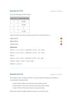
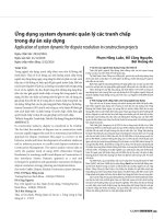
We firstly detected the level of IRFs in PC in Oncomine
database. The results were shown in Fig. 1 and Table S1.
We found that the level of IRF2, IRF6, IRF7, IRF8 and
IRF9 were upregulated in tumor tissues in PC (Fig. 1,
P < 0.05). In addition, we also noticed that no difference was found between tumor tissues and normal tissues about the level of IRF1/3/4/5/6 in PC (Fig. 1). To
be more specific, Malte’s dataset revealed that IRF2
Fig. 1 IRFs expression in pancreatic cancer at mRNA level. The number in the figure was the numbers of datasets with statistically significant mRNA
over-expression (red) or down-expression (blue) of IRFs, which was obtain with the P-value of 0.05 and fold change of 2. This Figure was plotted
using ONCOMINE (https://www.oncomine.org/)
Zhang et al. BMC Genomic Data
(2022) 23:5
expression was increased in Pancreatic Ductal Adenocarcinoma with a fold change (FC) of 2.051 [12].
According to the data of Huadong’s study, IRF6 was
upregulated in Pancreatic Carcinoma tissues and the
FC is 2.43 [13]. A total of two datasets demonstrated
the upregulation of IRF7 in PC [12, 14]. Moreover,
three datasets suggested that IRF8 expression was
increased in PC [15–17]. We also found that the level
of IRF9 was elevated in PC with the FC of 2.205 and
2095 [13, 17]. This is followed by the verification of the
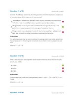
expression of IRFs in PC using the TCGA dataset. We
found that the mRNA level of IRF1, IRF2, IRF3, IRF5,
IRF6, IRF7, IRF8 and IRF9 (Fig. 2A-I) were upregulated
in PC (All p < 0.05). Therefore, we suggested that the
level of IRF3, IRF6, IRF7, IRF8 and IRF9 were upregulated in tumor tissues of PC.
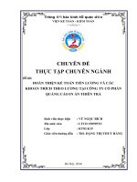
The association between the level of IRFs and patient’s
pathology stage in PC were also detected. Interestingly,
a significant association was obtained between IRF7
expression and patient’s pathology stage in PC (Fig. 3G,
p < 0.00908). Further analysis showed that the expression
of IRF7 is significantly higher in stage II compared with
stage I (p = 0.014). However, there was no association
Page 3 of 11
between IRF1/2/3/4/5/6/8/9 expression and patient’s
pathology stage in PC (Fig. 3, p > 0.05).
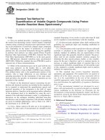
Prognostic value of IRFs in PC patients
The prognostic value of IRFs in PC was explored using
TCGA dataset. The data showed that PC patients with
high IRF2 (HR = 1.8, p = 0.0069) and low IRF3 expression (HR = 1.6, p = 0.031) were associated with poor
overall survival (Fig. 4A). Particularly, PC patients with
high IRF6 expression had both poor overall survival
(HR = 1.6, p = 0.03) (Fig. 4A) and poor disease-free survival (HR = 1.6, p = 0.028) (Fig. 4B).
Co‑expression, genetic alteration, and drug sensitivity
analyses of IRFs in PC patients
Comprehensive analyses were performed to explore
the molecular character of IRFs in PC using cBioportal. There was a low to moderate correlation among
the mRNA level of each IRFs member in patients with
PC (Fig. 5A). Moreover, the genetic alterations analysis revealed that IRF1, IRF2, IRF3, IRF4, IRF5, IRF6,
IRF7, IRF8 and IRF9 were altered in 6, 8, 8, 2.7, 6, 6,
4, 4, and 4% of the queried PC samples, respectively
Fig. 2 The mRNA level of IRFs in pancreatic cancer. The expression of IRF1 (A), IRF2 (B), IRF3 (C), IRF4 (D), IRF5 (E), IRF6 (F), IRF7 (G), IRF8 (H), IRF9 (I)
in pancreatic cancer tissues and normal tissues at mRNA level. This Figure was plotted using GEPIA (http://gepia.cancer-pku.cn/). *P < 0.05; T: tumor
tissues; N: normal tissues
Zhang et al. BMC Genomic Data
(2022) 23:5
Page 4 of 11
Fig. 3 Correlation between IRFs and the pathological stage of pancreatic cancer patients. The expression of IRF1 (A), IRF2 (B), IRF3 (C), IRF4 (D), IRF5
(E), IRF6 (F), IRF7 (G), IRF8 (H), IRF9 (I) in different pathological stage of pancreatic cancer patients at mRNA level. This Figure was plotted using GEPIA
(http://gepia.cancer-pku.cn/). *P < 0.05
(Fig. 5B). High mRNA expression, amplification and
deep deletion were the three most common type of
genetic alterations in these samples (Fig. 5B). To clarify whether these genetic alterations could affect the
prognosis of PC patients. Kaplan-Meier method was
drawn and revealed that genetic alterations of IRFs
could not affect the overall survival and disease-free
survival of PC patients (Fig. 5C, p > 0.05). Drug sensitivity analysis was also performed. And the results
suggested that low expression of IRF2/4/5/8 were
resistant to most of the drugs or small molecules
from GDSC (Fig. S1).
Immune cell infiltration analysis of IRFs in PC patients
Tumor-infiltrating lymphocytes could serve as a biomarker for predicting sentinel lymph node status and
cancer patients’ survival [18, 19]. The previous study has
revealed close correlation between immune infiltration
analysis and IRFs in cancers [20]. In our study, a comprehensive detection of the correlation between IRFs
and immune cell infiltration in PC was conducted using
TIMER. As shown in Fig. 6, the level of IRF7 was positively associated with the infiltration abundance of B cells
(Cor = 0.436, P = 2.40e-09), CD8+ T cells (Cor = 0.401,
P =
5.32e-08) macrophages (Cor
= 0.227, P = 2.84e-3),
Neutrophils (Cor = 0.471, P = 8.03e-11) and Dendritic
cells (Cor = 0.566, P = 6.71e-16) (Fig. 6A). Interestingly,
the expression of IRF2 and IRF6 also showed a positive
association with the infiltration abundance of these five
immune cells in PC (Fig. 6B and F, all p < 0.05). As for
IRF3, a positive correlation was obtained between IRF3
expression and the infiltration abundance of B cells,
CD8+ T cells and CD4+ T cells (Fig. 6C). Moreover,
the expression of IRF4 (Fig. 6D), IRF5 (Fig. 6E), IRF8
(Fig. 6H) and IRF9(Fig. 6I) was positively associated with
all these six immune cells, including B cells, CD8+ T
cells, CD4+ T cells, macrophages, Neutrophils and Dendritic cells (all p < 0.05). We also found that IRF7 expression was associated with the infiltration abundance of
CD8+ T cells (Cor = − 0.209, P = 6.07e-083), CD4+ T
cells (Cor = 0.389, P = 1.77e-7), Neutrophils (Cor = 0.252,
P = 8.72e-4) (Fig. 6G). We also explored the effect of copy
number alteration of IRF on the immune cell infiltration
in PC. As a result, copy number alteration of IRF could
suppress the infiltration level of immune cells to some
extent (Fig. S2).
IRFs‑associated biologic functions in PC
DAVID 6.8 and Metascape were utilized to explore the
biological functions of IRFs and their neighboring genes
(Table S2) in PC. As we could see in Fig. 7 the results
of functional analysis obtained from DAVID 6.8. The
item of GO enrichment analysis revealed that IRFs and
their neighboring genes were mainly involved in defense
response to virus, T cell receptor signaling pathway,
immune response, regulatory region DNA binding, protein binding, sequence-specific DNA binding, transcription factor activity, sequence-specific DNA binding,
cadherin binding involved in cell-cell adhesion and type I
interferon signaling pathway (Fig. 7A). The item of KEGG
pathway revealed that IRFs and their neighboring genes
were mainly linked to RIG-I-like receptor signaling pathway, T cell receptor signaling pathway, Toll-like receptor signaling pathway, Cell adhesion molecules (CAMs)
and Cytosolic DNA-sensing pathway (Fig. 7B). PPI network showed that IRFs were mainly involved in immune
Zhang et al. BMC Genomic Data
(2022) 23:5
Page 5 of 11
Fig. 4 The prognostic value of IRFs in pancreatic cancer. A The overall survival of pancreatic cancer patients with high/low mRNA level of IRFs.
B The disease-free survival of pancreatic cancer patients with high/low mRNA level of IRFs. All the analyses were performed with Kaplan-Meier
analysis. This Figure was plotted using GEPIA (http://gepia.cancer-pku.cn/). HR: Hazard Ratio
response, sequence-specific DNA binding, response to
Type I interferon (Fig. S3).
To further detect IRFs-associated functions in patients
with PC, Metascape was further used to perform enrichment analysis. Interestingly, the result suggested that
IRFs and their neighboring genes were mainly linked to
regulation of cytokine production, immune responseactivating signal transduction in GO function analysis
and type I interferon signaling pathway (Fig. S4A and
B, Table S3). The data of KEGG pathways analyses were
shown in Fig. S4C, D, and Table S4. As expected, IRFs
and their neighboring genes were involved in T cell
receptor signaling pathway, Cell adhesion molecules
(CAMs), Antigen processing (presentation) and Hippo
signaling pathway. Moreover, PPI network and Molecular Complex Detection (MCODE) components were isolated to identify the correlation between IRFs and their
neighboring genes. The result indicated the involvement
of IRFs in T cell receptor signaling pathway and Pertussis
(Fig. S4E and F).
Discussion
Increasing researches have reported the significant functions of IRFs in immune response [21]. IRFs also exert an
important function in basic cellular mechanisms, including cell invasion, proliferation, and apoptosis [22, 23].
Moreover, IRFs were also involved in the tumorigenesis
and progression of cancers, including colorectal cancer,
Zhang et al. BMC Genomic Data
(2022) 23:5
Page 6 of 11
Fig. 5 Co-expression and genetic alteration of IRFs in pancreatic cancer. A Correlation heat map of each member of IRFs in pancreatic cancer. B
Summary of genetic alterations of IRFs in pancreatic cancer. C Overall survival and disease-free survival of pancreatic cancer patients with/without
IRFs genetic alterations. This Figure was plotted using cBioportal (https://www.cbioportal.org/)
hepatocellular carcinoma, and esophageal cancer [24–
26]. In this study, we conducted a comprehensive analysis
to explore the specific role of IRFs in PC.
We first detected the mRNA level of IRFs in PC, revealing that the level of IRF2, IRF6, IRF7, IRF8 and IRF9
were elevated in tumor tissues in PC. Further prognosis
analysis revealed that high IRF2 expression, low IRF3
expression, and high IRF6 predict poor survival in PC.
Similarly, IRFs were also suggested to be prognosis biomarkers in various malignancies. It was reported that
low IRF3 was associated with poor disease free survival
and overall survival in urothelial carcinoma [27]. Another
study indicated high IRF2 expression independently predicts poor overall survival in colorectal cancer [28]. These
Zhang et al. BMC Genomic Data
(2022) 23:5
two were consistent with our study. Moreover, IRF3 and
IRF7 were linked to a poor prognosis in colon adenocarcinoma [20].
Another significant finding is that IRFs were correlated with the abundance of immune cells in PC, including B cells, CD8+ T cells, CD4+ T cells, macrophages,
Neutrophil and Dendritic cells. In fact, these immune
cells have been proved to be biomarker or involved in
the tumor progression of PC microenvironment. Mobilization of CD8 + T Cells could promote PD-1 checkpoint therapy in human PC by blockading CXCR4 [29].
Another study suggested infiltrating CD4/CD8 high T
cells as a biomarker involved in good prognosis in PC
[30]. Neutrophil extracellular traps could facilitate liver
micro metastasis by activating cancer-associated fibroblasts in PC [31]. Moreover, dendritic cell paucity could
result in dysfunctional immune surveillance in PC [32].
Enrichment analysis was performed, which revealed
that IRFs and their neighboring genes mainly associated
with T cell receptor signaling pathway, immune response,
Toll-like receptor signaling pathway, Cell adhesion molecules (CAMs), sequence-specific DNA binding, response
to Type I interferon, and Hippo signaling pathway. Interestingly, Toll-like receptor signaling pathway was associated with immune response and play an important
function in cancer initiation and progression [33, 34].
CAMs play a vital role in cancer progression and metastasis [35]. Increasing studies revealed that T cell receptor
signaling was involved in the control of regulatory T cell
differentiation and function, which plays an important
function in cancer initiation and progression [36].
Based on our results, we would like to emphasize the
potential roles of IRF2, IRF3, and IRF6. Generally, our
finding suggested that IRF2 functions as an oncoprotein,
Page 7 of 11
which is consistent with previous studies. IRF2 expression was increased in esophageal squamous cell carcinomas (ESCC) compared with matched normal esophageal
tissues. In addition, the tumorigenicity of ESCC cells was
enhanced with IRF2 overexpression in nude mice model
[37]. IRF2 could attenuated apoptosis through induction
of autophagy in acute myelocytic leukemia cells [38]. A
recent study found that Kras-IRF2 axis drives immune
suppression and immune therapy resistance in colorectal cancer [39]. Particularly, our finding was supported
by a previous study which reported that IRF2 expression
was upregulated and associated with tumor size, differentiation, pathology stage, and survival of PC patients
and knockdown on the expression of IRF2 inhibited cell
growth in PC cells [11]. Evidence above suggests that
IRF2 is a potential biomarker and therapeutic target in
PC and other malignancies.
IRF3 was reported to participant in the innate immune
response against cancer via STING pathway [40]. A
recent study revealed that IRF3 prevents colorectal
tumorigenesis via inhibiting the nuclear translocation of
β-catenin. Moreover, high expression of IRF3 correlated
with favorable survival in colorectal cancer, lung adenocarcinoma, and hepatocellular carcinoma patients [41].
Consistent with the literature above, our results showed
that IRF3 expression positively correlated with the infiltration abundance of B cells, CD8+ T cells and CD4+
T cells. Besides, high IRF3 expression level is associated
with better survival. These results indicated that IRF3
functions as a tumor suppressor.
Our results showed that IRF6 was overexpressed in
PC compared with normal tissue and high expression
level of IRF6 corelated with poor survival. It seems
that IRF6 plays a pro-cancer role and is a promising
Fig. 6 The correlation between IRFs and immune infiltration in pancreatic cancer. The correlation between the expression of IRF1 (A), IRF2 (B),
IRF3 (C), IRF4 (D), IRF5 (E), IRF6 (F), IRF7 (G), IRF8 (H), IRF9 (I) and the abundance of B cells, CD8+ T cells, CD4+ T cells, Macrophage, Neutrophils and
Dendritic cells. This Figure was plotted using TIMER (https://cistrome.shinyapps.io/timer/)
Zhang et al. BMC Genomic Data
(2022) 23:5
Page 8 of 11
Fig. 7 The enrichment analysis of IRFs and neighboring genes. A Bar plot of GO enrichment in cellular component terms, biological process terms,
and molecular function terms. B Bar plot of KEGG enriched terms. This Figure was plotted using David 6.8 (https://david.ncifcr f.gov/home.jsp)
therapeutic target in PC. However, previous studies
indicated that IRF6 acts as a tumor suppressor [42,
43]. And the decreased expression of IRF6 was clinically correlated with poor prognosis of Gastric cancer [44]. Our findings are contrary to previous studies
which have suggested further experimental and clinical
research to clarify the roles of IRF6 in PC.
Some limitations must be reported about our study.
Firstly, most analyses were performed at mRNA level but
not protein level and gene level. Secondly, immune suppressive cells, such as regulatory T cells (Tregs) and myeloid-derived suppressor cells (MDSCs) also defines the
microenvironment of PC [45]. These immune suppressive
cells may contribute to tumor progression and poor survival. Unfortunately, relevant data are temporarily unavailable. Furthermore, it would be better to validate our
results by performing in vivo and in vitro experiments.
Conclusion
This study comprehensively explored the expression
profile, prognostic value, and biological functions of
IRF family members in PC, providing insights of IRFs as
potential therapeutic targets and prognostic biomarker
for PC. Further basic and clinical studies are needed to
validate our findings and generalize the clinical application of IRFs in PC.
Methods
ONCOMINE
ONCOMINE (https://w ww.oncomine.org/) is an online
platform including oncogene expression signatures
from over 80,000 cancer samples [46]. We can analyze
the mRNA level of target genes in cancer and normal
tissues by using ONCOMINE database and the p-value
was 0.05, the fold change was 2 and the gene rank
Zhang et al. BMC Genomic Data
(2022) 23:5
Page 9 of 11
was10%, we analyzed the mRNA level of IRFs in PC and
normal tissue with student’s t-test.
somatic copy number alterations of IRFs. A P-value of
less than 0.05 meant significant difference existed.
GEPIA
David 6.8
GEPIA (http://gepia.cancer-pku.cn/) is a novel web portal
collecting mRNA data from The Cancer Genome Atlas
(TCGA) database [47]. A total of 186 complete TCGA PC
samples were involved in the following analyses. we further detected the mRNA level of IRFs in PC. Setting the
group cutoff as median, we explored the prognostic value
of IRFs in PC by using overall survival (OS) plots and
disease-free survival (DFS) plots. Hazard ratio (HR) and
log-rank P-value were also listed in the plots. Moreover,
correlation analysis was conducted to explore the genes
most associated with each member of IRFs in PC.
cBioPortal
cBioPortal (https://www.cbioportal.org/) is a comprehensive web portal that integrates genomic data from over
30,000 cancer samples of various cancer types [48]. Using
the TCGA datasets (N = 186), we performed gene alterations analysis of IRFs in PC samples, which was summarized by the “Oncoprint” module. Using cBioportal, we
also performed co-expression among IRFs in PC samples
in the “Co-expression” module with spearman’s correlation. In addition, we set a threshold as ±2.0 in mRNA
expression z-scores (RNA Seq V2 RSEM) and protein
expression z-scores (RPPA). Putative copy-number determined using GISTIC 2.0.
GSCALite
GSCALite (http://bioinfo.life.hust.edu.cn/web/GSCAL
ite/) is a novel web portal collecting mRNA data from
the TCGA database [49]. In drug sensitivity analysis,
the association between IRFs level and the drug using
the data from GDSC (Genomics of Drug Sensitivity
in Cancer) was analyzed with the spearman correlation. The positive correlation means that the gene high
expression is resistant to the drug, vise verse. These
analyses were performed with TCGA datasets (N = 186)
and a p-value < 0.05 indicates statistical significance.
TIMER
TIMER (https://cistrome.shinyapps.io/timer/) is a web
server for comprehensively analysis the relationship
between immune cells infiltration and gene expression
[50]. In the current study, we first evaluated the association between IRFs expression in PC and abundance of B
cell, CD8+ T cell, CD4+ T cell, Macrophage, Neutrophil,
and Dendritic cell according to TCGA datasets (N = 186).
In the “SCNA” module, we performed the comparison
of tumor infiltration levels among tumors with different
DAVID 6.8 (https://david.ncifcrf.gov/home.jsp) is a functional annotation tool providing the biological function
of submitted genes [51]. After isolated the genes most
associated with each member of IRFs in pancreatic adenocarcinoma, we performed ene Ontology (GO) [52, 53]
and Kyoto Encyclopedia of Genes and Genomes (KEGG)
[54–56] pathway enrichment analysis of these genes and
the result was visualized with R project using a “ggplot2”
package and a p < 0.05.
GeneMANIA
GeneMANIA (http://genemania.org/) is established to
predict the biological functions of target gene sets [57].
Protein protein interaction (PPI) networks of the IRFs
were constructed to indicate the relative relationships
and the potential functions of these gene sets.
Metascape
Metascape (http://metascape.org) is a reliable functional
annotation tool providing the biological function of submitted genes [58]. Based on the functional annotation of
gene/protein lists, Metascape can facilitate data-driven
decisions. After isolated the genes most associated with
each member of IRFs in pancreatic adenocarcinoma, we
further explored the function of IRFs and closely related
neighbor genes.
Abbreviations
CAMs: Cell adhesion molecules; CD: Cluster of differentiation; DFS: Diseasefree survival; GO: Gene ontology; HR: Hazard ratio; IRF: Interferon regulatory
factor; KEGG: Kyoto Encyclopedia of Genes and Genomes; MCODE: Molecular
Complex Detection; OS: Overall survival; PC: Pancreatic cancer; PD-1: Pro‑
grammed death-1; PPI: Protein-protein interaction; TCGA: The Cancer Genome
Atlas.
Supplementary Information
The online version contains supplementary material available at https://doi.
org/10.1186/s12863-021-01019-5.
Additional file 1.
Acknowledgments
The results shown here are in whole or part based upon data generated by
the TCGA Research Network: https://www.cancer.gov/tcga. We acknowl‑
edge TCGA program and other contributors for providing their platform and
datasets.
Authors’ contributions
KZ and PLX: performed the analysis and wrote the manuscript, YJL: performed
the analysis, SD and HFG: were responsible for writing, review, and editing,
LYC: was responsible for the supervision, HC and ZC: study concept and
Zhang et al. BMC Genomic Data
(2022) 23:5
design. The final manuscript was approved by all authors who agreed to be
accountable for the content of this work.
Funding
This work was supported by the National Natural Science Foundation of China
under Grant NO. 81973616. The funding bodies had no role in the design of
the study and collection, analysis, and interpretation of data and in writing the
manuscript.
Availability of data and materials
All data generated or analyzed during this study are included in the article and
its supplementary information files. The dataset supporting the conclusions
of this article is available in the TCGA repository, project identifier ‘TCGA-PAAD’
and hyperlink to dataset in https://portal.gdc.cancer.gov/repository.
Declarations
Ethics approval and consent to participate
The Cancer Genome Atlas (TCGA) and other databases used in this study
are public databases. Ethical approval has been obtained from the patients
involved in these databases. Users can download relevant data for free for
purpose of research and publishing articles. We state that all methods were
carried out in accordance with relevant guidelines and regulations.
Consent for publication
Not applicable.
Competing interests
The authors declare that the research was conducted in the absence of any
commercial or financial relationships that could be construed as a potential
conflict of interest.
Author details
1
Department of Integrative Oncology, Fudan University Shanghai Cancer
Center, Shanghai 200032, China. 2 Department of Oncology, Shanghai Medi‑
cal College, Fudan University, Shanghai 200032, China. 3 Chinese Integrative
Medicine Oncology Department, First Affiliated Hospital of Anhui Medical
University, Hefei 230000, Anhui, China.
Received: 5 May 2021 Accepted: 13 December 2021
References
1. Sung H, Ferlay J, Siegel RL, Laversanne M, Soerjomataram I, Jemal A,
et al. Global cancer statistics 2020: GLOBOCAN estimates of incidence
and mortality worldwide for 36 cancers in 185 countries. CA Cancer J
Clin. 2021;71:209.
2. Rahib L, Smith BD, Aizenberg R, Rosenzweig AB, Fleshman JM, Matri‑
sian LM. Projecting cancer incidence and deaths to 2030: the unex‑
pected burden of thyroid, liver, and pancreas cancers in the United
States. Cancer Res. 2014;74(11):2913–21.
3. Siegel RL, Miller KD, Fuchs HE, Jemal A. Cancer statistics, 2021. CA
Cancer J Clin. 2021;71(1):7–33.
4. Moore A, Donahue T. Pancreatic cancer. Jama. 2019;322(14):1426.
5. Tamura T, Yanai H, Savitsky D, Taniguchi T. The IRF family transcrip‑
tion factors in immunity and oncogenesis. Annu Rev Immunol.
2008;26:535–84.
6. Borden EC, Sen GC, Uze G, Silverman RH, Ransohoff RM, Foster GR, et al.
Interferons at age 50: past, current and future impact on biomedicine.
Nat Rev Drug Discov. 2007;6(12):975–90.
7. Yanai H, Negishi H, Taniguchi T. The IRF family of transcription factors:
inception, impact and implications in oncogenesis. Oncoimmunology.
2012;1(8):1376–86.
8. Yan Y, Zheng L, Du Q, Yan B, Geller DA. Interferon regulatory
factor 1 (IRF-1) and IRF-2 regulate PD-L1 expression in hepato‑
cellular carcinoma (HCC) cells. Cancer Immunol Immunother.
2020;69(9):1891–903.
Page 10 of 11
9. Armstrong MJ, Stang MT, Liu Y, Yan J, Pizzoferrato E, Yim JH. IRF-1
inhibits NF-κB activity, suppresses TRAF2 and cIAP1 and induces
breast cancer cell specific growth inhibition. Cancer Biol Ther.
2015;16(7):1029–41.
10. Chen YJ, Liang L, Li J, Wu H, Dong L, Liu TT, et al. IRF-2 inhibits gastric
cancer invasion and migration by down-regulating MMP-1. Dig Dis Sci.
2020;65(1):168–77.
11. Cui L, Deng Y, Rong Y, Lou W, Mao Z, Feng Y, et al. IRF-2 is over-expressed
in pancreatic cancer and promotes the growth of pancreatic cancer cells.
Tumour Biol. 2012;33(1):247–55.
12. Buchholz M, Braun M, Heidenblut A, Kestler HA, Klöppel G, Schmiegel W,
et al. Transcriptome analysis of microdissected pancreatic intraepithelial
neoplastic lesions. Oncogene. 2005;24(44):6626–36.
13. Pei H, Li L, Fridley BL, Jenkins GD, Kalari KR, Lingle W, et al. FKBP51 affects
cancer cell response to chemotherapy by negatively regulating Akt.
Cancer Cell. 2009;16(3):259–66.
14. Logsdon CD, Simeone DM, Binkley C, Arumugam T, Greenson JK,
Giordano TJ, et al. Molecular profiling of pancreatic adenocarcinoma and
chronic pancreatitis identifies multiple genes differentially regulated in
pancreatic cancer. Cancer Res. 2003;63(10):2649–57.
15. Segara D, Biankin AV, Kench JG, Langusch CC, Dawson AC, Skalicky DA,
et al. Expression of HOXB2, a retinoic acid signaling target in pancre‑
atic cancer and pancreatic intraepithelial neoplasia. Clin Cancer Res.
2005;11(9):3587–96.
16. Iacobuzio-Donahue CA, Maitra A, Olsen M, Lowe AW, van Heek NT, Rosty
C, et al. Exploration of global gene expression patterns in pancreatic ade‑
nocarcinoma using cDNA microarrays. Am J Pathol. 2003;162(4):1151–62.
17. Grützmann R, Pilarsky C, Ammerpohl O, Lüttges J, Böhme A, Sipos B, et al.
Gene expression profiling of microdissected pancreatic ductal carcino‑
mas using high-density DNA microarrays. Neoplasia. 2004;6(5):611–22.
18. Ohtani H. Focus on TILs: prognostic significance of tumor infiltrating
lymphocytes in human colorectal cancer. Cancer Immun. 2007;7:4.
19. Azimi F, Scolyer RA, Rumcheva P, Moncrieff M, Murali R, McCarthy SW,
et al. Tumor-infiltrating lymphocyte grade is an independent predictor
of sentinel lymph node status and survival in patients with cutaneous
melanoma. J Clin Oncol. 2012;30(21):2678–83.
20. Yuemaier M, Zhou Z, Zhou Y, Wu C, Li F, Liang X, et al. Identification of the
prognostic value and clinical significance of interferon regulatory factors
(IRFs) in colon adenocarcinoma. Med Sci Monit. 2020;26:e927073.
21. Battistini A. Interferon regulatory factors in hematopoietic cell differentia‑
tion and immune regulation. J Interf Cytokine Res. 2009;29(12):765–80.
22. Yi Y, Wu H, Gao Q, He HW, Li YW, Cai XY, et al. Interferon regulatory factor
(IRF)-1 and IRF-2 are associated with prognosis and tumor invasion in
HCC. Ann Surg Oncol. 2013;20(1):267–76.
23. Velloso FJ, Trombetta-Lima M, Anschau V, Sogayar MC, Correa RG. NODlike receptors: major players (and targets) in the interface between innate
immunity and cancer. Biosci Rep. 2019;39(4):BSR20181709.
24. Hong M, Zhang Z, Chen Q, Lu Y, Zhang J, Lin C, et al. IRF1 inhibits the
proliferation and metastasis of colorectal cancer by suppressing the RASRAC1 pathway. Cancer Manag Res. 2019;11:369–78.
25. Yu M, Xue H, Wang Y, Shen Q, Jiang Q, Zhang X, et al. miR-345 inhibits
tumor metastasis and EMT by targeting IRF1-mediated mTOR/STAT3/AKT
pathway in hepatocellular carcinoma. Int J Oncol. 2017;50(3):975–83.
26. Zhang M, Zhang L, Cui M, Ye W, Zhang P, Zhou S, et al. miR-302b inhibits
cancer-related inflammation by targeting ERBB4, IRF2 and CXCR4 in
esophageal cancer. Oncotarget. 2017;8(30):49053–63.
27. Wang LA, Yang B, Rao W, Xiao H, Wang D, Jiang J. The correlation of BER
protein, IRF3 with CD8+ T cell and their prognostic significance in upper
tract urothelial carcinoma. Onco Targets Ther. 2019;12:7725–35.
28. Mei Z, Wang G, Liang Z, Cui A, Xu A, Liu Y, et al. Prognostic value of IRF-2
expression in colorectal cancer. Oncotarget. 2017;8(24):38969–77.
29. Seo YD, Jiang X, Sullivan KM, Jalikis FG, Smythe KS, Abbasi A, et al.
Mobilization of CD8(+) T cells via CXCR4 blockade facilitates PD-1
checkpoint therapy in human pancreatic cancer. Clin Cancer Res.
2019;25(13):3934–45.
30. Wang Z, Zhao J, Zhao H, A S, Liu Z, Zhang Y, et al. Infiltrating CD4/CD8
high T cells shows good prognostic impact in pancreatic cancer. Int J Clin
Exp Pathol. 2017;10(8):8820–8.
31. Takesue S, Ohuchida K, Shinkawa T, Otsubo Y, Matsumoto S, Sagara A,
et al. Neutrophil extracellular traps promote liver micrometastasis in
Zhang et al. BMC Genomic Data
32.
33.
34.
35.
36.
37.
38.
39.
40.
41.
42.
43.
44.
45.
46.
47.
48.
49.
50.
51.
52.
53.
54.
55.
56.
(2022) 23:5
pancreatic ductal adenocarcinoma via the activation of cancer-associ‑
ated fibroblasts. Int J Oncol. 2020;56(2):596–605.
Hegde S, Krisnawan VE, Herzog BH, Zuo C, Breden MA, Knolhoff BL, et al.
Dendritic cell paucity leads to dysfunctional immune surveillance in
pancreatic cancer. Cancer Cell. 2020;37(3):289–307.e289.
Fitzgerald KA, Kagan JC. Toll-like receptors and the control of immunity.
Cell. 2020;180(6):1044–66.
Ohadian Moghadam S, Nowroozi MR. Toll-like receptors: the role in
bladder cancer development, progression and immunotherapy. Scand J
Immunol. 2019;90(6):e12818.
Beauchemin N, Arabzadeh A. Carcinoembryonic antigen-related cell
adhesion molecules (CEACAMs) in cancer progression and metastasis.
Cancer Metastasis Rev. 2013;32(3-4):643–71.
Li MO, Rudensky AY. T cell receptor signalling in the control of regulatory
T cell differentiation and function. Nat Rev Immunol. 2016;16(4):220–33.
Wang Y, Liu DP, Chen PP, Koeffler HP, Tong XJ, Xie D. Involvement of IFN
regulatory factor (IRF)-1 and IRF-2 in the formation and progression of
human esophageal cancers. Cancer Res. 2007;67(6):2535–43.
Zhang F, Li J, Zhu J, Liu L, Zhu K, Cheng S, et al. IRF2-INPP4B-mediated
autophagy suppresses apoptosis in acute myeloid leukemia cells. Biol
Res. 2019;52(1):11.
Liao W, Overman MJ, Boutin AT, Shang X, Zhao D, Dey P, et al. KRAS-IRF2
axis drives immune suppression and immune therapy resistance in
colorectal cancer. Cancer Cell. 2019;35(4):559–572.e557.
Woo SR, Fuertes MB, Corrales L, Spranger S, Furdyna MJ, Leung MY, et al.
STING-dependent cytosolic DNA sensing mediates innate immune
recognition of immunogenic tumors. Immunity. 2014;41(5):830–42.
Tian M, Wang X, Sun J, Lin W, Chen L, Liu S, et al. IRF3 prevents colorectal
tumorigenesis via inhibiting the nuclear translocation of beta-catenin.
Nat Commun. 2020;11(1):5762.
Restivo G, Nguyen BC, Dziunycz P, Ristorcelli E, Ryan RJ, Özuysal ÖY, et al.
IRF6 is a mediator of notch pro-differentiation and tumour suppressive
function in keratinocytes. EMBO J. 2011;30(22):4571–85.
Botti E, Spallone G, Moretti F, Marinari B, Pinetti V, Galanti S, et al. Devel‑
opmental factor IRF6 exhibits tumor suppressor activity in squamous cell
carcinomas. Proc Natl Acad Sci U S A. 2011;108(33):13710–5.
Li D, Cheng P, Wang J, Qiu X, Zhang X, Xu L, et al. IRF6 is directly regulated
by ZEB1 and ELF3, and predicts a favorable prognosis in gastric cancer.
Front Oncol. 2019;9:220.
Huber M, Brehm CU, Gress TM, Buchholz M, Alashkar Alhamwe B, von
Strandmann EP, et al. The immune microenvironment in pancreatic
cancer. Int J Mol Sci. 2020;21(19):7307.
Rhodes DR, Yu J, Shanker K, Deshpande N, Varambally R, Ghosh D, et al.
ONCOMINE: a cancer microarray database and integrated data-mining
platform. Neoplasia. 2004;6(1):1–6.
Tang Z, Li C, Kang B, Gao G, Li C, Zhang Z. GEPIA: a web server for cancer
and normal gene expression profiling and interactive analyses. Nucleic
Acids Res. 2017;45(W1):W98–w102.
Cerami E, Gao J, Dogrusoz U, Gross BE, Sumer SO, Aksoy BA, et al. The cBio
cancer genomics portal: an open platform for exploring multidimen‑
sional cancer genomics data. Cancer Discov. 2012;2(5):401–4.
Liu CJ, Hu FF, Xia MX, Han L, Zhang Q, Guo AY. GSCALite: a web server for
gene set cancer analysis. Bioinformatics. 2018;34(21):3771–2.
Li T, Fan J, Wang B, Traugh N, Chen Q, Liu JS, et al. TIMER: a web server for
comprehensive analysis of tumor-infiltrating immune cells. Cancer Res.
2017;77(21):e108–10.
Huang da W, Sherman BT, Lempicki RA. Bioinformatics enrichment tools:
paths toward the comprehensive functional analysis of large gene lists.
Nucleic Acids Res. 2009;37(1):1–13.
Ashburner M, Ball CA, Blake JA, Botstein D, Butler H, Cherry JM, et al. Gene
ontology: tool for the unification of biology. The gene ontology consor‑
tium. Nat Genet. 2000;25(1):25–9.
Gene Ontology Consortium. The Gene Ontology resource: enriching a
GOld mine. Nucleic Acids Res. 2021;49(D1):D325-34.
Kanehisa M, Goto S. KEGG: kyoto encyclopedia of genes and genomes.
Nucleic Acids Res. 2000;28(1):27–30.
Kanehisa M. Toward understanding the origin and evolution of cellular
organisms. Protein Sci. 2019;28(11):1947–51.
Kanehisa M, Furumichi M, Sato Y, Ishiguro-Watanabe M, Tanabe M.
KEGG: integrating viruses and cellular organisms. Nucleic Acids Res.
2021;49(D1):D545–d551.
Page 11 of 11
57. Warde-Farley D, Donaldson SL, Comes O, Zuberi K, Badrawi R, Chao P,
et al. The GeneMANIA prediction server: biological network integration
for gene prioritization and predicting gene function. Nucleic Acids Res.
2010;38(Web Server issue):W214–20.
58. Zhou Y, Zhou B, Pache L, Chang M, Khodabakhshi AH, Tanaseichuk O,
et al. Metascape provides a biologist-oriented resource for the analysis of
systems-level datasets. Nat Commun. 2019;10(1):1523.
Publisher’s Note
Springer Nature remains neutral with regard to jurisdictional claims in pub‑
lished maps and institutional affiliations.
Ready to submit your research ? Choose BMC and benefit from:
• fast, convenient online submission
• thorough peer review by experienced researchers in your field
• rapid publication on acceptance
• support for research data, including large and complex data types
• gold Open Access which fosters wider collaboration and increased citations
• maximum visibility for your research: over 100M website views per year
At BMC, research is always in progress.
Learn more biomedcentral.com/submissions