Whole exome sequencing of pediatric leukemia reveals a novel InDel within FLT-3 gene in AML patient from Mizo tribal population, Northeast India
Bạn đang xem bản rút gọn của tài liệu. Xem và tải ngay bản đầy đủ của tài liệu tại đây (1.17 MB, 9 trang )
(2022) 23:23
Vanlallawma et al. BMC Genomic Data
/>
BMC Genomic Data
Open Access
RESEARCH
Whole exome sequencing of pediatric
leukemia reveals a novel InDel within FLT‑3 gene
in AML patient from Mizo tribal population,
Northeast India
Andrew Vanlallawma1, Doris Lallawmzuali2, Jeremy L. Pautu3, Vinod Scaria4, Sridhar Sivasubbu4 and
Nachimuthu Senthil Kumar1*
Abstract
Background: Leukemia is the most common type of cancer in pediatrics. Genomic mutations contribute towards
the molecular mechanism of disease progression and also helps in diagnosis and prognosis. This is the first scientific
mutational exploration in whole exome of pediatric leukemia patients from a cancer prone endogamous Mizo tribal
population, Northeast India.
Result: Three non-synonymous exonic variants in NOTCH1 (p.V1699E), MUTYH (p.G143E) and PTPN11 (p.S502P) were
found to be pathogenic. A novel in-frame insertion-deletion within the juxtamembrane domain of FLT3 (p.Tyr589_
Tyr591delinsTrpAlaGlyAsp) was also observed.
Conclusion: These unique variants could have a potential mutational significance and these could be candidate
genes in elucidating the possibility of predisposition to cancers within the population. This study merits further investigation for its role in diagnosis and prognosis and also suggests the need for population wide screening to identify
unique mutations that might play a key role towards precision medicine.
Keywords: Pediatric leukemia, Exome sequencing, FLT3, PTPN11, Non-synonymous, Mizoram
Background
Leukemia is the most common type of childhood cancer and the incidence is estimated to be 3.1 per 100,000
cases worldwide [1]. Leukemia can be broadly classified
according to the type of hematopoietic lineage that turns
cancerous as lymphoid or myeloid leukemia and by the
progressiveness of the disease as acute or chronic. Previously, the causal root factor for leukemia was thought
to be chromosomal translocation [2], however, there are
reports that indicate that this translocation alone is not
*Correspondence:
1
Department of Biotechnology, Mizoram University, Aizawl, Mizoram
796004, India
Full list of author information is available at the end of the article
adequate for leukemiogenesis and are even observed during pregnancy [2–4]. Moreover, the translocation does
not define the progressiveness of ALL patients [5, 6].
Apart from the chromosomal translocation, studies
on nuclear mutational pattern revealed a crucial event
in the Acute Myeloid Leukemia (AML) pathogenesis
and its clinical significance [7, 8]. The two-hit model of
leukemiogenesis captures the key events in the genomic
alteration, where the two classes of mutations: one in the
genes responsible for growth or survival and the other
in the genes responsible for differentiation leading to
self-renewability were proposed for leukemiogenesis [9].
Identifying a specific gene mutation in leukemia plays a
© The Author(s) 2022. Open Access This article is licensed under a Creative Commons Attribution 4.0 International License, which
permits use, sharing, adaptation, distribution and reproduction in any medium or format, as long as you give appropriate credit to the
original author(s) and the source, provide a link to the Creative Commons licence, and indicate if changes were made. The images or
other third party material in this article are included in the article’s Creative Commons licence, unless indicated otherwise in a credit line
to the material. If material is not included in the article’s Creative Commons licence and your intended use is not permitted by statutory
regulation or exceeds the permitted use, you will need to obtain permission directly from the copyright holder. To view a copy of this
licence, visit http://creativecommons.org/licenses/by/4.0/. The Creative Commons Public Domain Dedication waiver (http://creativeco
mmons.org/publicdomain/zero/1.0/) applies to the data made available in this article, unless otherwise stated in a credit line to the data.
Vanlallawma et al. BMC Genomic Data
(2022) 23:23
vital role in its diagnosis, prognosis and also in predicting
the disease-free survival rate and recurrence [10].
Next Generation Sequencing (NGS) approach such as
Whole Exome Sequencing (WES) has been used in identifying the mutational profiles of different cancers and its
subtypes. The mutational profiles of pediatric leukemia
have also been studied in different ethnic groups revealing recurrent mutational hotspots, driver genes and
variants involved in different pathways: RTK/RAS signaling and its downstream MAPK/ERK signaling, PI3K/
AKT and MTOR, JAK/STAT signaling, Notch signaling,
WNT/β-catenin, CXCL12, NF-κB, Metabolic and other
pathways, including p53 [11–14]. The class of genes that
are frequently mutated includes lymphoid/myeloid differentiation, transcription factors, epigenetic regulators,
signal transduction, apoptotic regulators [15, 16]. FLT3 variants within a particular hotspot region have been
reported to be different across different ethnic groups
and various types of indels and internal tandem duplication have also been reported [17]. Hence, it is very much
essential to study unexplored ethnic groups with high
incidences of cancers.
Here, we report whole exome sequencing of pediatric
leukemic patients as the first scientific report from Mizo
endogamous tribal population, Northeast India wherein
the state has the highest incidences of various Cancers in
the country [18]. We hypothesize that the high incidence
Page 2 of 9
of cancer rate in the population might be a result of
unique mutations that are present within the coding
regions of the genome. To understand the germline
mutations in the population as well as to capture the variants that may be directly responsible for the disease, the
present study is a pilot approach to explore the pediatric
patient samples.
Results
Whole exome analysis of pediatric leukemia patients
identified 46 non-synonymous exonic variants with allele
frequency ≤ 0.05, out of which 16 variants have been
reported in ClinVar (Table 1). However, only MUTYH
variant (p.G143E; dbSNP id: rs730881833) present in
AML-M1 patient was reported as likely pathogenic for
MUTYH associated Polyposis and Hereditary Cancer
Predisposition Syndrome in ClinVar. Non-synonymous
exonic gene variants that are not present in ClinVar are
listed in Table 2. NOTCH1 variant (p.V1699E) in one
patient (AML-M1) was not reported in any database and
predicted as pathogenic by 7 different prediction tools
using VarSome [19]. PTPN11 variant (p.S502P) present
in one patient (AML-M1) was identified which was also
not present in ClinVar. Sanger Validation of point mutation observed in this study are shown in Supplementary
Figs. 1, 2 and 3.
Table 1 Non-synonymous exonic variants that matched with ClinVar with their clinical significance and disease associated
Chr Pos
Ref Alt Gene
Clinical Significance from ClinVar
Disease associated
11
108,098,555 A
G
ATM
Conflicting interpretations of Pathogenicity Ataxia-telangiectasia syndrome, Hereditary cancer-predisposing
syndrome
11
108,159,732 C
T
ATM
Benign / Likely Benign
Ataxia-telangiectasia syndrome, Hereditary cancer-predisposing
syndrome
11
119,156,193 C
T
CBL
Benign / Likely Benign
Rasopathy, Noonan-Like Syndrome Disorder
12
49,434,409
G
A
KMT2D
Benign
Kabuki syndrome
1
45,797,401
G
A
MUTYH
Conflicting interpretations of Pathogenicity MYH-associated polypopsis, Hereditary cancer-predisposing
syndrome
1
45,797,914 C
T
MUTYH Pathogenic / Likely Pathogenic
MYH-associated polypopsis, Hereditary cancer-predisposing
syndrome
1
45,800,146
C
T
MUTYH
Benign, Uncertain Significance
MYH-associated polypopsis, Hereditary cancer-predisposing
syndrome
1
45,800,167
G
A
MUTYH
Benign, Uncertain Significance
MYH-associated polypopsis, Hereditary cancer-predisposing
syndrome
18
42,643,270
G
T
SETBP1
likely Benign
Schinzel-Giedion syndrome
1
85,742,023
C
A
BCL10
Benign
Immunodeficiency 37
20
31,022,469
G
A
ASXL1
Benign
C-like syndrome
22
23,654,017
G
A
BCR
Uncertain Significance
ALL and AML
4
106,158,550 G
T
TET2
Not provided
4
55,589,830
A
G
KIT
Uncertain Significance
9
139,401,375 C
T
NOTCH1 Uncertain Significance
Adams-Oliver syndrome 5, Cardiovascular phenotype
9
139,410,139 T
C
NOTCH1 Uncertain Significance
Adams-Oliver syndrome 5
Chr Chromosome Number, Pos Position, Ref Reference Allele, Alt Alternate Allele
Gastrointestinal stroma tumor
A
T
G
BIRC3
NOTCH1
ATM
156 (83%)
A
107 (54%)
44 (72)
ATM
–
T
PTPN11
0 (0%)
BCR
C
BCL10
55 (51%)
11 (12%)
G
MUTYH
66 (55%)
FLT3
C
MUTYH
51 (49%)
72 (43%)
14 (47%)
49 (49%)
68 (46%)
47 (44%)
35 (49%)
14 (39%)
90 (49%)
57 (55%)
104 (75%)
58 (48%)
112 (53%)
16 (38%)
28 (52%)
28 (55%)
49 (43%)
Counts (%)
C
CCGGins
del and ins
C
A
A
T
T
T
T
T
G
G
A
G
A
G
T
A
T
C
G
A
A
Alt
91 (46%)
17 (27)
71 (78%)
32 (17%)
57 (97%)
52 (49%)
54 (45%)
53 (51%)
94 (56%)
16 (53%)
50 (51%)
80 (54%)
59 (56%)
37 (51%)
22 (61%)
95 (51%)
46 (45%)
35 (25%)
62 (51%)
99 (47%)
26 (62%)
26 (45%)
23 (45%)
64 (56%)
Counts (%)
198
61
85
188
59
107
121
104
167
30
99
149
106
72
36
185
103
139
121
211
42
54
51
114
Total reads
T1697P
S1092fs
YFY58991delWAGDins
S502P
A5S
P18L
G25D
Q757X
V1232M
E1466D
H1380Y
H24R
I438V
A230V
K260R
D1163N
K260R
V1699E
A5S
C1482F
I567V
K260R
A5S
A5S
AA change
Het
Het
Het
Het
Hom
Het
Het
Het
Het
Het
Het
Het
Het
Het
Het
Het
Het
Het
Het
Het
Het
Het
Het
Het
Hom /Het
T-cell prolymphocytic leukemia, somatic
ALL, CML somatic
ALL, AML
Leukemia, juvenile myelomonocytic, somatic
Male germ cell tumor, somatic
–
–
Myelodysplastic syndrome, somatic
–
–
T-cell prolymphocytic leukemia, somatic
T-cell prolymphocytic leukemia, somatic
Germ cell tumors, somatic, Leukemia, acute
myeloid (Smu,AD)
–
–
Myelodysplastic syndrome, somatic
–
–
Male germ cell tumor, somatic
T-cell prolymphocytic leukemia, somatic
–
–
Male germ cell tumor, somatic
Male germ cell tumor, somatic
OMIM phenotype and Mode of inheritance
T
0
0
T
T
T
T
T
T
T
T
T
T
T
T
T
T
D
T
T
T
T
T
T
S_P
B
0
0
P
B
B
P
0
P
B
B
B
B
P
B
B
B
D
B
B
B
B
B
B
P_P
N
0
0
D
N
D
N
D
N
N
N
N
D
D
N
N
N
D
N
N
D
N
N
N
MT_P
Ref Reference Allele, Alt Alternate Allele, Counts Read Counts, AA Change Amino acid Change, Hom/Het Homozygous/Heterozygous, S_P SIFT_Prediction, P_P PolyPhen2 Prediction and MT_P Mutation taster Prediction. B –
Benign, D – Damaging, P – Probably Damaging, N – Neutral, T- Tolerated, 0 – No prediction
GDN4262
GDN4261
GDN4260
C
G
SETBP1
C
C
ATM
ASXL1
A
ATM
NOTCH 1
A
KIT
GDN4259
G
MUTYH
G
ASXL1
GDN4258
A
BIRC3
A
A
NOTCH1
BIRC3
C
BCL10
GDN4256
GDN4255
C
BCL10
GDN4253
C
BCL10
GDN4252
Ref
Gene
Sample
Table 2 Non-synonymous exonic variants not matched in CIViC and ClinVar with their OMIM phenotype and pathogenicity prediction
Vanlallawma et al. BMC Genomic Data
(2022) 23:23
Page 3 of 9
Vanlallawma et al. BMC Genomic Data
(2022) 23:23
Page 4 of 9
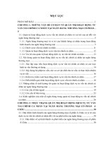
Fig. 1 Novel InDel in FLT-3 identified in AML-M1. A Wildtype FLT-3 (exon 14) depicting the genomic DNA with amino acid it encodes and the
position. Bases in lower script indicates the deleted bases (ttctac) in the Mutant type. B Mutant FLT-3 depicting the genomic DNA with amino acid it
encodes and the position. * Indicates the position of insertion and bases in lower script (gggcggggg) are the inserted bases
Identification of novel FLT3 InDel in PTPN11
mutation positive patient
Our study observed two tyrosine amino acid (in 589, 591
position) and phenylalanine (590 position) to be deleted
and an in-frame insertion consistent with ITD region
[17], four amino acids are inserted [tryptophan (W),
alanine (A), glycine (G), aspartic acid (D)- (p.Tyr589_
Tyr591delinsTrpAlaGlyAsp)] (Fig. 1). along with PTPN11
p.S502P from the same patient. NGS based evidence of
the indel and its Sanger validation is given in Supplementary Figures (Supplementary Figs. 4 and 5).
Discussion
Whole exome analysis performed in the germline
genomic mutational screening in pediatric leukemia
patients showed important heterozygous variants and
not in the corresponding mother samples suggesting that
it could be a de novo germline mutation or is inherited
from the father. The exception was for two homozygous
variants, BCL10: p.A5S and ASXL: p.G652 which were
reported as benign in ClinVar for immunodeficiency syndrome and C-like syndrome, respectively. Unreported
variants were observed in this study which could be population specific variant.
MUTYH encodes an enzyme DNA glycosylase that
functions in base excision repair when there is DNA
damage from oxidation. MUYTH variants are also found
in different types of cancers like gastric cancers [20],
pediatric high grade midline gliomas patients [21] and in
pediatric leukemia [22, 23]. However, a previously unreported variant G143E was found in a two years old girl
with AML-M1 subtype with a family history of gastric
cancer, but the mother did not carry the same mutation.
Nonetheless, as the variant was predicted as pathogenic
by three predicting softwares, as well as categorized as
MUTYH Associated Polyposis (MAP) and Hereditary
Cancer Predisposing Syndrome in ClinVar, the variant
might confer loss of the protein function.
NOTCH1 encodes a transmembrane receptor protein that is required in the differentiation and maturation process and is activated during early embryo or in
hematopoiesis [24, 25] Mutations in the PEST and heterodimer domains within NOTCH1 are found in 50% of
T-cell-ALL patients [26]. Mutations in the gene are likely
in ALL patients where its role is poorly understood in
myeloid malignancies. This may be because activation
of the Notch pathway varies between different cell types
[27] Fu et al. [28] first reported the NOTCH1 mutation
and even suggested that NOTCH1 mutations are rare
events in AML patients. Study reported that in vivo activation of NOTCH1 by its ligands arrest AML growth
while inhibition confers proliferation [29]. This suggested
that NOTCH1 plays a role as tumour suppressor in AML,
furthermore, a novel pathway that activates NOTCH1
for inhibiting cell growth was identified [30]. The mutation observed in this study as predicted by the prediction
softwares (SIFT, PolyPhen2 and Mutation Taster) was
deleterious suggesting that NOTCH1 p.V1699E mutation
might confer loss of function and its ability to suppress
tumour might be lost. From the aforementioned studies,
inactivation or loss of function aids in cell proliferation
suggesting that the patient in this study with AML-M1
subtype might have a proliferative advantage as extensive
expression of NOTCH1 especially in M1 and M0 – AML
patients with simultaneous expression of CD7 which is
a marker for immaturity was observed that reflects in a
poor overall survival rate [31].
FLT3 mutations can be classified into point mutations in the Tyrosine Kinase Domain (TKD) and Internal Tandem Duplications (ITD) in the juxtamembrane
domain with each accounting for 5 and 25% of patients
with AML, respectively. Both these types of mutations
resulted in constitutive activation of the gene where the
autoinhibitory mechanism is disrupted in the case of ITD
and turns to ligand independent FLT3 thereby promoting
cell proliferation. Similarly, point mutations in the TKD
are in the activation loop that stabilize the active kinase
conformation resulting in constitutive activation of its
kinase activity [32]. It was also highlighted that approximately 30% of ITDs insert in the TKD1 and not in the
JMD [33]. It was observed that 77 pediatric AML patients
out of 630 tested positive for ITD out of which 59 had a
single duplication and the rest 18 had 2 or 3 ITD’s [17].
Chow et al. [34] also showed that in 569 consecutive
adult AML patients 126 (22.1%) harbored FLT3-ITDs.
FLT3 mutations occurred in about 35–45% of AML
patients with normal karyotype [35]. Consistently, these
Vanlallawma et al. BMC Genomic Data
(2022) 23:23
FLT3-ITD are in-frame mutations with varying size that
ranges from 3 to > 1000 nucleotides [36].
Different types of FLT3-ITD within a hotspot region
have also been reported [35–37]. The InDel found in this
study have not been reported earlier. However, the site
of duplication observed in this study is fairly consistent
with other duplication site which is in the juxtamembrane domain, amino acid 591–599 [17, 34]. This study
identified an insertion deletion mutation, where amino
acids YFY (positions 589, 590 and 591) are deleted and 4
amino acids (WAGD) are inserted. Y589 and Y591 were
reported to be the STAT5 docking site [38] where it activates and expresses an antiapoptotic protein called BCLxL [39]. Though FLT3-ITD was reported to be a driver
mutation in AML patients’ initiation of leukemia by FLT3 through STAT pathway might not be the case for this
patient. However, evading cell death is not the only property of cancers, as acquiring a proliferative advantage is
also one of the natures of cancerous cells as proposed
in the “two hit model” [9]. The proliferative advantage
could be attained for this patient as the tyrosine residue at position 599 in FLT-3 is still intact and this residue was reported to be the interacting site of FLT-3 with
PTPN11. They also showed that the absence of tyrosine
residue (Y > F mutant) showed enhanced Erk activation
and acquired proliferation and survival advantages when
compared with WT-FLT-3 [40]. This could be a potential
pathway for its initiation as hyperactive PTPN11 deregulates the RAS pathway, thereby contributing to its growth
[41, 42]. This indel mutation generates a protein with one
amino acid longer than the wild type. Length mutation
of FLT-3 – ITD either by elongation or shortening of the
juxtamembrane domain results in gain-of-function and
could transform 32D cells, irrespective of the tyrosine
residues [43, 44].
Mutations in PTPN11 are found commonly in JMML
patients without RAS and NF1 mutation and are involved
in leukemiogenesis by negative regulation of the RAS
pathway by conferring growth advantage [45]. Most of
the mutations reported in PTPN11 are within the domain
N-terminal src-homology-2 (N-SH2) and protein tyrosine phosphatase (PTP) domain. The change of serine to
proline results in the loss of S502 – E76 H-bond that is
required for its auto-inhibition and thus acquiring an
open conformation exposing the catalytic site leading to
an increase by 8-fold turnover value of S502P when compared to wild type PTPN11 in their basal activity [46].
Consistent with other findings, GND4261 has a mutation in PTP domain (p.S502P) with no RAS mutation
but positive for FLT-3 mutants. PTPN11 mutation was
found to be seen more among boys [47], but in the present study, the mutation was found in a girl child. In contrast to adult AML patients, where there is no association
Page 5 of 9
observed between the two gene mutations, PTPN11 and
FLT-3-ITD [47]. However, the sample size is small to
define a true association for this population.
Conclusion
There are four different amino acid changes in the same
position of the PTPN11 (p.S502A, p.S502T, p.S502P,
p.S502L) that are reported in ClinVar. A change from
serine to alanine was interpreted as pathogenic with
clinical conditions like Rasopathy and Noonan Syndrome
[48], a change from serine to threonine was interpreted
as pathogenic with clinical conditions like Noonan Syndrome 1and Juvenile Myelomonocytic Leukemia [49] and
a change from serine to leucine was interpreted as pathogenic with clinical conditions like Noonan Syndrome
1 and Juvenile Myelomonocytic Leukemia [50]. Even
though, a change of serine to proline in the same position
was reported in few studies in AML and Myelodysplastic Syndrome (MDS) [51], there is no record of the variant’s pathogenicity in its clinical conditions in ClinVar.
However, as the other three changes p.S502A, p.S502T,
and p.S502L are interpreted as pathogenic, the chance of
p.S502P becoming pathogenic is also greatly increased.
Additionally, the amino acid residues that are close
by (p.R498W/L, p.R501K, p.G503R/V/A/E, p.M504V,
p.Q506P, p.T507K) are also reported for Noonan Syndrome in Human Gene Mutation Database (HGMD) [52]
which suggest the functional importance of this region.
The two mutations, NOTCH1 (p.V1699E), and FLT-3
(p.Tyr589_Tyr591delinsTrpAlaGlyAsp) observed in this
study have not been reported and the frequencies are
unknown as well. IndiGenomes is a database that had
over 1000 healthy Indian genomes where Mizo tribal
population are also included in the study [53]. South
Asian Genomes and Exomes (SAGE) database consists
of 1213 genomes and exome data sets from South Asians
comprising 154 million genetic variants [54]. The variants found in our study were not present in the IndiGenomes and SAGE database suggesting that these variants
observed might be a disease specific polymorphism
for the region. As the sample size of this study is small,
stressing the importance of these variants in the population might not be appropriate. However, these findings
could be a potential mutational uniqueness towards the
population that merits further investigation.
Materials and methods
Sample collection
All pediatric leukemia patients totaling to eleven
children between 2 and 16 years (median age = 11, 3
girls and 8 boys) who are diagnosed with leukemia
and undergoing treatment at Mizoram State Cancer Institute, Aizawl, Mizoram, Northeast India from
Vanlallawma et al. BMC Genomic Data
(2022) 23:23
Page 6 of 9
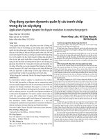
Fig. 2 Prioritization of variants for whole exome data. F1 to F4: Filter’s applied. 1: Raw VCF file annotated using ANNOVAR; 2: Selection of
non-Synonymous exonic variants from the annotated variants; 3: Selection of variants having allele frequency lower than 0.05; 4: Selection of
variants that are predicted as deleterious in any of the two-predicting software (SIFT, PolyPhen2, Mutation Taster); 5: Matching with frequently
mutated genes associate with leukemia; 6: Matching with CIViC and ClinVar database; 7: Interpreting using OMIM database
January–July 2018 were included in this study (Supplementary Table 1). After obtaining informed consent
from the parents, 2 ml of peripheral blood was drawn
from the patients. Blood sample was also collected from
four mothers who are willing to participate. Peripheral
blood was collected in EDTA coated vials and stored in
-20 °C for DNA isolation.
DNA isolation and whole exome sequencing
DNA was isolated from whole blood by using QIAamp
DNA Mini Kit (CA, USA) as per the manufacturer’s
protocol with some modifications. The quality of isolated DNA was checked using Nanodrop (NanoDrop™
1000 Spectrophotometer, Thermofisher) at optical density (OD) 260 nm. The purity of the isolated DNA was
checked by measuring OD at 260/280 for protein contamination as well as 260/230 for RNA contamination.
The quality of the isolated DNA was also checked by
0.8% Agarose Gel Electrophoresis. After the required
concentration of 100 ng for library preparation was
obtained, DNA library was prepared by using Illumina
v4 TruSeq Exome library prep as per the manufacturer’s
protocol. The sequencing and data analysis was carried
out at CSIR- IGIB, New Delhi.
WES data analysis
Whole Exome Sequencing was performed using Illumina
HiSeq 2500 and generated approximately 52.2 million
reads that passed Quality Control (QC) with 52.1 million
reads (99.97%) aligned to the reference genome (hg19)
per sample (Supplementary Table S2). GATK haplotype
caller was used for calling germline variants from the
generated BAM files [55]. The VCF file was annotated
using ANNOVAR [56].
Prioritization of variants
The quality of the raw read fastq files were checked
twice before and after trimming the adapter sequence
and the low-quality reads by Trimmomatic software [57] and FastQC [58]. Processed fastq files were
mapped on human reference genome (hg19) using
BWA-MEM [59]. Variant calling was done using
GATK haplotype caller [55] and the vcf file was annotated using ANNOVAR [56]. Prioritizations of variants
found in the whole exome data are shown in Fig. 2. The
number of variants after every filtering step is given in
Vanlallawma et al. BMC Genomic Data
(2022) 23:23
Supplementary Table S3. From the annotated variants:
the first filtering step (F1) variants that are non-synonymous and exonic were selected, the second filter (F2)
selected variants that have allele frequency ≤ 0.05, and
the third filtering step (F3) selected variants that are
predicted as deleterious by any two of the predicting
software (SIFT, PolyPhen2 or Mutation Taster) [60–62]
for further analysis. Frequently mutated genes which
are reported in leukemia patients were listed out after
performing data mining through literature survey as
well as which are catalogued in databases (Supplementary Table S4). F2 and F3 were then matched with the
list of frequently mutated genes in leukemia (F4). The
observed variants were interpreted using CIViC [63]
and ClinVar database [64], while variants not present in
CIViC and ClinVar were interpreted using dbSNP [65]
and OMIM database [66]. The allele frequency was also
compared using databases like ExAc [67], gnoMAD
[68], ESP6500 (https://e vs.gs.washington.edu/EVS/),
1000genomes [69], IndiGenomes [53] and SAGE [54].
Abbreviations
ALL: Acute Lymphoblastic Leukemia; AML: Acute Myeloid Leukemia; ASXL:
ASXL Transcriptional Regulator 1; BCL10: BCL10 immune signalling adaptor; CIViC: Clinical Interpretation of Variants in Cancer; CML: Chronic Myeloid
Leukemia; Erk: Extracellular Signal Regulated Kinase; ExAc: Exome Aggregation Consortium; FLT3-ITD: Fms Related Receptor Tyrosine Kinase 3; GATK:
Genome Analysis Toolkit; HGMD: Human Gene Mutation Database; JCML:
Juvenile Chronic Myelogenous Leukemia; MAP: MUTYH Associated Polyposis;
MLL: Myeloid Lymphoid Leukemia; MUTYH: MutY DNA Glycosylase; NGS: Next
Generation Sequencing; NOTCH1: Neurogenic locus notch homolog protein
1; OMIM: Online Mendelian Inheritance in Man; PEST: Proline (P), glutamic acid
(E), serine (S), and threonine (T); PTP: Protein Tyrosine Phosphatase; PTPN11:
Protein Tyrosine Phosphatase Non-Receptor Type 11; QC: Quality Control;
SAGE: South Asian Genomes and Exomes; SIFT: Sorting Intolerant From
Tolerant; STAT: Signal Transducer and Activator of Transcription proteins; VCF:
Variant Calling File; WES: Whole Exome Sequencing; WT-FLT3: Wildtype-Fms
Related Receptor Tyrosine Kinase 3.
Supplementary Information
The online version contains supplementary material available at https://doi.
org/10.1186/s12863-022-01037-x.
Additional file 1.
Additional file 2.
Acknowledgements
The authors acknowledge the research scholars from Department of Biotechnology, Mizoram University and research scholars from SSB and VS lab of CSIRIGIB, New Delhi. The authors also thank Mr. David K Zorinsanga, Department of
Biotechnology, Mizoram University for his help during the work.
Authors’ contributions
NSK, JLP, DL conceptualized and designed the work. JLP, DL and AV performed
sampling. AV did the literature search and experimental studies. SS, VK
performed whole exome sequencing and data acquisition. SS, VS and AV performed preliminary data analysis. AV and NSK carried out data analysis using
variants. All the authors contributed in manuscript preparation, manuscript
editing and manuscript review.
Page 7 of 9
Funding
The authors would like to acknowledge GUaRDIAN program, CSIR-Institute of
Genomics and Integrative Biology, New Delhi for the support. The work was
supported by Department of Science and Technology, New Delhi sponsored
Technology enabling Center, Mizoram University.
Availability of data and materials
Alignment files (.bam) that support the findings of this study have been
deposited in SRA with the accession codes PRJNA774922.
Declarations
Ethics approval and consent to participate
Ethical clearance was obtained from Institutional Ethics Committee, Civil
Hospital Aizawl (#No.B.12018/1/13-CH(A)/IEC/70).
Consent for publication
All the participants in this study gave their voluntary consent to publish.
Competing interests
The authors declare that there are no competing interests associated with the
manuscript.
Author details
1
Department of Biotechnology, Mizoram University, Aizawl, Mizoram 796004,
India. 2 Department of Pathology, Mizoram State Cancer Institute, Zemabawk,
Aizawl, Mizoram 796017, India. 3 Department of Medical Oncology, Mizoram
State Cancer Institute, Zemabawk, Aizawl, Mizoram 796017, India. 4 CSIR Institute of Genomics and Integrative Biology, South Campus, Mathura Road,
New Delhi 110025, India.
Received: 29 October 2021 Accepted: 9 March 2022
References
1. World Health Organization International Agency for Research on Cancer
(IARC). GLOBOCAN 2012: estimated cancer incidence, mortality and
prevalence worldwide in 2012.
2. Wiemels J. Chromosomal translocations in childhood leukemia: natural
history, mechanisms, and epidemiology. J Natl Cancer Inst Monogr.
2008;39:87–90.
3. Montes R, Ayllón V, Gutierrez-Aranda I, et al. Enforced expression of
MLL-AF4 fusion in cord blood CD34+ cells enhances the hematopoietic
repopulating cell function and clonogenic potential but is not sufficient
to initiate leukemia. Blood. 2011;117(18):4746–58.
4. McHale CM, Wiemels JL, Zhang L, et al. Prenatal origin of childhood acute
myeloid leukemias harboring chromosomal rearrangements t(15;17) and
inv(16). Blood. 2003;101(11):4640–1.
5. Pui CH, Frankel LS, Carroll AJ, et al. Clinical characteristics and treatment
outcome of childhood acute lymphoblastic leukemia with the t(4;11)
(q21;q23): a collaborative study of 40 cases. Blood. 1991;77(3):440–7.
6. Pui CH, Raimondi SC, Srivastava DK, et al. Prognostic factors in infants
with acute myeloid leukemia. Leukemia. 2000;14(4):684–7.
7. Boissel N, Leroy H, Brethon B, et al. Incidence and prognostic impact of
c-kit, FLT3, and Ras gene mutations in core binding factor acute myeloid
leukemia (CBF-AML). Leukemia. 2006;20(6):965–70.
8. Boissel N, Renneville A, Biggio V, et al. Prevalence, clinical profile, and
prognosis of NPM mutations in AML with normal karyotype. Blood.
2005;106(10):3618–20.
9. Kelly LM, Gilliland DG. Genetics of myeloid leukemias. Annu Rev Genomics Hum Genet. 2002;3:179–98.
10. Renneville A, Roumier C, Biggio V, et al. Cooperating gene mutations in acute myeloid leukemia: a review of the literature. Leukemia.
2008;22(5):915–31.
11. Bonaccorso P, Nellina A, Valeria I, et al. Molecular pathways in childhood
acute lymphoblastic leukemia: from the bench to the bedside. J Pediatr
Biochem. 2016;5(4):146–56.
Vanlallawma et al. BMC Genomic Data
(2022) 23:23
12. Farrar JE, Schuback HL, Ries RE, et al. Genomic profiling of pediatric acute
myeloid leukemia reveals a changing mutational landscape from disease
diagnosis to relapse. Cancer Res. 2016;76(8):2197–205.
13. Zhang J, Mullighan CG, Harvey RC, et al. Key pathways are frequently
mutated in high-risk childhood acute lymphoblastic leukemia: a report
from the Children’s oncology group. Blood. 2011;118(11):3080–7.
14. Mirabilii S, Ricciardi MR, Allegretti M, et al. Targeting metabolic pathways for leukemia treatment. Blood. 2012;120(21):1371.
15. Bolouri H, Farrar J, Triche T, et al. The molecular landscape of pediatric
acute myeloid leukemia reveals recurrent structural alterations and
age-specific mutational interactions. Nat Med. 2018;24:103–12.
16. Ding LW, Sun QY, Tan KT, et al. Mutational landscape of pediatric acute
lymphoblastic leukemia [published correction appears in Cancer res.
2017 Apr 15;77(8):2174]. Cancer Res. 2017;77(2):390–400.
17. Meshinchi S, Stirewalt DL, Alonzo TA, et al. Structural and numerical
variation of FLT3/ITD in pediatric AML. Blood. 2008;111(10):4930–3.
18. Mathur P, Sathishkumar K, Chaturvedi M, et al. Cancer statistics, 2020:
report from National Cancer Registry Programme, India. JCO Glob
Oncol. 2020;6:1063–75.
19. Kopanos C, Tsiolkas V, Kouris A, et al. VarSome: the human genomic
variant search engine. Bioinformatics. 2019;35(11):1978–80.
20. Kim CJ, Cho YG, Park CH, et al. Genetic alterations of the MYH gene in
gastric cancer. Oncogene. 2004;23(40):6820–2.
21. Kline CN, Joseph NM, Grenert JP, et al. Inactivating MUTYH germline
mutations in pediatric patients with high-grade midline gliomas.
Neuro-Oncology. 2016;18(5):752–3.
22. Stanczyk M, Sliwinski T, Cuchra M, et al. The association of polymorphisms in DNA base excision repair genes XRCC1, OGG1 and MUTYH
with the risk of childhood acute lymphoblastic leukemia. Mol Biol Rep.
2011;38(1):445–51.
23. Akyerli CB, Ozbek U, Aydin-Sayitoğlu M, Sirma S, Ozỗelik T. Analysis
of MYH Tyr165Cys and Gly382Asp variants in childhood leukemias. J
Cancer Res Clin Oncol. 2003;129(10):604–5.
24. Kojika S, Griffin JD. Notch receptors and hematopoiesis. Exp Hematol.
2001;29(9):1041–52.
25. Schroeder T, Kohlhof H, Rieber N, Just U. Notch signaling induces
multilineage myeloid differentiation and up-regulates PU.1 expression.
J Immunol. 2003;170(11):5538–48.
26. Weng AP, Ferrando AA, Lee W, et al. Activating mutations of
NOTCH1 in human T cell acute lymphoblastic leukemia. Science.
2004;306(5694):269–71.
27. Baldi A, De Falco M, De Luca L, et al. Characterization of tissue specific
expression of Notch-1 in human tissues. Biol Cell. 2004;96(4):303–11.
28. Fu L, Kogoshi H, Nara N, Tohda S. NOTCH1 mutations are rare in acute
myeloid leukemia. Leuk Lymphoma. 2006;47(11):2400–3.
29. Kannan S, Sutphin RM, Hall MG, et al. Notch activation inhibits AML
growth and survival: a potential therapeutic approach. J Exp Med.
2013;210(2):321–37.
30. Lobry C, Ntziachristos P, Ndiaye-Lobry D, et al. Notch pathway activation targets AML-initiating cell homeostasis and differentiation. J Exp
Med. 2013;210(2):301–19.
31. Sliwa T, Awsa S, Vesely M, et al. Hyperexpression of NOTCH-1 is
found in immature acute myeloid leukemia. Int J Clin Exp Pathol.
2014;7(3):882–9 Published 2014 Feb 15.
32. Gilliland DG, Griffin JD. The roles of FLT3 in hematopoiesis and leukemia. Blood. 2002;100(5):1532–42.
33. Bretenbuecher F, Shnittger S, Grundler R, et al. Identification of a novel
type of ITD mutations located in nonjuxtamembrane domains of the
FLT3 tyrosine kinase receptor. Blood. 2008;113:4074–7.
34. Chou WC, Hou HA, Liu CY, et al. Sensitive measurement of quantity
dynamics of FLT3 internal tandem duplication at early time points provides prognostic information. Ann Oncol. 2011;22(3):696–704. https://
doi.org/10.1093/annonc/mdq402.
35. Blau O, Berenstein R, Sindram A, Blau IW. Molecular analysis of different FLT3-ITD mutations in acute myeloid leukemia. Leuk Lymphoma.
2013;54(1):145–52.
36. Schnittger S, Bacher U, Haferlach C, Alpermann T, Kern W, Haferlach T.
Diversity of the juxtamembrane and TKD1 mutations (exons 13-15) in
the FLT3 gene with regards to mutant load, sequence, length, localization, and correlation with biological data. Genes Chromosomes Cancer.
2012;51(10):910–24.
Page 8 of 9
37. Kiyoi H, Naoe T, Yokota S, et al. Internal tandem duplication of FLT3
associated with leukocytosis in acute promyelocytic leukemia. Leukemia study Group of the Ministry of Health and Welfare (Kohseisho).
Leukemia. 1997;11(9):1447–52.
38. Rocnik JL, Okabe R, Yu JC, et al. Roles of tyrosine 589 and 591 in
STAT5 activation and transformation mediated by FLT3-ITD. Blood.
2006;108(4):1339–45.
39. Irish JM, Anensen N, Hovland R, et al. Flt3 Y591 duplication and Bcl-2
overexpression are detected in acute myeloid leukemia cells with high
levels of phosphorylated wild-type p53. Blood. 2007;109(6):2589–96.
40. Heiss E, Masson K, Sundberg C, et al. Identification of Y589 and Y599 in
the juxtamembrane domain of Flt3 as ligand-induced autophosphorylation sites involved in binding of Src family kinases and the protein
tyrosine phosphatase SHP2. Blood. 2006;108(5):1542–50.
41. Loh ML, Reynolds MG, Vattikuti S, et al. PTPN11 mutations in pediatric
patients with acute myeloid leukemia: results from the Children’s Cancer
group. Leukemia. 2004;18(11):1831–4.
42. Loh ML, Vattikuti S, Schubbert S, et al. Mutations in PTPN11 implicate the
SHP-2 phosphatase in leukemogenesis. Blood. 2004;103(6):2325–31.
43. Kiyoi H, Towatari M, Yokota S, et al. Internal tandem duplication of the
FLT3 gene is a novel modality of elongation mutation which causes
constitutive activation of the product. Leukemia. 1998;12(9):1333–7.
44. Kiyoi H, Ohno R, Ueda R, et al. Mechanism of constitutive activation of
FLT3 with internal tandem duplication in the juxtamembrane domain.
Oncogene. 2002;21(16):2555–63.
45. Tartaglia M, Martinelli S, Cazzaniga G, et al. Genetic evidence for lineagerelated and differentiation stage-related contribution of somatic PTPN11
mutations to leukemogenesis in childhood acute leukemia. Blood.
2004;104(2):307–13.
46. LaRochelle JR, Fodor M, Xu X, et al. Structural and functional consequences of three Cancer-associated mutations of the oncogenic phosphatase SHP2. Biochemistry. 2016;55(15):2269–77.
47. Kratz CP, Niemeyer CM, Castleberry RP, et al. The mutational spectrum of
PTPN11 in juvenile myelomonocytic leukemia and Noonan syndrome/
myeloproliferative disease. Blood. 2005;106(6):2183–5.
48. National Center for Biotechnology Information. ClinVar;
[VCV000040556.3], https://www.ncbi.nlm.nih.gov/clinvar/variation/
VCV000040556.3. Accessed June 10, 2021. PTPN11 S502A.
49. National Center for Biotechnology Information. ClinVar;
[VCV000013332.6], https://www.ncbi.nlm.nih.gov/clinvar/variation/
VCV000013332.6. Accessed June 10, 2021. PTPN11 S502T.
50. National Center for Biotechnology Information. ClinVar;
[VCV000040557.6], https://www.ncbi.nlm.nih.gov/clinvar/variation/
VCV000040557.6. Accessed June 11, 2021.
51. Aoki Y, Niihori T, Narumi Y, Kure S, Matsubara Y. The RAS/MAPK syndromes:
novel roles of the RAS pathway in human genetic disorders. Hum Mutat.
2008;29(8):992–1006.
52. Stenson PD, Mort M, Ball EV, et al. The human gene mutation database:
building a comprehensive mutation repository for clinical and molecular
genetics, diagnostic testing and personalized genomic medicine. Hum
Genet. 2014;133:1–9.
53. Jain A, Bhoyar RC, Pandhare K, et al. IndiGenomes: a comprehensive
resource of genetic variants from over 1000 Indian genomes. Nucleic
Acids Res. 2021;49(D1):D1225–32.
54. Hariprakash JM, Vellarikkal SK, Verma A, et al. SAGE: a comprehensive
resource of genetic variants integrating South Asian whole genomes and
exomes. Database (Oxford). 2018:1–10 Published 2018 Jan 1.
55. McKenna A, Hanna M, Banks E, et al. The genome analysis toolkit: a
MapReduce framework for analyzing next-generation DNA sequencing
data. Genome Res. 2010;20(9):1297–303.
56. Wang K, Li M, Hakonarson H. ANNOVAR: functional annotation of genetic
variants from high-throughput sequencing data. Nucleic Acids Res.
2010;38(16):e164.
57. Bolger AM, Lohse M, Usadel B. Trimmomatic: a flexible trimmer for Illumina sequence data. Bioinformatics. 2014:btu170.
58. Andrews S. (2010). FastQC: a quality control tool for high throughput
sequence data. Availableonline at: http://www.bioinformatics.babraham.
ac.uk/projects/fastqc.
59. Li H (2013) Aligning sequence reads, clone sequences and assembly
contigs with BWA-MEM. arXiv:1303.3997v1 [q-bio.GN].
Vanlallawma et al. BMC Genomic Data
(2022) 23:23
Page 9 of 9
60. Ng PC, Henikoff S. SIFT: predicting amino acid changes that affect protein
function. Nucleic Acids Res. 2003;31(13):3812–4.
61. Adzhubei I, Jordan DM, Sunyaev SR. Predicting functional effect of
human missense mutations using PolyPhen-2. Curr Protoc Hum Genet.
2013; Chapter7:Unit7.20.
62. Schwarz JM, Cooper DN, Schuelke M, Seelow D. MutationTaster2:
mutation prediction for the deep-sequencing age. Nat Methods.
2014;11(4):361–2.
63. Griffith M, Spies NC, Krysiak K, et al. CIViC is a community knowledgebase
for expert crowdsourcing the clinical interpretation of variants in cancer.
Nat Genet. 2017;49(2):170–4.
64. Landrum MJ, Lee JM, Benson M, Brown G, Chao C, Chitipiralla S, et al. ClinVar: public archive of interpretations of clinically relevant variants. Nucleic
Acids Res. 2016;44(D1):D862–8.
65. Sherry ST, Ward M, Sirotkin K. dbSNP-database for single nucleotide
polymorphisms and other classes of minor genetic variation. Genome
Res. 1999;9(8):677–9.
66. Hamosh A, Scott AF, Amberger JS, Bocchini CA, McKusick VA. Online
Mendelian inheritance in man (OMIM), a knowledgebase of human
genes and genetic disorders. Nucleic Acids Res. 2005;33(Database
issue):D514–7.
67. Karczewski KJ, Weisburd B, Thomas B, Solomonson M. The ExAC browser:
displaying reference data information from over 60 000 exomes. Nucleic
Acids Res. 2017;45(D1):D840–5.
68. Karczewski KJ, Francioli LC, Tiao G, et al. The mutational constraint
spectrum quantified from variation in 141,456 humans [published
correction appears in nature. 2021 Feb;590(7846):E53]. Nature.
2020;581(7809):434–43.
69. 1000 Genomes Project Consortium, Auton A, Brooks LD, et al. A global
reference for human genetic variation. Nature. 2015;526(7571):68–74.
Publisher’s note
Springer Nature remains neutral with regard to jurisdictional claims in published maps and institutional affiliations.
Ready to submit your research ? Choose BMC and benefit from:
• fast, convenient online submission
• thorough peer review by experienced researchers in your field
• rapid publication on acceptance
• support for research data, including large and complex data types
• gold Open Access which fosters wider collaboration and increased citations
• maximum visibility for your research: over 100M website views per year
At BMC, research is always in progress.
Learn more biomedcentral.com/submissions