Báo cáo khoa học: Enhanced sensitivity to hydrogen peroxide-induced apoptosis in Evi1 transformed Rat1 fibroblasts due to repression of carbonic anhydrase III docx
Bạn đang xem bản rút gọn của tài liệu. Xem và tải ngay bản đầy đủ của tài liệu tại đây (470.91 KB, 12 trang )
Enhanced sensitivity to hydrogen peroxide-induced
apoptosis in Evi1 transformed Rat1 fibroblasts due to
repression of carbonic anhydrase III
P. Roy
1
, E. Reavey
1
, M. Rayne
1
, S. Roy
1
, M. Abed El Baky
1
, Y. Ishii
2
and C. Bartholomew
1
1 Department of Biological & Biomedical Sciences, Glasgow Caledonian University, City Campus, Glasgow, UK
2 Graduate School of Pharmaceutical Sciences, Kyushu University, Fukuoka, Japan
Introduction
Multiple mechanisms have been proposed for Evi1’s
contribution to cancer progression, including enhanced
cell proliferation, impaired differentiation and evasion
of apoptosis [1]. Evasion of apoptosis has been
observed in both haemopoietic and epithelial cells with
a variety of agents and suggests that Evi1 is a survival
factor. For example, either deregulated or enforced
expression of Evi1 has been shown to protect
HEK293, HEC-1B and Jurkat cells from UV-induced
apoptosis [2], U937 cells from tumour necrosis factor-
a-induced apoptosis [2], SiHa cells from interferon-
a-induced apoptosis [3] and both rat intestinal epithe-
lial cells and HT-29 cells from transforming growth
factor-b (TGFb)- and paclitaxel-induced apoptosis [4].
Keywords
apoptosis; carbonic anhydrase III; Evi1;
H
2
O
2
Correspondence
C. Bartholomew, Department of Biological &
Biomedical Sciences, Glasgow Caledonian
University, City Campus, Cowcaddens
Road, Glasgow G4 OBA, UK
Fax: +44 141 331 3208
Tel: +44 141 331 3213
E-mail:
(Received 18 August 2009, revised
9 November 2009, accepted 16 November
2009)
doi:10.1111/j.1742-4658.2009.07496.x
EVI1 is a nuclear zinc finger protein essential to normal development,
which participates in acute myeloid leukaemia progression and transforms
Rat1 fibroblasts. In this study we show that enforced expression of Evi1 in
Rat1 fibroblasts protects from paclitaxel-induced apoptosis, consistent with
previously published studies. Surprisingly, however, these cells show
increased sensitivity to hydrogen peroxide (H
2
O
2
)-induced apoptosis, dem-
onstrated by elevated caspase 3 catalytic activity. This effect is caused by a
reduction in carbonic anhydrase III (caIII) production. caIII transcripts are
repressed by 92–97% by Evi1 expression, accompanied by a similar reduc-
tion in caIII protein. Reporter assays with the rat caIII gene promoter
show repressed activity, demonstrating that Evi1 either directly or indi-
rectly modulates transcription of this gene in Rat1 cells. Targeted knock-
down of caIII alone, with Dicer-substrate short inhibitory RNAs, also
increases the sensitivity of Rat1 fibroblasts to H
2
O
2
, which occurs in the
absence of any other changes mediated by Evi1 expression. Enforced
expression of caIII in Evi1-expressing Rat1 cells reverts the phenotype,
restoring H
2
O
2
resistance. Together these data show that Evi1 represses
transcription of caIII gene expression, leading to increased sensitivity to
H
2
O
2
-induced apoptosis in Rat1 cells and might suggest the basis for the
development of a novel therapeutic strategy for the treatment of leukae-
mias and solid tumours where EVI1 is overexpressed.
Abbreviations
AKT, protein kinase B; caIII, carbonic anhydrase III; DCF-DA, 2¢-7¢-dichloroflourescene diacetate; DMEM, Dulbecco’s modified Eagle’s
medium; DsiRNA, Dicer-substrate short inhibitory RNA; FACS, fluorescent activated cell sorter; H
2
O
2
, hydrogen peroxide;
MTT, 3-(4,5-dimethylthiazol-2-yl)-2,5-diphenyl-tetrazolium bromide; PI3K, phosphoinositide-3-kinase; ROS, reactive oxygen species;
RTQ, real-time quantitative; siRNA, short inhibitory RNA; TGFb, transforming growth factor-b.
FEBS Journal 277 (2010) 441–452 ª 2009 The Authors Journal compilation ª 2009 FEBS 441
Variant forms of Evi1 are also antiapoptotic, protect-
ing murine acute myeloid leukaemia cells that have
been treated with arsenic trioxide [5]: in this case the
agent actually targets degradation of AML1 ⁄ EVI1 in
order to induce programmed cell death. Recent evi-
dence also supports a survival role in nonpathological
conditions, as mice require Evi1 to maintain adequate
numbers of haemopoietic stem cells [6] and this is also
consistent with a more general requirement for Evi1
for cell survival during murine development [7].
Evi1 is a 145 kDa nuclear protein member of the
cys2his2 zinc finger family [8]. It possesses multiple
domains that have been identified by both sequence
homology and functional activity, including: two dis-
tinct zinc finger motifs of seven and three repeating
units at the N-terminus and towards the C-terminus,
respectively [8]; a central repressor domain [9] and a
C-terminal acidic domain [8]. These domains have been
shown to interact with other molecules, including DNA
[10,11] and proteins [1] and are responsible for mediat-
ing Evi1 inhibition of apoptosis. Interactions of various
molecules with these motifs enable Evi1 to impair or
activate particular signalling pathways, including TGFb
[12,13], c-Jun N-terminal kinase (JNK) [2] and phos-
phoinositide-3-kinase ⁄ protein kinase B (PI3K ⁄ AKT)
[4]. Intervention of critical signalling molecules are the
basis for Evi1-mediated enhanced cell survival.
A number of agents have been used to study the
impact of Evi1 on apoptosis in cells, including UV
light, tumour necrosis factor-a, TGFb, interferon-a,
arsenic trioxide and taxol (paclitaxel). Hydrogen per-
oxide (H
2
O
2
) also induces apoptosis, but the impact of
Evi1 expression on its apoptotic-inducing capability
has not been investigated previously. Either exoge-
nously supplied or endogenous H
2
O
2
generate reactive
oxygen species (ROS), which if unchecked cause oxida-
tive stress, resulting in damaged cellular DNA, lipids
and proteins that interfere with cell function. To com-
bat oxidative stress, complex antioxidant defence
mechanisms have evolved to protect cells from
oxidative injury. Established antioxidants include the
enzymatic systems catalase, superoxide dismutase, glu-
tathione peroxidases and peroxiredoxin III and nonen-
zymatic systems including vitamins C, E and B
2
,
coenzyme Q
10
, glutathione and carotene [14]. If the
amount of ROS exceeds the capacity of the antioxi-
dant machinery, then oxidative stress occurs [15].
The enzyme carbonic anhydrase III (caIII) (EC
4.2.1.1) is also thought to protect cellular proteins
from oxidation [16]. Carbonic anhydrases are a family
of 15 distinct isozymes that catalyse the reversible con-
version of H
2
O+CO
2
and H
+
+ HCO
3
)
[17]. caIII
is unique, very abundant in liver, skeletal muscle and
adipocytes and unlike other members of this family
has low hydratase catalytic activity [18]. The function
of caIII is unknown, but it is suggested that it has an
antioxidant function and it has been shown to protect
cells from H
2
O
2
-induced apoptosis [19,20].
In this study, we investigated H
2
O
2
-induced
apoptosis in Rat1 cells expressing an Evi1 transgene.
Surprisingly, we found that Evi1 expression increases
sensitivity to H
2
O
2
-mediated apoptosis in complete
contrast to the protective effect of other apoptosis-
inducing agents. Increased sensitivity is primarily due
to the transcriptional downregulation of caIII gene
expression mediated by Evi1.
Results
Evi1-expressing Rat1 fibroblasts are resistant to
taxol-induced apoptosis
Independent, stable populations of Rat1 cells express-
ing murine Evi1 were generated by infection with the
p50M5.6neo retrovirus (Fig. 1A), produced by tran-
sient transfection of EcoPak2Ô cells, and designated
5.61 and 5.62. Empty vector Neo1 and Neo2 cells were
similarly created with the p50MX-neo retrovirus. Evi1
expression was confirmed by western blot analysis with
a-Evi1 (1806), detecting a 145 kDa protein in 5.61 and
oenASDS Evi1 RTLRTL
p50M5.6neo
Rat1
Neo1
Neo2
5.61 cells
5.62 cells
35 kDa
145 kDa
α-Evi1
α-gapdh
A
B
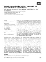
Fig. 1. Schematic representation of the murine Evi1-expressing
recombinant retroviral vector p50M5.6neo and production of Evi1 in
virus-infected Rat1 fibroblasts. (A) Viral long terminal repeats (LTR),
the murine Evi1 gene, including the N-terminal and C-terminal zinc
finger domains (black boxes), repressor domain (grey box) and
acidic domain (striped box), the Neo gene (neo) and splice donor
(SD) and splice acceptor (SA) sites for the production of subgenom-
ic transcripts for the expression of neo. (B) Western blot analysis
of whole cell protein extracts derived from the indicated cell lines
and populations using a-Evi1 (1806) and a-gapdh (6C5) antibodies.
The positions of 145 and 35 kDa Evi1 and gapdh proteins are
shown.
Evi1 enhanced oxidant-induced apoptosis P. Roy et al.
442 FEBS Journal 277 (2010) 441–452 ª 2009 The Authors Journal compilation ª 2009 FEBS
5.62 cells that is absent from Neo1, Neo2 and parental
Rat1 cells (Fig. 1B).
Previous studies have shown that Evi1 is a survival
factor, protecting cells from apoptosis induced by a
variety of agents. To determine if Evi1 also protects
Rat1 cells from apoptosis, we treated our panel of cell
populations with paclitaxel (taxol). Apoptosis was
monitored by measuring caspase 3 catalytic activity.
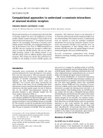
The results showed that taxol (1 lm, 16 h) induced sig-
nificantly higher caspase 3 catalytic activity in Rat1,
Neo1 and Neo2 cells than in Evi1-expressing 5.61 and
5.62 cells (Fig. 2). Taxol induced caspase 3 activity in
all cell populations, but to a much lesser extent in 5.61
and 5.62 cells. These data show that Evi1 protects
Rat1 cells from taxol-induced apoptosis, consistent
with previous studies in other cell types.
Rat1 fibroblasts expressing Evi1 have increased
sensitivity to H
2
O
2
-induced apoptosis
Although previous studies have shown that Evi1 pro-
tects cells from a variety of inducers of apoptosis, the
effects of H
2
O
2
have not yet been examined. Rat1 cells
and 5.61 cells were exposed to various concen-
trations of H
2
O
2
and cell viability monitored by 3-(4,5-
dimethylthiazol-2-yl)-2,5-diphenyl-tetrazolium bromide
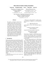
(MTT) assay. The results showed that cell viability
was reduced in a H
2
O
2
dose-dependent manner in both
Rat1 and 5.61 cells (Fig. 3A). Surprisingly, the viabil-
ity of 5.61 cells was significantly less than Rat1 cells at
each concentration of H
2
O
2
(Fig. 3A). The viability of
the entire panel of cell populations was examined by
MTT assay following H
2
O
2
(750 lm) treatment and
this confirmed that ectopic Evi1 expression decreased
survival in Rat1 cells (Fig. 3B). This was supported by
the dramatic morphological change observed in H
2
O
2
-
treated 5.61 cells compared with parental Rat1 or
empty vector control Neo cells treated with the same
concentration of reagent (Fig. 3C).
The morphological changes observed in 5.61 cells
treated with H
2
O
2
resembled apoptosis. Therefore, we
examined caspase 3 activation in cultures of the cell
populations. The results showed that H
2
O
2
induced
caspase 3 catalytic activity in all cell populations exam-
ined, but the level of activation was significantly
greater in 5.6 cell populations compared with parental
Rat1 and vector control cells (Fig. 3D).
Evi1 represses expression of the potential
antioxidant caIII in Rat1 cells
Previous studies have shown that arsenic trioxide
induces apoptosis in leukaemia cells by degrading the
Evi1 fusion protein, AML1 ⁄ EVI1 [5]. H
2
O
2
-induced
Evi1 degradation could reduce cell survival in the pres-
ence of this agent. Therefore, the stability of Evi1
transgene expression in 5.61 cells was examined by
western blot analysis (a-Evi1, 1806) following H
2
O
2
treatment for 4, 10 and 16 h. However, the abundance
of Evi1 protein remained unchanged during this time
period, confirming that H
2
O
2
had no effect on protein
stability (Fig. 4), eliminating this mechanism.
Recently, we used microarray technology to identify
Evi1-mediated induction and repression of gene tran-
scripts in Rat1 cells (E. R. Reavey & C. Bartholomew,
unpublished results). Inspection of these data revealed
transcriptional repression of caIII, which encodes a
protein that has previously been shown to protect cells
from H
2
O
2
-induced apoptosis [19,20].
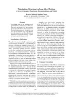
The microarray data were confirmed by real-time
quantitative RT-PCR (RTQ-RT-PCR) using total cel-
lular RNA from Rat1 and derivative Neo and 5.6
cells. These data showed that caIII mRNA transcripts
were repressed by 92–97% in 5.61 and 5.62 cells rela-
tive to Rat1 and empty vector control cells Neo1 and
Neo 2 (Fig. 5A). Western blot analysis with a-caIII
(E-19) confirmed that caIII protein levels were also
dramatically reduced in 5.61 and 5.62 cells (Fig. 5B),
consistent with the RTQ-RT-PCR data.
caIII gene promoter activity is repressed by Evi1
in Rat1 cells
To determine if Evi1-mediated caIII repression occurs
at the level of gene transcription, reporter assays were
160 000
180 000
200 000
***
80 000
100 000
120 000
140 000
Relative caspase 3 activity
(luminescence,RLU)
0
20 000
40 000
60 000
Fig. 2. Histogram showing relative caspase 3 catalytic activity of
the indicated cell lines and populations in the absence (grey col-
umns) or presence of 1 l
M paclitaxel for 16 h (black columns). The
columns represent the mean of an experiment performed in qua-
druplicate and error bars the standard deviation. ***P £ 0.0002.
P. Roy et al. Evi1 enhanced oxidant-induced apoptosis
FEBS Journal 277 (2010) 441–452 ª 2009 The Authors Journal compilation ª 2009 FEBS 443
performed. A pGL3-basic vector containing )1485 to
+55 of rat caIII gene promoter sequence [21], desig-
nated p-1485 ⁄ +55 caIII luc, was created (Y. Ishii,
unpublished data) and transfected into Rat1, Neo and
5.6 cells, together with the control vector pRLCMV.
The activity of p-1485 ⁄ +55 caIII luc in the various
cell types, normalized for pRLCMV activity, is shown
in Fig. 6A. The results showed that the caIII gene
promoter had at least 10-fold greater transcriptional
activity in Rat1, Neo1 and Neo2 cells compared with
the Evi1-expressing 5.61 and 5.62 cells. In contrast, the
activity of a minimal thymidine kinase gene promoter
construct (pGL2tkluc), normalized for pRLCMV, was
similar in all cell types examined (Fig. 6B). These
results show that Evi1 specifically repressed the
transcriptional activity of the caIII gene promoter in
Rat1 cells.
caIII knockdown alone enhances H
2
O
2
-induced
apoptosis in Rat1 cells and enhanced caIII
expression is protective
Short inhibitory RNAs (siRNA) were used to deter-
mine if repression of caIII gene expression alone sensi-
Cell viability (%)
0
20
40
60
80
100
120
Rat1
Neo 1
Neo 2
5.61 cells
5.62 cells
Rat1
Neo 1
Neo 2
5.61 cells
5.62 cells
0
20
40
60
80
100
120
UT 100 150 200 250 500 750
Cell viability (%)
H
2
O
2
(µM)
Rat1 Neo2 5.61 cells
750
M
H
2
O
2
Untreated
0
20 000
40 000
60 000
80 000
100 000
120 000
140 000
Rat1
Neo1
Neo2
5.61 cells
5.62 cells
Rat1
Neo1
Neo2
5.61 cells
5.62 cells
Relative caspase 3 activity
(luminescence, RLU)
**
***
C
AB
D
Fig. 3. Cell viability and caspase 3 catalytic activity of cell lines and populations following 16 h treatment with H
2
O
2
. (A) Histogram of the
percentage viability (MTT assay) of untreated cells (UT) and cells treated with the indicated concentration of H
2
O
2
for 16 h. Grey columns
are Rat1 cells and black columns are 5.61 cells. The columns represent the mean of an experiment performed in quadruplicate and error
bars the standard deviation. (B) Histogram of the percentage viability (MTT assay) of the indicated untreated (black columns) and 16 h of
750 l
M H
2
O
2
treated (grey columns) cell lines and populations. The columns represent the mean of an experiment performed in quadrupli-
cate and error bars the standard deviation. **P £ 0.002. (C) Photographs showing the morphology of either untreated or treated (16 h
750 l
M H
2
O
2
) indicated cell lines and populations. (D) Histogram showing relative caspase 3 catalytic activity of the indicated cell lines and
populations in the absence (grey columns) or presence of 750 l
M H
2
O
2
for 16 h (black columns). The columns represent the mean of an
experiment performed in quadruplicate and error bars the standard deviation. ***P < 0.0001.
UT 4 h 10 h 16 h UT 4 h 10 h 16 h
5.61 cells Neo1
α-Evi1
α-gapdh
145 kDa
35 kDa
Fig. 4. Western blot analysis of whole cell protein extracts derived
from Neo1 and 5.61 cell populations following treatment for 0 (UT),
4, 10 and 16 h with 750 l
M H
2
O
2
using a-Evi1 (1806) and a-gapdh
(6C5) antibodies. The positions of 145 kDa Evi1 and 35 kDa gapdh
proteins are shown.
Evi1 enhanced oxidant-induced apoptosis P. Roy et al.
444 FEBS Journal 277 (2010) 441–452 ª 2009 The Authors Journal compilation ª 2009 FEBS
tizes Rat1 cells to H
2
O
2
. Both RTQ-RT-PCR and wes-
tern blot analysis (a-caIII) were used to identify an
effective Dicer-substrate siRNA (DsiRNA) that inhib-
ited both caIII mRNA gene transcripts (98% reduc-
tion, Fig. 7A, 10 nm siRNA1) and caIII protein
(Fig. 7B, 10 nm siRNA1) when transfected into Rat1
cells. A control, nonspecific DsiRNA had no effect on
either caIII mRNA or caIII protein when transfected
into Rat1 cells at the same concentration (Fig. 7A,B,
Non sp siRNA).
The effect of caIII siRNA1 on H
2
O
2
sensitivity in
transfected Rat1 cells was then investigated by moni-
toring caspase 3 catalytic activity. siRNA1 transfected
Rat1 cells treated with 750 lm H
2
O
2
for 16 h had at
least double the caspase 3 activity observed with
750 lm H
2
O
2
-treated untransfected and nonspecific
DsiRNA transfected Rat1 control cells (Fig. 7C). caIII
knockdown with a second distinct siRNA (siRNA3)
produced the same phenotype (Fig. S1). The results
show that H
2
O
2
induced caspase 3 catalytic activity in
all cells, but the level of activation was significantly
greater in Rat1 cells transfected with a caIII-specific
siRNA (Fig. 7C). Furthermore, caIII knockdown only
sensitized Rat1 cells to H
2
O
2
treatment and had no
effect upon apoptosis induced by treatment with taxol
(Fig. S2).
caIII expression was restored in 5.61 cells to deter-
mine if the sensitivity to H
2
O
2
treatment could be
reverted. Rat1 cells were transiently transfected with a
caIII expression vector (pRC-sport 6caIII), which sig-
nificantly increased cellular levels of the caIII protein
(Fig. 8A). The increased levels of caIII protein in 5.61
cells protected them from H
2
O
2
treatment, compared
with untreated or empty vector control transfected
cells, as determined by measuring caspase 3 catalytic
activity (Fig. 8B).
Finally, we measured intracellular levels of ROS to
determine if the basal oxidized state varied between
Rat1 and 5.61 cells. Cells labelled with 2¢-7¢-dichloro-
flourescene diacetate (DCF-DA) were examined by
fluorescent automatic cell sorter (FACS). The results
2.00
***
1.20
1.60
0.40
0.80
0.00
Neo1 Neo2 5.61 cells 5.62 cells
caIII gene expression relative
to Rat1 cells
Rat1
Neo1
Neo2
5.61 cells
α-caIII
α-gapdh35 kDa
27 kDa
A
B
Fig. 5. caIII gene expression in Rat1 cells and derivative cell popu-
lations. (A) Histogram of caIII mRNA levels normalized for gapdh
mRNA relative to normalized caIII mRNA in Rat1 cells, determined
by RTQ-RT-PCR. The columns are the mean of an experiment per-
formed in quadruplicate and the error bars the standard deviation.
(B) Western blot analysis of whole cell protein extracts using a-caIII
(E-19) and a-gapdh (6C5) antibodies. The positions of 27 kDa caIII
and 35 kDa gapdh proteins are shown. ***P < 0.0001.
B
A
Fig. 6. Reporter assays showing the activity of the caIII and mini-
mal herpes simplex virus thymidine kinase (HSV tk) gene promot-
ers in Rat1 and derivative cells. (A) Histogram of caIII gene
promoter firefly luciferase reporter activity (p-1485 ⁄ +55 caIII luc)
normalized for cytomegalovirus (CMV) immediate early enhancer ⁄
promoter renilla luciferase reporter activity (pRLCMV). The columns
are the mean of an experiment performed in quadruplicate and
error bars the standard deviation. ***P < 0.0001. (B) Histogram of
HSV tk gene promoter firefly luciferase reporter activity (pGL2tkluc)
normalized for pRLCMV. The columns are the mean of an experi-
ment performed in quadruplicate and error bars the standard
deviation.
P. Roy et al. Evi1 enhanced oxidant-induced apoptosis
FEBS Journal 277 (2010) 441–452 ª 2009 The Authors Journal compilation ª 2009 FEBS 445
showed that the mean fluorescence of 5.61 cells was
significantly higher than that observed in Rat1 and
Neo1 cells (Fig. 9). This shows that ROS were elevated
in 5.61 cells, consistent with the observed reduction in
caIII expression.
Discussion
We show here for the first time that ectopic expression
of Evi1 sensitizes Rat1 cells to H
2
O
2
-induced apopto-
sis. This represents the first description that Evi1 can
actually stimulate cell death. Previous studies with a
variety of agents have shown that Evi1 protects cells
from apoptosis and functions as a survival factor, pro-
viding one of multiple suggested roles that contribute
to the development and progression of leukaemia.
Consistent with this view, we also show that Evi1 pro-
tects Rat1 cells from apoptosis induced by at least one
of these agents (taxol) and therefore probably also acts
as a survival factor in these cells. Evi1-mediated pro-
tection from taxol-induced apoptosis in rat intestinal
epithelial cells and colon cancer cells (HT-29) is due to
UT
Non sp siRNA
siRNA 1
α-
caIII
α-
gapdh
35 kDa
27 kDa
0
0.2
0.4
0.6
0.8
1
1.2
1.4
caIII gene expression
relative to Rat1 cells
**
Non sp siRNA 1
siRNA
Relative caspase 3 activity
(luminescence, RLU)
0
100 000
200 000
300 000
400 000
500 000
600 000
700 000
800 000
900 000
UT Non sp
siRNA
siRNA 1
UT
750 µM
***
A
B
C
Fig. 7. DsiRNA-mediated knockdown of caIII mRNA and protein
and enhanced caspase 3 catalytic activity in Rat1 cells. (A) Histo-
gram of caIII mRNA levels normalized for gapdh mRNA relative to
normalized caIII mRNA in untreated Rat1 cells, determined by QRT-
PCR in Rat1 cells transfected for 48 h with 10 n
M of either a non-
specific siRNA (Non sp siRNA) or a caIII-specific siRNA (siRNA
10 n
M). The columns are the mean of an experiment performed in
quadruplicate and the error bars the standard deviation.
**P £ 0.0041. (B) Western blot analysis of whole cell extracts
derived from Rat1 cells, transfected as described in (A), with a -caIII
(E-19) and a-gapdh (6C5) antibodies. The positions of 27 kDa caIII
and 35 kDa gapdh proteins are shown. (C) Relative caspase 3 cata-
lytic activity in Rat1 cells transfected as described in (A), either
treated (black columns) or untreated (grey columns) with 750 l
M
H
2
O
2
for 16 h. The columns are the mean of an experiment per-
formed in quadruplicate and error bars the standard deviation.
***P < 0.0001.
UT
pRC-sport6caIII
α-caIII
α-gapdh35 kDa
27 kDa
1400 000
1600 000
1800 000
800 000
1000 000
1200 000
***
200 000
400 000
600 000
Relative caspase 3 activity
(luminescence, RLU)
***
0
UT 750 µ
M
B
A
Fig. 8. 5.61 cell protection from H
2
O
2
-induced caspase 3 catalytic
activity by ectopic expression of caIII. (A) Western blot analysis of
whole cell extracts derived from untransfected 5.61 cells (UT),
empty vector transfected 5.61 cells (pRC CMV) and caIII expres-
sion vector transfected cells (pRC-sport6caIII) with a-caIII (E-19)
and a-gapdh (6C5) antibodies. The positions of 27 kDa caIII and
35 kDa gapdh proteins are shown. (B) Relative caspase 3 catalytic
activity in untransfected 5.61 cells (grey columns) and 5.61 cells
transfected with pRC CMV (white columns) or pRC-sport6caIII
(black columns) with (H
2
O
2
) or without (UT) 750 lM H
2
O
2
treat-
ment. ***P £ 0.0007.
Evi1 enhanced oxidant-induced apoptosis P. Roy et al.
446 FEBS Journal 277 (2010) 441–452 ª 2009 The Authors Journal compilation ª 2009 FEBS
stimulation of PI3K and its downstream effector AKT
[4]. The same mechanism probably also operates in
Rat1 cells, but was not examined in this study.
Sensitization to H
2
O
2
-induced apoptosis, determined
by caspase 3 catalytic activity, was seen in both Evi1-
expressing Rat1 cells (5.6 cells) and in caIII knockdown
Rat1 cells. Furthermore, Evi1-expressing Rat1 cells had
a 90% reduction in caIII gene transcripts and protein.
Together, these results confirm that Evi1-mediated stim-
ulation of H
2
O
2
-induced cell death is due to the reduc-
tion in cellular levels of the caIII protein.
The results presented here suggest that caIII protects
Rat1 cells from H
2
O
2
-derived ROS and therefore acts
as an antioxidant. However, the biological activity of
caIII is an enigma. Unlike other products of this gene
family, caIII has very low catalytic activity and so it is
unlikely that it functions in hydrating carbon dioxide
[18]. Furthermore, knockout mice, deficient in caIII,
have normal growth, development and lifespan under
laboratory conditions, suggesting that the protein is
not essential [22]. However, several recent observations
suggest that caIII is an important antioxidant, consis-
tent with the observations here. caIII is highly abun-
dant in skeletal muscle, a tissue of high oxygen
consumption and antioxidant activity. Microarray
analysis of skeletal muscle of wild-type and caIII-defi-
cient knockout mice revealed that caIII has a possible
role in the glutathione-mediated antioxidative system
[23]. This is supported by biochemical evidence show-
ing that caIII undergoes rapid reversible S-glutathiola-
tion or irreversible oxidation in mildly and
exhaustively stressed muscle, respectively [23]: in the
presence of glutathione, glutathione peroxidases restore
reversibly S-glutathiolated caIII. This mechanism
would explain both the protective effect of ectopic
caIII expression observed in NIH3T3 cells [20] and the
increased sensitivity of caIII knockdown Rat1 or Evi1-
expressing Rat1 cells exposed to H
2
O
2
.
Several possibilities exist to explain the caIII repres-
sion effect observed in the present study. Abundant
caIII gene transcripts and protein occur in Rat1 cells,
which, like all fibroblasts examined (C. Bartholomew,
unpublished results), normally express low levels of
endogenous evi1. Therefore, caIII gene expression and
protein production normally occur efficiently in the
presence of evi1 in Rat1 cells. The simplest explanation
is that merely elevated cellular levels of Evi1 are suffi-
cient to repress caIII transcription. Consistent with
this, previous studies have shown that the abundance
of Evi1 is crucial to 32Dcl3 granulocyte differentiation
[24], suggesting that differential changes in gene
expression must occur that are dependent on the quan-
tity of cellular levels of the Evi1 protein. However,
other possibilities exist. Multiple naturally occurring
evi1 isoforms occur and it might be that it is the relative
increase in the abundance of the Evi1 full-length form
[25] in Rat1 cells (5.6 cells), observed here, that signifi-
cantly represses caIII gene expression. It is possible that
only some isoforms of Evi1 repress caIII expression,
whereas perhaps other forms either have no effect or
the opposite effect to the full-length form. Some
studies have shown that the MDS1 ⁄ EVI1 isoform has
Rat1
MFI –250
ROS levels
Neo1
MFI –144
ROS levels
5.61 cells
MFI –905
ROS levels
10
0
10
1
10
2
10
3
10
4
10
0
10
1
10
2
FL1-H
FL1-H
10
3
10
4
10
0
10
0
10
1
10
1
10
2
10
2
FL1-H
10
3
10
3
10
4
10
4
10
5
10
0
10
1
10
2
10
3
10
4
10
5
10
0
10
1
10
2
10
3
10
4
10
5
M1
M1
M1
Fig. 9. Histogram of fluorescence intensity (x-axis) versus cell num-
ber (y-axis) of indicated DCF-DA-labelled cells analysed by FACS.
The mean fluorescence intensity (MFI) for the region designated
M1 is shown for each cell type.
P. Roy et al. Evi1 enhanced oxidant-induced apoptosis
FEBS Journal 277 (2010) 441–452 ª 2009 The Authors Journal compilation ª 2009 FEBS 447
the opposite effect of the full-length form. For example,
it is reported that the MDS1 ⁄ EVI1 isoform enhances
the growth inhibitory effects of TGFb [26], whereas the
full-length form blocks this response [12].
caIII is a very abundant protein, particularly in liver,
muscle and adipocytes. However, very little is known
about its transcriptional regulation. Transcription of the
rat caIII gene is inhibited by the aryl hydrocarbon recep-
tor ligand 3-methylchlanthrene in hepatocytes and in
the livers of rats fed an ethanol-supplemented diet
[21,27]. Human CAIII mRNA is induced in muscle of
athletes training under hypoxic conditions [28]. One
study has been conducted with the caIII gene promoter,
with a preliminary analysis of an active 2.8 kb human
CAIII gene promoter in myogenic cells and a significant
loss of activity upon deletion to )715 bp [29].
Reporter assays with the caIII gene promoter
()1485 ⁄ +55) showed that this region contains strong
promoter activity in Rat1 cells, consistent with a previ-
ous analysis of the human promoter [29]. This region
also has the cis-regulatory elements necessary for Evi1-
mediated transcriptional repression.
Evi1-mediated repression could be caused by binding
directly to promoter sequences. Previous studies with
artificial promoter reporter constructs have shown that
Evi1 can function as a DNA-binding transcriptional
repressor protein [9]. Evi1 binds several corepressor
molecules, CtBP, the histone methyltransferase
SUV39H1 and the histone deacetylase HDAC1
[30–32], each of which mediates transcriptional repres-
sion. However, to date no genes that are direct targets
for transcriptional repression have been identified.
Furthermore, very few genes have been identified that
are directly regulated and induced by Evi1; GATA2
being the best characterized [6,33]. Inspection of the
)1485 ⁄ +55 rat nucleotide sequence for Evi1 protein-
binding sites with matinspector software revealed mul-
tiple potential sites. This suggests that caIII may be a
direct target for Evi1-mediated transcriptional repres-
sion, although binding and biological activity of any of
these motifs require experimental investigation.
Alternatively, repression of caIII gene expression
could be indirect. Evi1 has been shown to interact with a
number of transcription factors, including PU.1,
RUNX1, GATA1, E2F1 and SMAD3 [12,34–37] and
signalling molecules such as JNK and PI3K ⁄ AKT [2,4]
to inhibit their biological activities. Therefore, it remains
possible that Evi1 might repress caIII gene expression
by interacting with a transcription factor or by inhibit-
ing a signalling pathway that is normally required for
the high level of expression observed in Rat1 cells.
Inspection of the caIII )1485 ⁄ +55 promoter region
with matinspector software showed several potential
binding sites for E2F family proteins, but not for any of
the other transcription factors Evi1 has been shown to
interact with. The precise mechanism by which Evi1
represses caIII gene expression awaits a more detailed
analysis of the )1485 ⁄ +55 gene promoter region.
These data show that Evi1 represses transcription of
caIII in Rat1 cells (5.6 cells) and as a consequence these
cells are vulnerable to oxidative stress. This raises the
possibility that Evi1 regulates caIII in other cell types
and if the caIII protein is an important antioxidant, then
they too would be vulnerable to oxidative stress.
EVI1 is overexpressed in some human neoplasias,
including acute myeloid leukaemias [38] and hepatocel-
lular carcinoma [4]. CAIII is also very abundant in nor-
mal liver and presumably is an important antioxidant in
this tissue. Interestingly, CAIII expression is reduced in
human hepatocellular carcinoma [39], although it is not
known which tumours overexpress EVI1. EVI1 might
be responsible for CAIII repression in some cases and
perhaps different mechanisms operate in others. CAIII
expression in haemopoietic cells and leukaemia cells has
not been described. There is some evidence that CAIII
might operate as an antioxidant in erythrocytes in cer-
tain anaemias [40], suggesting that it might be important
in protecting haemopoietic cells from oxidative stress. It
would be interesting to assess the expression level of
CAIII in normal haemopoetic cells and leukaemia cells
to determine if it is reduced in these neoplasias and if
levels are inversely proportional to EVI1 expression. If
this is the case, then tumours overexpressing EVI1 might
be vulnerable to therapeutic agents that induce oxidative
stress.
Materials and methods
Preparation of plasmid DNA
Plasmids p50MX-neo, p50M5.6neo, pGL2tkluc, pBluescript
KSII, pCMVcar3 (I.M.A.G.E. Id 4195712) and pRLCMV
have all been described previously [9] and were obtained
from Promega UK (Southampton, UK), Stratagene (La
Jolla, CA, USA) and Source Bioscience, geneservice (Cam-
bridge, UK). The construction of pGL3-caIII ()1485 ⁄ +55)
has not been published (Y. Ishii, unpublished data). Plas-
mid DNAs were prepared by affinity chromatography using
Nucleobond
Ò
PC500EF gravity flow columns according to
the manufacturer’s instructions (Macherey-Nagal, Du
¨
ren,
Germany).
Cell culture
Rat1 and EcoPak2 cells were cultured in complete medium
comprising Dulbecco’s modified Eagle’s medium (DMEM,
Evi1 enhanced oxidant-induced apoptosis P. Roy et al.
448 FEBS Journal 277 (2010) 441–452 ª 2009 The Authors Journal compilation ª 2009 FEBS
Lonza Group, Basel, Switzerland, BE12-604F) supple-
mented with 5% newborn calf serum (Sigma-Aldrich,
Poole, UK, N4637) or 10% fetal calf serum (Lonza
Group, DE14-801F), respectively, and 2.5 mm glutamine,
50 lgÆmL
)1
penicillin, 50 unitsÆmL
)1
streptomycin (Lonza
Group, BE17-605E and BE17-603E), 37 °C, 5% CO
2
. For
retrovirus production, EcoPak2 cells (Clontech-Takara Bio
Europe, Saint-Germain-en-Laye, France) were plated on
collagen (Sigma-Aldrich, C38671) coated dishes and tran-
siently transfected with either p50M5.6-neo or p50MX-neo
using the calcium phosphate coprecipitate method described
previously [41]. Virus was harvested and used to infect
Rat1 fibroblasts, as described previously [9]; infected cells
were selected in complete medium supplemented with
50 lgÆmL
)1
G418 (Invitrogen, Paisley, UK). For paclitaxel
and H
2
O
2
treatment, cells were incubated in complete med-
ium supplemented with either 1 lm paclitaxel (Sigma-
Aldrich, T7191) or 100–750 lm H
2
O
2
(Sigma-Aldrich,
21676) for 16 h.
Western blotting
Protein extracts, SDS ⁄ PAGE and western blotting were
performed as described previously [9] with either a-caIII
(Santa Cruz Biotechnology, Santa Cruz, CA, USA, E-19),
a-Evi1 (1806) or a-gapdh (Fitzgerald Industries, North
Acton, MA, USA, 6C5) and diluted 1 ⁄ 200 or 1 ⁄ 5000 (1806
and 6C5). Appropriate horseradish peroxidase-conjugated
anti-goat (Sigma-Aldrich, A5420), anti-rabbit (Sigma-
Aldrich, A9169) or anti-mouse (Sigma-Aldrich, A9044) IgG
secondary antibodies were used at 1 ⁄ 5000 dilutions and
detection was performed by enhanced chemiluminescence
(Pierce, Rockford, IL, USA, 32209).
DNA-mediated transfection and reporter assays
Rat1 cells and derivatives were transfected using Fugene6
Ò
(Roche Diagnostics, Mannheim, Germany, 11815091001).
For reporter assays, cells were transfected with recombinant
pGL3-caIII ()1485 ⁄ +55) and pRLCMV plasmid DNAs.
Constant DNA concentrations were maintained with pBlue-
script KSII. Cells (5 · 10
3
) were incubated with a 1 : 6 ratio
DNA : FuGENE6
Ò
, prepared as described by the manufac-
turer, in 96-well plates (Costar, New York, NY, USA,
3917) for 48 h. Cells were lysed, and luciferase activity
determined using the dual-luciferase reporter assay system
(Promega, TM046) in a Fluostar OPTIMA luminometer
(BMG LABTECH, Offenburg, Germany).
Oligonucleotides
Gene-specific oligonucleotides were designed using primer
express software version 3.0 (Applied Biosystems), synthe-
sized and supplied by Eurogentec (Seraing, Belgium):
5¢ rat caIII: ccgggactattggacctacca
3¢ rat caIII: cagtagcagccacacaatgca
5¢ HEX, 3¢ TAMRA rat caIII probe: cttcaccacgccaccctgc
gag
5¢ rat gapdh: gggcagcccagaacatca
3¢ rat gapdh: ccgttcagctctgggatgac
5¢ 6-FAM, 3¢ TAMRA rat gapdh probe: ccctgcatccactgg
tgctgcc
Preparation of total cellular RNA, cDNA synthesis
and RTQ-RT-PCR
RNA was prepared from semiconfluent cultures of cells using
the Trizol method (Invitrogen, 1559-026). Total cellular
RNA (1 lg) was used to synthesize cDNA with Supermix III
first-strand strand synthesis for QPCR according to the man-
ufacturer’s instructions (Invitrogen, 11752). Five per cent of
the cDNA reaction was used for RTQ-PCR using the ABso-
lute QPCR mix (ABgene, Epsom, UK, AB-4136), gene-spe-
cific oligonucleotide primers and dual-labelled probes, 95 °C,
15 min followed by 40 cycles 95 °C, 30 s, 60 °C, 30 s in an
OPTICON 2 DNA engine (MJ Research, Watertown, MA,
USA).
The efficiency of the RTQ-PCR reactions was calculated
using the formula efficiency = )1+10
()1 ⁄ slope)
against the
standard curve of each assay over a gradient of template
concentration with each gene. The efficiencies for caIII and
gapdh primers ⁄ probes were 90 and 101%, respectively. Rela-
tive expression levels between caIII and gapdh were deter-
mined using the arithmetic comparative 2
)DDCt
method [42]
and were determined relative to caIII in Rat1 cells (cali brator).
Caspase 3 assay
Cells were incubated in 96-well dishes (Costar 3917), trea-
ted with various agents and apoptosis determined using the
Caspase 3 ⁄ 7-Glo
Ò
assay according to the manufacturer’s
instructions (Promega, G8090), measuring luminescence
with a Fluostar OPTIMA luminometer (BMG LABTECH).
MTT assays
MTT assays were performed on cells grown in 96-well tissue
culture plates following treatment with H
2
O
2
. Cells were
treated with 500 lgÆmL
)1
MTT (Sigma-Aldrich, M5655) in
complete medium, 37 °C, 5% CO
2
, 1 h. The medium was
removed and replaced with 100 lL dimethylsulfoxide
(Sigma-Aldrich, 472301) and absorbance measured at
570 nm using an MRX plate reader (Dynatech Laboratories,
Guernsey Channel Island, UK). The final absorbance was
determined by subtracting the absorbance of treated wells
lacking cells. The formazan concentration was determined
using the formula: c (formazan concentration; lm)=A
(absorbance) ⁄ e (extinction coefficient) 1 (path length).
P. Roy et al. Evi1 enhanced oxidant-induced apoptosis
FEBS Journal 277 (2010) 441–452 ª 2009 The Authors Journal compilation ª 2009 FEBS 449
Knockdown of rat caIII
Rat caIII knockdown was achieved in Rat1 cells using Tri-
FECTa DsiRNA Kit RNC.RNAI.NO19292.9 (Integrated
DNA Technologies, Leuven, Belgium). SiRNA1 (5¢-CCA
UUGAACUGCAUACUAAAGACAT-3¢,5¢-AUGUCUU
UAGUAUGCAGUUCAAUGGGU-3¢) was found to be
the most effective and used for these studies. The control
DsiRNA sequence used was (5¢-CUUCCUCUCUUUCUC
UCCCUUGUGA-3¢,5¢ UCACAAGGGAGAGAAAGA
GAGGAAGGA-3¢). In total, 1 · 10
5
Rat1 cells per well
were seeded in a 12-well tissue culture plate in compete
medium and incubated, 37 °C, 5% CO
2
. Twenty-four hours
later the medium was removed and replaced with 600 lL
compete medium. After 1 h, 1.5 lL Silentfect
Ò
(BioRad,
Hercules, CA, USA, 170-3360) in 50 lL DMEM was mixed
with 50 lL DMEM containing DsiRNA and added to the
cells, giving a final DsiRNA concentration of 10 nm. The
cells were incubated, 37 °C, 5% CO
2
, for 48 h prior to
isolation of whole cell protein extracts or treatment with
H
2
O
2
or 24 h for isolation of total cellular RNA.
ROS assay
The ROS assay was performed by labelling cells with
DCF-DA [43]. Cells grown in complete medium were
labelled for 30 min with 20 lm DCF-DA (Sigma-Aldrich,
35845), 37 °C, 5% CO
2
. The cells were trypsinized, pelleted
and washed three times with ice-cold phosphate-buffered
saline (Lonza Group, BE17-516F), then analysed for
fluorescence by FACS (FACSCaliber, Becton Dickinson,
Oxford, UK).
Acknowledgements
This work was funded by a Glasgow Caledonian
University PhD studentship and Overseas Research
Student Award Scheme (PR) and in part by the
Leukaemia Research Fund (CB, 08018).
References
1 Weiser R (2007) The oncogene and developmental regu-
lator EVI1: expression, biochemical properties, and bio-
logical functions. Gene 396, 346–357.
2 Kurokawa M, Mitani K, Yamagata T, Takahashi T,
Izutsu K, Ogawa S, Moriguchi T, Nishida E, Yazaki Y
& Hirai H (2000) The evi-1 oncoprotein inhibits c-Jun
N-terminal kinase and prevents stress-induced cell
death. EMBO J 19, 2958–2968.
3 Buonamici S, Li D, Mikhail FM, Sassano A, Platanias
LC, Colamonici O, Anastasi J & Nucifora G (2005)
EVI1 abrogates interferon-alpha response by
selectively blocking PML induction. J Biol Chem 280,
428–436.
4 Liu Y, Chen L, Ko TC, Fields AP & Thompson EA
(2006) Evi1 is a survival factor which conveys resistance
to both TGFbeta- and taxol-mediated cell death via
PI3K ⁄ AKT. Oncogene 25, 3565–3575.
5 Shackelford D, Kenific C, Blusztajn A, Waxman S &
Ren R (2006) Targeted degradation of the AML1 ⁄ MD-
S1 ⁄ EVI1 oncoprotein by arsenic trioxide. Cancer Res
66, 11360–11369.
6 Yuasa H, Oike Y, Iwama A, Nishikata I, Sugiyama D,
Perkins A, Mucenski ML, Suda T & Morishita K
(2005) Oncogenic transcription factor Evi1 regulates
hematopoietic stem cell proliferation through GATA-2
expression. EMBO J 24, 1976–1987.
7 Hoyt PR, Bartholomew C, Davis AJ, Yutzey K, Gamer
LW, Potter SS, Ihle JN & Mucenski ML (1997) The
Evi1 proto-oncogene is required at midgestation for
neural, heart, and paraxial mesenchyme development.
Mech Dev 65, 55–70.
8 Morishita K, Parker DS, Mucenski ML, Jenkins NA,
Copeland NG & Ihle JN (1988) Retroviral activation
of a novel gene encoding a zinc finger protein in
IL-3-dependent myeloid leukemia cell lines. Cell 54,
831–840.
9 Bartholomew C, Kilbey A, Clark AM & Walker M
(1997) The Evi-1 proto-oncogene encodes a transcrip-
tional repressor activity associated with transformation.
Oncogene 14, 569–577.
10 Delwel R, Funabiki T, Kreider BL, Morishita K & Ihle
JN (1993) Four of the seven zinc fingers of the Evi-1
myeloid-transforming gene are required for sequence-
specific binding to GA(C ⁄ T)AAGA(T ⁄ C)AAGATAA.
Mol Cell Biol 13, 4291–4300.
11 Funabiki T, Kreider BL & Ihle JN (1994) The carboxyl
domain of zinc fingers of the Evi-1 myeloid transform-
ing gene binds a consensus sequence of GAAGAT-
GAG. Oncogene 9, 1575–1581.
12 Kurokawa M, Mitani K, Irie K, Matsuyama T,
Takahashi T, Chiba S, Yazaki Y, Matsumoto K &
Hirai H (1998) The oncoprotein Evi-1 represses
TGF-beta signalling by inhibiting Smad3. Nature 394,
92–96.
13 Izutsu K, Kurokawa M, Imai Y, Maki K, Mitani K &
Hirai H (2001) The corepressor CtBP interacts with
Evi-1 to repress transforming growth factor beta signal-
ing. Blood 97, 2815–2822.
14 Ames BN, Shigenaga MK & Hagen TM (1993)
Oxidants, antioxidants, and the degenerative diseases of
aging. Proc Natl Acad Sci USA 90, 7915–7922.
15 Wu WS (2006) The signalling of ROS in tumor progres-
sion. Cancer Metastasis Rev 25, 695–705.
16 Wistrand PJ (2002) Carbonic anhydrase III in liver and
muscle of male rats. Purification and properties. Ups J
Med Sci 107, 77–88.
17 Lehtonen J, Shen B, Vihinen M, Casini A, Scozzafava
A, Supuran CT, Parkkila AK, Saarnio J, Kivela
¨
AJ,
Evi1 enhanced oxidant-induced apoptosis P. Roy et al.
450 FEBS Journal 277 (2010) 441–452 ª 2009 The Authors Journal compilation ª 2009 FEBS
Waheed A et al. (2004) Characterization of CA XIII,
a novel member of the carbonic anhydrase isozyme
family. J Biol Chem 279, 2719–2727.
18 Carter ND, Lo
¨
nnerholm G, Meyerson BJ & Wistrand PJ
(2001) Androgen-linked control of carbonic anhydrase
III expression occurs in rat perivenous hepatocytes; an
immunocytochemical study. Ups J Med Sci 106, 67–76.
19 Parkkila S, Halsted CH, Villanevva JA, Va
¨
a
¨
na
¨
nen HK
& Niemela
¨
O (1999) Expression of testosterone-depen-
dent enzyme, carbonic anyhdrase III and oxidative
stress in experimental alcoholic liver disease. Dig Dis
Sci 44, 2205–2213.
20 Raisanen SR, Lehenkari P, Tasana M, Rahkila P,
Harkonen PL & Va
¨
a
¨
na
¨
nen HK (1999) Carbonic anhy-
drase III protects cells from hydrogen peroxide- induced
apoptosis. FASEB J 13, 513–522.
21 Ishii Y, Akazawa D, Aoki Y, Yamada H & Oguri K
(2005) Suppression of carbonic anhydrase III mRNA
level by an aryl hydrocarbon receptor ligand in primary
cultured hepatocytes of rat. Biol Pharm Bull 28, 1087–
1090.
22 Kim G, Lee TH, Wetzel P, Geers C, Robinson MA,
Myers TG, Owens JW, Wehr NB, Eckhaus MW, Gros
G et al. (2004) Carbonic anhydrase III is not required
in the mouse for normal growth, development, and life
span. Mol Cell Biol 24 , 9942–9947.
23 Zimmerman UJ, Wang P, Zhang X, Bogdanovich S &
Forster R (2004) Anti-oxidative response of carbonic an-
hydrase III in skeletal muscle. IUBMB Life 56, 343–347.
24 Khanna-Gupta A, Lopingco MC, Savinelli T, Zibello
T, Berliner N & Perkins AS (1996) Retroviral inser-
tional activation of the EVI1 oncogene does not pre-
vent G-CSF-induced maturation of the murine
pluripotent myeloid cell line 32Dcl3. Oncogene 12,
563–569.
25 Alzuherri H, McGilvray R, Kilbey A & Bartholomew C
(2006) Conservation and expression of a novel
alternatively spliced Evi1 exon. Gene 384, 154–162.
26 Sood R, Talwar-Trikha A, Chakrabarti SR &
Nucifora G (1999) MDS1 ⁄ EVI1 enhances TGF-beta1
signaling and strengthens its growth-inhibitory effect
but the leukemia-associated fusion protein
AML1 ⁄ MDS1 ⁄ EVI1, product of the t(3;21), abrogates
growth-inhibition in response to TGF-beta1. Leukemia
13, 348–357.
27 Kharbanda KK, Vigneswara V, McVicker BL,
Newlaczyl AU, Bailey K, Tuma D, Ray DE & Carter
WG (2009) Proteomics reveal a concerted upregulation
of methionine metabolic pathway enzymes, and downre-
gulation of carbonic anhydrase-III, in betaine supple-
mented ethanol-fed rats. Biochem Biophys Res Commun
381, 523–527.
28 Zoll J, Ponsot E, Dufour S, Doutreleau S, Ventura-
Clapier R, Vogt M, Hoppeler H, Richard R & Flu
¨
ck M
(2006) Exercise training in normobaric hypoxia in
endurance runners. III. Muscular adjustments
of selected gene transcripts. J Appl Physiol
100,
1258–1266.
29 Tweedie S, Morrison K, Charlton J & Edwards YH
(1991) CAIII a marker for early myogenesis: analysis of
expression in cultured myogenic cells. Somat Cell Mol
Genet 17, 215–228.
30 Palmer S, Brouillet JP, Kilbey A, Fulton R, Walker
M, Crossley M & Bartholomew C (2001) Evi-1
transforming and repressor activities are mediated
by CtBP co-repressor proteins. J Biol Chem 276,
25834–25840.
31 Spensberger D & Delwel R (2008) A novel interaction
between the proto-oncogene Evi1 and histone
methyltransferases, SUV39H1 and G9a. FEBS Lett 582,
2761–2767.
32 Vinatzer U, Taplick J, Seiser C, Fonatsch C & Wieser
R (2001) The leukaemia-associated transcription factors
EVI-1 and MDS1 ⁄ EVI1 repress transcription and
interact with histone deacetylase. Br J Haematol 114,
566–573.
33 Yatsula B, Lin S, Read AJ, Poholek A, Yates K, Yue
D, Hui P & Perkins AS (2005) Identification of binding
sites of EVI1 in mammalian cells. J Biol Chem 280,
30712–30722.
34 Laricchia-Robbio L, Premanand K, Rinaldi CR &
Nucifora G (2009) EVI1 impairs myelopoiesis by deregu-
lation of PU.1 function. Cancer Res 69, 1633–1642.
35 Senyuk V, Sinha KK, Li D, Rinaldi CR, Yanamandra
S & Nucifora G (2007) Repression of RUNX1 activity
by EVI1: a new role of EVI1 in leukemogenesis. Cancer
Res 67, 5658–5666.
36 Laricchia-Robbio L, Fazzina R, Li D, Rinaldi CR,
Sinha KK, Chakraborty S & Nucifora G (2006) Point
mutations in two EVI1 Zn fingers abolish EVI1-
GATA1 interaction and allow erythroid differentiation
of murine bone marrow cells. Mol Cell Biol 26, 7658–
7666.
37 Chi Y, Senyuk V, Chakraborty S & Nucifora G (2003)
EVI1 promotes cell proliferation by interacting with
BRG1 and blocking the repression of BRG1 on E2F1
activity. J Biol Chem 278 , 49806–49811.
38 Nucifora G, Laricchia-Robbio L & Senyuk V (2006)
EVI1 and hematopoietic disorders: history and perspec-
tives. Gene 368, 1–11.
39 Kuo WH, Chiang WL, Yang SF, Yeh KT, Yeh CM,
Hsieh YS & Chu SC (2003) The differential expression
of cytosolic carbonic anhydrase in human hepatocellu-
lar carcinoma. Life Sci 73, 2211–2223.
40 Kuo WH, Yang SF, Hsieh YS, Tsai CS, Hwang WL &
Chu SC (2005) Differential expression of carbonic anhy-
drase isoenzymes in various types of anemia. Clin Chim
Acta 351, 79–86.
41 Wigler M, Silverstein S, Lee LS, Pellicer A, Cheng Y &
Axel R (1977) Transfer of purified herpes virus
P. Roy et al. Evi1 enhanced oxidant-induced apoptosis
FEBS Journal 277 (2010) 441–452 ª 2009 The Authors Journal compilation ª 2009 FEBS 451
thymidine kinase gene to cultured mouse cells. Cell 1,
223–232.
42 Livak KJ & Schmittgen TD (2001) Analysis of relative
gene expression data using real-time quantitative PCR
and the 2(-Delta Delta C(T)) Method. Methods 25,
402–408.
43 Bass DA, Parce JW, Dechatelet LR, Szejda P, Seeds
MC & Thomas M (1983) Flow cytometric studies of
oxidative product formation by neutrophils: a graded
response to membrane stimulation. J Immunol 130,
1910–1917.
Supporting information
The following supplementary material is available:
Fig. S1. DsiRNA-mediated knockdown of caIII
mRNA and protein and enhanced caspase 3 catalytic
activity in Rat1 cells.
Fig. S2. DsiRNA-mediated knockdown of caIII
mRNA and protein and caspase 3 catalytic activity in
Rat1 cells following treatment with paclitaxel.
This supplementary material can be found in the
online version of this article.
Please note: As a service to our authors and readers,
this journal provides supporting information supplied
by the authors. Such materials are peer-reviewed and
may be re-organized for online delivery, but are not
copy-edited or typeset. Technical support issues arising
from supporting information (other than missing files)
should be addressed to the authors.
Evi1 enhanced oxidant-induced apoptosis P. Roy et al.
452 FEBS Journal 277 (2010) 441–452 ª 2009 The Authors Journal compilation ª 2009 FEBS