Báo cáo khoa học: Novel L-amino acid oxidase with antibacterial activity against methicillin-resistant Staphylococcus aureus isolated from epidermal mucus of the flounder Platichthys stellatus pptx
Bạn đang xem bản rút gọn của tài liệu. Xem và tải ngay bản đầy đủ của tài liệu tại đây (813.62 KB, 13 trang )
Novel L-amino acid oxidase with antibacterial activity
against methicillin-resistant Staphylococcus aureus
isolated from epidermal mucus of the flounder
Platichthys stellatus
Kosuke Kasai
1,2
, Takashi Ishikawa
1
, Takafumi Komata
3
, Kaori Fukuchi
4
, Mitsuru Chiba
5
,
Hiroyuki Nozaka
1,2
, Toshiya Nakamura
1,2
, Tatsusuke Sato
1,2
and Tomisato Miura
1,2
1 Division of Medical Life Sciences, Hirosaki University Graduate School of Health Sciences, Japan
2 Research Center for Biomedical Sciences, Hirosaki University, Japan
3 Clinical Laboratory, Shichinohe Hospital, Japan
4 Clinical Laboratory, Suzuki Lady’s Hospital, Kanazawa, Japan
5 Graduate School of Comprehensive Human Sciences, University of Tsukuba, Japan
Introduction
The mucus layer covering the body surface of many
animal species plays a defensive role as both a physical
and chemical barrier against bacterial and viral
infection. The mucus components are reported to vary
widely and to have a number of biological functions
for host defense [1–4]. Fish also produce such mucus
Keywords
antibacterial protein; methicillin-resistant
Staphylococcus aureus (MRSA);
Platichthys stellatus;
L-amino acid oxidase;
mucus
Correspondence
T. Miura, Division of Medical Life Sciences,
Hirosaki University Graduate School of
Health Sciences, 66-1 Hon-cho, Hirosaki,
Aomori 036-8564, Japan
Fax: +81 172 39 5966
Tel: +81 172 39 5966
E-mail:
(Received 26 August 2009, revised 26
October 2009, accepted 16 November
2009)
doi:10.1111/j.1742-4658.2009.07497.x
Fish produce mucus substances as a defensive outer barrier against envi-
ronmental xenobiotics and predators. Recently, we found a bioactive pro-
tein in the mucus layer of the flounder Platichthys stellatus, which showed
antibacterial activity against Staphylococcus epidermidis, Staphylococ-
cus aureus and methicillin-resistant S. aureus. In this study, we isolated and
identified the antibacterial protein from the mucus components of P. stella-
tus using a series of column chromatography steps. We then performed gel
electrophoresis and cDNA cloning to characterize the protein. The antibac-
terial protein in the mucus had a molecular mass of approximately 52 kDa
with an isoelectric point of 5.3, and cDNA sequencing showed that it cor-
responded completely with the peptide sequence of antibacterial protein
from the gill. A BLAST search suggested that the cDNA encoded an anti-
bacterial protein sharing identity with a number of l-amino acid oxidases
(LAAOs) and possessing several conserved motifs found in flavoproteins.
RT-PCR using a specific primer, and immunohistochemical analysis with
anti-LAAO IgG, demonstrated tissue-specific expression and localization in
the gill. Moreover, the anti-LAAO IgG was able to neutralize the antibac-
terial activity of the protein against methicillin-resistant S. aureus. Thus,
we demonstrated that this antibacterial protein, identified from P. stellatus-
derived epidermal mucus, is a novel LAAO-like protein with antibacterial
activity, similar to snake LAAOs.
Abbreviations
CFU, colony-forming units; GSP, gene-specific primer; HIO
4
⁄ Schiff, periodic acid ⁄ Schiff’s reagent; LAAO, L-amino acid oxidase; MRSA,
methicillin-resistant Staphylococcus aureus; PSEM, Platichthys stellatus-derived epidermal mucus; psLAAO, LAAO sequence of
Platichthys stellatus; PVDF, poly(vinylidene difluoride); TSA, trypticase soy agar; 6
M urea ⁄ PAGE, PAGE in the presence of 6 M urea.
FEBS Journal 277 (2010) 453–465 ª 2009 The Authors Journal compilation ª 2009 FEBS 453
substances for defense, as their environment is rich in
microorganisms [5]. Skin and gill mucus secretions of
fish are known to contain many substances that are
active against bacteria and viruses, including peptides,
lysozymes, lectins and proteases. These also play an
important role in innate immunity [6,7].
Antibacterial peptides isolated from the epidermal
mucus of several species of fish have already been
characterized. One type, the cathelicidins, act by dis-
rupting the bacterial cell membrane and are considered
to be important effectors of eukaryotic immunity [8].
Recently, it has been shown that infection with fish
pathogens causes up-regulation of cathelicidin mRNA
in various tissues such as the gill, spleen and head kid-
ney [9]. A 22-residue antibacterial peptide, moroneci-
din, isolated from the skin and gill of hybrid striped
bass, exhibits a broad spectrum of antibacterial activity
[10]. A lysozyme-like peptide from rainbow trout
(Oncorhynchus mykiss) demonstrates antibacterial
activity against gram-positive bacteria [11]. Also, an
antibacterial protein with ion channel activity against
both gram-negative and gram-positive bacteria has
been found in mucus extract from carp (Cyprinus
carpio) [12]. Pleurocidin, found in skin mucus secre-
tions of the winter flounder (Pleuronectes americanus),
has been shown to exhibit antibacterial activity against
both gram-negative and gram-positive bacteria [13].
In recent years, some reports have documented details
of high-molecular-mass antibacterial proteins in fish
mucus, such as that of the rockfish (Sebastes schegeli),
which demonstrates selective antibacterial activity
against gram-negative bacteria [14]. A pore-forming
65-kDa glycoprotein isolated from the rainbow trout
(O. mykiss, formerly Salmo gairdneri), has also been
found to have strong antibacterial properties [15].
Glycosylated proteins from the hydrophobic superna-
tant of mucus from tench (Tinca tinca), eel (Anguilla
anguilla) and rainbow trout (O. mykiss) show strong
activity against both gram-negative and gram-positive
bacteria [16].
In the present study, we found an antibacterial pro-
tein in the epidermal mucus of the flounder Platich-
thys stellatus. This species, which has a rich covering
of mucus on its body surface, inhabits brackish water
at the mouths of rivers. This mucus protein was shown
to exert antibacterial activity against Staphylococ-
cus epidermidis, Staphylococcus aureus and methicillin-
resistant S. aureus (MRSA). Moreover, we identified
this antibacterial protein as a novel l-amino acid
oxidase (LAAO; EC.1.4.3.2). LAAOs catalyze the
oxidative deamination of an l-amino acid substrate
and have been reported to exert antibacterial activity
in a variety of animal fluids, such as snake venom [17].
The present communication describes the isolation and
cloning of this LAAO-like antibacterial protein from
P. stellatus.
Results
Antibacterial activity of mucus
It is assumed that Platichthys stellatus-derived epider-
mal mucus (PSEM) includes antibacterial substances,
because the body surface, which is exposed to the
external environment, functions as the first barrier to
invasion by bacteria. Therefore, we analyzed the anti-
bacterial activity of PSEM against 19 different gram-
positive and gram-negative clinically pathogenic bacte-
ria using a growth-inhibition plate assay (Table 1).
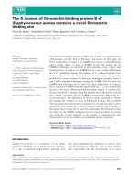
The PSEM inhibited the growth of all Staphylococcus
spp. (antibacterial score: 2+ to 3+). Proliferation of
S. epidermidis in particular was strongly suppressed,
the effect being most marked among all the bacteria
we studied (Fig. 1A). The PSEM had intermediate
Table 1. Antibacterial activity spectra of Platichthys stellatus-derived
epidermal mucus.
Species and strains
Diameter of
clear zone
(mm) Score
a
Gram-positive bacteria
Staphylococcus aureus NIHJ JC-1 8.5 + +
Staphylococcus aureus ATCC25923 6.3 + +
Staphylococcus epidermidis 18.1 + + +
Methicillin-resistant Staphylococcus
aureus 87-7920
8.2 + +
Methicillin-resistant Staphylococcus
aureus 87-7927
8.3 + +
Methicillin-resistant Staphylococcus
aureus 87-7928
8.1 + +
Methicillin-resistant Staphylococcus
aureus 87-7931
8.2 + +
Methicillin-resistant Staphylococcus
aureus 87-7958
8.1 + +
Streptococcus pyogenes 5.5 +
Streptococcus agalactiae 2.8 –
Enterococcus faecalis ATCC33186 2.8 –
Enterococcus faecium ATCC19434 2.8 –
Enterococcus faecium BM4147 (VanA
+
) 2.8 –
Enterococcus faecalis V583 (VanB
+
) 2.8 –
Enterococcus gallinarum BM4174 (VanC1
+
) 2.8 –
Gram-negative bacteria
Escherichia coli NIHJ JC-2 2.8 –
Serratia marcescens 2.8 –
Vibrio parahaemolyticus RIMD2210001 5.7 +
Pseudomonas aeruginosa ATCC27853 2.8 –
a
Clear zone £ 2.8 mm. +, clear zone < 6.0 mm; + +, clear zone
< 10.0 mm; + + +, clear zone ‡ 10.0 mm.
A flounder LAAO-like antibacterial protein K. Kasai et al.
454 FEBS Journal 277 (2010) 453–465 ª 2009 The Authors Journal compilation ª 2009 FEBS
antibacterial activity for S. aureus (Fig. 1B), although
the antibacterial activity of PSEM against two strains
of S. aureus was slightly different. The growth of
MRSA was also inhibited by PSEM (Fig. 1C) and
there was no marked difference in antibacterial activity
among the five MRSA strains tested (Table 1). Among
gram-positive cocci, except for the staphylococci,
PSEM weakly suppressed the growth of S. pyogenes
(1+). Among gram-negative bacilli, the proliferation
of Vibrio parahaemolyticus was weakly suppressed by
PSEM. However, PSEM showed no antibacterial activ-
ity against two strains of Streptococcus spp., five
strains of Enterococcus spp. [including vancomycin-
resistant Enterococcus (VRE)], Escherichia coli, Serra-
tia marcescens and Pseudomonas aeruginosa. In the
growth-inhibition plate assay, the agar medium in the
clear zone formed in the MRSA assay was collected
and cultured in trypticase soy agar (TSA) in order to
confirm the bactericidal activity of PSEM. It was clari-
fied that the PSEM had bactericidal activity against
MRSA because MRSA did not proliferate in TSA
after 96 h of culture.
Temperature sensitivity of PSEM for antibacterial
activity
Generally, proteins lose their activity when subjected to
heat treatment, and complement (which is a component
of blood) is inactivated by heating at 56 °C for 30 min.
Therefore, the antibacterial activity of PSEM was inves-
tigated after incubation at various temperatures, in
order to investigate the properties of the antibacterial
components. The antibacterial activity of PSEM for
MRSA 87-7928 was lowered slightly at 45 °C, markedly
at 56 °C and completely at 70 °C (Fig. 1D), suggesting
that the antibacterial component of PSEM is a protein.
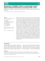
Purification of antibacterial protein from PSEM
The antibacterial protein in PSEM was separated by ul-
tracentrifugation and purified by hydrophobic chroma-
tography (Fig. 2A). Protein fractions were monitored by
measuring the absorbance at 280 nm, and antibacterial
activity was assayed using the growth-inhibition plate
method. Pooled antibacterial fractions were further
purified by gel filtration chromatography (Fig. 2B) and
chromatofocusing (Fig. 2C). In gel filtration chromato-
graphy and chromatofocusing steps, the antibacterial
activity was eluted as a single peak. SDS ⁄ PAGE of the
fractions containing antibacterial activity that had been
separated by chromatofocusing contained three main
bands with molecular masses of 39, 40 and 52 kDa
(Fig. 2D). Because of irreversible denaturation of the
protein, antibacterial activity was not detected in the
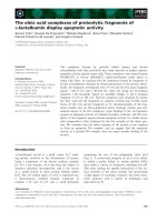
gels after SDS ⁄ PAGE. Therefore, we performed PAGE
in the presence of 6 m urea (6 m urea ⁄ PAGE) to sepa-
rate the antibacterial protein as remaining bioactivity.
Interestingly, the purified PSEM retained its bioactivity
after this step. The antibacterial activity of gel extracts
from the 6 m urea ⁄ PAGE was analyzed using the
growth-inhibition plate method, and the molecular mass
of the antibacterial protein was confirmed by
SDS ⁄ PAGE. Antibacterial protein was detected only in
fractions 19–22 (Fig. 3A), and its molecular mass was
estimated to be 52 kDa (Fig. 3B). Two lower-molecular-
mass proteins of 39 kDa (fractions 23–24) and 40 kDa
(fractions 15–16) did not show antibacterial activity.
Moreover, 2D gel electrophoresis revealed a single spot
at 52 kDa with an isoelectric point of 5.3 (Fig. 3C).
cDNA cloning and sequence analysis of
antibacterial protein
For cloning, the antibacterial protein was blotted onto
a poly(vinylidene difluoride) (PVDF) membrane after
2D gel electrophoresis, and the spot corresponding to
AB
CD
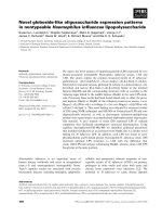
Fig. 1. Antibacterial activity of PSEM against (A) Staphylococcus
epidermidis, (B) Staphylococcus aureus NIHJ JC-1B and (C) MRSA,
clinical isolate 87-7928. Each bacterial strain was suspended in TSA
at a final concentration of 1 · 10
6
CFUÆmL
)1
. (c) Control buffer
without PSEM and (mu) PSEM were applied to holes in the agar.
Antibacterial activity was measured after overnight incubation at
37 °C. (D) Heat sensitivity of PSEM against MRSA, clinical isolate
87-7928. (c) Control buffer without PSEM at 0 °C. PSEM was
exposed to temperatures of 0, 25, 37, 45, 56, 70 and 100 °C for 1 h.
Each sample was applied to the holes in the agar, and antibacterial
activity was measured after overnight incubation at 37 °C.
K. Kasai et al. A flounder LAAO-like antibacterial protein
FEBS Journal 277 (2010) 453–465 ª 2009 The Authors Journal compilation ª 2009 FEBS 455
52 kDa was cut out. Then, the N-terminal peptide
sequence was analyzed by Edman degradation and the
inner peptide sequences were determined using an
amino acid sequencer. This showed that the N-terminal
peptide sequence was Leu-Ser-Phe-Arg-Ala-His-Leu-Ser-
Asp and that the internal peptide sequences were
Arg-Thr-Phe-Glu-Val-Asn-Ala-His-Pro-Asp-Ile-Leu,
Ser-Ala-Asp-Gln-Leu-Leu-Gln-Gln-Ala-Leu and Ser-Glu-
Gly-Arg-Leu-His-Phe-Ala-Gly-Glu-His-Thr. To deter-
mine the cDNA encoding the antibacterial protein of
PSEM, mRNA was prepared from skin and gill. PCR
was performed using degenerate primers based on the
N-terminal peptide sequence LSFRAHLSD and the
internal peptide sequence RTFEVNAHPDIL. Subse-
quently, the full-length cDNA was amplified by
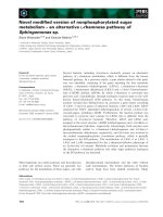
3¢-RACE and 5¢-RACE. Sequence analysis identified
two genes, which completely corresponded to the
peptides of antibacterial protein from the gill (Fig. 4),
and another highly homologous gene from skin (DDBJ
accession number AB495361). The full-length cDNA
found in the gill, which encodes an antibacterial pro-
tein, consisted of 2002 bp plus poly (A). The N-termi-
nal amino acid sequence of LSFRAHLSD was encoded
by nucleotides 183–209. The internal amino acid
sequences RTFEVNAHPDIL, SADQLLQQAL and
SEGRLHFAGEHT were found at positions 567–602,
636–675 and 1524–1559, respectively (Fig. 4). The ini-
tial codon, ATG, was found at positions 102–104, and
the open reading frame was composed of a 1566-bp
region, encoding a protein of 522 amino acid residues.
A BLAST search demonstrated that the encoded anti-
bacterial protein shared identity with a number of
LAAO flavoproteins. The gene encoding this antibacte-
rial protein had 71% identity with the skin mucus
antibacterial LAAO of S. schlegeli (NCBI accession
no. BAF43314) and 69% identity with the apoptosis-
A
B
C
D
Fig. 2. Purification of epidermal mucus protein. (A) Chromatography using a Phenyl Sepharose 6 Fast Flow high sub column. One-hundred and
thirty milliliters of PSEM was applied to the column at a flow rate of 30 mLÆh
)1
. The protein content of each fraction was monitored by measur-
ing the absorbance at 280 nm (s) and antibacterial activity (d) was assayed using the growth-inhibition plate method. Pooled fractions indicated
by the bar (I) were used for gel filtration chromatography. (B) Gel filtration chromatography using a Sephacryl S-100 HR column. The fraction vol-
ume was 2.5 mL and the flow rate was 8.0 mLÆh
)1
. The protein content of each fraction was monitored by measuring the absorbance at
280 nm (s) and antibacterial activity (d) was assayed using the growth-inhibition plate method. Pooled fractions indicated by bar (II) were used
for chromatofocusing. (C) The antibacterial protein was further purified by chromatofocusing on a PBE94 column at pH 7–4. The fraction volume
was 2.5 mL and the flow rate was 30 mLÆh
)1
. The protein content of each fraction was monitored by measuring the absorbance at 280 nm (s)
and antibacterial activity (d) was assayed using the growth-inhibition plate method. The pH of each fraction is indicated by a diamond. Pooled
fractions indicated by bar (III) were used for 6
M urea ⁄ PAGE. (D) SDS ⁄ PAGE of the antibacterial fractions at each chromatography step. C,
crude mucus protein; I, pooled antibacterial fractions from Phenyl Sepharose chromatography; II, pooled antibacterial fractions from gel filtration
chromatography; III, pooled antibacterial fractions from chromatofocusing. The positions of the molecular mass markers are indicated.
A flounder LAAO-like antibacterial protein K. Kasai et al.
456 FEBS Journal 277 (2010) 453–465 ª 2009 The Authors Journal compilation ª 2009 FEBS
inducing protein of Scomber japonicus (NCBI accession
no. CAC00499). A domain search showed that the
gene detected in the gill of P. stellatus contained a
dinucleotide-binding motif followed by a GG-motif
(R-x-G-G-R-x-x-T ⁄ S), which is typical of flavoproteins
[18]. RT-PCR using primers for the 5¢-UTR and
3¢-UTR regions of the LAAO sequence of P. stellatus
(psLAAO) was performed to examine the tissue-specific
expression. The results suggested that the psLAAO gene
was expressed in gill, but not in skin (Fig. 5).
Localization of psLAAO by immunohistochemistry
To identify the localization of psLAAO protein in
the gill of P. stellatus, immunohistochemistry was
performed with an anti-psLAAO IgG, obtained by
immunization of a Japanese white rabbit with insoluble
recombinant psLAAO purified from the E. coli expres-
sion extracts. The psLAAO cDNA sequence, without
the predicted signal peptide, was cloned into the
pET-20b vector and transformed into Rosetta2 (kDE3)
E. coli competent cells. In 5 L of Luria–Bertani (LB)
broth, about 1.4 mg of insoluble recombinant psLAAO
protein was expressed, but the protein was not detected
in soluble form by SDS ⁄ PAGE or western blotting
(Fig. 6). The insoluble recombinant psLAAO protein
was used for the preparation of antiserum. Immunohis-
tochemistry with the anti-psLAAO IgG showed a posi-
tive reaction in the undifferentiated cells surrounding
the vacuolated mucus-secreting cells of the gill (Fig. 7B),
principally within the epithelium of the primary lamellae
and secondary lamellae. The mucus-secreting cells
stained positively with periodic acid ⁄ Schiff’s reagent
(HIO
4
⁄ Schiff), alcian blue and alcian blue-HIO
4
⁄ Schiff.
Neutralization of antibacterial activity with anti-
psLAAO IgG
In order to confirm whether the antibacterial protein
was psLAAO, western blot analysis and a neutralization
Fig. 3. Identification of antibacterial protein
by 6
M urea ⁄ PAGE and 2D gel electrophore-
sis. (A) 6
M urea ⁄ PAGE after chromatofo-
cusing. The antibacterial activity of each gel
extract from a 2-mm-wide strip was
measured using the growth-inhibition plate
method and is indicated as a diagram.
(B) SDS ⁄ PAGE after 6
M urea ⁄ PAGE. Each
of the gel extracts (slice numbers 8–29) was
subjected to determination of the molecular
mass of the antibacterial protein. Antibacte-
rial fractions correspond to the upper
diagram and are indicated by ‘+’. The
asterisk indicates the specific band of the
antibacterial protein. (C) 2D gel electrophore-
sis shows a single spot of antibacterial
protein indicated by a circle. The positions
of the molecular mass markers are
indicated.
K. Kasai et al. A flounder LAAO-like antibacterial protein
FEBS Journal 277 (2010) 453–465 ª 2009 The Authors Journal compilation ª 2009 FEBS 457
assay of antibacterial activity were performed using the
anti-psLAAO IgG. In the western blot analysis,
psLAAO was detected in mucus and gill extract
(Fig. 8A). In the neutralization assay, an apparent dis-
tinction was not found between the anti-psLAAO IgG
free control and the normal rabbit immunoglobulin con-
trol (Fig. 8B). The neutralization activity of the anti-
psLAAO IgG increased in an antibody concentration-
dependent manner.
Discussion
In the present study, we showed that the epidermal
mucus of P. stellatus contains a protein with activity
against various pathogenic species and strains of
bacteria. We isolated this antibacterial protein by col-
umn chromatography through three different matrices
and gel electrophoresis. Furthermore, we detected the
Fig. 4. The cDNA and amino acid
sequences of Platichthys stellatus antibacte-
rial protein. The nucleotide sequence of
cDNA encoding the PSEM antibacterial
protein (DDBJ accession number
AB495360) and the derived amino acid
sequence are shown. The N-terminal and
internal peptide sequences of antibacterial
protein detected by amino acid sequencing
analysis are indicated by boxes. The
predicted dinucleotide-binding motif and the
GG-motif are indicated by a straight line and
a broken line, respectively.
Fig. 5. Tissue-specific expression of psLAAO mRNA by RT-PCR.
Tissues were collected from the same fish. Lane 1, total RNA from
gill; lane 2, total RNA from skin.
A flounder LAAO-like antibacterial protein K. Kasai et al.
458 FEBS Journal 277 (2010) 453–465 ª 2009 The Authors Journal compilation ª 2009 FEBS
N-terminal and internal peptide sequences of this pro-
tein and elucidated its complete mRNA sequence by
cDNA cloning. Because a BLAST search demonstrated
that the encoded antibacterial protein shared identity
with a number of LAAO flavoproteins, and a domain
search showed that the gene contained typical flavo-
protein motifs, the protein was suggested to be a new
member of the LAAO family. RT-PCR and immuno-
histochemical analysis demonstrated tissue-specific
expression and localization in the gill. Western blot
analysis with an anti-psLAAO IgG detected the pro-
tein in mucus and gill extract. Moreover, a neutraliza-
tion assay of antibacterial activity against MRSA
demonstrated that the clear zone was slightly reduced
depending on the volume of anti-psLAAO IgG
employed. Thus, we confirmed that the protein present
in PSEM was a novel LAAO-like antibacterial protein.
LAAOs are flavoenzymes that catalyze the oxidation
of l-amino acids, resulting in the production of a-keto
acids, ammonia and hydrogen peroxide [19]. It has
AB
Fig. 6. Recombinant protein expression in the transfected bacteria.
(A) SDS ⁄ PAGE and (B) western blot analysis of the bacterial
extracts. Lane 1, soluble cytoplasmic fraction; lane 2, insoluble
cytoplasmic fraction. The positions of the molecular mass markers
are indicated. M, positions of the molecular mass markers.
AB
CD
EF
Fig. 7. Immunohistochemical analysis of
Platichthys stellatus gill tissues with anti-
psLAAO IgG. Gill sections of P. stellatus
were stained with (A) nonimmune control
immunoglobulin, (B) anti-psLAAO IgG
(C) hematoxylin & eosin, (D) HIO
4
⁄ Schiff,
(E) alcian blue and (F) alcian blue-
HIO
4
⁄ Schiff. Arrows denote the mucous
cells. Scale bar, 50 lm.
K. Kasai et al. A flounder LAAO-like antibacterial protein
FEBS Journal 277 (2010) 453–465 ª 2009 The Authors Journal compilation ª 2009 FEBS 459
been reported that LAAOs have bioactivities as anti-
bacterial, antiviral and cytotoxic agents in a variety of
animal fluids, such as snake venom [20–25], mouse
milk [26,27], fish epidermal mucus and extract [28,29],
body surface mucus of the giant African snail [30] and
the ink of the sea hare [31,32]. Previous studies have
suggested that the bioactivity of LAAO is elicited by
hydrogen peroxide generated from l-amino acid oxida-
tion [25,32] and the binding of LAAO to bacterial cells
and viruses [33,34]. Achacin, an antibacterial protein
in the mucus of the giant African snail, also shows
significant bacterial-binding and LAAO activity
against S. aureus and E. coli [33]. Escapin, from the
ink of the sea hare, has an l-lysine-dependent antibac-
terial effect and a broad antimicrobial spectrum, being
most effective against S. aureus [32]. Moreover, the
antimicrobial and antiparasitic LAAO isolated from
Bothrops jararaca has the highest effectiveness against
S. aureus [25]. These findings suggest that the anti-
bacterial effect is dependent on hydrogen peroxide
production, because the antibacterial activity was
abolished by catalase. In the present study, PSEM also
showed specific antibacterial activity against S. aureus,
and MRSA was significantly suppressed depending on
the dose of catalase employed (data not shown). Thus,
psLAAO in PSEM exerts antibacterial activity through
hydrogen peroxide generated from the catalytic oxida-
tion of l-amino acid, although details of the selective
effect against bacteria are still unclear.
In the cloning analysis, we identified a cDNA corre-
sponding to the peptide sequence of the antibacterial
protein. RT-PCR analysis suggested that psLAAO
mRNA was specifically expressed in the gill, and
immunohistochemistry with anti-psLAAO IgG also
showed that psLAAO-positive cells were present in the
gill. These results suggest that psLAAO has tissue-
specific expression and is localized in gill. Interestingly,
using cloning analysis, we identified a highly homolo-
gous gene that was expressed in the skin. A domain
search analysis suggested that this homologous gene
also has a dinucleotide-binding motif and a GG motif,
which are characteristic of the LAAO family. Further-
more, a BLAST search demonstrated high identity
with the antibacterial protein of S. schlegeli and other
members of the LAAO family. Immunohistochemical
staining also showed a positive reaction with anti-
psLAAO IgG in skin tissue (data not shown) because
the anti-psLAAO IgG was cross-reactive with highly
homologous LAAO extracted from skin mucus. These
results suggest that some types of LAAO are expressed
in different tissues of fish epidermis.
The gill has a very important function as the main
respiratory organ of fish and it also has an additional
role in defense by secreting a mucus layer, which
includes antibacterial proteins, as it is constantly
exposed to bacteria in the external environment [6,7].
Fig. 8. Reaction of anti-psLAAO IgG with antibacterial protein. (A)
SDS ⁄ PAGE and western blot analysis. Lanes 1 and 3, PSEM; lanes 2
and 4, gill extract. The 52 kDa band is indicated by an asterisk. (B) Neu-
tralization of antibacterial activity with anti-psLAAO IgG. The MRSA
clinical isolate 87-7928 was suspended in TSA at a final concentration
of 1 · 10
6
CFUÆmL
)1
. Ten microliters of PSEM (upper panel) or gill
extract (lower panel) with the indicated volume (0–10 lL) of anti-
psLAAO IgG were applied to each hole in the agar after incubation at
37 °C for 1 h. Control immunoglobulin (10 lL) was applied with 10 lL
of PSEM or gill extract to the holes, as indicated by the hole labelled
‘C’. The total volume was adjusted with NaCl ⁄ P
i
to 20 lL. PSEM and
gill extract protein in the clear zone on the growth-inhibition plate are
indicated as a diagram. M, positions of the molecular mass markers.
A flounder LAAO-like antibacterial protein K. Kasai et al.
460 FEBS Journal 277 (2010) 453–465 ª 2009 The Authors Journal compilation ª 2009 FEBS
The biological importance of the mucus interface
between the body and the aqueous environment
includes functions such as physiological and chemical
protection. In the present study, the N-terminal peptide
sequence of psLAAO was found to start with a leucine
residue, not a methionine residue. Moreover, the
complete psLAAO sequence was 1566 bp in length,
encoding a protein of 522 amino acid residues and with
an expected molecular mass of higher than 52 kDa. The
antibacterial protein we isolated was estimated to have
a molecular mass of approximately 52 kDa. Therefore,
psLAAO may be cleaved at Ala27 to become a mature
protein and secreted from the gill into the extracellular
matrix, and the antibacterial protein starting at Leu28
may be a component of the mucus covering the body
surface and acting as a barrier against bacteria.
We found that psLAAO is effective against various
species of bacteria, suggesting its potential use against
clinical pathogens. MRSA is a major cause of hospi-
tal-acquired infections and a matter of serious public-
health concern worldwide [35], including the UK [36],
Japan [37] and the USA [38]. The appearance of such
multidrug-resistant bacteria has made it imperative to
develop effective and novel antimicrobial agents that
could be used to treat infection with these pathogens.
We speculate that the psLAAO included in PSEM
could be one such agent because it has activity against
MRSA. Our future work will be aimed at improving
the expression of bioactive recombinant psLAAO and
evaluating the mechanism of its antibacterial effect.
Experimental procedures
Collection of epidermal mucus
P. stellatus was caught in the brackish-water region of
Jusanko Lake, in Goshogawara City, Aomori, Japan. After
rinsing the body surface with distilled water, the epidermal
mucus was scraped off with a rubber spatula and frozen at
) 80 °C. The PSEM was then thawed and centrifuged at
105 000 g for 1 h. The supernatant was stored at ) 80 °C.
Bacterial species and strains
Nineteen species or strains of bacteria were used to test the
antibacterial activity of PSEM: the gram-positive bacteria
S. aureus (ATCC25923 and NIHJ JC-1), S. epidermidis
(community isolate), MRSA (clinical isolates 87-7920,
87-7927, 87-7928, 87-7931 and 87-7958), Streptococcus
pyogenes (clinical isolate), Streptococcus agalactiae (clinical
isolate), Enterococcus faecalis ATCC33186, Enterococcus
faecium ATCC19434, E. faecium BM4147 (VanA
+
, clinical
isolate), E. faecalis V583 (VanB
+
, clinical isolate) and Entero-
coccus gallinarum BM4174 (VanC1
+
, clinical isolate); and the
gram-negative bacteria E. coli NIHJ JC-2, S. marcescens
(clinical isolate), V. parahaemolyticus RIMD2210001 and
P. aeruginosa ATCC27853. All clinical isolates were provided
by Hirosaki University School of Medicine and Hospital.
Antibacterial assay
The antimicrobial effects of PSEM were determined using a
growth-inhibition plate assay. The various bacterial species
and strains were cultured in TSA (Difco, Detroit, MI,
USA) for 16 h at 37 °C, except for V. parahaemolyticus,
which was cultured in trypticase soy broth supplemented
with 0.5% NaCl. The cell culture density was measured at
655 nm in a spectrophotometer and then adjusted to
approximately 1 · 10
8
colony-forming units (CFU)ÆmL
)1
with phosphate-buffered saline (NaCl ⁄ P
i
), based on the
standard curve. In order to prepare pour plates, bacteria
were suspended in TSA at a final concentration of
1 · 10
6
CFUÆmL
)1
. Next, a hole of 2.8 mm in diameter
was punched in the pour plate and filled with 12 lLof
mucus or fractions from each of the purification steps.
After overnight incubation at 37 °C, the clear zone around
the hole was measured. To examine heat resistance, the
PSEM was incubated for 1 h at 25, 37, 45, 56, 70 and
100 °C. Each PSEM sample that had been subjected to the
heating treatment was then applied to each hole. After
incubation overnight at 37 °C, the diameter of the clear
zone around each spot was then measured.
Purification of antibacterial protein from
epidermal mucus
Unless indicated otherwise, all procedures were performed at
4 °C. One-hundred and thirty milliliters of PSEM was
thawed and dialyzed against 1 m (NH
4
)
2
SO
4
in 50 mm phos-
phate buffer (pH 7.0), then applied to a column of Phenyl
Sepharose 6 Fast Flow high sub (1.0 · 25 cm; GE Health-
care UK Ltd., Little Chalfont, Bucks, UK), equilibrated pre-
viously with the same buffer, and the column was then
washed with the buffer. The flow rate of the column was
30 mLÆh
)1
and the fraction volume was 10 mL. The protein
concentration in each fraction was monitored by measuring
the absorbance at 280 nm. Adsorbed proteins were eluted
from the column using a linear gradient of 1–0 m (NH
4
)
2
SO
4
in 50 mm phosphate buffer, followed by elution with 50 mm
phosphate buffer and 10 mm phosphate buffer. Antibacterial
activity was assayed using the growth-inhibition plate
method. The fractions with antibacterial activity were col-
lected and the solution was subjected to 80% ammonium
sulfate fractionation. After centrifugation, the resulting pre-
cipitate was dissolved in a small quantity of 0.1 m NaCl in
20 mm Tris ⁄ HCl buffer (pH 7.5) and dialyzed against the
same buffer. The collected proteins were subjected to gel
K. Kasai et al. A flounder LAAO-like antibacterial protein
FEBS Journal 277 (2010) 453–465 ª 2009 The Authors Journal compilation ª 2009 FEBS 461
filtration chromatography on a column of Sephacryl S-100
HR (1.2 · 147cm; GE Healthcare) equilibrated with the
same buffer. The fraction volume was 2.5 mL and the flow
rate was 8.0 mLÆh
)1
. Antibacterial protein was further
purified by chromatofocusing at pH 7–4. The protein in the
antibacterial activity fraction was concentrated by 80%
ammonium sulfate fractionation, as described above. The
resulting precipitate was dialyzed against 25 mm imidazole-
HC1 (pH 7.4) and applied to a column of PEB94 polybuffer
exchanger (1.0 · 27 cm; GE Healthcare) equilibrated with
25 mm imidazole-HC1 (pH 7.4). The fraction was eluted
with polybuffer 74 (pH 4.0), diluted 12-fold with de-aerated
water and further eluted with 0.5 m NaCl. The fraction
volume was 2.5 mL and the flow rate was 30 mLÆh
)1
.
Tissue collection and purification of antibacterial
protein from gill
After rinsing P. stellatus in distilled water, the gill tissue was
harvested and ground into powder using a mortar and
pestle under liquid nitrogen. Proteins were extracted in
the CytoBuster Protein Extraction Reagent (Novagen,
Madison, WI, USA) containing the protease inhibitor by
incubation at room temperature for 5 min. After centrifuga-
tion, the supernatants were collected. Extracted protein
from the gill was thawed and dialyzed against 1 m
(NH
4
)
2
SO
4
in 50 mm phosphate buffer (pH 7.0), then
applied to a column of HiTrap Phenyl FF high sub
(1.6 · 2.5 cm; GE Healthcare) equilibrated with the same
buffer, and the column was then washed with the buffer.
The flow rate of the column was 1 mL ⁄ min and the fraction
volume was 1 mL. Proteins were eluted stepwise from the
column using 1–0 m (NH
4
)
2
SO
4
in 50 mm phosphate buffer,
followed by elution with 50 mm phosphate buffer. Antibac-
terial activity was assayed using growth-inhibition plates.
The fractions with antibacterial activity were collected.
Electrophoresis
SDS ⁄ PAGE was performed according to the method of
Laemmli [39]. The samples were heated in 10% glycerol,
2% SDS, 6% 2-mercaptoethanol and 0.05 m Tris ⁄ HCl buf-
fer (pH 6.8) for 3 min in a boiling water bath and subjected
to SDS ⁄ PAGE with a 10% polyacrylamide gel. Protein was
stained with Coomassie Brilliant Blue R-250. The antibacte-
rial protein fraction separated by chromatofocusing was
subjected to 6 m urea ⁄ PAGE at room temperature. The
lower gel consisted of 7.5% acrylamide, 6 m urea, 0.06%
ammonium persulfate, 0.15% N,N,N¢, N¢ -tetramethyl ethy-
lenediamine (TEMED) and 0.3 m acetate buffer (pH 4.8),
while the upper gel consisted of 5.0% acrylamide, 6 m urea,
0.002% riboflavin 0.015% TEMED and 0.2 m acetate buf-
fer (pH 5.0). The reservoir buffer was composed of 0.35 m
b-alanine and 0.136 m acetate buffer (pH 4.8). The upper
gel was polymerized by illumination with a fluorescent
light. After electrophoresis, the lower gel was cut into strips
2 mm wide. Then, 40 lLof10mm phosphate buffer was
added and the gel was broken into small pieces. The super-
natant obtained by centrifugation was then used to measure
antibacterial activity or to determine the molecular mass of
antibacterial protein by SDS ⁄ PAGE. 2D gel electrophoresis
was performed according to the method of O’Farrell [40],
as modified by Hirsch et al. [41]. Protein was stained with
Coomassie Brilliant Blue R-250. The second-dimension
electrophoresis was carried out on a 10% acrylamide gel.
Amino acid sequencing
After 2D gel electrophoresis, proteins in the gel were
blotted onto a PVDF membrane (Millipore Corp., Bedford,
MA, USA) using a semidry-type blotting apparatus, and
the target protein spot was cut out. The N-terminal amino
acid sequence was analyzed using the Edman degradation
method. An inner peptide amino acid sequence analysis
was also performed. Peptidase digestion using lysyl end-
peptidase, separation of the fragments by RP-HPLC and
amino acid sequence analysis were assigned to the APRO
Life Science Institute Inc. (Naruto, Tokushima, Japan).
mRNA extraction and degenerate PCR
Total RNA was extracted from the epidermis and gill tissues
of P. stellatus using an RNeasy Mini kit (Qiagen, Valencia,
CA, USA) in accordance with the manufacturer’s instruc-
tions. Total RNA was transcribed to cDNA at 42 °C for
60 min in the presence of the oligo (dT)
15
Primer (Promega,
Madison, WI, USA) and Primescript Reverse Transcriptase
(Takara, Tokyo, Japan). Degenerate oligonucleotide primers
were designed on the basis of the determined amino
acid sequences of the peptide fragments. The forward degen-
erate primers were 5¢-YTITCITTYCGIGIGCNCAY-3¢,
5¢-YTIAGYTTYCGIGCNCAY-3¢,5¢-YTITCITTYAGRG
CNCAY-3¢ and 5¢-YTIAGYTTYAGRGCNCAY-3¢ (corre-
sponding to LSFRAHLSD). The reverse degenerate primer
was 5¢-RTGIGCRTTIACYTCRAANGT-3¢ (corresponding
to RTFEVNAHPDIL). Amplification was carried out using
Ex Taq polymerase (Takara) under the following condi-
tions: 95 °C for 5 min; 35 cycles of 95 °C for 1 min, 48 °C
for 1 min and 72 °C for 1 min; 72 °C for 9 min. All PCR
products were subcloned into the T-vector prepared by dT
addition on EcoRV-digested blunt ends of pBluescript II
SK+ (Stratagene, LA Jolla, CA, USA). DNA sequences
were determined using an abi prism 310 Genetic Analyzer
(Applied Biosystems, Foster City, CA, USA).
5¢-RACE and 3¢-RACE
5¢-RACE was carried out according to the procedure of the
5¢-RACE System for Rapid Amplification of cDNA Ends
A flounder LAAO-like antibacterial protein K. Kasai et al.
462 FEBS Journal 277 (2010) 453–465 ª 2009 The Authors Journal compilation ª 2009 FEBS
(Invitrogen, Carlsbad, CA, USA) using a gene-specific pri-
mer (GSP) (5¢-CATTCCTGTACGTCTCCACTC-3¢) and a
nested GSP (5¢-GTTCTCTACTGTCTGCAGCAG-3¢). 3¢-
RACE was carried out using the procedure of the 3¢-RACE
System for Rapid Amplification of cDNA Ends (Invitrogen)
using a GSP (5¢-GGAATGAGCAAGGCTGGTAC-3¢) and
a nested GSP (5¢-CTCTTCTTCGTGGAGTTACTC-3¢).
The GSPs used for 5¢-RACE and 3¢-RACE were designed
on the basis of the determined sequences of degenerate
PCR clones. Amplification was carried out using AmpliTaq
Gold DNA polymerase (Applied Biosystems) under the
following conditions: 95 °C for 10 min; 35 cycles of 95 °C
for 1 min, 48 °C for 1 min and 72 °C for 1 min; 72 °C for
9 min. The PCR products were subcloned into the T-vector
and sequenced. The nucleotide sequence of the full-length
cDNA was amplified by RT-PCR using the forward primer
5¢-GAAGTTTCTCTACGGACTGC-3¢ and the reverse
primer 5¢-CAACCATCGATTGTGTCCAG-3¢. The beta-
actin primer pair (forward primer 5¢-CATGTACGTTGC
CATCCAAG-3¢ and reverse primer 5¢-TCTCAGCTGTGG
TGGTGAAG-3¢) was designed on the basis of the Euro-
pean flounder (P. flesus) beta-actin gene sequence (NCBI
accession number AF135499). Amplification was carried
out using AmpliTaq Gold DNA polymerase (Applied Bio-
systems) under the following conditions: 95 °C for 10 min;
35 cycles of 95 °C for 1 min, 55 °C for 1 min and 72 °C for
1.5 min; 72 °C for 9 min. PCR products were subcloned
into the T-vector and sequenced.
Recombinant protein expression
Primers were designed to amplify the active form without
the secretory signal sequence, so that antibacterial protein
could be expressed in E. coli as a His-tagged fusion protein.
The forward primer included an NdeI restriction site
(5¢-CC GCATATGCTCA GCTTCAGGGCA CATCTG-3¢)
and the reverse degenerate primer included a XhoI restric-
tion site (5¢-GCACTCGAGGGTGTGTTCAACCAGCAA
AG-3¢). Amplification was carried out using AmpliTaq
Gold DNA polymerase under the following conditions:
95 °C for 10 min; 35 cycles of 95 °C for 1 min, 55 ° C for
1 min and 72 °C for 1.5 min; 72 °C for 9 min. The PCR
products were then cleaved with restriction enzymes and
the gene was subcloned into the pET-20b expression vector
(Novagen) using the same enzymes. The DNA sequence
was determined using an abi prism 310 Genetic Analyzer
(Applied Biosystems). For protein expression, the plasmid
was transformed into E. coli strain Rosetta 2 (kDE3).
Five liters of these bacterial cells were grown in LB broth
(Difco) at 37 °C until the culture reached a D at 600 nm of
approximately 0.5, and proteins were induced with 0.4 mm
isopropyl thio-b-d-galactoside at 15 °C for 16 h. The
expressed His-tagged fusion proteins were isolated by
means of Ni-nitrilotriacetic acid agarose (Qiagen), in accor-
dance with the manufacturer’s instructions. The purified
His-tagged fusion protein was digested with trypsin and the
amino acid sequence was analyzed using nanoFrontier
nLC-Linear-Trap-TOF MS (Hitachi, Tokyo, Japan).
Antiserum preparation and IgG purification
An antiserum against the antibacterial protein was obtained
by injecting a Japanese white rabbit (Kitayama Labes Co.
Ltd., Nagano, Japan) with the insoluble recombinant pro-
tein of His-tagged psLAAO purified from the E. coli
expression extracts. The recombinant psLAAO (300 lg)
was emulsified with 1.5 mL of Freund’s complete adjuvant
(Difco) and injected subcutaneously into each animal.
Booster injections of 100 lg of recombinant psLAAO in an
emulsion of Freund’s incomplete adjuvant (Difco) were
then given at 2, 5 and 8 weeks after the primary immuniza-
tion. At 9 weeks, the antiserum was obtained. Control
serum was obtained from a naive Japanese white rabbit.
The IgG fraction was purified according to the recom-
mended procedure for the ImmunoPure Melon Gel IgG
Spin Purification kit (Pierce, Rockford, IL, USA).
Western blot analysis
PSEM, gill extract and His-tagged recombinant psLAAO
protein were individually heated at 100 °C in 10% glycerol,
2% SDS, 6% 2-mercaptoethanol and 0.05 m Tris ⁄ HCl buf-
fer (pH 6.8) and subjected to SDS ⁄ PAGE (10% polyacryl-
amide gel). After electrophoresis, the proteins were
electrically transferred from the gel onto a PVDF mem-
brane (GE Healthcare). The membrane was blocked with
20 mm Tris ⁄ HCl (pH 7.4), 125 mm NaCl, 0.2% Tween 20
and 5% skim milk (Yotsuba, Sapporo, Japan). psLAAO in
the PSEM and gill extracts was detected using the anti-
psLAAO IgG (1 : 2000 dilution). His-tagged fusion proteins
were detected using a mouse monoclonal anti-His IgG
(1 : 3000 dilution; GE Healthcare). Horseradish peroxidase-
conjugated secondary antibody mouse anti-rabbit IgG
(1 : 5000 dilution; GE Healthcare) or sheep anti-mouse IgG
(1 : 10000 dilution; GE Healthcare) was used for detection,
followed by enhanced ECL Plus Western blotting detection
reagents (GE Healthcare).
Histology
Gill tissues of P. stellatus were fixed in 4% paraformalde-
hyde and embedded in paraffin. Sections (4 lm thick) were
mounted on Mac-coated slides (Matsunami Trading Co.
Ltd., Osaka, Japan). Deparaffinized and rehydrated sections
were stained with hematoxylin and eosin. Immunohisto-
chemical staining for antibacterial protein was performed
using the avidin–biotin–peroxidase complex method using a
Histofine SAB-PO (MULTI) kit (Nichirei, Tokyo, Japan) in
accordance with the manufacturer’s instructions. Sections
K. Kasai et al. A flounder LAAO-like antibacterial protein
FEBS Journal 277 (2010) 453–465 ª 2009 The Authors Journal compilation ª 2009 FEBS 463
were counterstained with hematoxylin for microscopic
examination. Anti-psLAAO IgG or control IgG was used as
the primary antibody (1 : 1000 dilution). To characterize the
psLAAO-positive cells, HIO
4
⁄ Schiff, alcian blue (pH 2.6)
and alcian blue-HIO
4
⁄ Schiff staining reactions were
performed.
Neutralization assay of antibacterial activity
Samples of MRSA (clinical isolates) were suspended in
TSA at a final concentration of 1 · 10
6
CFUÆmL
)1
. Then,
1–10 lL of anti-psLAAO IgG was added to 10 lL of either
mucus or gill extract and applied to holes 2.8 mm in diame-
ter. After overnight incubation at 37 °C, the diameter of
the clear zone around each hole was measured.
Acknowledgements
We are grateful to Dr M. Senda for advice, to J. Oikawa
for technical assistance, to APRO Life Science Institute
Inc. (Naruto, Tokushima, Japan) for performing amino
acid sequence analysis and to the Jusanko fishermen’s
cooperative association (Goshogawara, Aomori, Japan)
for provision of P. stellatus. This work was supported,
in part, by a Grant for Priority Research Designated by
the Japan Science and Technology Agency.
References
1 Kubota Y, Watanabe Y, Otsuka H, Tamiya T, Tsu-
chiya T & Matsumoto JJ (1985) Purification and char-
acterization of an antibacterial factor from snail mucus.
Comp Biochem Physiol C 82, 345–348.
2 Simmaco M, Mignogna G & Barra D (1998) Antimi-
crobial peptides from amphibian skin: what do they tell
us? Biopolymers 47, 435–450.
3 Andreu D & Rivas L (1998) Animal antimicrobial pep-
tides: an overview. Biopolymers 47, 415–433.
4 Bulet P, Sto
¨
cklin R & Menin L (2004) Anti-microbial
peptides: from invertebrates to vertebrates. Immunol
Rev 198, 169–184.
5 Hellio C, Pons AM, Beaupoil C, Bourgougnon N &
Gal YL (2002) Antibacterial, antifungal and cytotoxic
activities of extracts from fish epidermis and epidermal
mucus. Int J Antimicrob Agents 20, 214–219.
6 Ellis AE (2001) Innate host defense mechanisms of fish
against viruses and bacteria. Dev Comp Immunol 25,
827–839.
7 Magnado
´
ttir B (2006) Innate immunity of fish (over-
view). Fish Shellfish Immunol 20, 137–151.
8 Zanetti M, Gennaro R & Romeo D (1995)
Cathelicidins: a novel protein family with a common
proregion and a variable C-terminal antimicrobial
domain. FEBS Lett 374, 1–5.
9 Chang CI, Zhang YA, Zou J, Nie P & Secombes CJ
(2006) Two cathelicidin genes are present in both rain-
bow trout (Oncorhynchus mykiss) and atlantic salmon
(Salmo salar). Antimicrob Agents Chemother 50, 185–195.
10 Lauth X, Shike H, Burns JC, Westerman ME, Ostland
VE, Carlberg JM, Van Olst JC, Nizet V, Taylor SW,
Shimizu C et al. (2002) Discovery and characterization
of two isoforms of moronecidin, a novel antimicrobial
peptide from hybrid striped bass. J Biol Chem 277,
5030–5039.
11 Smith VJ, Fernandes JM, Jones SJ, Kemp GD & Tat-
ner MF (2000) Antibacterial proteins in rainbow trout,
Oncorhynchus mykiss. Fish Shellfish Immunol 10, 243–
260.
12 Lemaıˆ tre C, Orange N, Saglio P, Saint N, Gagnon J &
Molle G (1996) Characterization and ion channel activi-
ties of novel antibacterial proteins from the skin mucosa
of carp (Cyprinus carpio). Eur J Biochem 240, 143–149.
13 Cole AM, Weis P & Diamond G (1997) Isolation and
characterization of pleurocidin, an antimicrobial peptide
in the skin secretions of winter flounder. J Biol Chem
272, 12008–12013.
14 Nagashima Y, Kikuchi N, Shimakura K & Shiomi K
(2003) Purification and characterization of an antibacte-
rial protein in the skin secretion of rockfish Sebastes
schlegeli. Comp Biochem Physiol C 136, 63–71.
15 Molle V, Campagna S, Bessin Y, Ebran N, Saint N &
Molle G (2008) First evidence of the pore-forming
properties of a keratin from skin mucus of rainbow
trout (Oncorhynchus mykiss, formerly Salmo gairdneri).
Biochem J 411, 33–40.
16 Ebran N, Julien S, Orange N, Auperin B & Molle G
(2000) Isolation and characterization of novel glycopro-
teins from fish epidermal mucus: correlation between
their pore-forming properties and their antibacterial
activities. Biochim Biophys Acta 1467, 271–280.
17 Du XY & Clemetson KJ (2002) Snake venom L-amino
acid oxidases. Toxicon 40
, 659–665.
18 Vallon O (2000) New sequence motifs in flavoproteins:
evidence for common ancestry and tools to predict
structure. Proteins 38, 95–114.
19 Wellner D & Meister A (1961) Studies on the mecha-
nism of action of L-amino acid oxidase. J Biol Chem
236, 2357–2364.
20 Franc¸ a SC, Kashima S, Roberto PG, Marins M, Ticli
FK, Pereira JO, Astolfi-Filho S, Sta
´
beli RG, Magro AJ,
Fontes MR et al. (2007) Molecular approaches for
structural characterization of Bothrops L-amino acid
oxidases with antiprotozoal activity: cDNA cloning,
comparative sequence analysis, and molecular modeling.
Biochem Biophys Res Commun 355, 302–306.
21 Tempone AG, Andrade HF Jr, Spencer PJ, Lourenc¸ o
CO, Rogero JR & Nascimento N (2001) Bothrops
moojeni venom kills Leishmania spp. with hydrogen
A flounder LAAO-like antibacterial protein K. Kasai et al.
464 FEBS Journal 277 (2010) 453–465 ª 2009 The Authors Journal compilation ª 2009 FEBS
peroxide generated by its L-amino acid oxidase.
Biochem Biophys Res Commun 280, 620–624.
22 Sta
´
beli RG, Marcussi S, Carlos GB, Pietro RC,
Selistre-de-Arau´ jo HS, Giglio JR, Oliveira EB & Soares
AM (2004) Platelet aggregation and antibacterial effects
of an l-amino acid oxidase purified from Bothrops
alternatus snake venom. Bioorg Med Chem 12,
2881–2886.
23 Zhang YJ, Wang JH, Lee WH, Wang Q, Liu H, Zheng
YT & Zhang Y (2003) Molecular characterization of
Trimeresurus stejnegeri venom L-amino acid oxidase
with potential anti-HIV activity. Biochem Biophys Res
Commun 309, 598–604.
24 Sant’Ana CD, Menaldo DL, Costa TR, Godoy H, Mul-
ler VD, Aquino VH, Albuquerque S, Sampaio SV,
Monteiro MC, Sta
´
beli RG et al. (2008) Antiviral and
antiparasite properties of an L-amino acid oxidase from
the snake Bothrops jararaca: cloning and identification
of a complete cDNA sequence. Biochem Pharmacol 76,
279–288.
25 Ciscotto P, Machado de Avila RA, Coelho EA, Oliveira
J, Diniz CG, Farı
´
as LM, de Carvalho MA, Maria WS,
Sanchez EF, Borges A et al. (2009) Antigenic, microbi-
cidal and antiparasitic properties of an l-amino acid oxi-
dase isolated from Bothrops jararaca snake venom.
Toxicon 53, 330–341.
26 Sun Y, Nonobe E, Kobayashi Y, Kuraishi T, Aoki F,
Yamamoto K & Sakai S (2002) Characterization and
expression of L-amino acid oxidase of mouse milk. J
Biol Chem 277, 19080–19086.
27 Nagaoka K, Aoki F, Hayashi M, Muroi Y, Sakurai T,
Itoh K, Ikawa M, Okabe M, Imakawa K & Sakai S
(2009) L-amino acid oxidase plays a crucial role in host
defense in the mammary glands. FASEB J 23, 2514–
2520.
28 Kitani Y, Tsukamoto C, Zhang G, Nagai H, Ishida M,
Ishizaki S, Shimakura K, Shiomi K & Nagashima Y
(2007) Identification of an antibacterial protein as
L-amino acid oxidase in the skin mucus of rockfish
Sebastes schlegeli. FEBS J 274, 125–136.
29 Jung SK, Mai A, Iwamoto M, Arizono N, Fujimoto D,
Sakamaki K & Yonehara S (2000) Purification and
cloning of an apoptosis-inducing protein derived from
fish infected with Anisakis simplex, a causative nema-
tode of human anisakiasis. J Immunol 165, 1491–1497.
30 Obara K, Otsuka-Fuchino H, Sattayasai N, Nonomura
Y, Tsuchiya T & Tamiya T (1992) Molecular cloning of
the antibacterial protein of the giant African snail,
Achatina fulica Fe
´
russac. Eur J Biochem 209, 1–6.
31 Petzelt C, Joswig G, Stammer H & Werner D (2002)
Cytotoxic cyplasin of the sea hare, Aaplysia punctata,
cDNA cloning, and expression of bioactive recombi-
nants in insect cells. Neoplasia 4, 49–59.
32 Yang H, Johnson PM, Ko KC, Kamio M, Germann
MW, Derby CD & Tai PC (2005) Cloning, character-
ization and expression of escapin, a broadly antimicro-
bial FAD-containing L-amino acid oxidase from ink of
the sea hare Aplysia californica. J Exp Biol 208, 3609–
3622.
33 Ehara T, Kitajima S, Kanzawa N, Tamiya T &
Tsuchiya T (2002) Antimicrobial action of achacin is
mediated by L-amino acid oxidase activity. FEBS Lett
531, 509–512.
34 Kitani Y, Kikuchi N, Zhang G, Ishizaki S, Shimakura
K, Shiomi K & Nagashima Y (2008) Antibacterial
action of L-amino acid oxidase from the skin mucus of
rockfish Sebastes schlegelii. Comp Biochem Physiol B
149, 394–400.
35 Boyce JM, Cookson B, Christiansen K, Hori S,
Vuopio-Varkila J, Kocago
¨
z S, Oztop AY,
Vandenbroucke-Grauls CM, Harbarth S & Pittet D
(2005) Meticillin-resistant Staphylococcus aureus.
Lancet Infect Dis 5, 653–663.
36 Wyllie DH, Peto TE & Crook D (2005) MRSA bactera-
emia in patients on arrival in hospital: a cohort study in
Oxfordshire 1997-2003. BMJ 331, 992–997.
37 Takeda S, Yasunaka K, Kono K & Arakawa K (2000)
Methicillin-resistant Staphylococcus aureus (MRSA)
isolated at Fukuoka University Hospital and hospitals
and clinics in the Fukuoka city area. Int J Antimicrob
Agents 14, 39–43.
38 National Nosocomial Infections Surveillance System.
(2004) National Nosocomial Infections Surveillance
(NNIS) System Report, data summary from January
1992 through June 2004, issued October 2004. Am J
Infect Control 32, 470–485.
39 Laemmli UK (1970) Cleavage of structural proteins
during the assembly of the head of bacteriophage T4.
Nature 227, 680–685.
40 O’Farrell PH (1995) High resolution two-dimensional
electrophoresis of proteins. J Biol Chem 250, 4007–
4021.
41 Hirsch FW, Nall KN, Busch FN, Morris HP & Busch
H (1978) Comparison of abundant cytosol proteins in
rat liver, Novikoff hepatoma, and Morris hepatoma by
two-dimensional gel electrophoresis. Cancer Res 38,
1514–1522.
K. Kasai et al. A flounder LAAO-like antibacterial protein
FEBS Journal 277 (2010) 453–465 ª 2009 The Authors Journal compilation ª 2009 FEBS 465